Case Report

Rehabilitation of a Wide Buccal Recession Using a Combination of Adhesive Prosthetic Procedures and Transmucosal Convergent Neck Implant to Replace a Lower Fractured Canine: Case Report with 6 Years Follow-Up

by
Carlo Prati
1,*,
Andrea Spinelli
1,
Maria Giovanna Gandolfi
2 and
Fausto Zamparini
1,2
1
Endodontic Clinical Section, Department of Biomedical and Neuromotor Sciences, Dental School, University of Bologna, 40125 Bologna, Italy
2
Laboratory of Biomaterials and Oral Pathology, Department of Biomedical and Neuromotor Sciences, School of Dentistry, University of Bologna, 40125 Bologna, Italy
*
Author to whom correspondence should be addressed.
Prosthesis 2025, 7(5), 117; https://doi.org/10.3390/prosthesis7050117
Submission received: 1 July 2025 / Revised: 13 August 2025 / Accepted: 26 August 2025 / Published: 10 September 2025

Abstract

Objectives: The presence of gingival buccal recession is a frequent problem especially in the canine area. The cortical buccal bone may be absent in presence of health normal lingual/palatal bone and of other periodontal pockets. The present case report describes a minimally invasive approach in a 76-year-old patient with previously endodontically treated lower canine affected by root fracture and by a serious chronic buccal recession. Methods: The tooth was characterized by a deep vestibular bone defect, lack of buccal bone and acute periapical lesion. After extraction, Maryland bridge was positioned on the edentulous area. A two-piece convergent neck transmucosal implant was inserted with a flapless approach after 6 months. Maryland bridge was left in place for additional 3 months. After this time, digital impressions were taken, and a customized abutment was positioned. A provisional crown was designed according to Biologically Oriented Preparation Technique (BOPT) concept and maintained for 6 months. A zirconia definitive crown was digitally designed and cemented with a polycarboxylate-based cement. The Pink Esthetic Score (PES) was used as an index to assess peri-implant soft tissue stability over time (preoperatively, at 9 months, at 12 months and 72 months). Results: The patient was followed for 6 years under a conventional hygienic recall program. No complications occurred, and the PES improved from 4 preoperatively to 8 at 9 months, 10 at 12 months and 13 at 72 months. Conclusions: The use of Maryland bridge prevented occlusal trauma on healing tissues and appeared to support bone and soft tissue healing for transmucosal implant placement. A stable aesthetic rehabilitation was achieved up to 6 years.

1. Introduction

Implant rehabilitations showed predictable results in healthy patients. Several recent reviews and meta-analyses highlight high survival rates of implant rehabilitations in the first 5–10 years post-insertion [1,2]. In the esthetic zone, however, outcomes depend on preserving adequate hard and soft tissue volumes. Thin buccal bone plates—often less than 1 mm—are prone to significant resorption within weeks after extraction, and current techniques such as immediate placement, socket grafting or augmentation do not fully prevent this remodeling [3,4]. The initial phases of soft and hard tissue management after extraction [5,6] and early osseointegration are indeed critical for long-term success [7,8].
Animal studies, along with human histological and micromorphological analyses, have shown varying patterns of bone remodeling and mineralization following extraction and implant placement in the mandible and maxilla, revealing that timing of implant placement and the loading protocol have a critical influence on the phases of osseointegration [9,10,11].
In elderly patients, extensive bone and soft tissue deficiencies are common due to advanced periodontal disease, tooth fractures or failed endodontic therapy. Complex grafting procedures may be poorly tolerated because of longer recovery times, higher complication risks and greater treatment discomfort [12,13]. Minimally invasive protocols, particularly those avoiding multiple surgical stages, can therefore be advantageous in this population.
Presence of chronic periapical lesions or exacerbated abscesses are also common in elderly patients. These conditions do not also allow immediate implant placement or bone grafting procedures, due to significant increase in surgical site infection and low residual bone volume areas [14,15]. The post-extractive site should be protected to allow healing without any occlusal stress. The adoption of provisional bonded restorations showed to guide soft and hard tissues healing during both post-extraction and implant osseointegration phases [16].
Non-submerged transmucosal protocols may be particularly beneficial for elderly patients. In this approach, an implant with a long, convergent neck is placed at the crestal level, leaving the neck exposed to guide soft tissue healing at the transmucosal interface. This procedure avoids surgical re-entry before the prosthetic phases and may reduce the chances of early bone remodeling during this critical period. The minimally invasive nature of this approach may also allow for the formation of stable peri-implant tissue over time with reduced chances of biological complications.
This case report presents a minimally invasive approach—extraction site management, bonded provisionalization and a non-submerged transmucosal implant protocol—for the rehabilitation of an unrestorable mandibular canine in a 76-year-old patient. The goal was to achieve functional stability and satisfactory esthetics without additional grafting procedures.

2. Materials and Methods

The work was part of a project approved by the institutional ethical committee (CE AVEC 33-2023-OSS-AUSLBO, Acronym “Endo-Implant Retro 2023”). The present case report was described following the CARE Checklist (accessed on 6 February 2024). Study timeline is reported in Figure 1.

2.1. Patient Information

A 76-year-old non-smoker patient was referred to the Endodontic Clinical Section, University of Bologna. An informed consent form was signed at the time of the dental examination. The patient had no significant medical, family, or psycho-social history contributing to the current dental issue. The affected tooth, identified as mandibular left canine (#33), had undergone a non-surgical root canal retreatment 10 years prior. The procedure involved shaping with Nickel titanium (NiTi) and manual instruments, followed by obturation using a calcium silicate-based sealer (Tech Biosealer Endo, Isasan, Rovello, Italy) and a carrier-based technique (Thermafil, Dentsply, Konstanz, Germany). The tooth was then restored with Se Bond (Kuraray, Osaka, Japan) and Gradia composite (GC Europe, Leuven, Belgium).
The primary concern was persistent pain and swelling in the mandibular anterior region, significantly impacting the patient’s quality of life. Upon visual inspection, tooth #33 exhibited a root fracture, with a noticeable loss of attachment on the buccal aspect. The tooth showed a 14 mm soft tissue recession that extends to or beyond the mucogingival junction, with periodontal attachment loss in the interdental area or mispositioning of teeth. (Figure 2) Oral examination revealed recessions on both adjacent teeth (Clinical Attachment Loss of 4 mm) but no signs of Pocket Probing Depth > 4 mm or Bleeding.

2.2. Tooth Extraction and Resin-Bonded Bridge Preparation

The sudden tooth fracture and apical infection required the extraction of the tooth. Root extraction was performed without raising any flap. All granulation tissue was meticulously removed from the apical portion of the socket. Soft tissues were finally sutured with Vicryl 4/0 to allow alveolar socket healing with no risks of infections.
Due to the presence of an active periapical lesion and the absence of a cortical buccal wall, there was no possibility to position bone graft and membranes on the post-extractive sites. A provisional resin-bonded bridge (Maryland bridge) was prepared prior to the extraction as temporary aesthetic restoration, to guide and protect both hard and soft tissues during the healing time [16].
The Maryland bridge was positioned as follows: the palatal/lingual enamel surfaces of the adjacent teeth were etched with 33% orthophosphoric acid gel (3M ESPE, St. Paul, MN, USA) and rinsed gently for 20 s. Universal Scotchbond adhesive (3M ESPE, St. Paul, MN, USA) was applied to the enamel surface with a micro brush, followed by the application of dual-cure Relyx Ultimate cement (3M ESPE, St. Paul, MN, USA) to affix the Maryland bridge (Figure 3).

2.3. Implant Insertion

Implant placement was positioned 6 months after extraction and after Maryland bridge was removed (Figure 4). The post-extractive site underwent clinical and radiographic assessment, including baseline periapical X-rays to assess mesio-distal and vertical dimensions. Additional volumetric assessment of the healed ridge was obtained through direct clinical measurements with a periodontal probe and the use of digital impressions models, allowing evaluation of the alveolar crest width and guiding the selection of implant diameter and position. During flapless implant site preparation, a 1.2 mm pilot bur was used, following the residual lingual bony wall as a guide, with subsequent preparation using calibrated burs at 225 rpm under abundant saline irrigation.
A 4.25 mm Zirconium oxide-blasted acid-etched titanium (Zir-Ti) implant (Prama, Sweden & Martina, Due Carrare, Padova, Italy) characterized by a 2.8 mm machined convergent neck profile was inserted. Implant placement involved partially submerging the transmucosal neck in soft tissues, with approximately 1 mm of the neck exposed above the soft tissues [17,18]. In this way, all the rough surface was placed in contact with the mesial, distal and lingual bone tissues and the microtextured surface of the neck was placed intramucosally and partially exposed. Subsequently, a 1.0 mm cover screw was placed and maintained throughout the healing phases. The Maryland bridge was modified in order to avoid any contact with the exposed implant neck.
Follow-up visits were scheduled at 1 and 3 months to check the patient’s hygienic compliance and to monitor soft tissues and mesial and distal crestal bone levels.
After 3 months from insertion, the soft and hard tissues showed no inflammation and radiographically stable bone levels. Prosthetic procedures were then scheduled using a digital intraoral scanning workflow (Figure 4).

2.4. Digital Workflow for Prosthetic Rehabilitation

Intraoral digital impressions were obtained using an intraoral scanner (Carestream CS3600, Carestream, Rochester, NY, USA).
A full-mouth digital impression was taken after the removal of Maryland bridge. The acquisition started from the occlusal surface of the most distant elements (i.e., molars) to the contralateral teeth. Subsequently, the vestibular and lingual/palatal parts of the elements were scanned, with attention in detecting the interproximal zone of the adjacent teeth to the implant rehabilitation. The same procedure was used to scan the maxillary arch. Occlusion was then taken through intercuspidation in the molar–premolar and frontal regions.
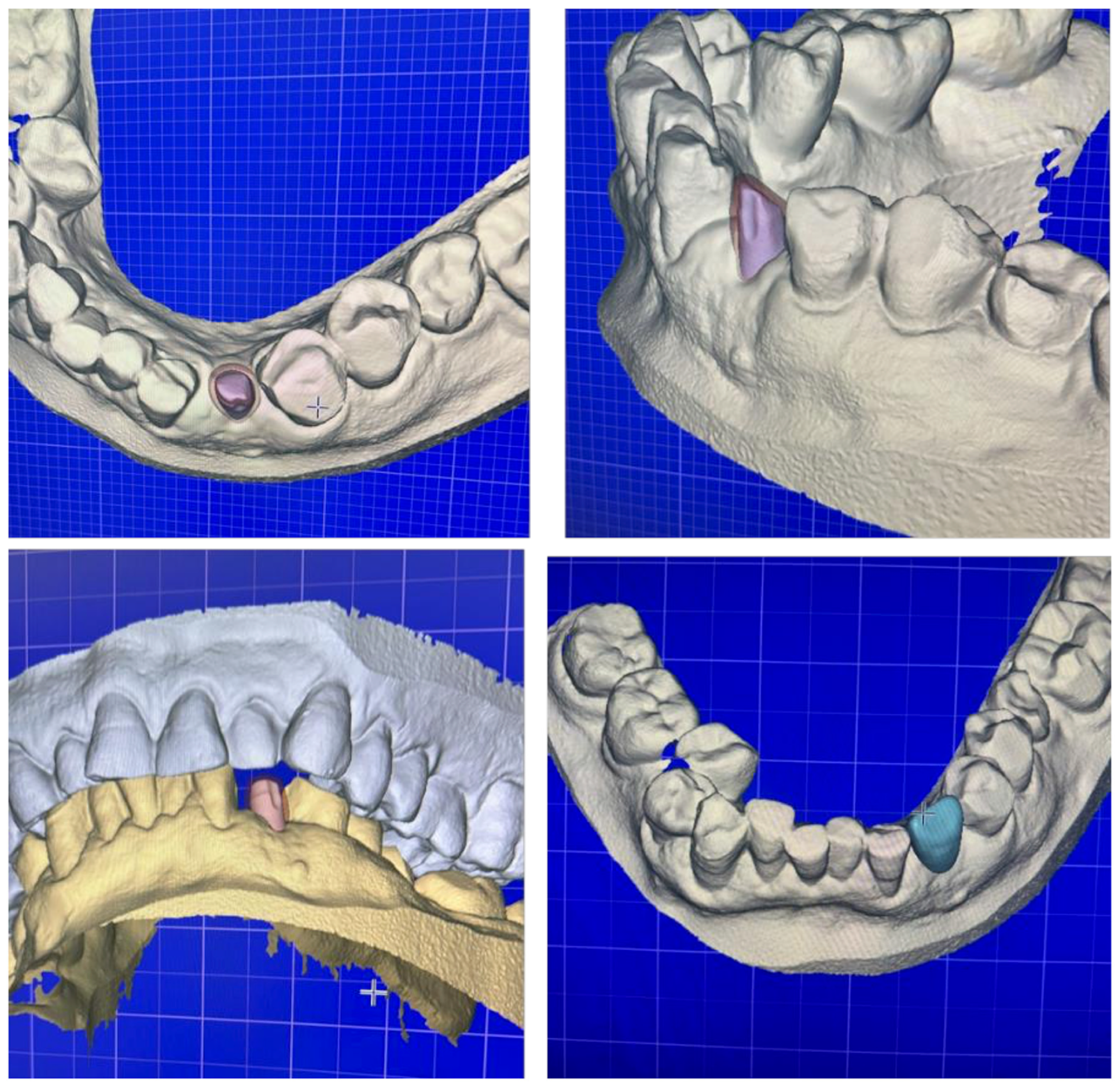
The next phase involved the scan body acquisition (Figure 5). From the obtained impression, a digital cut of the implant emergence area was applied. The two-piece Prama implant eliminated the need for a second surgery or other techniques to expose the implant neck. Moreover, this ensured accurate positioning and reading of the scan body without risk of misplacement or misreading. The scan body was placed on the implant emergence so that its grooved portion is located vestibular and scanned. After that, impressions were e-mailed to the dental technician and Maryland bridge was cemented again.
The obtained impression was processed using dedicated software (Exocad Dental CAD, Woburn, MA, USA). Briefly, the scan was matched with the dedicated abutment from the library. The shape and angulation of the abutment were then computer-designed (Figure 6).
The titanium abutment was applied after approximately 15 days. The temporary resin crown was placed with zinc oxide eugenol cement (Temp Bond, Kerr, Scafati, Italy) for initial prosthetic rehabilitation.
The crown was designed following the concepts of Biologically Oriented Preparation Technique (BOPT). BOPT has been described in the context of natural tooth-supported restorations, where crown is positioned on a previously prepared tooth with no finishing line, creating a new prosthetic cementoenamel junction and allowing the crown’s gingival margin to be adequately shaped [19]. Following this concept, the implant–abutment connection resulted internal to the crown [17,18].
Six months later, the definitive zirconia prosthetic rehabilitation was placed and fixed with a powder/liquid polycarboxylate cement (Heraeus Kulzer GmbH, Hanau, Germany). The margin of the prosthetic restoration was designed to gently compress the peri-implant soft tissues, ending on the implant’s hyperbolic neck (Figure 7).

2.5. Follow-Up

The patient was followed-up with and soft tissue and hard tissues were clinically and radiographically monitored in order to assess both esthetic and functional outcome.
A routine hygienic recall was performed twice per year, comprising dental scaling, periapical radiographs and intraoral photographs. After 60 and 72 months, soft tissues were stable and healthy, with no signs of inflammation, mobility or suppuration (Figure 8 and Figure 9). Bone levels were also stable.
Pink Esthetic Score (PES): PES was assessed following Furhauser et al. [20] as index of success of rehabilitation preoperatively, at 9 months, 12 months and at 72 months.
Briefly, one blinded trained examiner evaluated patient photographs. The examiner performed two assessments for each photograph at an interval of 4 weeks; at the second assessment, the photographs were scored in the reverse order. Seven variables were evaluated vs. a natural reference tooth using a 0-1-2 scoring system (0 being the lowest and 2 being the highest value):
Mesial papilla—Presence and completeness of the papilla between implant crown and adjacent tooth on the mesial side.
Distal papilla—Same as above, but on the distal side.
Curvature of the facial mucosa—The gingival curvature compared with the contralateral natural tooth.
Level of the facial mucosa—Vertical position of the gingival margin relative to the contralateral tooth.
Root convexity/soft tissue color—Convexity of the buccal soft tissue in the cervical third compared with the contralateral tooth.
Soft tissue texture—Surface texture and stippling compared with the reference tooth.
Soft tissue color match—Color harmony of peri-implant mucosa with adjacent natural gingiva. The maximum achievable PES was 14.
Case report success was assessed according to Misch et al. (2008) criteria [21], namely absence of pain, infection or peri-implant radiolucency; vertical bone loss < 2 mm from placement; healthy peri-implant soft tissues.

3. Results

Following the atraumatic extraction of the fractured tooth and placement of a Maryland bridge, the post-extractive site healed without complications. No signs of infection or adverse tissue response were observed, consistent with the defined success criteria.
At 9 months, clinical examination revealed increased soft tissue thickness and stability, with no loss of alveolar ridge height; the PES was 8, indicating a moderate outcome with partial papilla fill and early contour recovery (Table 1). At 12 months, the PES improved to 10, reflecting a good esthetic outcome with further enhancement of mucosal contour, soft tissue texture and papilla presence. Long-term evaluations at 72 months confirmed stable bone levels and healthy peri-implant tissues; the final PES at 72 months was 13, indicating an excellent esthetic result with complete papilla fill, harmonious gingival levels and optimal texture and color match, maintaining both esthetic and functional success over time.

4. Discussion

The proposed protocol aimed to integrate minimally invasive techniques—namely the placement of a two-piece implant with a convergent neck featuring an Ultrathin Threaded Microsurface (UTM) and a flapless approach—into a digital workflow that eliminated the need for a second surgery to expose the implant neck. The protocol allowed the rehabilitation with good 6-year clinical outcome in a 76-year-old elderly patient with minimal surgical steps. The study wishes to emphasize that the Maryland bridge technique allowed preservation of ridge dimensions sufficient for implant placement after several months, as reflected in the improvement of the Pink Esthetic Score (PES) from 4 preoperatively (indicating a poor esthetic and structural site) to 8 at 6 months (corresponding to a moderate outcome with partial papilla fill, improved soft tissue contour and alveolar process deficiency). These findings suggest a potential effect of the Maryland bridge on peri-implant tissues; however, they represent observations from a single case and should not be considered definitive conclusions.
The protocol resolved both esthetic and functional concerns in a mandibular canine with complete vertical root fracture and severe vestibular recession. The tooth was unrestorable due to fracture in the apical and middle thirds. Although calcium silicate–based materials show high long-term survival, rare complications such as sudden vertical root fractures may occur [22]. The absence of buccal bone, soft tissue recession and acute infection posed significant challenges for implant rehabilitation in the esthetic zone, increasing the risk of poor esthetic outcomes, inflammation and bone loss [23]. Immediate regenerative procedures were avoided due to active infection, as these are associated with higher complication rates in unhealed post-extractive sites [24]. A healing period of approximately six months was selected to allow complete resolution of infection, maturation of granulation tissue into well-vascularized connective tissue and partial bone fill of the socket, processes that are generally completed within 4–6 months in non-grafted sites according to clinical studies [25].
Different concepts were applied in order to finalize the present case.
The management of post-extraction site and implant healing time with the use of Maryland bridge has been widely described as provisional resin-bonded restoration only for occlusal functionality and aesthetic performances of the patients with single edentulism. The use of the Maryland bridge guided the soft and hard tissue healing through a slight compression of the soft tissues and the occlusal protection of the post-extractive site, being a valid tool to preserve post-extractive bone levels and to support bone level stability during the first phases of healing of non-submerged implants [16]. At the end of the post-extraction period, a thick, soft tissue was observed in the edentulous area (evident in Figure 3). Obviously, at the moment of implant insertion, the Maryland bridge was modified to avoid any compression on the neck and to guarantee cleanability of the site.
It should be highlighted that no alveolar ridges preservation procedures or other invasive procedures were performed after tooth extraction. A recent review highlighted the benefits of alveolar socket regeneration procedures when compared to unassisted healing of the post-extractive sockets but concluded that the most accentuated benefits were encountered when soft tissues of thin phenotypes (<1 mm) are present [26]. Moreover, systemic and patient-related variables could also significantly affect the outcome of this procedure [26].
Thicker, soft tissues are advantageous for wound healing and reducing restorative trauma in both periodontal and implant surgery [26,27]. Soft tissue thickening in thin bone phenotypes contributes to favorable esthetic outcomes, minimizing the need for additional soft tissue grafting and reducing morbidity and treatment costs [28]. The cellular and molecular mechanisms driving soft tissue thickening are not well understood. Following tooth extraction, fibroblasts migrate into the wound and some differentiate into myofibroblasts, important for wound stabilization [29,30].
In the context of this case report, the use of a Maryland bridge as provisional restoration may have contributed to maintaining soft tissue architecture and to protect the healing site from functional loading. This approach has been shown only in a previous study to help preserve post-extraction ridge contour and facilitate later implant placement by guiding soft tissue maturation [16]. In our patient, this strategy resulted in stable ridge dimensions and satisfactory PES value after six months, without the need for grafting procedures.
The use of the convergent neck implant with partially exposed protocol and the application of a digital workflow for the subsequent prosthetic phases could also have added some advantages in the prosthetic rehabilitation and soft tissue maturation and stability observed during the follow-up.
The implant was positioned partially exposed with the convergent neck approximately 1.0 mm above the gingival mucosa. The 2.8 mm neck is characterized by an Ultrathin Threaded Microsurface [17] and allows the ZirTi rough surface to be positioned at bone level, while the UTM surface contacts the soft tissues. The neck’s micromorphology facilitates fibroblast and cell attachment when exposed partially submerged [31] and could be particularly indicated when a thick soft tissue is present. The convergent neck associated with UTM micromorphology also showed a faster fibroblast adhesion and proliferation when compared to divergent neck implants with standard (smooth) micromorphology, as demonstrated in a recent human histomorphological study [32].
Less recent histological studies also showed that in the initial months following implant insertion, non-submerged exposed implants experience less marginal bone loss compared to crestal and subcrestal placements [33,34]. Compared to a subcrestal placement, it does not require a second surgery that creates a deep soft tissue tunnel during prosthetic phases [35], which might interfere with the mucosal seal around the implant and potentially trigger peri-implant tissue inflammation [36,37]. Moreover, a more coronal implant abutment connection reduces stress near the bone and minimizes bacterial contamination. These data are clinically validated by investigations on Prama implants placed with the neck partially exposed, showing stable results up to 6 years [16,17,38]
The prosthetic rehabilitation protocol was digitally designed and was supported by the adopted non-submerged protocol. The development of new intraoral scanners and digital software generally simplified acquisition techniques, increased the precision and accuracy of digital impressions and allowed to prepare customized abutments, printing the 3D model and designing both provisional and final restoration [39,40,41].
The design of the provisional and final crowns was planned and executed in the STL files. The provisional and definitive crowns were designed with a modest compression of peri-implant soft tissues, achieved by placing the finishing margin of the crowns at the level of the implant hyperbolic neck. The images reported in Figure 8 demonstrated the soft tissue growth occurred in the first months of loading and the overall stability of the tissue up to 6 years.
To summarize, the described protocol results were particularly effective for elderly patients by reducing surgical morbidity, preserving existing tissues and optimizing comfort. The combination of a flapless approach, non-submerged transmucosal implant and avoidance of grafting minimized surgical steps, lowered complication risks and promoted stable peri-implant tissue maturation, all key factors in geriatric implant rehabilitation where treatment tolerance and long-term stability are important. Although this protocol was designed with consideration for elderly patients, its principles may also apply to younger individuals presenting with similar localized defects, active infection, or contraindications for immediate grafting.
The protocol described has some limitations. The preparation of the Maryland bridge should be carefully analyzed to allow adequate plaque control for patients. It allows also further customization after implant placement in order to avoid compressions of the transmucosal portion of the neck. The flapless technique should be performed by experienced surgeons, as there may be a greater risk for implant malposition. In the present study, no CBCT was used as the volume of post-extractive site was analyzed through model cast and 3D model previously acquired to prepare the Maryland bridge and to evaluate the socket modification after extractions. In addition, some studies also reported distortions and a blooming effect when trying to analyze bone levels at the buccal lingual portions close to the implants [42,43].

5. Conclusions

The present case report demonstrated the possibility of having a suitable implant rehabilitation of a complex case with a minimally invasive protocol. The use of a Maryland bridge supported the formation of a thick buccal soft tissue. The combination of a minimally invasive flapless technique with a long neck transmucosal implant appeared to support soft tissue maturation during the loading phase and was associated with stable clinical outcomes over 6 years, although such findings should be interpreted within the context of a single case report.

Author Contributions

Conceptualization, C.P.; methodology, C.P.; software, A.S. and F.Z.; validation, A.S. and F.Z.; investigation, C.P., A.S. and F.Z.; resources, C.P.; writing—original draft preparation, F.Z., M.G.G. and C.P.; writing—review and editing, M.G.G.; visualization, M.G.G.; supervision, C.P.; project administration, C.P. All authors have read and agreed to the published version of the manuscript.

Funding

This research received no external funding.

Institutional Review Board Statement

The work was part of a project approved by the institutional ethical committee (CE AVEC 33-2023-OSS-AUSLBO, Acronym “Endo-Implant Retro 2023”).

Informed Consent Statement

Informed consent was obtained from the subject involved in the study. Written informed consent has been obtained from the patient to publish this paper.

Data Availability Statement

Data available upon reasonable request.

Conflicts of Interest

The authors declare no conflicts of interest.

References

  1. Jung, R.E.; Zembic, A.; Pjetursson, B.E.; Zwahlen, M.; Thoma, D.S. Systematic review of the survival rate and the incidence of biological, technical, and aesthetic complications of single crowns on implants reported in longitudinal studies with a mean follow-up of 5 years. Clin. Oral Implant. Res. 2012, 23, 2–21. [Google Scholar] [CrossRef] [PubMed]
  2. Pjetursson, B.E.; Thoma, D.; Jung, R.; Zwahlen, M.; Zembic, A. A systematic review of the survival and complication rates of implant-supported fixed dental prostheses (FDPs) after a mean observation period of at least 5 years. Clin. Oral Implant. Res. 2012, 23, 22–38. [Google Scholar] [CrossRef] [PubMed]
  3. Araújo, M.G.; Lindhe, J. Dimensional ridge alterations following tooth extraction: An experimental study in the dog. J. Clin. Periodontol. 2005, 32, 212–218. [Google Scholar] [CrossRef]
  4. Belser, U.C.; Buser, D.; Hess, D.; Schmid, B.; Bernard, J.P.; Lang, N.P. Aesthetic implant restorations in partially edentulous patients—A critical appraisal. Periodontology 2000 1998, 17, 132–150. [Google Scholar] [CrossRef] [PubMed]
  5. Cardaropoli, G.; Araujo, M.; Lindhe, J. Dynamics of bone tissue formation in tooth extraction sites: An experimental study in dogs. J. Clin. Periodontol. 2003, 30, 809–818. [Google Scholar] [CrossRef]
  6. Trombelli, L.; Farina, R.; Marzola, A.; Bozzi, L.; Liljenberg, B.; Lindhe, J. Modeling and remodeling of human extraction sockets. J. Clin. Periodontol. 2008, 35, 630–639. [Google Scholar] [CrossRef]
  7. Galindo-Moreno, P.; León-Cano, A.; Ortega-Oller, I.; Monje, A.; OValle, F.; Catena, A. Marginal bone loss as success criterion in implant dentistry: Beyond 2 mm. Clin. Oral Implant. Res. 2015, 26, 28–34. [Google Scholar] [CrossRef]
  8. Thoma, D.S.; Mühlemann, S.; Jung, R.E. Critical soft-tissue dimensions with dental implants and treatment concepts. Periodontology 2000 2014, 66, 106–118. [Google Scholar] [CrossRef]
  9. Botticelli, D.; Berglundh, T.; Lindhe, J. Hard-tissue alterations following immediate implant placement in extraction sites. J. Clin. Periodontol. 2004, 31, 820–828. [Google Scholar] [CrossRef] [PubMed]
  10. Romanos, G.; Zamparini, F.; Spinelli, A.; Prati, C.; Gandolfi, M.G. ESEM-EDX Microanalysis at Bone-Implant Region on Immediately Loaded Implants Retrieved Postmortem. Int. J. Oral Maxillofac. Implant. 2022, 37, e51–e60. [Google Scholar] [CrossRef] [PubMed]
  11. Gandolfi, M.G.; Zamparini, F.; Iezzi, G.; Degidi, M.; Botticelli, D.; Piattelli, A.; Prati, C. Microchemical and Micromorphologic ESEM-EDX Analysis of Bone Mineralization at the Thread Interface in Human Dental Implants Retrieved for Mechanical Complications After 2 Months to 17 Years. Int. J. Periodontics Restor. Dent. 2018, 38, 431–441. [Google Scholar] [CrossRef] [PubMed]
  12. Sato, Y.; Kitagawa, N.; Isobe, A. Implant treatment in ultra-aged society. Jpn. Dent. Sci. Rev. 2018, 54, 45–51. [Google Scholar] [CrossRef] [PubMed] [PubMed Central]
  13. Ogawa, H.; Yoshihara, A.; Hirotomi, T.; Ando, Y.; Miyazaki, H. Risk factors for periodontal disease progression among elderly people. J. Clin. Periodontol. 2002, 29, 592–597. [Google Scholar] [CrossRef]
  14. Quirynen, M.; Vogels, R.; Alsaadi, G.; Naert, I.; Jacobs, R.; van Steenberghe, D. Predisposing conditions for retrograde peri-implantitis, and treatment suggestions. Clin. Oral Implant. Res. 2005, 16, 599–608. [Google Scholar] [CrossRef] [PubMed]
  15. Zhao, D.; Wu, Y.; Xu, C.; Zhang, F. Immediate dental implant placement into infected vs. non-infected sockets: A meta-analysis. Clin. Oral Implant. Res. 2016, 27, 1290–1296. [Google Scholar] [CrossRef] [PubMed]
  16. Prati, C.; Zamparini, F.; Azizi, A.; Spinelli, A.; Gandolfi, M.G. Resin-Bonded Prosthesis in Posterior Area to Prevent Early Marginal Bone Resorption in Implants Placed at Tissue Level. Prosthesis 2022, 4, 575–588. [Google Scholar] [CrossRef]
  17. Prati, C.; Zamparini, F.; Canullo, L.; Pirani, C.; Botticelli, D.; Gandolfi, M.G. Factors Affecting Soft and Hard Tissues Around Two-Piece Transmucosal Implants: A 3-Year Prospective Cohort Study. Int. J. Oral Maxillofac. Implant. 2020, 35, 1022–1036. [Google Scholar] [CrossRef]
  18. Prati, C.; Zamparini, F.; Spinelli, A.; Lenzi, J.; Gandolfi, M.G. The Use of Two-Piece Transmucosal Implants Designed with a Convergent Neck: A 6-Year Clinical Prospective Cohort Study Evaluating the Impact on Soft and Hard Tissues. Int. J. Oral Maxillofac. Implant. 2024, 21, 396–408. [Google Scholar] [CrossRef] [PubMed]
  19. Loi, I.; Di Felice, A. Biologically oriented preparation technique (BOPT): A new approach for prosthetic restoration of periodontically healthy teeth. Eur. J. Esthet. Dent. 2013, 8, 10–23. [Google Scholar]
  20. Fürhauser, R.; Florescu, D.; Benesch, T.; Haas, R.; Mailath, G.; Watzek, G. Evaluation of soft tissue around single-tooth implant crowns: The pink esthetic score. Clin. Oral Implant. Res. 2005, 16, 639–644. [Google Scholar] [CrossRef] [PubMed]
  21. Misch, C.E.; Perel, M.L.; Wang, H.L.; Sammartino, G.; Galindo-Moreno, P.; Trisi, P.; Steigmann, M.; Rebaudi, A.; Palti, A.; Pikos, M.A.; et al. Implant success, survival, and failure: The International Congress of Oral Implantologists (ICOI) Pisa Consensus Conference. Implant. Dent. 2008, 17, 5–15. [Google Scholar] [CrossRef] [PubMed]
  22. Pace, R.; Giuliani, V.; Nieri, M.; Di Nasso, L.; Pagavino, G. Mineral trioxide aggregate as apical plug in teeth with necrotic pulp and immature apices: A 10-year case series. J. Endod. 2014, 40, 1250–1254. [Google Scholar] [CrossRef] [PubMed]
  23. Sculean, A.; Gruber, R.; Bosshardt, D.D. Soft tissue wound healing around teeth and dental implants. J. Clin. Periodontol. 2014, 41, S6–S22. [Google Scholar] [CrossRef]
  24. Barootchi, S.; Tavelli, L.; Majzoub, J.; Stefanini, M.; Wang, H.L.; Avila-Ortiz, G. Alveolar ridge preservation: Complications and cost-effectiveness. Periodontology 2000 2023, 92, 235–262. [Google Scholar] [CrossRef] [PubMed]
  25. de Oliveira-Neto, O.B.; Lemos, C.A.; Barbosa, F.T.; de Sousa-Rodrigues, C.F.; Camello de Lima, F.J. Immediate dental implants placed into infected sites present a higher risk of failure than immediate dental implants placed into non-infected sites: Systematic review and meta-analysis. Med. Oral Patol. Oral Cir. Bucal 2019, 24, e518–e528. [Google Scholar] [CrossRef]
  26. Hwang, D.; Wang, H.L. Flap thickness as a predictor of root coverage: A systematic review. J. Periodontol. 2006, 77, 1625–1634. [Google Scholar] [CrossRef]
  27. Evans, C.D.; Chen, S.T. Esthetic outcomes of immediate implant placements. Clin. Oral Implant. Res. 2008, 19, 73–80. [Google Scholar] [CrossRef]
  28. Buser, D.; Chappuis, V.; Kuchler, U.; Bornstein, M.M.; Wittneben, J.G.; Buser, R.; Cavusoglu, Y.; Belser, U.C. Long-term stability of early implant placement with contour augmentation. J. Dent. Res. 2013, 92, 176S–182S. [Google Scholar] [CrossRef]
  29. Gurtner, G.; Werner, S.; Barrandon Longaker, M.T. Wound repair and regeneration. Nature 2008, 453, 314–321. [Google Scholar] [CrossRef] [PubMed]
  30. Klingberg, F.; Hinz, B.; White, E.S. The myofibroblast matrix: Implications for tissue repair and fibrosis. J. Pathol. 2013, 229, 298–309. [Google Scholar] [CrossRef]
  31. Palombo, D.; Rahmati, M.; Vignoletti, F.; Sanz-Esporrin, J.; Haugen, H.J.; Sanz, M. Hard and soft tissue healing around implants with a modified implant neck configuration: An experimental in vivo preclinical investigation. Clin. Oral Implant. Res. 2021, 32, 1127–1141. [Google Scholar] [CrossRef]
  32. Canullo, L.; Giuliani, A.; Furlani, M.; Menini, M.; Piattelli, A.; Iezzi, G. Influence of abutment macro- and micro-geometry on morphologic and morphometric features of peri-implant connective tissue. Clin. Oral Implant. Res. 2023, 34, 920–933. [Google Scholar] [CrossRef] [PubMed]
  33. Valles, C.; Rodríguez-Ciurana, X.; Nart, J.; Santos, A.; Galofre, M.; Tarnow, D. Influence of Implant Neck Surface and Placement Depth on Crestal Bone Changes Around Platform-Switched Implants: A Clinical and Radiographic Study in Dogs. J. Periodontol. 2017, 88, 1200–1210. [Google Scholar] [CrossRef]
  34. Alomrani, A.N.; Hermann, J.S.; Jones, A.A.; Buser, D.; Schoolfield, J.; Cochran, D.L. The effect of a machined collar on coronal hard tissue around titanium implants: A radiographic study in the canine mandible. Int. J. Oral Maxillofac. Implant. 2005, 20, 677–686. [Google Scholar]
  35. Schwarz, F.; Mihatovic, I.; Golubovich, V.; Schär, A.; Sager, M.; Becker, J. Impact of abutment microstructure and insertion depth on crestal bone changes at nonsubmerged titanium implants with platform switch. Clin. Oral Implant. Res. 2015, 26, 287–292. [Google Scholar] [CrossRef]
  36. Chan, D.; Pelekos, G.; Ho, D.; Cortellini, P.; Tonetti, M.S. The depth of the implant mucosal tunnel modifies the development and resolution of experimental peri-implant mucositis: A case-control study. J. Clin. Periodontol. 2019, 46, 248–255. [Google Scholar] [CrossRef] [PubMed]
  37. Derks, J.; Schaller, D.; Hakansson, J.; Wennstrom, J.L.; Tomasi, C.; Berglundh, T. Effectiveness of implant therapy analyzed in a swedish population: Prevalence of peri-implantitis. J. Dent. Res. 2016, 95, 43–49. [Google Scholar] [CrossRef] [PubMed]
  38. Morón-Conejo, B.; Sanz-Sánchez, I.; Salido, M.P.; Martínez-Rus, F.; Pradíes, G. The effect of a convergent transmucosal neck on soft tissues and radiographic outcomes: A 1-year follow-up randomized controlled trial. Clin. Oral Investig. 2023, 27, 2923–2933. [Google Scholar] [CrossRef] [PubMed] [PubMed Central]
  39. van der Meer, W.J.; Andriessen, F.S.; Wismeijer, D.; Ren, Y. Application of intra-oral dental scanners in the digital workflow of implantology. PLoS ONE 2012, 7, e43312. [Google Scholar] [CrossRef] [PubMed] [PubMed Central]
  40. Lee, S.J.; Betensky, R.A.; Gianneschi, G.E.; Gallucci, G.O. Accuracy of digital versus conventional implant impressions. Clin. Oral Implant. Res. 2015, 26, 715–719. [Google Scholar] [CrossRef]
  41. Mangano, F.G.; Hauschild, U.; Veronesi, G.; Imburgia, M.; Mangano, C.; Admakin, O. Trueness and precision of 5 intraoral scanners in the impressions of single and multiple implants: A comparative in vitro study. BMC Oral Health 2019, 19, 101. [Google Scholar] [CrossRef] [PubMed]
  42. Schulze, R.; Heil, U.; Gross, D.; Bruellmann, D.D.; Dranischnikow, E.; Schwanecke, U.; Schoemer, E. Artefacts in CBCT: A review. Dentomaxillofac. Radiol. 2011, 40, 265–273. [Google Scholar] [CrossRef] [PubMed]
  43. Wanderley, V.A.; de Faria Vasconcelos, K.; Leite, A.F.; Pauwels, R.; Shujaat, S.; Jacobs, R.; Oliveira, M.L. Impact of the blooming artefact on dental implant dimensions in 13 cone-beam computed tomography devices. Int. J. Implant. Dent. 2021, 7, 67. [Google Scholar] [CrossRef] [PubMed]
Figure 1. Timeline of the case report.
Prosthesis 07 00117 g001
Figure 2. Preoperative images. Root fracture of a previously retreated lower canine filled with carrier-based technique and Tech Bioseal Endo (wide apex). The recession affected two-thirds of the root, while the root fracture was evident in the buccal view. Tooth was considered hopeless and extraction was scheduled.
Prosthesis 07 00117 g002
Figure 3. Application of the Maryland bridge immediately after extraction. Follow-up after 1 month and 6 months from extractions. No bone or soft tissue grafts were placed in the post-extractive socket due to the presence of acute infection. Patient was instructed to clean the Maryland bridge.
Prosthesis 07 00117 g003
Figure 4. Implant insertion using a flapless technique. Image after 1 week after surgery and re-application of customized Maryland bridge. Please note that no compression was applied in this case (arrow). A space was provided in order to allow cleaning and maintenance of the Maryland bridge.
Prosthesis 07 00117 g004
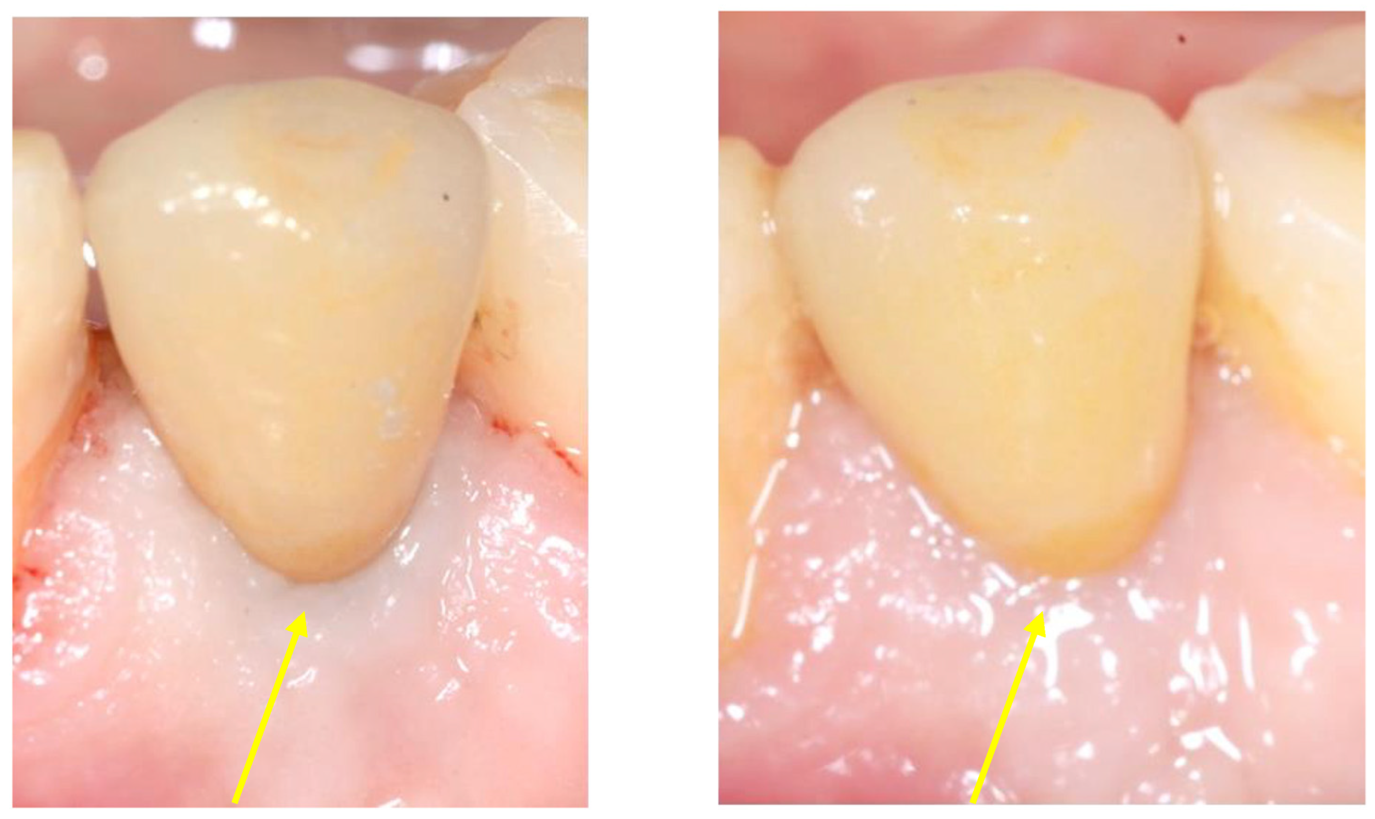
Figure 5. Digital acquisition of the implant through a scanbody. Abutment positioning and provisional crown cementation. Please note the buccal soft tissue growth which occurred during the healing period (arrow). Absence of inflammation was observed.
Prosthesis 07 00117 g005
Figure 6. Digital workflow of the cemented crown. Slight soft tissue compression was provided to the provisional crown, which was left positioned for 6 months.
Prosthesis 07 00117 g006
Figure 7. Definitive crown cementation and follow-up after 6 months from loading. Note the soft tissue improvement during time, demonstrated by the disappearance of the grayish contour due to the thickening of the soft tissues. PES improved from 8 (cementation) to 10 (12 months follow-up). Arrows show the improvement of soft tissue texture and color.
Prosthesis 07 00117 g007
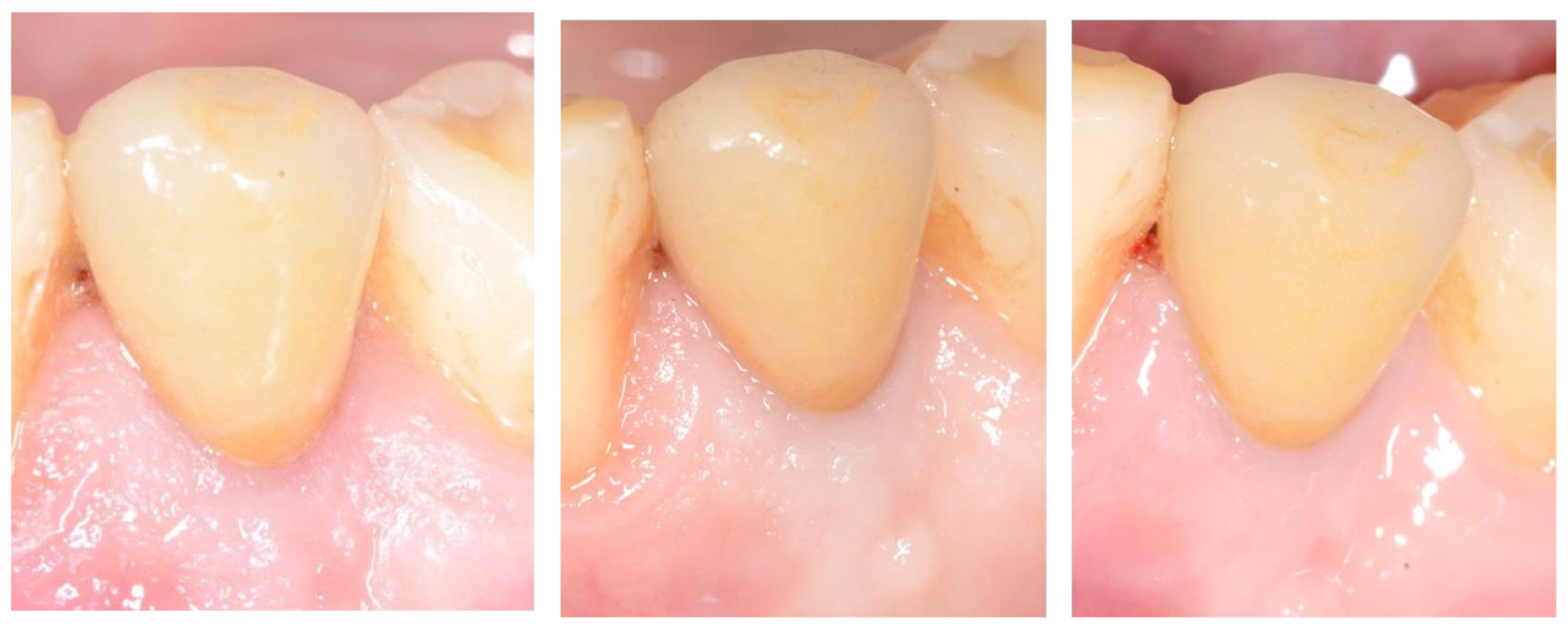
Figure 8. Intraoral images taken during the hygienic recalls at 18 months, 24 months and 36 months from implant insertion. The soft tissues remained healthy and stable.
Prosthesis 07 00117 g008
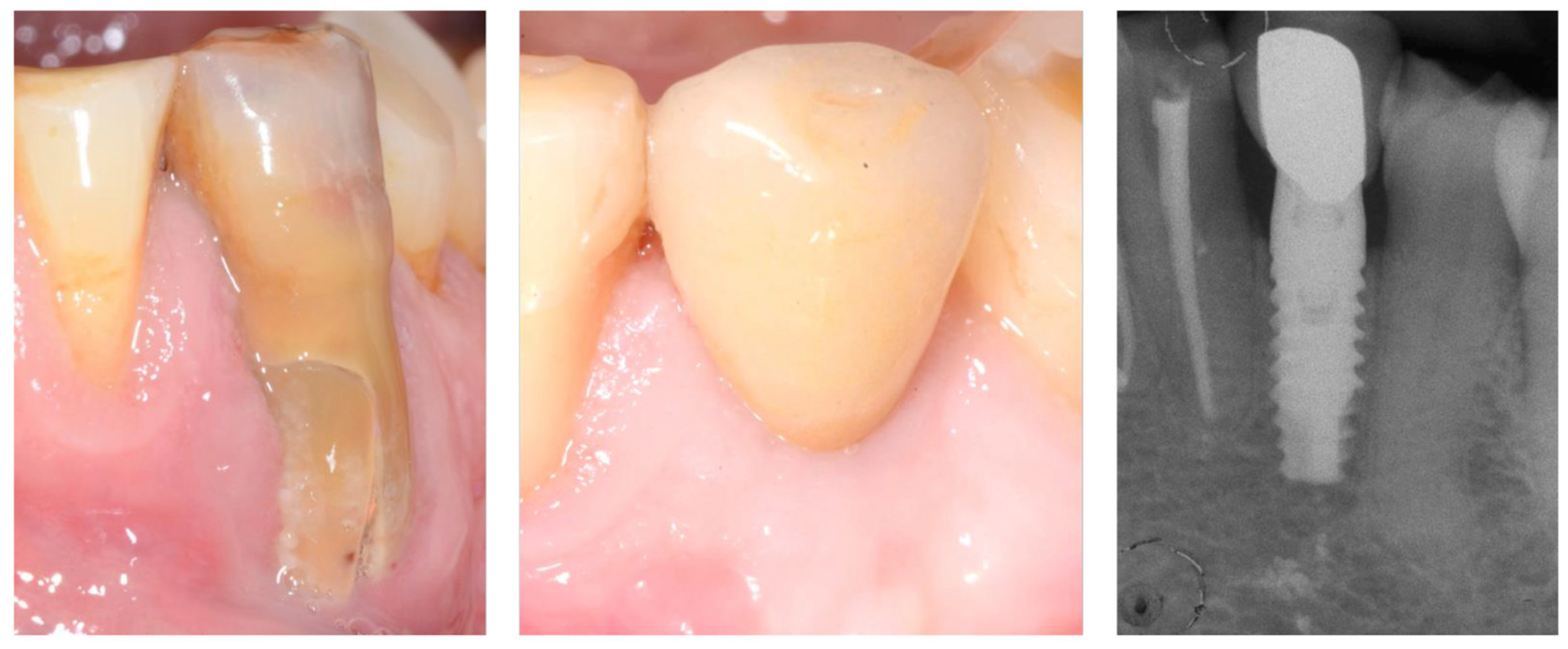
Figure 9. Preoperative view and last follow-up (image and periapical radiographs) (72 months from insertion). Hard and bone tissues were defined as healthy as no bone loss was radiographically detected and tissues showed no signs of inflammation. Final PES was 13.
Prosthesis 07 00117 g009
Table 1. Pink Esthetic Score (PES) Parameters with Endpoints.
QParameterScoring (0–2)Preoperative
(Before Extraction)
9 Months12 Months72 Months
Q1Mesial Papilla0 = absent, 1 = incomplete, 2 = complete2112
Q2Distal Papilla0 = absent, 1 = incomplete, 2 = complete2111
Q3Soft tissue contour0 = poor match, 1 = moderate match, 2 = perfect match0122
Q4Soft tissue level0 = >2 mm discrepancy, 1 = 1–2 mm discrepancy, 2 = <1 mm discrepancy0112
Q5Alveolar process deficiency0 = poor match, 1 = moderate match, 2 = perfect match0112
Q6Soft Tissue Texture0 = poor match, 1 = moderate match, 2 = perfect match0122
Q7Soft Tissue Color 0 = poor match, 1 = moderate match, 2 = perfect match0222
Total12–14 Excellent outcome
9–11 Good outcome
6–8 Moderate outcome
0–5 Poor esthetic outcome
481013
Disclaimer/Publisher’s Note: The statements, opinions and data contained in all publications are solely those of the individual author(s) and contributor(s) and not of MDPI and/or the editor(s). MDPI and/or the editor(s) disclaim responsibility for any injury to people or property resulting from any ideas, methods, instructions or products referred to in the content.