Article

Retrospective Clinical Study of Resin Composite and Ceram-Ic Indirect Posterior Restorations up to 11 Years

by
Nikolina Spyropoulou
1,
Sofia Diamantopoulou
1,
Stavros Patrinos
2 and
Efstratios Papazoglou
1,*
1
Department of Operative Dentistry, National and Kapodistrian University of Athens, 2 Thivon Str., 11527 Athens, Greece
2
Private Practice, Loutraki, 20300 Corinthia, Greece
*
Author to whom correspondence should be addressed.
Prosthesis 2025, 7(5), 108; https://doi.org/10.3390/prosthesis7050108 (registering DOI)
Submission received: 27 June 2025 / Revised: 17 August 2025 / Accepted: 20 August 2025 / Published: 25 August 2025

Abstract

Objective: The aim of this study was to evaluate and compare the clinical behavior of indirect onlays/overlays made of lithium disilicate and composite resin and to investigate risk factors associated with restoration failures. Methods: 112 indirect partial coverage posterior restorations (onlays and overlays) placed in 51 adult patients between January 2014 and December 2020 were examined. The restorations were evaluated using selected FDI criteria (color match, surface gloss, anatomic form, fracture of restorative material, tooth cracks and fractures, marginal discoloration, marginal integrity and recurrence of initial pathology). The survival of the restorations was analyzed using Kaplan–Meier method. Risk factors (type of restoration, type of tooth, tooth vitality, smoking) were investigated using Cox regression analysis. Risk estimation was conducted for each evaluated criterion (p < 0.05). Results: For composite restorations, the estimated survival rate was 94.2% after 5 years, dropping to 74.3% in 7.9 years and continued falling to less than 60% after 8.2 years. On the contrary, for lithium disilicate restorations the estimated survival rate was 90.9% after 5 years, dropped to 85.2% after 5.5 years remaining stable thereafter. Lithium disilicate onlays demonstrated significantly better performance than lithium disilicate overlays. Cox regression analysis did not reveal any significant association between the survival of the indirect partial restorations and restoration material, tooth type, restoration type and history of endodontic treatment. However, smoking was found to be a statistically significant risk factor (p < 0.05). Conclusions: Lithium disilicate and composite indirect restorations exhibited comparable survival rates at the early observation period. However, lithium disilicate partial coverage restorations demonstrated more favorable clinical behavior compared to composite in the long term, with statistical significance observed in ceramic vs. composite onlays.

1. Introduction

Extensive restorations of posterior teeth have long presented a clinical challenge for dentists. The choice of restoration type is often influenced by clinical judgment, practitioner experience, and individual skill. Partial-coverage posterior restorations—such as onlays, overlays, and vonlays (veneer-onlays)—offer the advantage of preserving healthy dental tissue [1]. In recent years, advancements in dental materials, coupled with increasing patient demand for esthetics, have contributed to the widespread adoption of esthetic materials for posterior tooth restorations, primarily composite resins and ceramics.
Numerous studies [2,3,4,5,6,7,8,9,10,11,12,13] have evaluated the longevity of indirect posterior partial-coverage restorations fabricated from ceramic or composite materials. While most investigations assess each material independently, some studies have compared the clinical performance of composite and ceramic partial-coverage posterior restorations.
A short-term (2-year) randomized controlled clinical trial using a split-mouth design [14] compared lithium disilicate and hybrid resin nano-ceramic CAD/CAM onlay restorations, reporting a 90% survival rate for both materials after 2 years of clinical follow-up. In a 10-year prospective clinical study [15] evaluating lithium disilicate and composite resin partial indirect restorations with deep margin elevation, an overall survival rate of 95.9% was observed, with no significant difference between the two materials. A retrospective study [16] examining the longevity of lithium disilicate ceramics and laboratory-processed resin-based composite inlays/onlays/overlays reported survival rates of 96.8% and 84.9%, respectively, after a mean observation period of 7.8 ± 3.3 years. Notably, both materials demonstrated survival rates exceeding 98% during the first 6 years; however, composite restorations declined to 60% by the 15th year, while lithium disilicate maintained a high long-term survival rate (>95%).
One of the earliest systematic reviews and meta-analyses on this topic [17] concluded that there is limited evidence suggesting ceramics outperform composite materials for inlays in the short term (3 years). Another systematic review and meta-analysis [1] reported survival rates of 95% and 91% for feldspathic porcelain and glass-ceramics at the 5-and 10-year follow-ups, respectively. Specifically, feldspathic porcelain exhibited survival rates of 92% at 5 years and 91% at 10 years, while glass-ceramics showed survival rates of 96% and 93%, respectively. A meta-analysis of composite resin restorations was not feasible due to a lack of studies involving overlays, onlays, and inlays made from composite resin at the time of data collection. More recently, another systematic review and meta-analysis [18] concluded that lithium disilicate and indirect composite materials exhibit comparable short-term survival rates, based on medium-quality evidence.
Despite several clinical trials evaluating the longevity of ceramic and composite resin onlays, the current evidence remains inconclusive. Therefore, the objective of this clinical study is to compare the survival of indirect onlays and overlays fabricated from lithium disilicate and composite resin, and to assess the types of failures that may occur. The first null hypothesis is that there is no significant difference in the long-term survival of ceramic and resin composite indirect restorations. The second null hypothesis is that the evaluated risk factors (restorative material, restoration type, tooth type, history of endodontic treatment, and smoking status) do not significantly affect restoration survival.

2. Materials and Methods

This retrospective, non-interventional clinical study was conducted in accordance with research guidelines involving human subjects at the Postgraduate Clinic of the Department of Restorative Dentistry, Dental School, National and Kapodistrian University of Athens. The study protocol received approval from the Research Ethics Committee of the Dental School, National and Kapodistrian University of Athens (No. 629/23 February 2024).

2.1. Selection of Patients

A total of 185 indirect partial-coverage posterior restorations were placed in 76 adult patients at the Postgraduate Clinic of the Department of Restorative Dentistry, Dental School, National and Kapodistrian University of Athens. All restorations were performed by postgraduate students in the Restorative Dentistry program, under faculty supervision. Each patient received at least one onlay or overlay made from either composite resin or lithium disilicate. The evaluated restorations were placed between January 2014 and December 2020, providing a minimum observation period of 3 years and a maximum of 11 years.
All patients were contacted by telephone and invited to attend a follow-up examination, scheduled between January and June 2024. Fifty-one patients agreed to participate, while the remaining 25 either declined or could not be reached. Written informed consent was obtained from all participants prior to the clinical evaluation. Additionally, relevant data on the restored teeth—including restorative material used and date of delivery—were collected before the follow-up visit. Due to the retrospective design of the study and the extended observation period, various brands of composite materials, adhesives, and cements were used, depending on clinical indication and availability at the time of placement.
Exclusion criteria for study participation were as follows:
  • Presence of systemic health conditions affecting salivary flow or quality.
  • Teeth in the terminal stage of periodontal disease.
Restoration evaluations were performed using a mirror and a new sharp metal explorer (Reicodent, Tuttlingen, Germany). Radiographic imaging was not routinely used, except in cases where ethical considerations warranted it, such as the clinical diagnosis of carious lesions requiring intervention.

2.2. Evaluation of Restorations

Restorations were assessed by two examiners using the FDI World Dental Federation Clinical Criteria [19] for the evaluation of direct and indirect restorations. Examiners underwent calibration training to ensure agreement on the evaluation criteria, achieving an inter-examiner agreement level exceeding 90%. Any disagreements during data collection were resolved by consensus. The evaluated criteria included fracture of restorative material and retention, tooth cracks and fractures, and recurrence of the initial pathology. The overall rating for each restoration was determined following the completion of individual scores for each clinical criterion. Each criterion was scored by both examiners on a 1–5 scale. A score of 4 or 5 was considered a failure. The selected criteria are further detailed in Table 1.
Tooth-related risk factors included tooth type (premolar vs. molar) and vitality status (vital vs. endodontically treated). Restoration-related factors included the restorative material (ceramic vs. composite), restoration extension (inlay vs. onlay/overlay), and restoration age (3–11 years). The patient-related risk factor considered was smoking status.

2.3. Statistical Analysis

Kaplan–Meier methodology was used to calculate the success rates of each restorative material over the observation period. Survival probabilities were also estimated for the two restoration types (onlays vs. overlays), tooth types (molars vs. premolars), pulp status (vital vs. endodontically treated), and smoking status (smokers vs. non-smokers). Cox regression analysis was conducted to investigate associations between restoration survival and the following variables: restorative material, restoration type, endodontic status, and smoking. The likelihood ratio (LR) test was used in the Cox model. All variables were treated as categorical. Statistical analyses were performed using STATA software (Version 12.1; StataCorp, College Station, TX, USA).

3. Results

A total of 112 indirect partial-coverage restorations were placed in 51 patients, resulting in a response rate of 67.1%. The observation period ranged from 31 to 132 months, with a mean follow-up duration of 69 months. Participant characteristics, including gender, restoration type, tooth type, restorative material, pulp vitality, and smoking status, are presented in Table 2.

3.1. Failure Criteria and Overview

Restorations were classified as failures if they received an FDI score of 4 or 5 in any of the following categories: fracture and retention of the restorative material, tooth cracks or fractures, or recurrence of the initial pathology. The distribution of the scores of the indirect restorations in the study population according to the selected FDI criteria is presented in Table 3. According to these criteria, 33 out of 112 restorations (29.5%) were classified as failures (Table 4). Among the lithium disilicate group, five failures were observed—all in vital teeth restored with overlays. In contrast, 28 failures were recorded among the composite restorations, affecting both vital and endodontically treated teeth and involving both onlays and overlays. No failures were observed prior to 39 months. Accordingly, 1-, 2-, and 3-year survival was 100%.

3.2. Material Performance

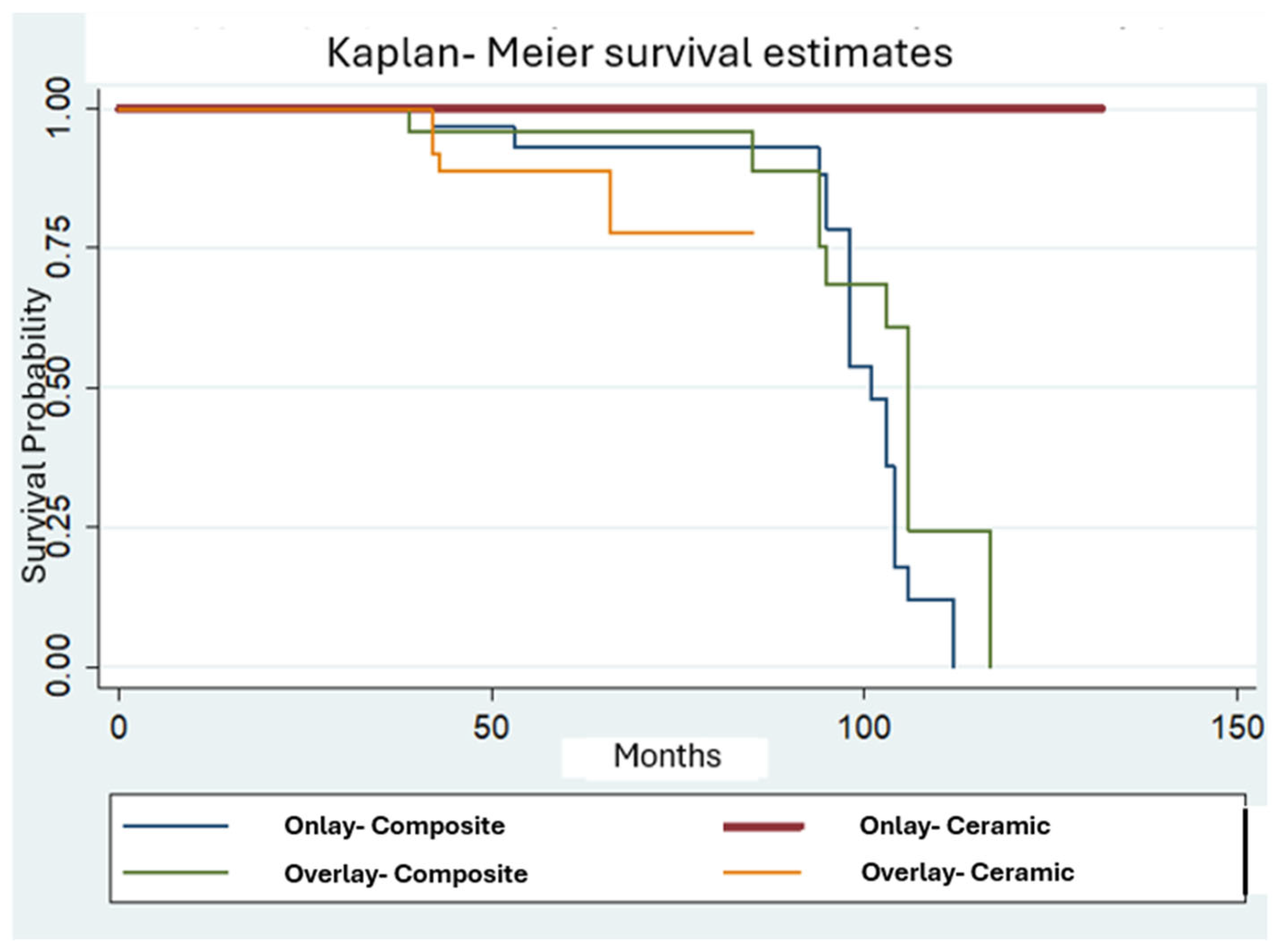
Kaplan–Meier survival analysis comparing lithium disilicate and composite indirect posterior restorations revealed that composite restorations had a 4-year survival rate of 96.3% and a 5-year survival rate of 94.2%, which decreased to 91.5% at 7 years and further decreased at 74.3% at 95 months and fell below 60% beyond 98 months. In contrast, ceramic restorations demonstrated a 4-year survival rate of 90.8% and a 5-year survival rate of 90.9%, which declined slightly to 85.2% after 5.5 years and remained stable thereafter (Figure 1). Although ceramic restorations tended to exhibit more favorable long-term performance, the difference was not statistically significant, likely due to the lower number of ceramic restorations. However, when considering both material and restoration type, ceramic onlays significantly outperformed composite onlays across the observation period (p < 0.005) (Figure 2).

3.3. Restoration Type

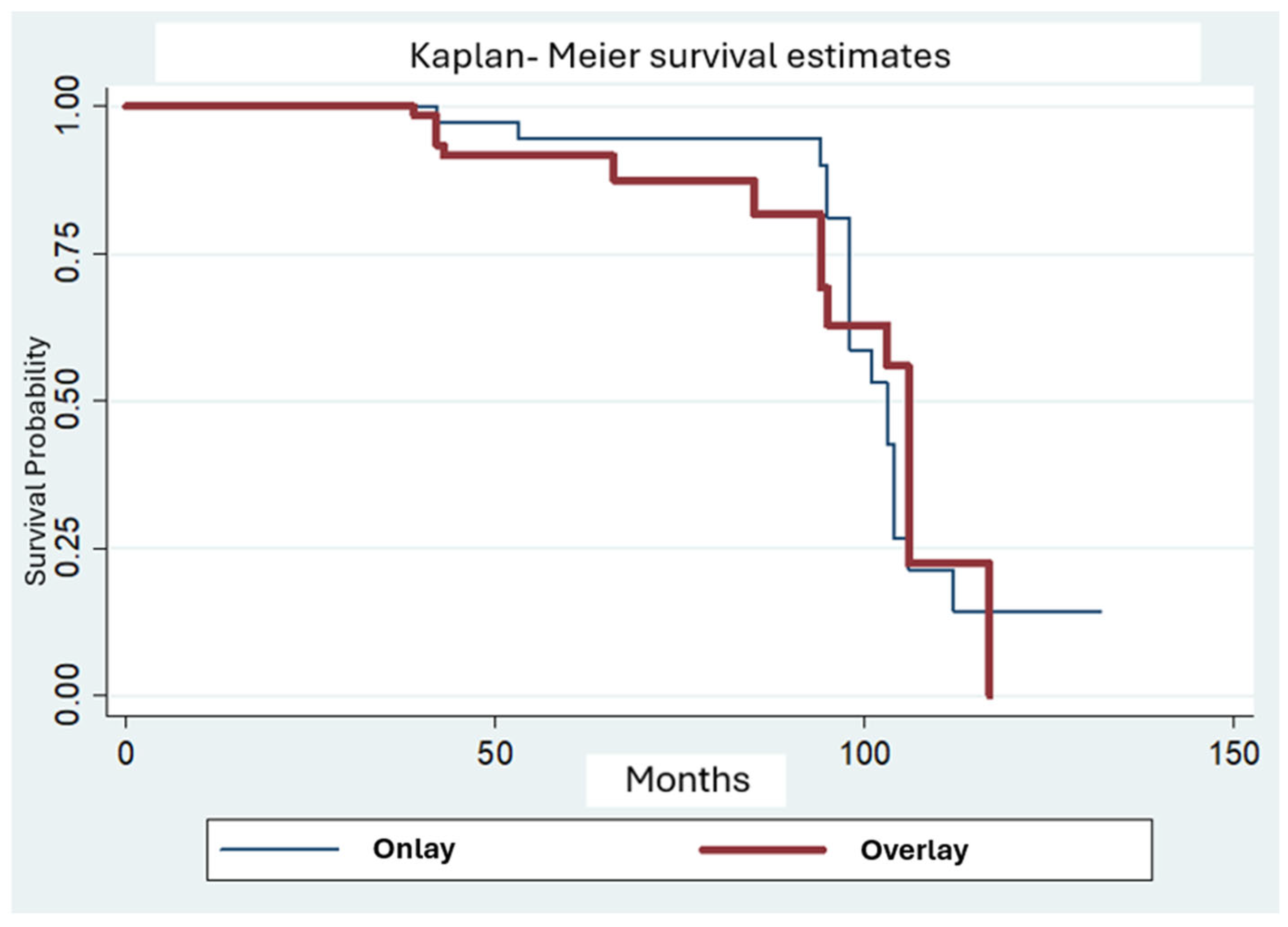
Kaplan–Meier analysis comparing onlays and overlays indicated that onlays initially demonstrated slightly better survival but experienced a more pronounced decline over time. Onlays had a 4-year survival rate of 97.4% and a 5-year survival rate of 94.6%, which remained stable at 7 years, decreased to 58.5% at 8 years and dropped below 50% by 8.5 years. Overlays showed a 4-year and 5-year survival rate of 91.6%, which declined to 81.7% at 7 years and declined further under 60% at 8.5 years (Figure 3). The difference between restoration types was not statistically significant.

3.4. Tooth Type

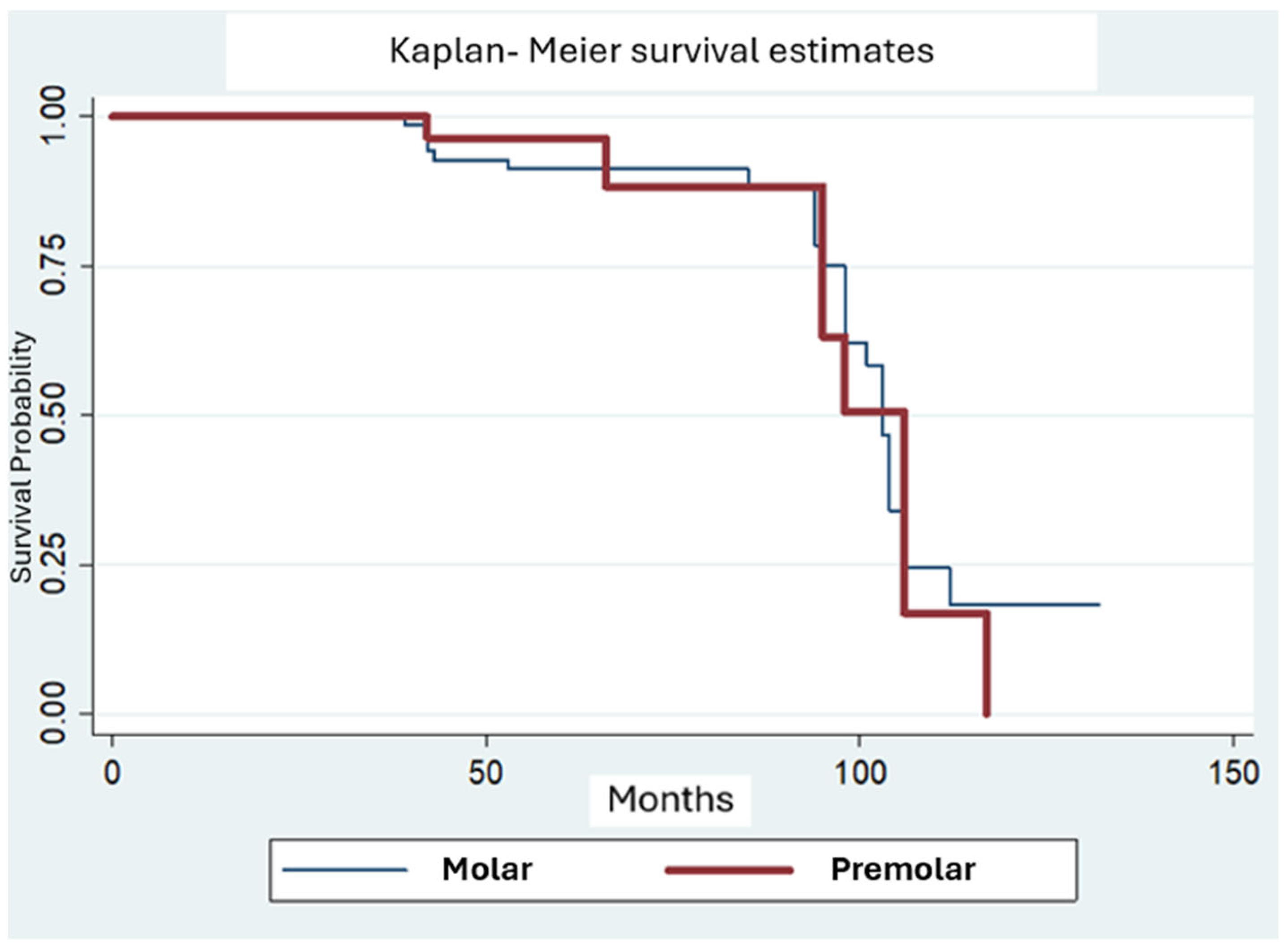
Kaplan–Meier curves comparing molars and premolars showed that molars had a 4-year survival rate of 92.8% and a 5-year survival rate of 91.2%. The 7-year survival rate was 88.2%, declined below 70% after 8 years and dropped to under 50% by 102 months. Premolars exhibited slightly lower initial survival (4-year survival rate: 96.4%, 5-year survival rate <90%) and a comparable long-term decline, falling below 50% after 8 years (Figure 4). Differences in survival rates between molars and premolars were not statistically significant.

3.5. Pulp Vitality

Kaplan–Meier analysis was also performed based on pulp vitality (Figure 5). Restorations on vital teeth had a 4-year and 5-year survival rate of 92.8%, which decreased to 88% by year 7 and declined to below 50% by 8.5 years. Endodontically treated teeth showed a 4-year survival rate of 100%, a 5-year survival rate of 92.3%, which remained stable up to 7 years. However, they experienced a steeper decline, reaching 55% at around 8 years. No statistically significant difference was found between the two groups.

3.6. Smoking Status

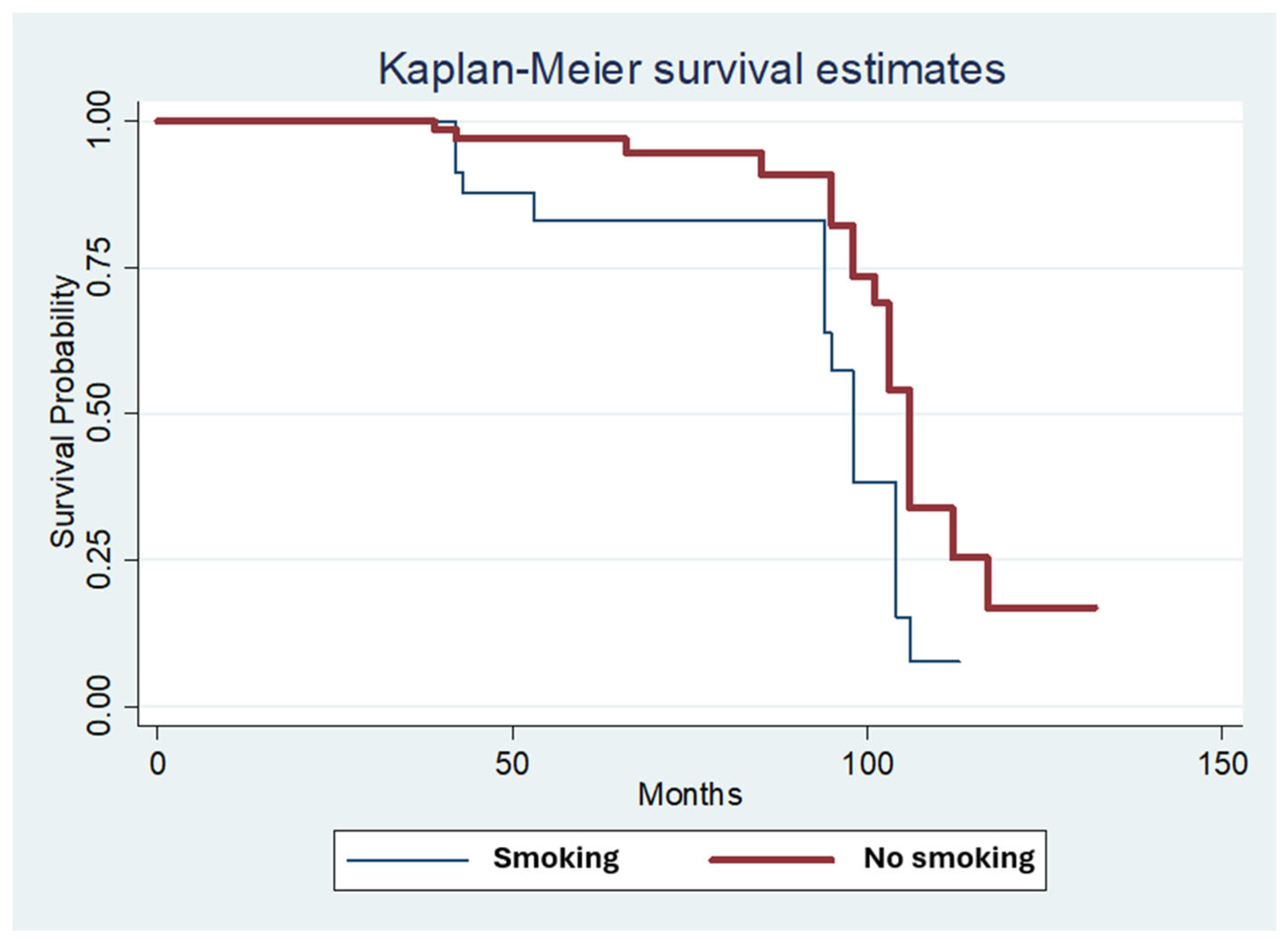
Smoking status was the only covariate significantly associated with restoration survival (p = 0.019; Figure 6, Table 4). Patients who smoked exhibited notably lower restoration longevity over time, underscoring the negative clinical impact of smoking.

3.7. Multivariate Analysis

Cox regression analysis failed to identify statistically significant associations between restoration survival and material type (p = 0.625), restoration type (p = 0.993), tooth type (p = 0.456), or endodontic status (p = 0.967). However, smoking remained a significant predictor of reduced restoration survival (p < 0.05) (Table 5).

4. Discussion

This retrospective clinical study evaluated the long-term performance of ceramic and composite indirect restorations. Although ceramics tended to perform better, the difference was not statistically significant, so the first null hypothesis was accepted. Among the examined risk factors, only smoking showed a statistically significant negative effect on restoration survival, leading to partial rejection of the second null hypothesis.
In this study, indirect composite restorations had a survival rate of 94.2% at 5 years, decreasing to 74.3% at 7.9 years and below 60% by 8.17 years. Material fracture and caries were the primary causes of failure. These findings align with previous retrospective studies: one reported a 91.1% 5-year survival rate [4], with secondary caries—often interproximal—being the main cause of failure, and another found an 81% 10-year survival rate [6], with fractures as the predominant issue. For ceramic restorations, survival was 90.9% at 5 years, 85.2% at 5.5 years, and remained unchanged thereafter. Ceramic fracture was the only failure type observed. These results are consistent with earlier research: a 4-year study reported a 92.7% survival rate for ceramic onlays on molars and premolars [10]; a long-term study found rates of 92.3% at 10 years and 83.8% at 14 years [11], with fractures and chipping most common; another study reported 91.8% survival after 23.5 years [13]; and a separate analysis documented rates of 96.3%, 91.5%, and 67.4% at 2, 4, and 6 years, respectively [12].
Although many clinical trials have examined ceramic and composite indirect restorations separately [2,3,4,5,6,7,8,9,10,11,12,13,14], long-term comparative data remain limited. Consistent with our findings, previous studies [14,15,16] reported no significant differences in survival between the two materials. In the present study, composite restorations declined from 74.3% survival at nearly 8 years to below 60% at 98 months, whereas ceramics decreased to 85.2% at 5.5 years and then remained stable. Thus, ceramics generally demonstrated more favorable long-term performance. Kaplan–Meier analysis by material and restoration type revealed a significant advantage for ceramic onlays over composite onlays. This agrees with a previous retrospective study [16], where annual failure rates for LiSi and resin composite restorations were 0.2% and 1%, respectively. That study also found survival above 98% for both materials in the first 6 years, dropping to 60% for composite by year 15, while LiSi maintained >95% long-term survival. Similarly, a systematic review and meta-analysis [1] reported a higher failure risk for composites versus ceramics, although meta-analysis of composites was limited due to insufficient overlay/onlay/inlay studies. Another review [18] found short-term survival rates for LiSi and indirect composites to be comparable, with no significant advantage for leucite ceramics or composites in short- to medium-term follow-up. A larger sample size in our study might have influenced these results.
Cox regression analysis showed no significant association between restoration survival and tooth type (molar/premolar), consistent with previous studies [1,7,8,9,11,12,13]. However, Ravasini et al. [6] reported a lower failure risk in premolars, likely due to reduced occlusal loads compared to molars. Similarly, Murgueitio et al. [20] found second molars to be five times more failure-prone than first molars, with no failures in premolars, while Naeselius et al. [10] observed failures only in molars after 4 years, without statistical significance.
Restoration type (onlay/overlay) also did not influence survival, aligning with prior findings [4,6]. Several studies [2,8,11] report similar results for onlays and inlays. In contrast, one prospective trial [3] found an eightfold higher failure risk for overlays, primarily due to fractures occurring shortly after placement—possibly linked to the mechanical limitations of composite materials, particularly in full cuspal coverage restorations without immediate dentin sealing.
Tooth vitality did not significantly affect survival rates in this study, consistent with previous findings [3,7,15]. It should be noted, however, that the number of endodontically treated teeth included in the study was rather low (16 out of 112). A 15-year retrospective study [9] reported higher failure rates in non-vital teeth (39.0%) than in vital teeth (20.9%), attributed to reduced retention area and differences in dentin substrate. to which the hydrophilic primers were applied. Dentin is hydrophilic in vital teeth vs. more sclerotic less-water containing endodontically treated teeth. In addition, in another study [21] it was found that non-vital teeth had significantly higher possibility of failure than vital teeth, possibly because of higher cuspal deflection and reduced stiffness of the non-vital teeth due to endodontic access and restorative procedures. According to a systematic review and metanalyses [21] indirect partial restorations on endodontically treated teeth showed acceptable clinical performance for a medium follow-up period of 2 to 4 years. Failures increased considerably after 7 years and up to 12–30 years. However, most failures were restorable, preventing tooth loss. Another systematic review [1] reported that the chance of failure was 80% lower in vital teeth compared to endodontically treated teeth, reinforcing tooth vitality as a key factor for indirect restoration survival.
In this study, Cox regression analysis revealed a statistically significant influence of smoking on the restorations’ survival (p < 0.05). This finding is novel and not easily explained clinically or biologically. While smoking is known to affect periodontal health and implant survival, its direct impact on indirect posterior restorations is less clear. Possible indirect mechanisms include an increased risk of caries in smokers, as suggested by a systematic review, although the overall evidence is weak [22]. Behavioral factors, such as reduced dental care attendance and poorer oral hygiene among smokers, may also contribute. The present findings align with a study where smoking nearly doubled the failure risk for indirect composite restorations, though not reaching full statistical significance. In contrast, another study [11] found no difference for ceramic restorations, possibly due to material differences. In vitro research [23] indicates composite resins are more susceptible to cigarette effects than ceramics, suggesting ceramics may maintain better color stability in smokers. In this study, no restoration failed due to discoloration. Further clinical research is needed to confirm smoking as a long-term risk factor for indirect restoration survival.
Several limitations should be noted. As this was a retrospective study, it was not possible to compare the current clinical quality of restorations—placed up to 11 years ago—with their initial state, limiting assessment of changes over time. The participant recall rate was 67.1%, introducing potential bias, as respondents may have been more motivated to maintain oral health. Additionally, varied restorative materials, adhesives, and cements were used over the extended observation period, preventing consistent data collection and direct material comparisons. As a result, all patients were not treated with the same restorative materials, bonding protocols or resin cements. Thus, it was not feasible to collect consistent or complete data regarding the specific materials used in each case. It is not possible therefore to make comparisons and draw conclusions on the effect of the materials used.
Despite these limitations, the study has strengths. All restorations were examined by two independent and calibrated dentists, minimizing examiner bias. Moreover, the patient sample was representative of the population since no exclusion criteria were applied to the recruited participants. The restorations have been placed by post-graduate students with various levels of experience demonstrated a good clinical performance and lifespan, comparable with the findings of research conducted by experienced operators.

Clinical Relevance

This study highlights the long-term performance of indirect partial coverage restorations in posterior teeth. Both composite and lithium disilicate materials provided acceptable survival rates over time, with lithium disilicate onlays showing significantly better outcomes compared to composite onlays. The results suggest that restoration material and type of restoration may influence longevity, especially in the case of onlays, where ceramics offer superior durability. However, factors such as pulp vitality and tooth type did not significantly affect survival. These findings support the clinical use of lithium disilicate onlays when long-term stability is a priority, while composite restorations remain a viable option in suitable cases.

5. Conclusions

Within the limitations of this retrospective study, indirect partial coverage restorations showed overall satisfactory survival rates over an extended follow-up period. Lithium disilicate, showed superior long-term performance compared to composites, with statistical significance observed only in ceramic vs. composite onlays. Restoration type, tooth type, and pulp vitality had no significant effect on survival, whereas smoking emerged as a significant risk factor warranting further investigation in future studies.

Author Contributions

Conceptualization, S.D. and E.P.; methodology, S.D., N.S. and E.P.; software, S.P.; validation, S.D., S.P. and E.P.; formal analysis, S.D. and S.P.; investigation, N.S. and S.D.; resources, S.D. and E.P.; data curation, N.S. and S.D.; writing—original draft preparation, N.S. and S.D.; writing—review and editing, S.D. and E.P.; visualization, N.S., S.D. and S.P.; supervision, S.D. and E.P.; project administration, S.D. and E.P. All authors have read and agreed to the published version of the manuscript.

Funding

This research received no external funding.

Institutional Review Board Statement

This retrospective non-interventional clinical study was carried out according to research guidelines involving human subjects at the postgraduate clinic of the Department of Restorative Dentistry (Dental School, National and Kapodistrian University of Athens). The study protocol was approved by the Research Ethics Committee of the Dental School of National and Kapodistrian University of Athens (No. 629/23 February 2024).

Informed Consent Statement

Informed consent was obtained from all subjects involved in the study.

Data Availability Statement

The original contributions presented in this study are included in the article. Further inquiries can be directed to the corresponding author(s).

Conflicts of Interest

The authors declare no conflict of interest.

References

  1. Morimoto, S.; Rebello de Sampaio, F.B.W.; Braga, M.M.; Sesma, N.; Özcan, M. Survival rate of resin and ceramic inlays, onlays, and overlays: A systematic review and meta-analysis. J. Dent. Res. 2016, 95, 985–994. [Google Scholar] [CrossRef]
  2. Barabanti, N.; Preti, A.; Vano, M.; Derchi, G.; Mangani, F.; Cerutti, A. Indirect composite restorations luted with two different procedures: A ten years follow up clinical trial. J. Clin. Exp. Dent. 2015, 7, e54. [Google Scholar] [CrossRef]
  3. Özcan, M. Clinical Performance of Posterior Indirect Resin Composite Restorations with the Proximal Box Elevation Technique: A Prospective Clinical Trial up to 3 Years. J. Adhes. Dent. 2024, 26, 19–30. [Google Scholar]
  4. D’Arcangelo, C.; Zarow, M.; De Angelis, F.; Vadini, M.; Paolantonio, M.; Giannoni, M.; D’Amario, M. Five-year retrospective clinical study of indirect composite restorations luted with a light-cured composite in posterior teeth. Clin. Oral Investig. 2014, 18, 615–624. [Google Scholar] [CrossRef] [PubMed]
  5. Hofsteenge, J.W.; Fennis, W.M.M.; Kuijs, R.H.; Özcan, M.; Cune, M.S.; Gresnigt, M.M.; Kreulen, C.M. Clinical survival and performance of premolars restored with direct or indirect cusp-replacing resin composite restorations with a mean follow-up of 14 years. Dent. Mater. 2023, 39, 383–390. [Google Scholar] [CrossRef] [PubMed]
  6. Ravasini, F.; Bellussi, D.; Pedrazzoni, M.; Ravasini, T.; Orlandini, P.; Meleti, M.; Bonanini, M. Treatment Outcome of Posterior Composite Indirect Restorations: A Retrospective 20-Year Analysis of 525 Cases with a Mean Follow-Up of 87 Months. Int. J. Periodontics Restor. Dent. 2018, 38, 655–663. [Google Scholar] [CrossRef]
  7. Van den Breemer, C.R.; Buijs, G.J.; Cune, M.S.; Özcan, M.; Kerdijk, W.; Van der Made, S.; Gresnigt, M.M. Prospective clinical evaluation of 765 partial glass-ceramic posterior restorations luted using photo-polymerized resin composite in conjunction with immediate dentin sealing. Clin. Oral Investig. 2021, 25, 1463–1473. [Google Scholar] [CrossRef] [PubMed]
  8. Frankenberger, R.; Taschner, M.; Garcia-Godoy, F.; Petschelt, A.; Krämer, N. Leucite-reinforced glass ceramic inlays and onlays after 12 years. J. Adhes. Dent. 2008, 10, 393–398. [Google Scholar]
  9. van Dijken, J.W.; Hasselrot, L. A prospective 15-year evaluation of extensive dentin–enamel-bonded pressed ceramic coverages. Dent. Mater. 2010, 26, 929–939. [Google Scholar] [CrossRef] [PubMed]
  10. Naeselius, K.; Arnelund, C.F.; Molin, M.K. Clinical evaluation of all-ceramic onlays: A 4-year retrospective study. Int. J. Prosthodont. 2008, 21, 40–44. [Google Scholar]
  11. Strasding, M.; Sebestyén-Hüvös, E.; Studer, S.; Lehner, C.; Jung, R.E.; Sailer, I. Long-term outcomes of all-ceramic inlays and onlays after a mean observation time of 11 years. Quintessence Int. 2020, 51, 566–576. [Google Scholar] [PubMed]
  12. Archibald, J.J.; Santos Jr, G.C.; Santos, M.J.M.C. Retrospective clinical evaluation of ceramic onlays placed by dental students. J. Prosthet. Dent. 2018, 119, 743–748. [Google Scholar] [CrossRef]
  13. Krug, R.; Droste, L.; Schreiber, C.; Reichardt, E.; Krastl, G.; Hahn, B.; Soliman, S. Long-term performance of ceramic in/-onlays vs. cast gold partial crowns–a retrospective clinical study. Clin. Oral Investig. 2024, 28, 298. [Google Scholar] [CrossRef]
  14. Saavedra, G.D.S.F.A.; Tribst, J.P.M.; Ramos, N.D.C.; Melo, R.M.D.; Rodrigues, V.A.; Ramos, G.F.; Bottino, M.A. Feldspathic and lithium disilicate onlays with a 2-year follow-up: Split-mouth randomized clinical trial. Braz. Dent. J. 2021, 32, 53–63. [Google Scholar] [CrossRef]
  15. Bresser, R.A.; Gerdolle, D.; Van den Heijkant, I.A.; Sluiter-Pouwels, L.M.A.; Cune, M.S.; Gresnigt, M.M.M. Up to 12 years clinical evaluation of 197 partial indirect restorations with deep margin elevation in the posterior region. J. Dent. 2019, 91, 103227. [Google Scholar] [CrossRef]
  16. Lempel, E.; Gyulai, S.; Lovász, B.V.; Jeges, S.; Szalma, J. Clinical evaluation of lithium disilicate versus indirect resin composite partial posterior restorations–A 7.8-year retrospective study. Dent. Mater. 2023, 39, 1095–1104. [Google Scholar] [CrossRef]
  17. Chabouis, H.F.; Faugeron, V.S.; Attal, J.P. Clinical efficacy of composite versus ceramic inlays and onlays: A systematic review. Dent. Mater. 2013, 29, 1209–1218. [Google Scholar] [CrossRef]
  18. Bresser, R.A.; Hofsteenge, J.W.; Wieringa, T.H.; Braun, P.G.; Cune, M.S.; Özcan, M.; Gresnigt, M.M.M. Clinical longevity of intracoronal restorations made of gold, lithium disilicate, leucite, and indirect resin composite: A systematic review and meta-analysis. Clin. Oral Investig. 2023, 27, 4877–4896. [Google Scholar] [CrossRef]
  19. Hickel, R.; Peschke, A.; Tyas, M.; Mjör, I.; Bayne, S.; Peters, M.; Hiller, K.A.; Randall, R.; Vanherle, G.; Heintze, S.D. FDI World Dental Federation: Clinical criteria for the evaluation of direct and indirect restorations—update and clinical examples. Clin. Oral Investig. 2010, 14, 349–366. [Google Scholar] [CrossRef] [PubMed]
  20. Murgueitio, R.; Bernal, G. Three-year clinical follow-up of posterior teeth restored with leucite-reinforced IPS Empress onlays and partial veneer crowns. J. Prosthodont. Implant. Esthet. Reconstr. Dent. 2012, 21, 340–345. [Google Scholar] [CrossRef] [PubMed]
  21. Fan, J.; Xu, Y.; Si, L.; Li, X.; Fu, B.; Hannig, M. Long-term clinical performance of composite resin or ceramic inlays, onlays, and overlays: A systematic review and meta-analysis. Oper. Dent. 2021, 46, 25–44. [Google Scholar] [CrossRef] [PubMed]
  22. Benedetti, G.; Campus, G.; Strohmenger, L.; Lingstrom, P. Tobacco and dental caries: A systematic review. Acta Odontol. Scand. 2013, 71, 363–371. [Google Scholar] [CrossRef] [PubMed]
  23. Vohra, F.; Andejani, A.F.; Alamri, O.; Alshehri, A.; Al-Hamdan, R.S.; Almohareb, T.; Abduljabbar, T. Influence of electronic nicotine delivery systems (ENDS) in comparison to conventional cigarette on color stability of dental restorative materials. Pak. J. Med. Sci. 2020, 36, 993. [Google Scholar] [CrossRef] [PubMed]
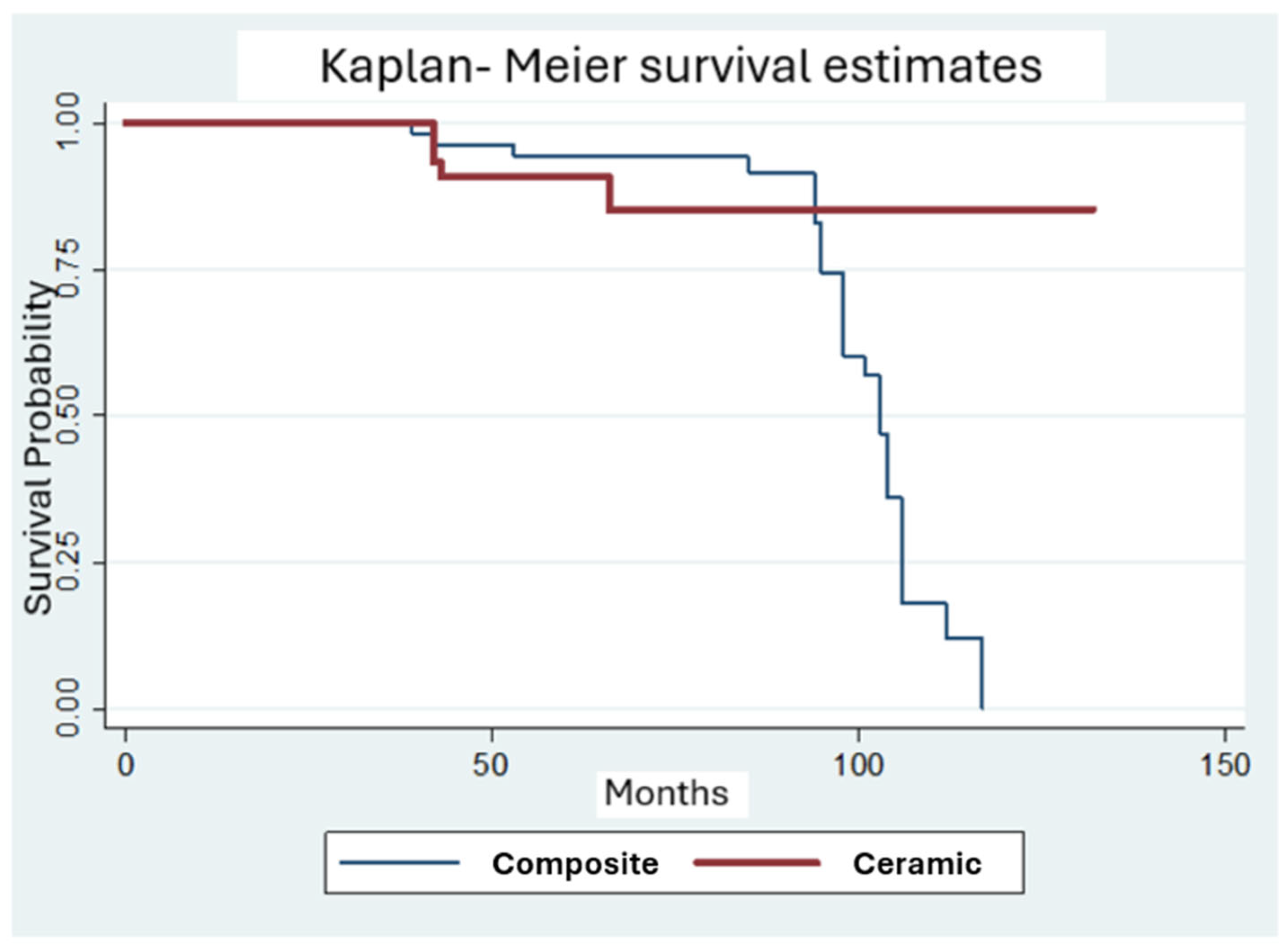
Figure 1. Kaplan–Meier survival rates for indirect partial coverage restorations according to the restoration material (composite vs. ceramic).
Prosthesis 07 00108 g001
Figure 2. Kaplan–Meier survival rates for indirect partial coverage restorations according to the restoration type and the restoration material (composite onlay vs. composite overlay vs. ceramic onlay vs. ceramic overlay).
Prosthesis 07 00108 g002
Figure 3. Kaplan–Meier survival rates for indirect partial coverage restorations according to the restoration type (onlay vs. overlay).
Prosthesis 07 00108 g003
Figure 4. Kaplan–Meier survival rates for indirect partial coverage restorations according to the tooth type (molar vs. premolar).
Prosthesis 07 00108 g004
Figure 5. Kaplan–Meier survival rates for indirect partial coverage restorations according to the presence of endodontic treatment (vital pulp vs. endodontic treatment).
Prosthesis 07 00108 g005
Figure 6. Kaplan–Meier survival rates for indirect partial coverage restorations according to smoking (smoking vs. no smoking).
Prosthesis 07 00108 g006
Table 1. Selected FDI criteria.
Fracture of Restorative Material and Restoration RetentionTooth Cracks and FracturesRecurrence of Initial Pathology
1. No fractures/cracks1. Complete integrity or small marginal fracture or hairline crack on enamel (<150 μm)1. No secondary or primary caries
2. Small hairline crack2. Small marginal fracture or hairline crack on enamel (<150 μm)2. Small/localized area of demineralization or erosion or abfraction
3. Larger hairline cracks and/or limited material chipping not affecting marginal quality or proximal contacts3.1 Marginal enamel defect or crack (<250 μm)
3.2 Enamel chipping
3.3 Multiple cracks
3. Larger areas or demineralization or erosion or abfraction (on dentine not exposed)
Only preventive measures
4.1. Material chip fractures affecting marginal quality and/or proximal contacts
4.2. Bulk fractures affecting less than half of the restoration
4.1. Enamel marginal gap >250 μm or dentin or base exposure
4.2. Large cracks >250 μm probe penetrates
4.3 Large enamel chipping or wall fracture
4. Cavitation caries and suspected undermining caries. Erosion and/or abfraction in dentine that can be repaired
5. Partial or complete loss of restoration or multiple fractures5. Cusp or tooth fracture5. Deep caries or exposed dentine, not accessible for repair
Table 2. Details about the distribution of the indirect restorations in the study population in relation to tooth type, restoration type, material, pulp vitality and smoking habits of patients.
MaleFemaleMolarPremolarOnlayOverlayCompositeCeramicEndoVitalSmokingNo Smoking
Number179579334072625016963676
Total112112112112112112
Table 3. Distribution of the scores of the indirect restorations in the study population according to the selected FDI criteria.
FDI CFDI Criteria/Score 12345TOTAL
Fracture of restorative material and restoration retention 8334517112
Recurrence of initial pathology 97-1311112
Tooth cracks and fractures 110-1-1112
Table 4. Data of failed restorations.
Time
Interval
n%
Failures
CariesFractureCompositeLiSiOnlayOverlayMolarPremolarVitalEndo
0–4 y618.21542155160
4–6 y26.11111111111
6–8 y721.21570345252
8–10 y1854.5810180135144153
>10 y000000000000
Table 5. Results of the Cox regression analysis for the success and the survival of the restorations with different covariates (p < 0.05).
CovariatesSignificance Values for Survival
Restoration material0.625
Restoration type0.993
Tooth0.456
Endodontic treatment0.967
Smoking0.019
Disclaimer/Publisher’s Note: The statements, opinions and data contained in all publications are solely those of the individual author(s) and contributor(s) and not of MDPI and/or the editor(s). MDPI and/or the editor(s) disclaim responsibility for any injury to people or property resulting from any ideas, methods, instructions or products referred to in the content.