Next Issue
Volume 13, July
Previous Issue
Volume 13, May


Biomedicines, Volume 13, Issue 6 (June 2025) – 242 articles

Cover Story (view full-size image): 
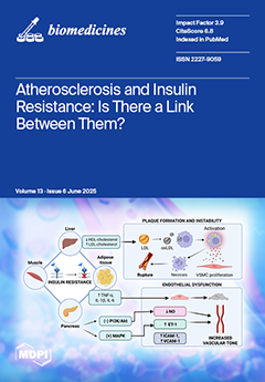
Cardiovascular disease is the leading global cause of death, with atherosclerosis responsible for 80% of cases, particularly in Eastern Europe, South Asia, and Latin America. Atherosclerosis involves lipid accumulation and chronic inflammation in arterial walls, driven by endothelial dysfunction and risk factors such as hypertension and dyslipidemia. Insulin resistance—impaired insulin-mediated glucose uptake in liver, muscle, and adipose tissue—often precedes diabetes and is now recognized as an independent predictor of atherosclerosis. Early detection of subclinical atherosclerosis and insulin resistance is possible through non-invasive imaging (coronary calcium scoring, carotid ultrasound) and surrogate indices such as HOMA-IR, TyG, Matsuda and triglyceride/HDL-cholesterol ratio. View this paper
  • Issues are regarded as officially published after their release is announced to the table of contents alert mailing list.
  • You may sign up for e-mail alerts to receive table of contents of newly released issues.
  • PDF is the official format for papers published in both, html and pdf forms. To view the papers in pdf format, click on the "PDF Full-text" link, and use the free Adobe Reader to open them.
Order results
Result details
Section
Select all
Export citation of selected articles as:
12 pages, 1336 KiB  
Article
Evaluating Genomic and Clinical Risk Factors for Alzheimer’s Disease in Individuals with Hypertension
by Elizabeth Kim, Kevin Zhang, Miski Abdi, Wei Tse Li, Ruomin Xin, Jessica Wang-Rodriguez and Weg M. Ongkeko
Biomedicines 2025, 13(6), 1508; https://doi.org/10.3390/biomedicines13061508 - 19 Jun 2025
Viewed by 358
Abstract
Background/Objectives: Alzheimer’s disease (AD) is a progressive neurodegenerative condition whose growing prevalence has become an increasingly important public health concern as the population ages. The lack of a definitive cure elevates the importance of identifying risk factors that are crucial for prevention efforts. [...] Read more.
Background/Objectives: Alzheimer’s disease (AD) is a progressive neurodegenerative condition whose growing prevalence has become an increasingly important public health concern as the population ages. The lack of a definitive cure elevates the importance of identifying risk factors that are crucial for prevention efforts. Hypertension (HTN) and obesity have emerged as two highly widespread, interrelated conditions that have independently been associated with AD risk. Despite extensive research into AD pathology, the impact of obesity in a hypertensive population is not well explored. This study aims to investigate how obesity and blood pressure control within a hypertensive population may interact with genomic risk and environmental factors to influence AD incidence. Methods: A retrospective cohort of matched AD and normal patients diagnosed with HTN and taking anti-HTN drugs (n = 1862) from the All of Us database was analyzed. In this hypertensive cohort, obesity was significantly associated with increased AD risk. Genome-wide association studies (GWASs) were conducted on hypertensive AD individuals (n = 1030) and identified six single nucleotide variants (SNVs) that were associated with AD development in this population. Results: Obesity and Area Deprivation Index, a measure of socioeconomic status, were significantly associated with elevated AD risk within the hypertensive cohort. GWAS analysis identified six SNVs significantly associated with AD development among the hypertensive cohort. Conclusions: Our findings suggest that among hypertensive individuals, comorbid obesity and the Area Deprivation Index confer greater AD risk. These results highlight the critical need for obesity prevention and management strategies as part of Alzheimer’s risk reduction efforts. Full article
(This article belongs to the Section Neurobiology and Clinical Neuroscience)
Show Figures

Figure 1

25 pages, 540 KiB  
Review
Malignancies in Celiac Disease—A Hidden Threat with Diagnostic Pitfalls
by Aleksandra Kubas and Ewa Małecka-Wojciesko
Biomedicines 2025, 13(6), 1507; https://doi.org/10.3390/biomedicines13061507 - 19 Jun 2025
Viewed by 394
Abstract
Celiac disease (CeD) is an autoimmune disorder that is triggered by gluten ingestion in genetically predisposed individuals. Untreated or poorly controlled CeD leads to various disease complications, such as malnutrition, osteoporosis, autoimmune diseases, or refractory celiac disease (RCD). Accumulating recent research has highlighted [...] Read more.
Celiac disease (CeD) is an autoimmune disorder that is triggered by gluten ingestion in genetically predisposed individuals. Untreated or poorly controlled CeD leads to various disease complications, such as malnutrition, osteoporosis, autoimmune diseases, or refractory celiac disease (RCD). Accumulating recent research has highlighted the association between CeD and the development of malignancies, particularly enteropathy-associated T-cell lymphoma (EATL) and small bowel carcinoma (SBC), which are neoplasms with extremely poor prognoses. Genetic alterations in the JAK1–STAT3 pathway and the high prevalence of microsatellite instability may be the main drivers of CeD-associated lymphomagenesis and small bowel oncogenesis and therefore could be an attractive therapeutic target to block cancer transformation. However, to date, the risk factors and exact mechanisms underlying malignancy development in patients with CeD remain unclear, and prospective cohort studies that include molecular profiling are needed. Moreover, current guidelines on the management of CeD do not provide standardized protocols for cancer surveillance—particularly regarding screening intervals, risk stratification, and monitoring strategies for high-risk patients such as those with RCD. This paper reviews the existing knowledge on malignancies in CeD, highlights diagnostic challenges, and discusses future perspectives on the early detection, monitoring, and treatment of CeD-associated neoplasms. Full article
(This article belongs to the Section Cancer Biology and Oncology)
Show Figures

Figure 1

21 pages, 3504 KiB  
Article
Genotype-Based Housing as a Potential Confounder in Studies Using Transgenic Mouse Models—Insight from the A53T Mouse Model of Parkinson’s Disease
by Olga Dubljević, Miodrag Dragoj, Milica Potrebić Stefanović, Maja Srbovan, Miloš Stanojlović and Željko Pavković
Biomedicines 2025, 13(6), 1506; https://doi.org/10.3390/biomedicines13061506 - 19 Jun 2025
Viewed by 295
Abstract
Background/Objectives: Environmental factors, including the differences in genotype-based housing (GbH), can act as confounding variables in studies using transgenic mouse models, potentially influencing experimental outcomes and limiting their reproducibility and translational value. Despite the widespread use of transgenic models in preclinical studies, [...] Read more.
Background/Objectives: Environmental factors, including the differences in genotype-based housing (GbH), can act as confounding variables in studies using transgenic mouse models, potentially influencing experimental outcomes and limiting their reproducibility and translational value. Despite the widespread use of transgenic models in preclinical studies, the extent to which housing conditions can affect the behavioral and molecular parameters of interest remains poorly understood. This study aims to investigate how different GbH conditions influence visuo-spatial memory and gene expression in the A53T mouse model (JAX006823) of Parkinson’s disease (PD) during the pre-motor phase. Methods: A53T+ transgenic male mice and their non-transgenic littermates (A53T−) were housed in either mixed-genotype (MGH) or single-genotype (SGH) environments from postnatal day (PND) 30, with C57BL/6J mice serving as the controls. A behavioral assessment using the Novel Object Recognition and Object Location Tests was conducted at PND 180, followed by a qPCR analysis of Iba1, Gfapα, Bdnf, Tnfα, Il-1β, and Il-6 expression in the medial prefrontal cortex and the hippocampus. Results: The variations in GbH influenced behavior and mRNA expression differently in the A53T+ and A53T− animals. Specifically, the A53T− mice in SGH environments displayed behavioral and molecular profiles similar to the C57BL/6J controls, while the same was not evident in the MGH environments. In the A53T+ mice, the mRNA expression of Iba1, Gfapα, Bdnf, and Tnfα was sensitive to variations in GbH, while memory impairment was not. Conclusions: This study highlights the importance of considering environmental factors in studies using transgenic animal models. The obtained data suggests that GbH can influence the parameters of interest in preclinical research, implicating the need for the optimization of future study designs. Full article
Show Figures

Figure 1

15 pages, 1291 KiB  
Systematic Review
Vaginal Cleansing and Post-Cesarean Infectious Morbidity? Updated Systematic Review and Meta-Analysis of Randomized Trials
by Marco La Verde, Marco Torella, Irene Iavarone, Rossella Molitierno, Antonio Cerillo, Margherita Casillo, Maria Maddalena Marrapodi, Mario Fordellone, Liliana Mariani, Chiara Melito, Barbara Gardella and Mattia Dominoni
Biomedicines 2025, 13(6), 1505; https://doi.org/10.3390/biomedicines13061505 - 19 Jun 2025
Viewed by 325
Abstract
Background: Endometritis, maternal fever and wound infection represent the most frequent post-cesarean complications. The aim of the present research was to evaluate the incidence of post-cesarean infections after vaginal cleansing. Materials and methods: The databases analyzed were MEDLINE, Scopus, EMBASE, CENTRAL, [...] Read more.
Background: Endometritis, maternal fever and wound infection represent the most frequent post-cesarean complications. The aim of the present research was to evaluate the incidence of post-cesarean infections after vaginal cleansing. Materials and methods: The databases analyzed were MEDLINE, Scopus, EMBASE, CENTRAL, Google Scholar, Clinicaltrials.gov and the Register of Controlled Trials. No language or geographical restrictions were applied. We included only randomized controlled trials that analyzed various vaginal antiseptic solutions to reduce postpartum endometritis. The terms employed were as follows: vaginal solution, cesarean section, endometritis, wound infection, chlorhexidine, povidone, metronidazole, cetrimide, and pregnancy. The PICO categorization was as follows: P—population: pregnant women; I—intervention: vaginal antiseptic; C—control: hands-off or routine care; O—outcome: post-cesarean endometritis, wound infection and postoperative fever; S—study design: randomized controlled trials. Results: A total of 32 articles, including 13,853 participants, were selected. The vaginal cleansing group showed a low incidence of endometritis. The chlorhexidine group had an OR of 0.56 (95% CI 0.45–0.70, p = 0.010). The povidone group had an OR of 0.47 (95% CI 0.37–0.59, p = 0.002). Considering maternal fever, 2598 patients from 5 studies in the chlorhexidine group were analyzed, alongside 6965 patients from 18 trials in the povidone group. The povidone group presented an Odds ratio of 0.47 (95% CI 0.38–0.57, p = 0.0001). A reduction in wound infection incidence was observed in the povidone group (OR = 0.59, 95% CI = 0.42–0.82, p < 0.05). Conclusions: Vaginal cleansing before cesarean section, particularly with povidone solutions, reduces the incidence of postoperative endometritis and maternal fever. Full article
(This article belongs to the Special Issue High-Risk Pregnancy, Labor and Delivery)
Show Figures

Figure 1

33 pages, 2546 KiB  
Review
A Review of the Relationship Between Insulin and Bone Health
by Sivasree Ravindran, Sok Kuan Wong, Nur-Vaizura Mohamad and Kok-Yong Chin
Biomedicines 2025, 13(6), 1504; https://doi.org/10.3390/biomedicines13061504 - 19 Jun 2025
Viewed by 401
Abstract
Insulin, a key hormone primarily involved in glucose metabolism, has emerged as a crucial modulator of bone metabolism. Increasing evidence suggests that insulin influences bone health, but its precise mechanism of action remains unestablished. This review explores the intricate relationship between insulin and [...] Read more.
Insulin, a key hormone primarily involved in glucose metabolism, has emerged as a crucial modulator of bone metabolism. Increasing evidence suggests that insulin influences bone health, but its precise mechanism of action remains unestablished. This review explores the intricate relationship between insulin and bone health, as well as elucidating the mechanism of action involved. Animal models of type 1 diabetes mellitus (T1DM) and type 2 diabetes mellitus (T2DM) demonstrated distinct skeletal alterations, largely attributed to differences in insulin availability and associated metabolic dysfunction. Insulin deficiency in T1DM was associated with the deterioration of trabecular and cortical bone, whereas insulin resistance in T2DM primarily compromised trabecular bone quality. The route, frequency, and duration of insulin administration have been shown to influence bone-related outcomes. Studies involving insulin receptor silencing have suggested that insulin signalling is essential for normal bone development and maintenance. In humans, inconsistent findings on the effects of circulating insulin levels and insulin resistance on bone health were mainly attributed to heterogeneity in age, gender, metabolic status, study designs, population characteristics, and assessment methods. This review also highlights current knowledge gaps and underscores the need for longitudinal studies and mechanistic research. A clearer understanding of the insulin–bone axis may guide the development of targeted strategies to mitigate skeletal complications in individuals with diabetes mellitus. Full article
(This article belongs to the Section Endocrinology and Metabolism Research)
Show Figures

Figure 1

29 pages, 1900 KiB  
Article
MSC1 Cells Suppress Colorectal Cancer Cell Growth via Metabolic Reprogramming, Laminin–Integrin Adhesion Signaling, Oxidative Stress Resistance, and a Tumor-Suppressive Secretome
by Panagiota-Angeliki Galliou, Niti Argyri, Papaioannou Maria, George Koliakos and Nikolaos A. Papanikolaou
Biomedicines 2025, 13(6), 1503; https://doi.org/10.3390/biomedicines13061503 - 19 Jun 2025
Viewed by 396
Abstract
Background/Objectives: Mesenchymal stem cells (MSCs) possess immunomodulatory properties, tumor-homing, and low immunogenicity, making them attractive for cell-based cancer therapies, but their role in colorectal cancer (CRC) remains controversial. The MSC1 phenotype, a pro-inflammatory, tumor-suppressive state induced by short-term, low-dose LPS activation via TLR4, [...] Read more.
Background/Objectives: Mesenchymal stem cells (MSCs) possess immunomodulatory properties, tumor-homing, and low immunogenicity, making them attractive for cell-based cancer therapies, but their role in colorectal cancer (CRC) remains controversial. The MSC1 phenotype, a pro-inflammatory, tumor-suppressive state induced by short-term, low-dose LPS activation via TLR4, has shown therapeutic promise but remains poorly characterized in CRC. We aimed to elucidate MSC1’s tumor-suppressive mechanisms and validate its activity against CRC cells using an integrated bioinformatics and in vitro approach. Methods: We constructed a high-confidence protein-protein interaction (PPI) network in Wharton’s jelly-derived MSCs (WJ-MSCs) following TLR4 activation to uncover enriched signaling pathways, transcriptional regulators, and secreted factors. Functional and transcriptional enrichment analyses pinpointed key mechanisms. We then co-cultured MSC1 cells with CRC cells to assess effects on proliferation and metabolism. Results: Network analysis revealed six tumor-suppressive mechanisms of MSC1 cells: (i) Metabolic reprogramming via enhanced glucose and lipid uptake, phosphoinositide signaling, and membrane/protein recycling, (ii) Robust antioxidant defenses, including SOS signaling and system xc⁻, (iii) Extracellular matrix stabilization and laminin-111–integrin-mediated adhesion, (iv) Secretome with direct anti-cancer effects, (v) Regulation of survival and cancer-associated fibroblasts (CAFs) formation inhibition through balanced proliferation, apoptosis, and epigenetic signals, (vi) Controlled pro-inflammatory signaling with anti-inflammatory feedback. In vitro, MSC1 cells significantly suppressed CRC cell proliferation and metabolic activity versus controls. Conclusions: This study provides the first mechanistic map of MSC1’s tumor-suppressive functions in CRC, extending beyond immunomodulation to include metabolic competition, ECM stabilization, and anti-cancer secretome activity. These findings establish MSC1 cells as a novel therapeutic strategy for CRC in cell-based cancer therapies. Full article
(This article belongs to the Section Cell Biology and Pathology)
Show Figures

Graphical abstract

11 pages, 2775 KiB  
Article
Pyridostigmine Mitigates Methotrexate-Induced Liver Fibrosis in Rats: Association with Changes in BMP-9, SIRT1, and Endoglin Expression
by Mehmet Ulusan, Mumin Alper Erdogan, Ozkan Simsek, Hilal Ustundag, Zafer Dogan, Bertug Bekir Ciftci, Mesih Kocamuftuoglu, Imdat Orhan and Oytun Erbas
Biomedicines 2025, 13(6), 1502; https://doi.org/10.3390/biomedicines13061502 - 19 Jun 2025
Viewed by 335
Abstract
Background and Objectives: Methotrexate (MTX) is a widely utilised pharmaceutical agent in the treatment of various malignancies and inflammatory diseases. However, its clinical utility is often constrained by its potential for hepatotoxicity. Although pyridostigmine is a well-established reversible acetylcholinesterase inhibitor, its potential therapeutic [...] Read more.
Background and Objectives: Methotrexate (MTX) is a widely utilised pharmaceutical agent in the treatment of various malignancies and inflammatory diseases. However, its clinical utility is often constrained by its potential for hepatotoxicity. Although pyridostigmine is a well-established reversible acetylcholinesterase inhibitor, its potential therapeutic role in preventing hepatic injury remains incompletely defined. The present study aimed to investigate whether pyridostigmine provides protective effects against MTX-triggered liver damage in a rat model. Methods: Thirty-six female Wistar albino rats randomly assigned to three groups: control (n = 12), MTX + saline (n = 12), and MTX + pyridostigmine (n = 12). Hepatotoxicity was induced by a single-dose MTX injection (20 mg/kg), followed by daily oral administration of either pyridostigmine (5 mg/kg) or saline for ten consecutive days. Hepatic function markers, oxidative stress parameters, fibrosis-associated mediators, and histopathological changes were assessed. Results: Pyridostigmine significantly attenuated MTX-induced elevations in plasma alanine aminotransferase (p < 0.05) and cytokeratin-18 levels (p < 0.001), and reduced liver and plasma malondialdehyde (MDA) levels (p < 0.05). Additionally, pyridostigmine treatment resulted in reduced levels of transforming growth factor-beta (p < 0.05), bone morphogenetic protein-9 (p < 0.001), and endoglin levels (p < 0.05), as well as increased sirtuin 1 level (p < 0.05). Histopathological examination revealed that pyridostigmine treatment significantly reduced MTX-induced hepatocyte necrosis, fibrosis, and cellular infiltration. Conclusions: Pyridostigmine exerted hepatoprotective effects against MTX-induced liver injury by attenuating oxidative stress, restoring SIRT1 expression, and suppressing pro-fibrotic signaling. These findings indicate that pyridostigmine may hold therapeutic potential for the prevention of MTX-associated hepatotoxicity. Full article
(This article belongs to the Section Cell Biology and Pathology)
Show Figures

Figure 1

13 pages, 968 KiB  
Article
Analysis of Tacrolimus Clearance in Patients with Kidney Transplants from Romania
by Corina Andreea Rotarescu, Ion Maruntelu, Ion Rotarescu, Alexandra-Elena Constantinescu and Ileana Constantinescu
Biomedicines 2025, 13(6), 1501; https://doi.org/10.3390/biomedicines13061501 - 18 Jun 2025
Viewed by 250
Abstract
Background/Objectives: Tacrolimus is a key immunosuppressant in kidney transplantation, but its high interindividual pharmacokinetic variability complicates dosing. This study aimed to develop a population pharmacokinetic model and identify the factors explaining variability to optimize tacrolimus therapy in Romanian kidney transplant recipients. Methods [...] Read more.
Background/Objectives: Tacrolimus is a key immunosuppressant in kidney transplantation, but its high interindividual pharmacokinetic variability complicates dosing. This study aimed to develop a population pharmacokinetic model and identify the factors explaining variability to optimize tacrolimus therapy in Romanian kidney transplant recipients. Methods: The study included 106 kidney transplant recipients treated at Fundeni Clinical Institute (2022–2024). Tacrolimus blood levels were measured using immunoassays, while gene polymorphisms of CYP3A4, CYP3A5, and ABCB1 were identified by real-time polymerase chain reaction. Results: Patients with CYP3A4*1/*1.001 impact clearance (RSE = 11.8%), while hematocrit was a significant covariate for intercompartmental clearance (RSE = 6.14%). Conclusions: Incorporating CYP3A4*1/*1.001 genotype and hematocrit into dosing strategies can improve therapeutic drug monitoring and personalize immunosuppressive therapy. Full article
(This article belongs to the Special Issue An Update on Transplantation Immunology)
Show Figures

Figure 1

19 pages, 2824 KiB  
Article
Regulation of Stemness by NR1D2 in Colorectal Cancer
by Sandra Alonso-García, Paula Sánchez-Uceta, Sara Moreno-SanJuan, Jorge Casado, Jose D. Puentes-Pardo, Huda Khaldy, David Lopez-Pérez, María Sol Zurita-Saavedra, Cristina González-Puga, Angel Carazo and Josefa León
Biomedicines 2025, 13(6), 1500; https://doi.org/10.3390/biomedicines13061500 - 18 Jun 2025
Viewed by 381
Abstract
Background: Nuclear Receptor Subfamily 1 Group D Member 2 (NR1D2), a transcription factor that regulates the circadian clock, has been described as an oncogene in colorectal cancer (CRC). In several types of cancer, NR1D2 regulates cancer progression and relapse through cancer stem [...] Read more.
Background: Nuclear Receptor Subfamily 1 Group D Member 2 (NR1D2), a transcription factor that regulates the circadian clock, has been described as an oncogene in colorectal cancer (CRC). In several types of cancer, NR1D2 regulates cancer progression and relapse through cancer stem cells (CSCs), although this aspect has not been studied in CRC. On the other hand, p53 is a tumour suppressor gene that appears mutated in approximately a 50% CRCs. Interestingly, p53 is considered to be a crucial nexus between circadian clock deregulation and cancer. In addition, p53 regulates CSC phenotypes. Methods: We developed an in vitro model in which NR1D2 was silenced in three isogenic cell lines with different p53 status. In addition, we analysed the expression of NR1D2 in a cohort of patients and determined its relationship with the characteristics of patients and tumours. Results: In the in vitro model, NRID2 silencing reduces cell growth and decreases stemness, although only in cells harbouring a wild type p53. In contrast, in cells lacking a functional p53 or harbouring a mutated one, NR1D2 knockout increases cell growth and stemness. In patients, NR1D2 expression correlates with poorly differentiated tumours and high expression of CSCs markers, although only in tumours with a wild type p53, corroborating the results obtained in the in vitro model. Conclusions: Although more research is needed to analyse the mechanism by which NR1D2 regulates stemness in a p53-dependent manner, our results highlight the possibility of using NR1D2 antagonists for treating this type of patient and to develop personalised medicine. Full article
(This article belongs to the Special Issue New Insights in Gastric, Colorectal, and Pancreatic Cancer)
Show Figures

Figure 1

14 pages, 1167 KiB  
Article
Role of Extracellular Vesicles in Chronic Post-Embolic Pulmonary Hypertension: Data from an Experimental Animal Model and Patients
by Elva Mendoza-Zambrano, Verónica Sánchez-López, Belén Gómez-Rodríguez, Inés García-Lunar, Daniel Pereda-Arnau, Luis Jara-Palomares, Teresa Elías-Hernández, Ana García-Álvarez and Remedios Otero-Candelera
Biomedicines 2025, 13(6), 1499; https://doi.org/10.3390/biomedicines13061499 - 18 Jun 2025
Viewed by 317
Abstract
Background: The pathogenesis of chronic thromboembolic pulmonary hypertension (CTEPH) involves a multifaceted interplay of factors, including incomplete thrombus resolution, endothelial dysfunction, and vascular remodeling. Recent studies have highlighted the role of extracellular vesicles (EVs) in vascular diseases, suggesting their potential involvement in [...] Read more.
Background: The pathogenesis of chronic thromboembolic pulmonary hypertension (CTEPH) involves a multifaceted interplay of factors, including incomplete thrombus resolution, endothelial dysfunction, and vascular remodeling. Recent studies have highlighted the role of extracellular vesicles (EVs) in vascular diseases, suggesting their potential involvement in CTEPH progression. This study aims to investigate the role of EVs from various cellular sources in the development of CTEPH. Methods: An experimental study was conducted using 11 male three-month-old Large-White pigs. The EVs of endothelial origin (EEVs; CD146+), leukocyte-derived EVs (LEVs; CD45+, CD44+), and consistent with mesenchymal-origin EVs (CD90+, CD105+) were quantified. Measurements were taken at baseline, after the first embolization, and prior to each subsequent weekly embolization. Embolizations were repeated until chronic pulmonary hypertension (PH) was generated. Based on these findings, a clinical case-control study was performed involving nine patients previously diagnosed with CTEPH and 18 patients with pulmonary embolism who did not develop CTEPH after two years of follow-up. Results: The experimental study, consistent with the mesenchymal-origin EVs, exhibited a progressive decrease below baseline levels; LEVs decreased after PH was established, while EEVs remained elevated throughout the study. Subsequently, in the clinical case-control study, CD45+ LEVs emerged as a significant association of CTEPH, with an odds ratio (OR) of 21.25 (95% CI: 1.91–236.00; p = 0.013). Conclusions: Inflammation involving LEVs and EEVs plays a crucial role in sustaining the vascular alterations leading to pulmonary vasculature remodeling in CTEPH. Full article
(This article belongs to the Special Issue Molecular and Translational Research in Cardiovascular Disease)
Show Figures

Figure 1

19 pages, 2909 KiB  
Article
Optimization, Characterization and Pharmacological Validation of the Endotoxin-Induced Acute Pneumonitis Mouse Model
by Emese Ritter, Kitti Hohl, László Kereskai, Ágnes Kemény, Dóra Hargitai, Veronika Szombati, Anikó Perkecz, Eszter Pakai, Andras Garami, Ákos Zsembery, Zsuzsanna Helyes and Kata Csekő
Biomedicines 2025, 13(6), 1498; https://doi.org/10.3390/biomedicines13061498 - 18 Jun 2025
Viewed by 397
Abstract
Background/Objectives: In preclinical research of airway inflammation, the endotoxin (lipopolysaccharide: LPS)–induced acute interstitial pneumonitis is the most commonly used mechanism model. However, studies apply different LPS serotypes, doses, administration routes, and reference compounds, making result interpretation challenging and drawing conclusions difficult. Therefore, [...] Read more.
Background/Objectives: In preclinical research of airway inflammation, the endotoxin (lipopolysaccharide: LPS)–induced acute interstitial pneumonitis is the most commonly used mechanism model. However, studies apply different LPS serotypes, doses, administration routes, and reference compounds, making result interpretation challenging and drawing conclusions difficult. Therefore, here we aimed to optimize, characterize, and validate this model with dexamethasone in mice. Methods: Pneumonitis was induced by intratracheal LPS (0.25, 1, 2.5, 5 mg/kg; E. coli O111:B4) in C57BL/6J and NMRI mice; controls received phosphate-buffered saline (PBS). Dexamethasone (5 mg/kg i.p.) was used as a positive control. Respiratory functions were measured by restrained plethysmography 24 h after induction, and core body temperature was monitored. Lungs were excised and weighed, and then myeloperoxidase (MPO) activity and histopathological analysis were performed to assess pulmonary inflammation. Results: LPS-induced significant body weight loss, perivascular pulmonary edema, MPO activity increase, neutrophil infiltration, and respiratory function impairment in a dose-independent manner. However, LPS-induced hypothermia dynamics and duration were dose-dependent. The inhibitory effects of the reference compound dexamethasone were only detectable in the case of the 0.25 mg/kg LPS dose on most inflammatory parameters. These results did not differ substantially between C57BL/6J and NMRI mouse strains. Conclusions: Very low doses of LPS induce characteristic functional and morphological inflammatory alterations in the lung, which do not worsen in response to even 20 times higher doses. Since the effect of pharmacological interventions is likely to be detectable in the case of the 0.25 mg/kg LPS dose, we suggest this protocol for testing novel anti-inflammatory agents. Full article
Show Figures

Graphical abstract

13 pages, 528 KiB  
Article
Neutrophil–Lymphocyte Ratio in Fibromyalgia and Axial Spondyloarthritis: A Potential Biomarker for Diagnosis and Disease Activity
by Miriam Almirall, Esther Espartal, Xabier Michelena, Carlos Suso-Ribera, Mayte Serrat, Sara Marsal and Alba Erra
Biomedicines 2025, 13(6), 1497; https://doi.org/10.3390/biomedicines13061497 - 18 Jun 2025
Viewed by 325
Abstract
Objective: The Neutrophil–Lymphocyte Ratio (NLR) has been proposed as an inflammatory biomarker in several diseases, including Fibromyalgia, with controversial results. The objectives of this study were to: (1) compare NLR values among participants with Fibromyalgia, Axial Spondyloarthritis, and healthy controls; (2) assess [...] Read more.
Objective: The Neutrophil–Lymphocyte Ratio (NLR) has been proposed as an inflammatory biomarker in several diseases, including Fibromyalgia, with controversial results. The objectives of this study were to: (1) compare NLR values among participants with Fibromyalgia, Axial Spondyloarthritis, and healthy controls; (2) assess the relationship between NLR and disease activity; and (3) establish diagnostic and activity cut-off values. Methods: A total of 112 age and gender-matched participants were included in each group. NLR values were compared between groups, correlations with disease activity were analyzed, and cut-off values were calculated using Receiver Operating Characteristic (ROC) curves. Results: The NLR was significantly higher in Fibromyalgia patients compared with healthy controls (1.8 ± 0.5 vs. 1.4 ± 0.2; p < 0.001) and in Axial Spondyloarthritis patients compared with both Fibromyalgia patients (2.1 ± 0.3 vs. 1.8 ± 0.5; p < 0.001) and healthy controls (2.1 ± 0.3 vs. 1.4 ± 0.2; p < 0.001). Within disease groups, the NLR was also significantly higher in patients with severe Fibromyalgia (FIQ ≥ 59) compared with non-severe cases (1.9 ± 0.5 vs. 1.7 ± 0.4; p = 0.008) and in patients with high/very high Axial Spondyloarthritis activity compared with those with low/inactive disease (2.3 ± 0.3 vs. 1.9 ± 0.2; p < 0.001). ROC analysis identified the NLR cut-off values of 1.54 for Fibromyalgia diagnosis, 1.64 for severe disease, 1.61 for Axial Spondyloarthritis diagnosis and 1.95 for high/very high disease activity. Conclusions: The NLR may serve as a cost-effective, rapid, and accessible biomarker for establishing diagnosis and disease activity in Axial Spondyloarthritis and, to a lesser extent, in Fibromyalgia. Further research is needed to validate these findings and explore NLR’s role alongside other inflammatory markers. Full article
Show Figures

Figure 1

14 pages, 1248 KiB  
Article
Multi-Metal Exposure Profiling in ALS Patients in South Korea via Hair Analysis: A Cross-Sectional Study
by Jae-Kook Yoo, Soon-Hee Kwon, Sul-Hee Yoon, Jeong-Eun Lee, Jong-Un Chun, Je-Hyuk Chung, Sang-Yoon Lee, Jeong-Hwan Lee and Yu-Ra Chae
Biomedicines 2025, 13(6), 1496; https://doi.org/10.3390/biomedicines13061496 - 18 Jun 2025
Viewed by 325
Abstract
Objectives: Amyotrophic lateral sclerosis (ALS) is a progressive neurodegenerative disease with an unclear etiology. This study aimed to assess chronic heavy metal exposure in ALS patients in South Korea by comparing hair concentrations of common (Hg, Pb, Cd) and rare (U, Th, Pt) [...] Read more.
Objectives: Amyotrophic lateral sclerosis (ALS) is a progressive neurodegenerative disease with an unclear etiology. This study aimed to assess chronic heavy metal exposure in ALS patients in South Korea by comparing hair concentrations of common (Hg, Pb, Cd) and rare (U, Th, Pt) metals with healthy controls. Methods: Hair samples were collected from 66 ALS patients and 70 healthy individuals at Rodem Hospital between 2022 and 2025. Metal concentrations were measured using inductively coupled plasma mass spectrometry (ICP-MS) following standardized washing and digestion protocols. Results: ALS patients showed significantly higher levels of Hg, Pb, Cd, Al, As, and U than controls (p < 0.05). Notably, 40% of ALS patients had Hg levels exceeding 50% of the reference upper limit, compared to only 10% of controls. Elevated levels of uranium and other rare metals were also observed in specific ALS cases. Conclusions: These findings suggest a possible association between heavy metal exposure and ALS in South Korea. Hair analysis may serve as a useful tool for identifying environmental factors contributing to ALS pathogenesis. Full article
(This article belongs to the Special Issue Advanced Molecular Mechanisms and Treatment of Neurological Diseases)
Show Figures

Figure 1

14 pages, 593 KiB  
Review
Hashimoto’s Thyroiditis and Female Fertility: Endocrine, Immune, and Microbiota Perspectives in Assisted Reproduction—A Narrative Review
by Emilia Cristina Popa, Laura Maghiar, Teodor Andrei Maghiar, Ilarie Brihan, Laura Monica Georgescu, Bianca Anamaria Toderaș, Liliana Sachelarie and Anca Huniadi
Biomedicines 2025, 13(6), 1495; https://doi.org/10.3390/biomedicines13061495 - 18 Jun 2025
Viewed by 372
Abstract
Hashimoto’s thyroiditis is the most prevalent autoimmune thyroid disorder, and it disproportionately affects women of reproductive age. Its impact on fertility and assisted reproductive technologies [ART] has become an area of growing clinical interest. Thyroid autoimmunity can influence female reproductive health through multiple [...] Read more.
Hashimoto’s thyroiditis is the most prevalent autoimmune thyroid disorder, and it disproportionately affects women of reproductive age. Its impact on fertility and assisted reproductive technologies [ART] has become an area of growing clinical interest. Thyroid autoimmunity can influence female reproductive health through multiple interconnected mechanisms, including subtle thyroid hormone imbalances, reduced ovarian reserve, altered endometrial receptivity, and dysregulated immune responses. Subclinical hypothyroidism and the presence of anti-thyroid antibodies have been linked to increased miscarriage risk and reduced success rates in ART, particularly in intracytoplasmic sperm injection (ICSI) cycles. Although levothyroxine supplementation is widely used, its benefits in euthyroid women remain uncertain. Recent studies suggest that gut microbiota may modulate immune function and affect fertility outcomes among women with autoimmune thyroid conditions. This narrative review synthesizes findings from a broad literature base of over 40 peer-reviewed publications published between 2010 and 2025, with 30 of the most relevant and methodologically robust studies selected for detailed analysis. The review integrates clinical, endocrine, immunological, and microbiome-related perspectives. The evidence supports the need for personalized fertility management in women with Hashimoto’s thyroiditis and highlights directions for future research into immune and microbiota-targeted therapies. Full article
Show Figures

Figure 1

14 pages, 1728 KiB  
Article
Auto Machine Learning and Convolutional Neural Network in Diabetes Mellitus Research—The Role of Histopathological Images in Designing and Exploring Experimental Models
by Iulian Tătaru, Simona Moldovanu, Oana-Maria Dragostin, Carmen Lidia Chiţescu, Alexandra-Simona Zamfir, Ionut Dragostin, Liliana Strat and Carmen Lăcrămioara Zamfir
Biomedicines 2025, 13(6), 1494; https://doi.org/10.3390/biomedicines13061494 - 18 Jun 2025
Viewed by 269
Abstract
Histopathological images represent a valuable data source for pathologists, who can provide clinicians with essential landmarks for complex pathologies. The development of sophisticated computational models for histopathological images has received significant attention in recent years, but most of them rely on free datasets. [...] Read more.
Histopathological images represent a valuable data source for pathologists, who can provide clinicians with essential landmarks for complex pathologies. The development of sophisticated computational models for histopathological images has received significant attention in recent years, but most of them rely on free datasets. Materials and Methods: Motivated by this drawback, the authors created an original histopathological image dataset that resulted from an animal experimental model, acquiring images from normal female rats/rats with experimentally induced diabetes mellitus (DM)/rats who received an antidiabetic therapy with a synthetic compound (AD_SC). Images were acquired from vaginal, uterine, and ovarian samples from both MD and AD_DC specimens. The experiment received the approval of the Medical Ethics Committee of the “Gr. T. Popa” University of Medicine and Pharmacy, Iași, Romania (Approval No. 169/22.03.2022). The novelty of the study consists of the following aspects. The first is the use of a diabetes-induced animal model to evaluate the impact of an antidiabetic therapy with a synthetic compound in female rats, focusing on three distinct organs of the reproductive system (vagina, ovary, and uterus), to provide a more comprehensive understanding of how diabetes affects female reproductive health as a whole. The second comprises image classification with a custom-built convolutional neural network (CB-CNN), the extraction of textural features (contrast, entropy, energy, and homogeneity), and their classification with PyCaret Auto Machine Learning (AutoML). Results: Experimental findings indicate that uterine tissue, both for MD and AD_DC, can be diagnosed with an accuracy of 94.5% and 85.8%, respectively. The Linear Discriminant Analysis (LDA) classifier features indicate a high accuracy of 86.3% when supplied with features extracted from vaginal tissue. Conclusions: Our research underscores the efficacy of classifying with two AI algorithms, CNN and machine learning. Full article
(This article belongs to the Special Issue Artificial Intelligence Applications in Cancer and Other Diseases)
Show Figures

Figure 1

11 pages, 954 KiB  
Article
Serum NT-ProBNP/Chloride Ratio Predicts Adverse Cardiovascular Outcomes in Patients with Acute Heart Failure
by Victor José Leal-Alcántara, Eder González-Macedo, Ana Cristina Maldonado-May, Alberto Santiago-Hernández, Eder Jonathan Amaro-Palomo, Sarai Hernandez-Pastrana, Anna Elisa Adib-Gracia, Rodrigo Gopar-Nieto, Daniel Sierra-Lara Martínez, José Luis Briseño-De la Cruz, Héctor González-Pacheco, Alexandra Arias-Mendoza and Diego Araiza-Garaygordobil
Biomedicines 2025, 13(6), 1493; https://doi.org/10.3390/biomedicines13061493 - 18 Jun 2025
Viewed by 320
Abstract
Background: Heart failure (HF) is a public health issue. It represents the second most common cause of hospitalization and the leading cause in individuals over 60 years old. Tools that predict adverse outcomes in patients with HF are needed. Objective: This study [...] Read more.
Background: Heart failure (HF) is a public health issue. It represents the second most common cause of hospitalization and the leading cause in individuals over 60 years old. Tools that predict adverse outcomes in patients with HF are needed. Objective: This study analyzed the prognostic role of the serum NT-proBNP/chloride ratio as a predictor of major cardiovascular events in patients with acute decompensated HF. Methods: Patients with a confirmed diagnosis of acute decompensated heart failure were retrospectively enrolled in the study; admission NT-proBNP/chloride ratio was used to stratify patients above or below the median (>/<83). The primary composite endpoint consisted of cardiovascular mortality, decompensated HF readmission, and unplanned emergency department visits. Results: A total of 197 individuals were included, of whom 100 (50.7%) were classified above and 97 (49.2%) below the median. Patients showing a high ratio had a lower LVEF (31 vs. 39%), a higher proportion of previous MI (30 vs. 15%), a lower diastolic blood pressure (73 vs. 80 mmHg), and higher BUN (38 vs. 23 mg/dL) and creatinine (1.6 vs. 1.1 mg/dL). After a follow-up period of 92 ± 3 days, 46 patients (23%) presented the primary endpoint; those with a high NT-proBNP/chloride ratio showed an increased risk (HR 3.18, 95% CI 1.55–6.52, p = 0.0015) of the primary endpoint. After multivariate analysis, only serum NT-proBNP/chloride ratio (p = 0.02) and diastolic pressure (0.037) remained significant. The area under the ROC curve for the NT-proBNP/chloride ratio for predicting the primary composite endpoint was significantly superior when compared with AUC for NT-proBNP or chloride alone. Conclusions: The serum NT-proBNP/chloride ratio is a novel, easy to use predictor of short- and medium-term cardiovascular events in patients with acute decompensated HF. Full article
(This article belongs to the Special Issue Heart Failure: New Diagnostic and Therapeutic Approaches)
Show Figures

Figure 1

9 pages, 998 KiB  
Article
Enteroviral Transverse Myelitis Presenting as Acute Ataxia in Children: A Case Series
by Luka Švitek, Dominik Ljubas, Nina Krajcar, Maja Vrdoljak Pažur, Ana Tripalo Batoš, Irena Tabain, Srđan Roglić and Lorna Stemberger Marić
Biomedicines 2025, 13(6), 1492; https://doi.org/10.3390/biomedicines13061492 - 18 Jun 2025
Viewed by 286
Abstract
Background: Enteroviruses, members of the Picornaviridae family, typically cause asymptomatic or mild infections. However, they can also result in central nervous system (CNS) involvement, with transverse myelitis (TM) occurring only on rare occasions. TM is a syndrome characterized by acute or subacute [...] Read more.
Background: Enteroviruses, members of the Picornaviridae family, typically cause asymptomatic or mild infections. However, they can also result in central nervous system (CNS) involvement, with transverse myelitis (TM) occurring only on rare occasions. TM is a syndrome characterized by acute or subacute spinal cord dysfunction, leading to neurological deficits below the level of the lesion. Case report: We report a case series of eight pediatric patients admitted over a three-month period, June to August 2024. All patients presented with ataxia and/or other neurological symptoms, alongside abnormal cerebrospinal fluid (CSF) findings. Although ataxia is commonly associated with cerebellitis, magnetic resonance imaging (MRI) in this cohort revealed findings consistent with TM. Notably, all patients demonstrated similar MRI abnormalities. The onset of symptoms occurred over a short time during an enterovirus epidemic. Enteroviral RNA was detected, or the virus was isolated in seven patients, while one patient had a close epidemiological link to the virus. All patients achieved full recovery following immunomodulatory therapy. Conclusions: This case series underscores that ataxia may be an atypical symptom associated with TM. Furthermore, there was a notable distinction between the clinical presentation and neuroradiological findings. Immunomodulatory therapy with immunoglobulins and corticosteroids has been shown to be effective and safe, supporting the hypothesis of an immune-mediated pathogenesis in these patients. Full article
(This article belongs to the Special Issue Pathogenesis, Diagnosis and Treatment of Infectious Diseases)
Show Figures

Figure 1

24 pages, 3748 KiB  
Article
Prescribing Antidiabetic Medications Among GPs in Croatia—A Real-Life Cross-Sectional Study
by Tomislav Kurevija, Ema Schönberger, Matea Matić Ličanin, Ines Bilić-Ćurčić, Ljiljana Trtica-Majnarić and Silvija Canecki-Varžić
Biomedicines 2025, 13(6), 1491; https://doi.org/10.3390/biomedicines13061491 - 17 Jun 2025
Viewed by 305
Abstract
Background: Advances in the treatment of type 2 diabetes (T2D) in recent decades have been primarily focused on its broader understanding in the context of the possibility of preventing the development and progression of the disease and of cardiovascular (CV) complications. Nevertheless, [...] Read more.
Background: Advances in the treatment of type 2 diabetes (T2D) in recent decades have been primarily focused on its broader understanding in the context of the possibility of preventing the development and progression of the disease and of cardiovascular (CV) complications. Nevertheless, worldwide research indicates that individuals with T2D are still under-regulated, both in terms of glycemic control and in preventing CV complications. The aim of this study was to examine Croatian general practitioners (GPs)’ practice and patterns in prescribing antidiabetic medications and their understanding of guidelines. Methods: Research was conducted using a self-designed anonymous survey, which was delivered to the e-mail addresses of GPs throughout Croatia in digital format. Respondents were solely GPs, without any restrictions with regard to their characteristics. Data on the number of individuals diagnosed with T2D and prescribed a specific medication were based on declarations by respondents from their e-health records. Results: Approximately 59% of individuals with T2D are cared for solely by GPs. In terms of achieving targeted values of HbA1c, 47% of individuals with T2D are well regulated. Almost all the respondents claim that they review prescribed T2D therapy at least once a year. A total of 47.6% of respondents have read and entirely understood the EASD/ADA guidelines, but 58.3% apply the dual principles of controlling HbA1c levels and CV risk in the treatment of T2D. In individuals with associated CV comorbidity, SGLT2ins were the most frequently prescribed. Conclusions: The results indicate that Croatian GPs are still inclined to apply outdated paradigms of T2D treatment but that they are gradually accepting new regimens of care and recommendations for prescribing novel, more effective medications. Full article
(This article belongs to the Special Issue Diabetes: Comorbidities, Therapeutics and Insights (2nd Edition))
Show Figures

Figure 1

14 pages, 2539 KiB  
Article
Sinusoidal Extremely Low-Frequency Electromagnetic Stimulation (ELF-EMS) Promotes Angiogenesis In Vitro
by Lena Perez Font, Amanda Moya-Gomez, Hannelore Kemps, Ivo Lambrichts, Jean-Michel Rigo, Bert Brône and Annelies Bronckaers
Biomedicines 2025, 13(6), 1490; https://doi.org/10.3390/biomedicines13061490 - 17 Jun 2025
Viewed by 275
Abstract
Background/Objectives: Angiogenesis is the multistep process of the formation of new blood vessels. It is beneficial in scenarios that require tissue repair and regeneration, such as wound healing, bone fracture repair, and recovery from ischemic injuries like stroke, where new blood vessel [...] Read more.
Background/Objectives: Angiogenesis is the multistep process of the formation of new blood vessels. It is beneficial in scenarios that require tissue repair and regeneration, such as wound healing, bone fracture repair, and recovery from ischemic injuries like stroke, where new blood vessel formation restores oxygen and nutrient supply to damaged areas. Extremely low-frequency electromagnetic stimulation (ELF-EMS), which involves electromagnetic fields in the frequency range of 0–300 Hz, have been shown to reduce ischemic stroke volume by improving cerebral blood flow and recovery effects that are dependent on eNOS. Based on previous results, we herein explore the effects of ELF-EMS treatment (13.5 mT/10 and 60 Hz) on the activation of angiogenic processes in vitro in homeostatic conditions. Methods: Using human microvascular endothelial cells (HMEC-1), we studied cell proliferation, migration, and tube formation in vitro, as well as nitric oxide production and the effect of calcium and nitric oxide (NO) on these processes. Moreover, blood vessel formation was studied using a chicken chorioallantoic membrane (CAM) assay. Results: Our results showed that ELF-EMS increases proliferation, tube formation, and both the migration and transmigration of these cells, the latter of which was mediated via NO. In turn, calcium inhibition decreased ELF-EMF-induced NO production. Furthermore, ELF-EMS significantly increased blood vessel formation in the CAM assay. Conclusions: Our results indicated that ELF-EMS exposure (13.5 mT/10 and 60 Hz) significantly induces angiogenesis in vitro and in ovo, underscoring its potential application in the treatment of conditions characterized by insufficient blood supply. Full article
(This article belongs to the Section Cell Biology and Pathology)
Show Figures

Graphical abstract

26 pages, 6060 KiB  
Article
Identification Exploring the Mechanism and Clinical Validation of Mitochondrial Dynamics-Related Genes in Membranous Nephropathy Based on Mendelian Randomization Study and Bioinformatics Analysis
by Qiuyuan Shao, Nan Li, Huimin Qiu, Min Zhao, Chunming Jiang and Cheng Wan
Biomedicines 2025, 13(6), 1489; https://doi.org/10.3390/biomedicines13061489 - 17 Jun 2025
Viewed by 275
Abstract
Background: Membranous nephropathy (MN), a prevalent glomerular disorder, remains poorly understood in terms of its association with mitochondrial dynamics (MD). This study investigated the mechanistic involvement of mitochondrial dynamics-related genes (MDGs) in the pathogenesis of MN. Methods: Comprehensive bioinformatics analyses—encompassing Mendelian randomization, machine-learning [...] Read more.
Background: Membranous nephropathy (MN), a prevalent glomerular disorder, remains poorly understood in terms of its association with mitochondrial dynamics (MD). This study investigated the mechanistic involvement of mitochondrial dynamics-related genes (MDGs) in the pathogenesis of MN. Methods: Comprehensive bioinformatics analyses—encompassing Mendelian randomization, machine-learning algorithms, and single-cell RNA sequencing (scRNA-seq)—were employed to interrogate transcriptomic datasets (GSE200828, GSE73953, and GSE241302). Core MDGs were further validated using reverse-transcription quantitative polymerase chain reaction (RT-qPCR). Results: Four key MDGs—RTTN, MYO9A, USP40, and NFKBIZ—emerged as critical determinants, predominantly enriched in olfactory transduction pathways. A nomogram model exhibited exceptional diagnostic performance (area under the curve [AUC] = 1). Seventeen immune cell subsets, including regulatory T cells and activated dendritic cells, demonstrated significant differential infiltration in MN. Regulatory network analyses revealed ATF2 co-regulation mediated by RTTN and MYO9A, along with RTTN-driven modulation of ELOA-AS1 via hsa-mir-431-5p. scRNA-seq analysis identified mesenchymal–epithelial transitioning cells as key contributors, with pseudotime trajectory mapping indicating distinct temporal expression profiles: NFKBIZ (initial upregulation followed by decline), USP40 (gradual fluctuation), and RTTN (persistently low expression). RT-qPCR results corroborated a significant downregulation of all four genes in MN samples compared to controls (p < 0.05). Conclusions: These findings elucidate the molecular underpinnings of MDG-mediated mechanisms in MN, revealing novel diagnostic biomarkers and therapeutic targets. The data underscore the interplay between mitochondrial dynamics and immune dysregulation in MN progression, providing a foundation for precision medicine strategies. Full article
(This article belongs to the Section Gene and Cell Therapy)
Show Figures

Figure 1

12 pages, 527 KiB  
Review
Catheter Ablation of Frequent PVCs in Structural Heart Disease: Impact on Left Ventricular Function and Clinical Outcomes
by Nikias Milaras, Nikolaos Ktenopoulos, Paschalis Karakasis, Aikaterini-Eleftheria Karanikola, Vasileios Michopoulos, Konstantinos Pamporis, Panagiotis Dourvas, Anastasios Apostolos, Zoi Sotiriou, Stefanos Archontakis, Athanasios Kordalis, Konstantinos Gatzoulis and Skevos Sideris
Biomedicines 2025, 13(6), 1488; https://doi.org/10.3390/biomedicines13061488 - 17 Jun 2025
Viewed by 315
Abstract
Background: Frequent premature ventricular complexes (PVCs) are associated with adverse outcomes in patients with structural heart disease (SHD), including increased risk of mortality and impaired left ventricular ejection fraction (LVEF). While radiofrequency ablation (RFA) of idiopathic PVCs is well established, its role in [...] Read more.
Background: Frequent premature ventricular complexes (PVCs) are associated with adverse outcomes in patients with structural heart disease (SHD), including increased risk of mortality and impaired left ventricular ejection fraction (LVEF). While radiofrequency ablation (RFA) of idiopathic PVCs is well established, its role in patients with SHD remains less clear. Objective: To review the evidence on the efficacy of RFA for PVC suppression in patients with SHD, specifically evaluating its impact on LVEF and clinical outcomes. Methods: A review of the literature was conducted using PubMed and the Cochrane Library, focusing on studies published after 2010 that included adult patients with SHD and a PVC burden >4% on 24 h Holter monitoring. Studies including patients with presumed PVC-induced cardiomyopathy without underlying SHD were excluded. Key outcomes were LVEF recovery, functional status, and procedural success rates. Results: In ischemic cardiomyopathy, RFA reduced PVC burden significantly and resulted in modest but significant LVEF improvement. In non-ischemic cardiomyopathy, successful ablation improved LVEF by 8–12% on average and enhanced NYHA class. Across mixed cohorts, patients with sustained PVC suppression showed significant improvements in LVEF, functional status, which, in many cases, removed the indication for implantable cardioverter-defibrillators. Notably, procedural success rates ranged from 60 to 94%, and the high baseline PVC burden (>13–20%) consistently predicted LVEF recovery regardless of SHD etiology. Conclusions: RFA of frequent PVCs in patients with SHD leads to meaningful improvements in systolic function and symptoms, particularly in those with high PVC burden. These benefits are seen across ischemic and non-ischemic substrates, although procedural complexity and recurrence rates may be higher. PVC burden, rather than SHD presence alone, should guide patient selection for ablation. Full article
Show Figures

Figure 1

20 pages, 3733 KiB  
Article
Targeting Biomarkers of Proliferation and Inflammation (Ki67, p53, and COX-2) in Actinic Keratoses with Photodynamic Therapy
by Justyna Ceryn, Aleksandra Lesiak, Magdalena Ciążyńska, Dorota Sobolewska-Sztychny, Marcin Noweta, Olga Stasikowska-Kanicka, Karol Ciążyński, Iris Zalaudek and Joanna Narbutt
Biomedicines 2025, 13(6), 1487; https://doi.org/10.3390/biomedicines13061487 - 17 Jun 2025
Viewed by 358
Abstract
Background: Actinic keratoses (AKs) are common pre-neoplastic lesions that may progress to cutaneous squamous cell carcinoma (cSCC). Photodynamic therapy (PDT) is an effective field-directed treatment for AK, but its impact on key biomarkers remains unclear. This study evaluates the clinical, dermatoscopic, and [...] Read more.
Background: Actinic keratoses (AKs) are common pre-neoplastic lesions that may progress to cutaneous squamous cell carcinoma (cSCC). Photodynamic therapy (PDT) is an effective field-directed treatment for AK, but its impact on key biomarkers remains unclear. This study evaluates the clinical, dermatoscopic, and immunohistochemical effects of PDT on AK, with a focus on proliferation (Ki67, p53) and inflammation (COX-2) markers, to assess its efficacy in delaying carcinogenesis. Methods: In our prospective one-center study, we enrolled 31 patients with AK, with no history of previous AK treatment. They underwent three PDT sessions at four-week intervals, with follow-up eight weeks after the final session. Clinical, dermatoscopic, and immunohistochemical analyses of Ki67, p53, and COX-2 expression were performed before and after treatment. Results: Clinically, 54.8% of patients achieved complete lesion clearance, with no residual severe AK lesions. Ki67 and p53 immunoexpression significantly decreased post-PDT (p < 0.05), confirming its antiproliferative effect. COX-2 expression also declined significantly (p < 0.05), supporting PDT’s anti-inflammatory role. However, COX-2 remained stable or increased in 35.48% of cases, possibly due to inflammation-induced regeneration. There is a positive correlation between the reduction in Ki67, p53, and COX-2 immunoexpression and the decrease in AK severity (both according to Olsen grade and dermatoscopic grade). Conclusions: PDT effectively reduces AK severity, proliferation, and inflammation markers, potentially delaying carcinogenesis. However, residual biomarker expression suggests that additional treatment sessions or combination therapies may be necessary for complete lesion clearance. Further studies are required to optimize PDT protocols. Full article
Show Figures

Figure 1

13 pages, 2939 KiB  
Article
Predictive Vascular Changes in OCTA in Diabetic Patients
by Jelena Cuk, Dejana Stanisavljevic, Jelena Vasilijevic, Milica Jeremic Kaplarevic, Milica Micovic, Aleksandar Risimic and Dijana Risimic
Biomedicines 2025, 13(6), 1486; https://doi.org/10.3390/biomedicines13061486 - 17 Jun 2025
Viewed by 253
Abstract
Background/Objectives: The aim of this study was to investigate quantitative differences in optical coherence tomography angiography (OCTA) between diabetic patients and healthy controls and to identify the early OCTA biomarkers for diabetic macular changes. Methods: Ophthalmological examination and OCTA were performed [...] Read more.
Background/Objectives: The aim of this study was to investigate quantitative differences in optical coherence tomography angiography (OCTA) between diabetic patients and healthy controls and to identify the early OCTA biomarkers for diabetic macular changes. Methods: Ophthalmological examination and OCTA were performed on two groups of diabetic patients (with and without mild diabetic retinopathy) and healthy controls. Macular, foveal, perifoveal, and parafoveal vessel density (VD) in the superficial capillary plexus (SCP) and deep capillary plexus (DCP), foveal avascular zone (FAZ), and flow area in the choriocapillaris were calculated. Results: A total of 431 eyes of 233 participants were analyzed. The VD in the SCP in the whole macula was the lowest in the DM + DR group and lower than in the DMnoDR group; however, in the fovea, it was the highest in the DM + DR group and higher than in the DMnoDR group. The VD in the SCP in the parafovea was lower in the DM + DR group than in the DMnoDR group, and in the perifovea, it was lower in the DMnoDR group than in the control group. The VD in the DCP in the macula, parafovea, and perifovea was lower in the DM + DR group than in the DMnoDR and control groups. The FAZ and flow areas in the choriocapillaris were smaller in the DM + DR group than in both the DMnoDR and control groups. Conclusions: VD reduction in the SCP and the DCP of the macular and parafoveal regions, as well as in the DCP of the perifoveal region, may indicate progression of diabetic retinopathy from subclinical to clinical stages; however, an increase in the foveal region in the SCP can be a compensatory mechanism. VD reduction in the perifovea and whole macula in the SCP can be a screening factor for subclinical macular changes. FAZ reduction before clinical signs of retinopathy may be an early compensatory vascular mechanism. Full article
Show Figures

Figure 1

21 pages, 4208 KiB  
Review
Pulmonary Involvement in Systemic Lupus Erythematosus: A Potentially Overlooked Condition
by Ilaria Mormile, Gerardo Nazzaro, Marco Filippelli, Francesca Della Casa, Mauro Mormile, Amato de Paulis and Francesca Wanda Rossi
Biomedicines 2025, 13(6), 1485; https://doi.org/10.3390/biomedicines13061485 - 16 Jun 2025
Viewed by 603
Abstract
Systemic lupus erythematosus (SLE) is a pleiotropic disease that can present in numerous forms, ranging from mild mucocutaneous symptoms to severe manifestations affecting multiple organs. SLE has the potential to impact any segment of the respiratory system, exhibiting a range of severity levels [...] Read more.
Systemic lupus erythematosus (SLE) is a pleiotropic disease that can present in numerous forms, ranging from mild mucocutaneous symptoms to severe manifestations affecting multiple organs. SLE has the potential to impact any segment of the respiratory system, exhibiting a range of severity levels throughout the different stages of the disease. Pulmonary manifestations in SLE patients can be classified as primary (i.e., directly related to SLE and to immune-mediated damage), secondary to other SLE manifestations (e.g., nephrotic syndrome, renal failure, congestive heart failure), and comorbidities (e.g., infections, cancers, overlapping primary respiratory diseases). Understanding and correctly managing lung involvement in SLE is crucial because pulmonary complications are common and can significantly impact morbidity and mortality in affected patients. Early recognition and appropriate treatment can prevent irreversible lung damage, improve quality of life, and reduce the risk of life-threatening complications. Treatment algorithms are based on the suppression of inflammation, with or without the need for dedicated, supportive care. According to disease severity, available treatments include nonsteroidal anti-inflammatory drugs, corticosteroids, immunosuppressants, and biological agents. In this review, we aim to summarize the current knowledge on lung involvement in SLE and then focus on the management and treatment approaches available for the different forms. Full article
(This article belongs to the Section Molecular and Translational Medicine)
Show Figures

Figure 1

17 pages, 1171 KiB  
Review
Ketone Body Induction: Insights into Metabolic Disease Management
by Byung Min Yoo, So Ra Kim and Byung-Wan Lee
Biomedicines 2025, 13(6), 1484; https://doi.org/10.3390/biomedicines13061484 - 16 Jun 2025
Viewed by 499
Abstract
Ketone bodies (KBs), particularly β-hydroxybutyrate, are crucial metabolites that provide clean and efficient energy, especially during periods of low glucose availability. Ketogenesis is a promising therapeutic avenue for conditions such as obesity, metabolic syndrome, and diabetes. This review aims to summarize the current [...] Read more.
Ketone bodies (KBs), particularly β-hydroxybutyrate, are crucial metabolites that provide clean and efficient energy, especially during periods of low glucose availability. Ketogenesis is a promising therapeutic avenue for conditions such as obesity, metabolic syndrome, and diabetes. This review aims to summarize the current evidence on ketogenesis across different health conditions and therapeutic modalities, highlighting the potential to mitigate metabolic disorders and diabetes-related complications. By reducing inflammation and oxidative stress, increased KB production provides cardiovascular and neuroprotective benefits. Ketogenesis is enhanced under physiological conditions like pregnancy and fasting, as well as in pathophysiological states such as diabetes and heart failure. Various interventions, including the promotion of endogenous ketogenesis through diet and exercise, drug-induced ketogenesis via sodium-glucose cotransporter 2 inhibitors, and exogenous ketone supplementation, have demonstrated favorable effects on metabolic health. However, challenges remain, including risks such as pathological ketoacidosis and dyslipidemia. In specific populations, such as lean mass hyper-responders, laboratory lipid profiles might reflect the metabolic privilege. This review will assist in the future clarification of individual differences and optimized therapeutic approaches leveraging ketogenesis for the personalized management of metabolic disorders. Full article
(This article belongs to the Section Molecular and Translational Medicine)
Show Figures

Figure 1

28 pages, 27676 KiB  
Article
An Explainable Fuzzy Framework for Assessing Preeclampsia Classification
by Matías Salinas, Daira Velandia, Leondry Mayeta-Revilla, Ayleen Bertini, Marvin Querales, Fabian Pardo and Rodrigo Salas
Biomedicines 2025, 13(6), 1483; https://doi.org/10.3390/biomedicines13061483 - 16 Jun 2025
Viewed by 295
Abstract
Background: Preeclampsia remains a leading cause of maternal morbidity worldwide. There is a critical need for predictive systems that not only perform accurately but also provide interpretable insights for clinical decision-making. This work introduces SK-MOEFS, an explainable framework based on fuzzy logic and [...] Read more.
Background: Preeclampsia remains a leading cause of maternal morbidity worldwide. There is a critical need for predictive systems that not only perform accurately but also provide interpretable insights for clinical decision-making. This work introduces SK-MOEFS, an explainable framework based on fuzzy logic and multi-objective evolutionary optimization, designed to classify preeclampsia risk while generating clinically interpretable rules. Methods: The model integrates fuzzy decision trees with a genetic algorithm to identify a compact and relevant set of rules, optimized for both accuracy and interpretability. The system was trained and evaluated on third-trimester pregnancy data from a publicly available, multi-ethnic cohort comprising 574 individuals. All processes, including preprocessing, training, and evaluation, were conducted using open-source tools, ensuring reproducibility. Results: SK-MOEFS achieved 91% classification accuracy, an AUC of 0.89, and a recall of 0.88—outperforming other standard interpretable models while maintaining high transparency. The model emphasizes minimizing false negatives, which is critical in clinical risk stratification for preeclampsia. Conclusions: Beyond predictive performance, SK-MOEFS offers a rule translation and defuzzification layer that outputs probabilistic interpretations in natural language, enhancing its suitability for clinical use. This framework provides an effective bridge between algorithmic inference and human clinical judgment, supporting transparent and reliable decision-making in maternal care. Full article
Show Figures

Figure 1

30 pages, 1777 KiB  
Review
Post-COVID Metabolic Fallout: A Growing Threat of New-Onset and Exacerbated Diabetes
by Shaghayegh Hemat Jouy, Harry Tonchev, Sarah M. Mostafa and Abeer M. Mahmoud
Biomedicines 2025, 13(6), 1482; https://doi.org/10.3390/biomedicines13061482 - 16 Jun 2025
Viewed by 819
Abstract
Emerging evidence highlights the profound and lasting impact of severe illnesses such as COVID-19, particularly among individuals with underlying comorbidities. Patients with pre-existing conditions like diabetes mellitus (DM) are disproportionately affected, facing heightened risks of both disease exacerbation and the onset of new [...] Read more.
Emerging evidence highlights the profound and lasting impact of severe illnesses such as COVID-19, particularly among individuals with underlying comorbidities. Patients with pre-existing conditions like diabetes mellitus (DM) are disproportionately affected, facing heightened risks of both disease exacerbation and the onset of new complications. Notably, the convergence of advanced age and DM has been consistently associated with poor COVID-19 outcomes. However, the long-term metabolic consequences of SARS-CoV-2 infection, especially its role in disrupting glucose homeostasis and potentially triggering or worsening DM, remain incompletely understood. This review synthesizes current clinical and experimental findings to clarify the bidirectional relationship between COVID-19 and diabetes. We critically examine literature reporting deterioration of glycemic control, onset of hyperglycemia in previously non-diabetic individuals, and worsening of metabolic parameters in diabetic patients after infection. Furthermore, we explore proposed mechanistic pathways, including pancreatic β-cell dysfunction, systemic inflammation, and immune-mediated damage, that may underpin the development or progression of DM in the post-COVID setting. Collectively, this work underscores the urgent need for continued research and clinical vigilance in managing metabolic health in COVID-19 survivors. Full article
(This article belongs to the Section Endocrinology and Metabolism Research)
Show Figures

Figure 1

31 pages, 1785 KiB  
Review
Chemerin as a Driver of Cardiovascular Diseases: New Perspectives and Future Directions
by Anna M. Imiela, Jan Stępnicki, Patrycja Sandra Zawadzka, Angelika Bursa and Piotr Pruszczyk
Biomedicines 2025, 13(6), 1481; https://doi.org/10.3390/biomedicines13061481 - 16 Jun 2025
Viewed by 497
Abstract
In recent years, the immune system has emerged as a key player in the development of atherosclerosis, heart failure, venous thromboembolism, and systemic hypertension. Obesity and related cardiovascular diseases (CVDs) remain the leading global cause of death. Adipokines—hormones produced by adipose tissue—exert diverse [...] Read more.
In recent years, the immune system has emerged as a key player in the development of atherosclerosis, heart failure, venous thromboembolism, and systemic hypertension. Obesity and related cardiovascular diseases (CVDs) remain the leading global cause of death. Adipokines—hormones produced by adipose tissue—exert diverse endocrine and immunomodulatory effects. Among them, chemerin, discovered in the early 20th century, is a chemotactic molecule that recruits dendritic cells, endothelial cells, macrophages, and lymphocytes during early immune responses. It regulates cell migration and vascular homeostasis. Dysregulated adipokine profiles contribute to chronic inflammation, insulin resistance, metabolic syndrome, and impaired blood pressure control. This review explores chemerin’s potential role in CVD pathogenesis, focusing on its immunomodulatory functions, impact on vascular inflammation, and endothelial dysfunction. The presented work also examines recent findings on chemerin’s diagnostic and therapeutic potential in cardiovascular health. Full article
(This article belongs to the Special Issue The Role of Chemerin in Human Disease2nd Edition)
Show Figures

Figure 1

16 pages, 1850 KiB  
Article
Immune System Alterations in the Development of Three Urological Cancers: Insights from Large-Sample Mendelian Randomization
by Zhijian Chen, Ye Xie, Xiong Chen, Guibin Hong, Runnan Shen, Haishan Lin, Fan Jiang, Yun Wang, Mengyi Zhu, Yixuan Liu, Haoxuan Wang, Hongkun Yang, Tianxin Lin and Shaoxu Wu
Biomedicines 2025, 13(6), 1480; https://doi.org/10.3390/biomedicines13061480 - 16 Jun 2025
Viewed by 325
Abstract
Background: Urological cancers (UCs) greatly impact global public health. While immunity plays an important role, the contribution of specific immune cell traits to the development of UCs remains unclear. In our study, we employed Mendelian randomization (MR) to elucidate the causal relationship between [...] Read more.
Background: Urological cancers (UCs) greatly impact global public health. While immunity plays an important role, the contribution of specific immune cell traits to the development of UCs remains unclear. In our study, we employed Mendelian randomization (MR) to elucidate the causal relationship between 731 immune cell traits and three common UCs, namely kidney cancer (KC), bladder cancer (BC), and prostate cancer (PC). Methods: In our research, we adopted and preprocessed the statistics of 731 immune cell types from the GWAS Catalog. The data of three common UCs were acquired from two databases, FinnGen and IEU. Five MR analysis models, including random-effect inverse-variance weighted, weighted median, MR Egger, weighted mode, and simple mode, were used to assess the association between 731 immune cell traits and UCs. Subsequently, a meta-analysis of the IVW method was performed, and the significant results were analyzed using the reverse MR method. Sensitivity analyses, including leave-one-out analysis, were also performed. Results: When analyzing the two datasets separately, 25, 41, and 23 immune phenotypes were found to be significantly associated with BC, PC, and KC, respectively. When applying meta-analysis, the combined results showed that a total of 18 immune cell types manifested the significant association, including 4 and 14 immune cell traits regarding BC and PC, respectively. Utilizing reverse MR analysis on the combined results, we found that two immune cell traits, namely lymphocyte absolute cell counts and CX3CR1 on CD14+ CD16- monocytes, showed a reverse causal relationship with PC. Conclusions: Our research depicts the immune landscape for these three common UCs, highlighting their strong genetic associations with immune cells. It provides valuable insights for identifying the systemic immunological context of cancer susceptibility and the development of blood-based immunological biomarkers and therapeutic targets. Full article
(This article belongs to the Special Issue Advanced Research on Genitourinary Cancer)
Show Figures

Figure 1

12 pages, 1031 KiB  
Article
IDH1 Mutation Impacts DNA Repair Through ALKBH2 Rendering Glioblastoma Cells Sensitive to Artesunate
by Olivier Switzeny, Stefan Pusch, Markus Christmann and Bernd Kaina
Biomedicines 2025, 13(6), 1479; https://doi.org/10.3390/biomedicines13061479 - 16 Jun 2025
Viewed by 470
Abstract
Background: Isocitrate dehydrogenase 1 and 2 (IDH1 and IDH2) are enzymes that catalyze the oxidative decarboxylation of isocitrate to alpha-ketoglutarate (α-KG), which is essential for many metabolic processes, including some steps in DNA repair. In tumors, notably in gliomas, IDH1 and IDH2 [...] Read more.
Background: Isocitrate dehydrogenase 1 and 2 (IDH1 and IDH2) are enzymes that catalyze the oxidative decarboxylation of isocitrate to alpha-ketoglutarate (α-KG), which is essential for many metabolic processes, including some steps in DNA repair. In tumors, notably in gliomas, IDH1 and IDH2 are frequently mutated. The mutation found in different cancers is functionally active, causing, instead of α-KG, the formation of 2-hydroxyglutarate (2-HG), which inhibits α-KG-dependent enzymes. Gliomas harboring mutated IDH1/2 show a better prognosis than IDH1 wild-type (wt) tumors of the same grade, which might result from the inhibition of DNA repair functions. A DNA repair enzyme dependent on α-KG is alkB homolog 2 (ALKBH2), which removes several lesions from DNA. These findings prompted us to investigate the response of glioma cells to artesunate (ART), a plant ingredient with genotoxic and anticancer activity currently used in several trials. Materials and Methods: We used isogenic glioblastoma cell lines that express IDH1 wild-type or, based on a TET-inducible system, the IDH1 mutant (mt) protein, and treated them with increasing doses of artesunate. We also treated glioblastoma cells with 2-HG, generated ALKBH2 knockout cells, and checked their sensitivity to the cytotoxic effects of artesunate. Results: We show that the cell-killing effect of ART is enhanced if the IDH1 mutant (R132H) is expressed in glioblastoma cells. Further, we show that 2-HG imitates the effect of IDH1mt as 2-HG ameliorates the cytotoxicity of ART. Finally, we demonstrate that the knockout of ALKBH2 causes the sensitization of glioblastoma cells to ART. Conclusions: The data indicate that ALKBH2 protects against the anticancer effect of ART, and the mutation of IDH1/2 commonly occurring in low-grade gliomas sensitizes to ART via an ALKBH2-dependent mechanism. The data support the use of ART in the therapy of IDH1/2-mutated cancers both in combination with chemotherapy and adjuvant treatment. Full article
(This article belongs to the Special Issue Glioma Therapy: Current Status and Future Prospects)
Show Figures

Figure 1

Previous Issue
Next Issue
Back to TopTop