
Auto Machine Learning and Convolutional Neural Network in Diabetes Mellitus Research—The Role of Histopathological Images in Designing and Exploring Experimental Models


 ,
,  ,
,
Abstract
1. Introduction
2. Literature Review
2.1. Research Gap and Contribution
2.2. Novelty and Contribution
- 1.
- The acquisition of four tissue types (healthy, uterine, vaginal, and ovarian) and the creation of a histopathological image dataset for both MD and AD_DC specimens;
- 2.
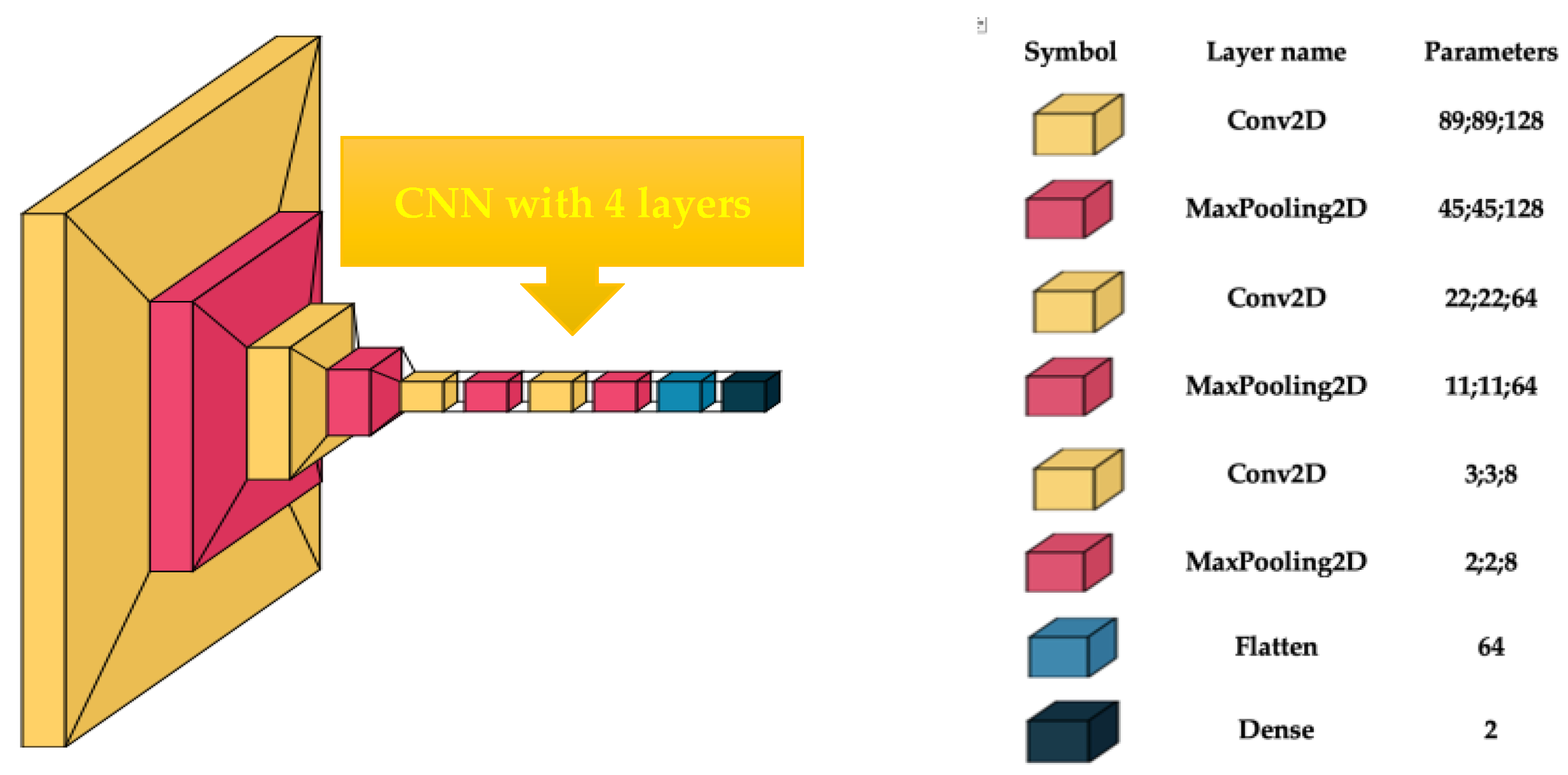
- Building, optimizing, and training a custom-built CNN with four layers;
- 3.
- Extracting second-order features (contrast, entropy, energy, and homogeneity) from the co-occurrence matrix;
- 4.
- Training the end-to-end PyCaret AutoML algorithm with correlation, energy, contrast, and homogeneity textural features;
- 5.
- A performance evaluation of both the CNN and AutoML in terms of accuracy, F1-score, and area under the curve (AUC).
3. Materials and Methods
3.1. Microscope
3.2. Hardware and Software
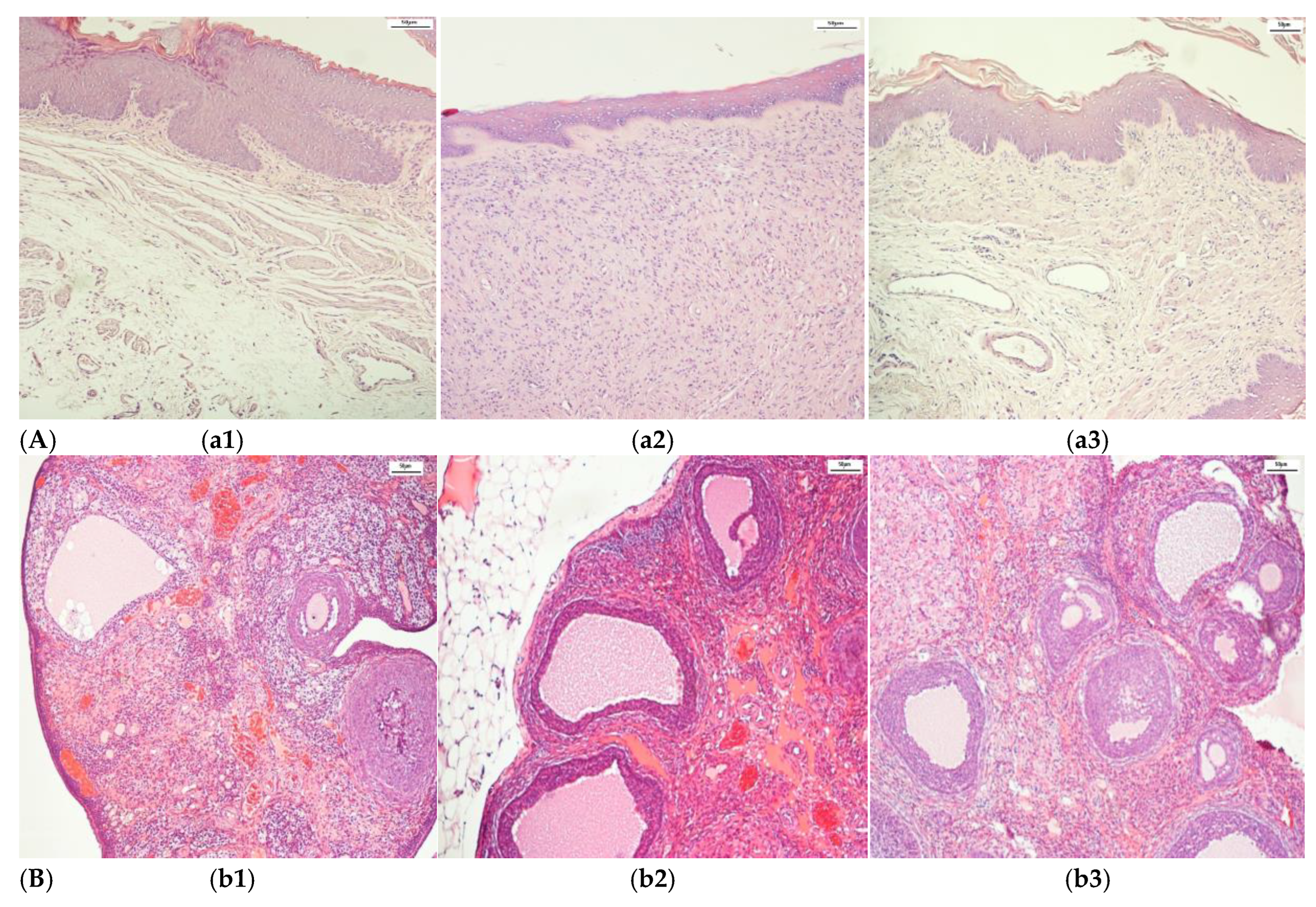
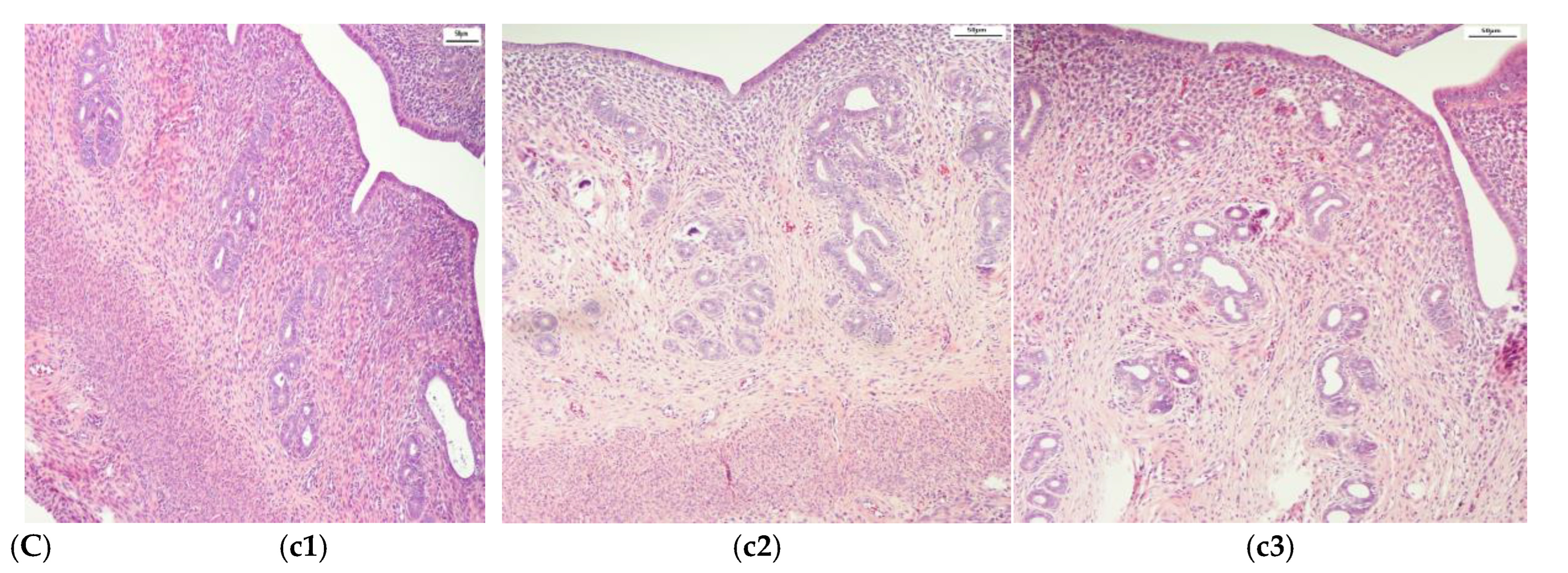
3.3. Histopathological Image Dataset and Augmentation
- (i)
- Random rotations and affine transformations. The images were manipulated with random rotations of up to 10 degrees, and affine transformations were employed to implement scaling and translations, in order to enhance the model’s robustness to spatial transformation;
- (ii)
- Random zoom. This creates surrounding pixel values in the image or interpolates pixel values according to the established percent; in this case, 30% was proposed;
- (iii)
- Random horizontal and vertical flips. These were applied with probabilities of 30% for the horizontal orientation [37].
3.4. PyCaret AutoML
3.5. Custom-Built CNN
3.6. Co-Occurrence Matrix
3.7. Performance Evaluation Metrics
4. Results and Discussion
Histopathological Image Dataset and Augmentation
5. Conclusions and Future Scope
Author Contributions
Funding
Institutional Review Board Statement
Informed Consent Statement
Data Availability Statement
Conflicts of Interest
Abbreviations
| DM | diabetes mellitus |
| AD-SC | antidiabetic therapy with a synthetic compound |
| CB-CNN | custom-built convolutional neural network |
| AutoML | PyCaret Auto Machine Learning |
| LDA | Linear Discriminant Analysis |
| DL | deep learning |
| ML | machine learning |
| AI | artificial intelligence |
| PCOS | polycystic ovary syndrome |
| XGBoost | extreme gradient boosting |
| KNN | k-nearest neighbor |
| RESNET | residual networks |
| RF | random forest |
References
- Ciarambino, T.; Crispino, P.; Leto, G.; Mastrolorenzo, E.; Para, O.; Giordano, M. Influence of Gender in Diabetes Mellitus and Its Complications. Int. J. Mol. Sci. 2022, 23, 8850. [Google Scholar] [CrossRef] [PubMed]
- Wing, R.R.; Bond, D.S.; Gendrano, I.N., III; Wadden, T.; Bahnson, J.; Lewis, C.E.; Brancati, F.; Schneider, S.; Kitabchi, A.E.; Van Dorsten, B.; et al. Effect of intensive lifestyle intervention on sexual dysfunction in women with type 2 diabetes: Results from an ancillary LookAHEAD study. Diabetes Care 2013, 36, 2937–2944. [Google Scholar] [CrossRef]
- Ahsan, M.M.; Luna, S.A.; Siddique, Z. Machine-Learning-Based Disease Diagnosis: A Comprehensive Review. Healthcare 2022, 10, 541. [Google Scholar] [CrossRef]
- Patil, V.I.; Patil, S.R. Optimized Transfer Learning With Hybrid Feature Extraction for Uterine Tissue Classification Using Histopathological Images. Microsc. Res. Tech. 2025, 88, 1582–1598. [Google Scholar] [CrossRef]
- Sun, H.; Zeng, X.; Xu, T.; Peng, G.; Ma, Y. Computer-aided diagnosis in histopathological images of the endometrium using a convolutional neural network and attention mechanisms. IEEE J. Biomed. Health Inform. 2019, 24, 1664–1676. [Google Scholar] [CrossRef] [PubMed]
- Kitaya, K.; Yasuo, T.; Yamaguchi, T.; Morita, Y.; Hamazaki, A.; Murayama, S.; Mihara, T.; Mihara, M. Construction of Deep Learning-based Convolutional Neural Network Model for Automatic Detection of Fluid Hysteroscopic Endometrial Micropolyps in Infertile Women with Chronic Endometritis. Eur. J. Obstet. Gynecol. Reprod. Biol. 2024, 297, 249–253. [Google Scholar] [CrossRef] [PubMed]
- Asadpour, V.; Puttock, E.J.; Getahun, D.; Fassett, M.J.; Xie, F. Automated placental abruption identification using semantic segmentation, quantitative features, SVM, ensemble and multi-path CNN. Heliyon 2023, 9, e13577. [Google Scholar] [CrossRef]
- Song, J.; Im, S.; Lee, S.H.; Jang, H.J. Deep Learning-Based Classification of Uterine Cervical and Endometrial Cancer Subtypes from Whole-Slide Histopathology Images. Diagnostics 2022, 12, 2623. [Google Scholar] [CrossRef]
- Zhao, F.; Dong, D.; Du, H.; Guo, Y.; Su, X.; Wang, Z.; Xie, X.; Wang, M.; Zhang, H.; Cao, X.; et al. Diagnosis of Endometrium Hyperplasia and Screening of Endometrial Intraepithelial Neoplasia in Histopathological Images Using a Global-to-Local Multi-Scale Convolutional Neural Network. Comput. Methods Programs Biomed. 2022, 221, 106906. [Google Scholar] [CrossRef]
- Li, T.; Liao, R.; Chan, C.; Greenblatt, E.M. Deep learning analysis of endometrial histology as a promising tool to predict the chance of pregnancy after frozen embryo transfers. J. Assist. Reprod. Genet. 2023, 40, 901–910. [Google Scholar] [CrossRef]
- Zafar, M.M.; Javaid, N.; Shaheen, I.; Alrajeh, N.; Aslam, S. Enhancing clinical decision support with explainable deep learning framework for C-Section forecasting. Computing 2025, 107, 20. [Google Scholar] [CrossRef]
- Peng, S.; Huang, H.; Cheng, M.; Yang, Y.; Li, F. Efficiently recognition of vaginal micro-ecological environment based on Convolutional Neural Network. In Proceedings of the 2020 IEEE International Conference on E-Health Networking, Application & Services (HEALTHCOM), Shenzhen, China, 1–2 March 2021; IEEE: Piscataway, NJ, USA, 2021; pp. 1–6. [Google Scholar]
- Onishi, S.; Egami, R.; Nakamura, Y.; Nagashima, Y.; Nishihara, K.; Matsuo, S.; Murai, A.; Hayashi, S.; Uesumi, Y.; Kato, A.; et al. Digital workflows for pathological assessment of rat estrous cycle stage using images of uterine horn and vaginal tissue. J. Pathol. Inform. 2022, 13, 100120. [Google Scholar] [CrossRef] [PubMed]
- Volinsky-Fremond, S.; Horeweg, N.; Andani, S.; Barkey Wolf, J.; Lafarge, M.W.; de Kroon, C.D.; Ørtoft, G.; Høgdall, E.; Dijkstra, J.; Jobsen, J.J.; et al. Prediction of recurrence risk in endometrial cancer with multimodal deep learning. Nat. Med. 2024, 30, 1962–1973. [Google Scholar] [CrossRef]
- Jeleń, U.; Stankiewicz-Antosz, I.; Chosia, M.; Jeleń, M. Cervical Cancer Diagnosis with Feature Selection and Deep Learning. Appl. Sci. 2025, 15, 1458. [Google Scholar] [CrossRef]
- Singh, J.; Kaliyar, R.K.; Kumari, R.; Sharma, N.; Manjunath, S.; Dharani Prasad, P. Advancements in Early Detection of Cervical Cancer using Machine Learning and Deep Learning Models for Cervicography Analysis. In Proceedings of the International Conference on Emerging Innovations and Advanced Computing (INNOCOMP), Sonipat, India, 25–26 May 2024. [Google Scholar]
- Rajan, B.K.; Harshan, M.H.; Swaminathan, R. Augmenting interpretation of vaginoscopy observations in cycling bitches with deep learning model. BMC Vet. Res. 2024, 20, 401. [Google Scholar] [CrossRef]
- Zhou, R.; Zhao, B.; Ding, H.; Fu, Y.; Li, H.; Wei, Y.; Xie, J.; Chen, C.; Yin, F.; Huang, D. Survival Prediction of Ovarian Serous Carcinoma Based on Machine Learning Combined with Pathological Images and Clinical Information. AIP Adv. 2024, 14, 045324. [Google Scholar] [CrossRef]
- El-Latif, E.I.A.; El-Dosuky, M.; Darwish, A.; Hassanien, A.E. A deep learning approach for ovarian cancer detection and classification based on fuzzy deep learning. Sci. Rep. 2024, 14, 26463. [Google Scholar] [CrossRef]
- Wang, C.W.; Lee, Y.C.; Chang, C.C.; Lin, Y.J.; Liou, Y.A.; Hsu, P.C.; Chang, C.C.; Sai, A.K.O.; Wang, C.H.; Chao, T.K. A weakly supervised deep learning method for guiding ovarian cancer treatment and identifying an effective biomarker. Cancers 2022, 14, 1651. [Google Scholar] [CrossRef] [PubMed]
- Behera, S.K.; Das, A.; Sethy, P.K. Deep fine-KNN classification of ovarian cancer subtypes using efficientNet-B0 extracted features: A comprehensive analysis. J. Cancer Res. Clin. Oncol. 2024, 150, 361. [Google Scholar] [CrossRef]
- Kodipalli, A.; Guha, S.; Dasar, S.; Ismail, T. An inception-ResNet deep learning approach to classify tumours in the ovary as benign and malignant. Expert Syst. 2022, e13215. [Google Scholar] [CrossRef]
- Kwatra, C.V.; Kaur, H. A comprehensive investigation into the use of machine learning to forecast ovarian cancer. In Proceedings of the 14th International Conference on Computing Communication and Networking Technologies (ICCCNT), Delhi, India, 6–8 July 2023; pp. 1–8. [Google Scholar]
- Radhakrishnan, M.; Sampathila, N.; Muralikrishna, H.; Swathi, K.S. Advancing ovarian cancer diagnosis through deep learning and explainable AI: A multiclassification approach. IEEE Access 2024, 12, 116968–116986. [Google Scholar] [CrossRef]
- Lăzărescu, A.M.D.; Moldovanu, S.; Moraru, L. Iris-Based Biometric Identification Using a Combination of the Right-Left Iris Statistical Features. J. Phys. Conf. Ser. 2024, 2701, 012006. [Google Scholar] [CrossRef]
- Tabacaru, G.; Moldovanu, S.; Răducan, E.; Barbu, M. A Robust Machine Learning Model for Diabetic Retinopathy Classification. J. Imaging 2024, 10, 8. [Google Scholar] [CrossRef]
- Evrimler, S.; Gedik, M.A.; Serel, T.A.; Ertunc, O.; Ozturk, S.A.; Soyupek, S. Bladder Urothelial Carcinoma: Machine Learning-based Computed Tomography Radiomics for Prediction of Histological Variant. Acad. Radiol. 2022, 29, 1682–1689. [Google Scholar] [CrossRef]
- Andlib, N.; Sajad, M.; Thakur, S.C. Association of diabetes mellitus with risk of reproductive impairment in females: A comprehensive review. Acta Histochem. 2024, 126, 152173. [Google Scholar] [CrossRef]
- Tariq, S.; Nurulain, S.M.; Rashed, H.; Lotfy, M.; Emerald, S.B.; Koturan, S.; Tekes, K.; Adeghate, E. Diabetes-induced changes in the morphology and nociceptinergic innervation of the rat uterus. J. Mol. Histol. 2016, 47, 21–33. [Google Scholar] [CrossRef]
- Montero-Pena, I.; Morales-De Pando, J.M.; Diaz-Gomez, A.; Gonzalez-Medina, G.; Ribelles-Garcia, A.; Perez-Arana, G.; Prada-Oliveira, J.A. Characterization of the histological changes in ovaries of Goto-Kakizaki diabetic rats. Eur. J. Anat. 2024, 28, 77–81. [Google Scholar] [CrossRef]
- Ali, E.M.T.; Abdalllah, H.I.; El-Sayed, S.M. Histomorphological, VEGF and TGF- β immunoexpression changes in the diabetic rats ovary and the potential amelioration following treatment with metformin and insulin. J. Mol. Histol. 2020, 51, 287–305. [Google Scholar] [CrossRef] [PubMed]
- Escobar-Morreale, H.F.; Roldan-Martin, M.B. Type 1 diabetes and Polycystic Ovary Sindrome: Systematic review and Meta-analysis. Diabetes Care 2016, 39, 639–648. [Google Scholar] [CrossRef]
- Rocha, D.S.; Dentz, M.V.; Model, J.F.A.; Vogt, E.L.; Ohlweiler, R.; Lima, M.V.; de Souza, S.K.; Kucharski, L.C. Female Wistar rats present particular glucose flux when submitted to classic protocols of experimental diabetes. Biomed. J. 2023, 46, 100539. [Google Scholar] [CrossRef]
- Kottaisamy, C.P.D.; Raj, D.S.; Prasanth Kumar, V.; Sankaran, U. Experimental animal models for diabetes and its related complications—A review. Lab. Anim. Res. 2021, 37, 23. [Google Scholar] [CrossRef] [PubMed]
- Yagihashi, S. Contribution of animal models to diabetes research: Its history, significance, and translation to humans. J. Diabetes Investig. 2023, 14, 1015–1037. [Google Scholar] [CrossRef]
- Tătaru, I.; Gardikiotis, I.; Dragostin, O.-M.; Confederat, L.; Gîrd, C.; Zamfir, A.-S.; Morariu, I.D.; Chiţescu, C.L.; Dinu (Iacob), A.; Costea Popescu, L.; et al. Hormonal status and oxidative stress monitoring in an in vivo 2 model of experimentally induced diabetes in female rats. Biomedicines 2025, 13, 922. [Google Scholar]
- Xu, M.; Yoon, S.; Fuentes, A.; Park, D.S. A comprehensive survey of image augmentation techniques for deep learning. Pattern Recognit. 2023, 137, 10934. [Google Scholar] [CrossRef]
- Ihme, M.; Chung, W.T.; Mishra, A.A. Combustion machine learning: Principles, progress and prospects. Prog. Energy Combust. Sci. 2022, 91, 101010. [Google Scholar] [CrossRef]
- Narayanan, A.N.; Das, S.S.; Mirnalinee, T. Evaluation of AutoML Frameworks for Computational ADMET Screening in Drug Discovery & Development. In Proceedings of the IEEE International Conference on Bioinformatics and Biomedicine, BIBM, Istanbul, Turkiye, 5–8 December 2023; IEEE: Piscataway, NJ, USA, 2023; pp. 4929–4931, ISBN 9798350337488. [Google Scholar]
- Chu, C.S.; Simpson, J.D.; O’Neill, P.M.; Berry, N.G. Machine learning–Predicting Ames mutagenicity of small molecules. J. Mol. Graph. Model. 2021, 109, 108011. [Google Scholar] [CrossRef]
- Das, D.; Naskar, R. Image splicing detection using low-dimensional feature vector of texture features and Haralick features based on Gray Level Co-occurrence Matrix. Signal Process. Image Commun. 2024, 125, 117134. [Google Scholar] [CrossRef]
- Moraru, L.; Moldovanu, S.; Culea-Florescu, A.-L.; Bibicu, D.; Dey, N.; Ashour, A.S.; Sherratt, R.S. Texture spectrum coupled with entropy and homogeneity image features for myocardium muscle characterization. Curr. Bioinform. 2019, 14, 295–304. [Google Scholar] [CrossRef]
- Luo, X.; Wang, W.; Xu, Y.; Lai, Z.; Jin, X.; Zhang, B.; Zhang, D. A deep convolutional neural network for diabetic retinopathy detection via mining local and long-range dependence. CAAI Trans. Intell. Technol. 2023, 1, 153–166. [Google Scholar] [CrossRef]


| References/Year | Weakness | AI Algorithms | Results Accuracy (%) |
|---|---|---|---|
| Patil and Patil [4]/2025 | -focuses on enhancing a CNN type -time-consuming | TL-CNN | 89.59% |
| Sun et al. [5]/2019 | -low accuracy -a limited ability to model | CNN | 84.50% |
| Kitaya et al. [6]/2024 | -custom-built CNN | CNN | 92.8% |
| Asadpour et al. [7]/2024 | -focuses on using a pre-trained CNN -low accuracy | ResNet-50 | 82.88% |
| Song et al. [8]/2022 | -only a custom-built DNN | DNN | 91.7% |
| Zhao et al. [9]/2022 | -global-to-local multi-scale CNN (G2LNet) | CNN | 95.34% |
| Li et al. [10]/2023 | -only a custom-built DNN -low accuracy | DNN | 75% |
| Zafar et al. [11]/2025 | -ML uterine tissue highly correlated 65 features. | Gated Highway Multi-layer-perceptron (GHiM) | 86.6% |
| Peng et al. [12]/2020 | -only a pre-trained CNN | ResNet V2 | 86% (AUC) |
| Onishi et al. [13]/2022 | -low accuracy -without fine-tuning | 10 conventional ML | 84% |
| Volinsky-Fremond et al. [14]/2024 | -low accuracy | Multimodal DNN | 78.9% |
| Jeleń et al. [15]/2025 | -without fine-tuning | NN, SVM | 93.3% |
| Jagendra et al. [16]/2024 | -only a neural network and a pre-trained CNN | VGG 16, Recurrent neural network (RNN) | 94.65% 87.65% |
| Rajan et al. [17]/2024 | -time-consuming | Inception v3, ResNet, XGBoost, SVM, KNN | 90.37% |
| Zhou et al. [18]/2024 | -time-consuming | SVM, RFXGBoost, CNN | 94.6% |
| El-Latif et al. [19]/2024 | -only a pre-trained CNN | ResNet-5 | 98.99% |
| Wang et al. [20]/2022 | -low accuracy | InceptionV3, ViT | 76% |
| Behera et al. [21]/2024 | -only a pre-trained CNN -low accuracy | EfficientNet-B0 | 78% |
| Kodipalli [22]/2022 | -only a pre-trained CNN -low accuracy | ResNet v2 | 67% |
| Kwatra and Kaur [23]/2023 | -pre-trained -time-consuming | MobileNetV3 ResNet50 | 96.3% 92.08% |
| Radhakrishnan et al. [24]/2024 | -time-consuming | MobileNetV2, VGG19, ResNet18, ResNeXt, Xception, EfficientNet | 97.96% |
| Classes | Test Set | Accuracy | F1-Score | AUC | Confusion Matrices [[TP FP][FN TN]] |
|---|---|---|---|---|---|
| Ovarian DC | 133 | 0.842 | 0.857 | 0.842 | [[63 9][12 49]] |
| Uterine DC | 55 | 0.945 | 0.950 | 0.943 | [[29 2][1 23]] |
| Vaginas DC | 62 | 0.75 | 0.75 | 0.739 | [[26 8][8 20]] |
| Ovarian AD_DC | 44 | 0.771 | 0.733 | 0.736 | [[22 5][6 11]] |
| Uterine AD_DC | 92 | 0.858 | 0.853 | 0.858 | [[38 7][6 41]] |
| Vaginas AD_DC | 82 | 0.771 | 0.853 | 0.786 | [[37 6][12 27]] |
| Classes | Accuracy | F1-Score | The Selected Classifier |
|---|---|---|---|
| Ovarian DC | 0.785 | 0.837 | ExtraTreesClassifier(criterion = ‘gini’, estimators = 100, random_state = 123) |
| Uterine DC | 0.615 | 0.573 | KNeighborsClassifier(algorithm = ‘auto’, leaf_size = 30, metric = ‘minkowski’, n_neighbors = 5) |
| Vaginas DC | 0.775 | 0.788 | LinearDiscriminantAnalysis(solver = ‘svd’, tol = 0.0001) |
| Ovarian AD_DC | 0.751 | 0.822 | RandomForestClassifier(criterion = ‘gini’, n_estimators = 100) |
| Uterine AD_DC | 0.7 | 0.53 | LinearDiscriminantAnalysis(solver = ‘svd’, tol = 0.0001) |
| Vaginas AD_DC | 0.86 | 0.88 | LinearDiscriminantAnalysis (solver = ‘svd’, tol = 0.0001) |
Disclaimer/Publisher’s Note: The statements, opinions and data contained in all publications are solely those of the individual author(s) and contributor(s) and not of MDPI and/or the editor(s). MDPI and/or the editor(s) disclaim responsibility for any injury to people or property resulting from any ideas, methods, instructions or products referred to in the content. |
© 2025 by the authors. Licensee MDPI, Basel, Switzerland. This article is an open access article distributed under the terms and conditions of the Creative Commons Attribution (CC BY) license (https://creativecommons.org/licenses/by/4.0/).
Share and Cite
Tătaru, I.; Moldovanu, S.; Dragostin, O.-M.; Chiţescu, C.L.; Zamfir, A.-S.; Dragostin, I.; Strat, L.; Zamfir, C.L. Auto Machine Learning and Convolutional Neural Network in Diabetes Mellitus Research—The Role of Histopathological Images in Designing and Exploring Experimental Models. Biomedicines 2025, 13, 1494. https://doi.org/10.3390/biomedicines13061494
Tătaru I, Moldovanu S, Dragostin O-M, Chiţescu CL, Zamfir A-S, Dragostin I, Strat L, Zamfir CL. Auto Machine Learning and Convolutional Neural Network in Diabetes Mellitus Research—The Role of Histopathological Images in Designing and Exploring Experimental Models. Biomedicines. 2025; 13(6):1494. https://doi.org/10.3390/biomedicines13061494
Chicago/Turabian StyleTătaru, Iulian, Simona Moldovanu, Oana-Maria Dragostin, Carmen Lidia Chiţescu, Alexandra-Simona Zamfir, Ionut Dragostin, Liliana Strat, and Carmen Lăcrămioara Zamfir. 2025. "Auto Machine Learning and Convolutional Neural Network in Diabetes Mellitus Research—The Role of Histopathological Images in Designing and Exploring Experimental Models" Biomedicines 13, no. 6: 1494. https://doi.org/10.3390/biomedicines13061494
APA StyleTătaru, I., Moldovanu, S., Dragostin, O.-M., Chiţescu, C. L., Zamfir, A.-S., Dragostin, I., Strat, L., & Zamfir, C. L. (2025). Auto Machine Learning and Convolutional Neural Network in Diabetes Mellitus Research—The Role of Histopathological Images in Designing and Exploring Experimental Models. Biomedicines, 13(6), 1494. https://doi.org/10.3390/biomedicines13061494