Next Issue
Volume 11, September
Previous Issue
Volume 11, July


Biosensors, Volume 11, Issue 8 (August 2021) – 49 articles

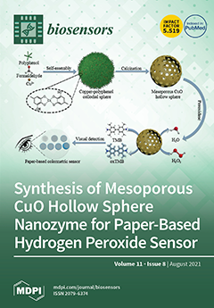
Cover Story (view full-size image): Point-of-care monitoring of hydrogen peroxide is important due to its wide usage in biomedicine, households and industry. Wei and co-authors from Xi’an Jiaotong University have developed a paper sensor for sensitive, visual and selective detection of H2O2 using mesoporous metal oxide hollow spheres as a nanozyme. The obtained paper sensor can be effectively used for detection of H2O2 in the range of 2.4–150 µM. This work provides a low-cost paper sensor fabricated from a mesoporous metal oxide nanozyme. These sensors could be potentially used in bioanalysis, food security and environmental protection. View this paper.
  • Issues are regarded as officially published after their release is announced to the table of contents alert mailing list.
  • You may sign up for e-mail alerts to receive table of contents of newly released issues.
  • PDF is the official format for papers published in both, html and pdf forms. To view the papers in pdf format, click on the "PDF Full-text" link, and use the free Adobe Reader to open them.
Order results
Result details
Section
Select all
Export citation of selected articles as:
25 pages, 1620 KiB  
Review
PDMS Bonding Technologies for Microfluidic Applications: A Review
by Alexandra Borók, Kristóf Laboda and Attila Bonyár
Biosensors 2021, 11(8), 292; https://doi.org/10.3390/bios11080292 - 23 Aug 2021
Cited by 83 | Viewed by 14235
Abstract
This review summarizes and compares the available surface treatment and bonding techniques (e.g., corona triggered surface activation, oxygen plasma surface activation, chemical gluing, and mixed techniques) and quality/bond-strength testing methods (e.g., pulling test, shear test, peel test, leakage test) for bonding PDMS (polydimethylsiloxane) [...] Read more.
This review summarizes and compares the available surface treatment and bonding techniques (e.g., corona triggered surface activation, oxygen plasma surface activation, chemical gluing, and mixed techniques) and quality/bond-strength testing methods (e.g., pulling test, shear test, peel test, leakage test) for bonding PDMS (polydimethylsiloxane) with other materials, such as PDMS, glass, silicon, PET (polyethylene terephthalate), PI (polyimide), PMMA (poly(methyl methacrylate)), PVC (polyvinyl chloride), PC (polycarbonate), COC (cyclic olefin copolymer), PS (polystyrene) and PEN (polyethylene naphthalate). The optimized process parameters for the best achievable bond strengths are collected for each substrate, and the advantages and disadvantages of each method are discussed in detail. Full article
(This article belongs to the Collection Recent Developments in Microfluidics)
Show Figures

Figure 1

26 pages, 6636 KiB  
Review
Electroanalytical Overview: Electrochemical Sensing Platforms for Food and Drink Safety
by Alejandro Garcia-Miranda Ferrari, Robert D. Crapnell and Craig E. Banks
Biosensors 2021, 11(8), 291; https://doi.org/10.3390/bios11080291 - 23 Aug 2021
Cited by 19 | Viewed by 4146
Abstract
Robust, reliable, and affordable analytical techniques are essential for screening and monitoring food and water safety from contaminants, pathogens, and allergens that might be harmful upon consumption. Recent advances in decentralised, miniaturised, and rapid tests for health and environmental monitoring can provide an [...] Read more.
Robust, reliable, and affordable analytical techniques are essential for screening and monitoring food and water safety from contaminants, pathogens, and allergens that might be harmful upon consumption. Recent advances in decentralised, miniaturised, and rapid tests for health and environmental monitoring can provide an alternative solution to the classic laboratory-based analytical techniques currently utilised. Electrochemical biosensors offer a promising option as portable sensing platforms to expedite the transition from laboratory benchtop to on-site analysis. A plethora of electroanalytical sensor platforms have been produced for the detection of small molecules, proteins, and microorganisms vital to ensuring food and drink safety. These utilise various recognition systems, from direct electrochemical redox processes to biological recognition elements such as antibodies, enzymes, and aptamers; however, further exploration needs to be carried out, with many systems requiring validation against standard benchtop laboratory-based techniques to offer increased confidence in the sensing platforms. This short review demonstrates that electroanalytical biosensors already offer a sensitive, fast, and low-cost sensor platform for food and drink safety monitoring. With continued research into the development of these sensors, increased confidence in the safety of food and drink products for manufacturers, policy makers, and end users will result. Full article
(This article belongs to the Special Issue Sensors for Environmental Monitoring and Food Safety)
Show Figures

Figure 1

22 pages, 9379 KiB  
Review
Spectrophotometric Assays for Sensing Tyrosinase Activity and Their Applications
by Yu-Fan Fan, Si-Xing Zhu, Fan-Bin Hou, Dong-Fang Zhao, Qiu-Sha Pan, Yan-Wei Xiang, Xing-Kai Qian, Guang-Bo Ge and Ping Wang
Biosensors 2021, 11(8), 290; https://doi.org/10.3390/bios11080290 - 23 Aug 2021
Cited by 20 | Viewed by 6726
Abstract
Tyrosinase (TYR, E.C. 1.14.18.1), a critical enzyme participating in melanogenesis, catalyzes the first two steps in melanin biosynthesis including the ortho-hydroxylation of L-tyrosine and the oxidation of L-DOPA. Previous pharmacological investigations have revealed that an abnormal level of TYR is tightly associated [...] Read more.
Tyrosinase (TYR, E.C. 1.14.18.1), a critical enzyme participating in melanogenesis, catalyzes the first two steps in melanin biosynthesis including the ortho-hydroxylation of L-tyrosine and the oxidation of L-DOPA. Previous pharmacological investigations have revealed that an abnormal level of TYR is tightly associated with various dermatoses, including albinism, age spots, and malignant melanoma. TYR inhibitors can partially block the formation of pigment, which are always used for improving skin tone and treating dermatoses. The practical and reliable assays for monitoring TYR activity levels are very useful for both disease diagnosis and drug discovery. This review comprehensively summarizes structural and enzymatic characteristics, catalytic mechanism and substrate preference of TYR, as well as the recent advances in biochemical assays for sensing TYR activity and their biomedical applications. The design strategies of various TYR substrates, alongside with several lists of all reported biochemical assays for sensing TYR including analytical conditions and kinetic parameters, are presented for the first time. Additionally, the biomedical applications and future perspectives of these optical assays are also highlighted. The information and knowledge presented in this review offer a group of practical and reliable assays and imaging tools for sensing TYR activities in complex biological systems, which strongly facilitates high-throughput screening TYR inhibitors and further investigations on the relevance of TYR to human diseases. Full article
(This article belongs to the Special Issue Enzyme-Based Biosensors and Their Applications)
Show Figures

Figure 1

13 pages, 1914 KiB  
Article
Stability Assessment of Four Chimeric Proteins for Human Chagas Disease Immunodiagnosis
by Paola Alejandra Fiorani Celedon, Leonardo Maia Leony, Ueriton Dias Oliveira, Natália Erdens Maron Freitas, Ângelo Antônio Oliveira Silva, Ramona Tavares Daltro, Emily Ferreira Santos, Marco Aurélio Krieger, Nilson Ivo Tonin Zanchin and Fred Luciano Neves Santos
Biosensors 2021, 11(8), 289; https://doi.org/10.3390/bios11080289 - 22 Aug 2021
Cited by 6 | Viewed by 2624
Abstract
The performance of an immunoassay relies on antigen-antibody interaction; hence, antigen chemical stability and structural integrity are paramount for an efficient assay. We conducted a functional, thermostability and long-term stability analysis of different chimeric antigens (IBMP), in order to assess effects of adverse [...] Read more.
The performance of an immunoassay relies on antigen-antibody interaction; hence, antigen chemical stability and structural integrity are paramount for an efficient assay. We conducted a functional, thermostability and long-term stability analysis of different chimeric antigens (IBMP), in order to assess effects of adverse conditions on four antigens employed in ELISA to diagnose Chagas disease. ELISA-based immunoassays have served as a model for biosensors development, as both assess molecular interactions. To evaluate thermostability, samples were heated and cooled to verify heat-induced denaturation reversibility. In relation to storage stability, the antigens were analyzed at 25 °C at different moments. Long-term stability tests were performed using eight sets of microplates sensitized. Antigens were structurally analyzed through circular dichroism (CD), dynamic light scattering, SDS-PAGE, and functionally evaluated by ELISA. Data suggest that IBMP antigens are stable, over adverse conditions and for over a year. Daily analysis revealed minor changes in the molecular structure. Functionally, IBMP-8.2 and IBMP-8.3 antigens showed reactivity towards anti-T. cruzi antibodies, even after 72 h at 25 °C. Long-term stability tests showed that all antigens were comparable to the control group and all antigens demonstrated stability for one year. Data suggest that the antigens maintained their function and structural characteristics even in adverse conditions, making them a sturdy and reliable candidate to be employed in future in vitro diagnostic tests applicable to different models of POC devices, such as modern biosensors in development. Full article
(This article belongs to the Special Issue Biosensing and Bioimaging: Trends and Perspective)
Show Figures

Figure 1

10 pages, 1882 KiB  
Article
A Rapid Single-Cell Antimicrobial Susceptibility Testing Workflow for Bloodstream Infections
by Britney Forsyth, Peter Torab, Jyong-Huei Lee, Tyler Malcom, Tza-Huei Wang, Joseph C. Liao, Samuel Yang, Erik Kvam, Chris Puleo and Pak Kin Wong
Biosensors 2021, 11(8), 288; https://doi.org/10.3390/bios11080288 - 22 Aug 2021
Cited by 8 | Viewed by 3939
Abstract
Bloodstream infections are a significant cause of morbidity and mortality worldwide. The rapid initiation of effective antibiotic treatment is critical for patients with bloodstream infections. However, the diagnosis of bloodborne pathogens is largely complicated by the matrix effect of blood and the lengthy [...] Read more.
Bloodstream infections are a significant cause of morbidity and mortality worldwide. The rapid initiation of effective antibiotic treatment is critical for patients with bloodstream infections. However, the diagnosis of bloodborne pathogens is largely complicated by the matrix effect of blood and the lengthy blood tube culture procedure. Here we report a culture-free workflow for the rapid isolation and enrichment of bacterial pathogens from whole blood for single-cell antimicrobial susceptibility testing (AST). A dextran sedimentation step reduces the concentration of blood cells by 4 orders of magnitude in 20–30 min while maintaining the effective concentration of bacteria in the sample. Red blood cell depletion facilitates the downstream centrifugation-based enrichment step at a sepsis-relevant bacteria concentration. The workflow is compatible with common antibiotic-resistant bacteria and does not influence the minimum inhibitory concentrations. By applying a microfluidic single-cell trapping device, we demonstrate the workflow for the rapid determination of bacterial infection and antimicrobial susceptibility testing at the single-cell level. The entire workflow from blood to categorical AST result can be completed in less than two hours. Full article
Show Figures

Figure 1

15 pages, 2577 KiB  
Article
Developing Activated Carbon Veil Electrode for Sensing Salivary Uric Acid
by Maria A. Bukharinova, Natalia Yu. Stozhko, Elizaveta A. Novakovskaya, Ekaterina I. Khamzina, Aleksey V. Tarasov and Sergey V. Sokolkov
Biosensors 2021, 11(8), 287; https://doi.org/10.3390/bios11080287 - 20 Aug 2021
Cited by 8 | Viewed by 2540
Abstract
The paper describes the development of a carbon veil-based electrode (CVE) for determining uric acid (UA) in saliva. The electrode was manufactured by lamination technology, electrochemically activated and used as a highly sensitive voltammetric sensor (CVEact). Potentiostatic polarization of the electrode [...] Read more.
The paper describes the development of a carbon veil-based electrode (CVE) for determining uric acid (UA) in saliva. The electrode was manufactured by lamination technology, electrochemically activated and used as a highly sensitive voltammetric sensor (CVEact). Potentiostatic polarization of the electrode at 2.0 V in H2SO4 solution resulted in a higher number of oxygen and nitrogen-containing groups on the electrode surface; lower charge transfer resistance; a 1.5 times increase in the effective surface area and a decrease in the UA oxidation potential by over 0.4 V, compared with the non-activated CVE, which was confirmed by energy dispersive X-ray spectroscopy, electrochemical impedance spectroscopy, chronoamperometry and linear sweep voltammetry. The developed sensor is characterized by a low detection limit of 0.05 µM and a wide linear range (0.09–700 µM). The results suggest that the sensor has perspective applications for quick determination of UA in artificial and human saliva. RSD does not exceed 3.9%, and recovery is 96–105%. UA makes a significant contribution to the antioxidant activity (AOA) of saliva (≈60%). In addition to its high analytical characteristics, the important advantages of the proposed CVEact are the simple, scalable, and cost-effective manufacturing technology and the absence of additional complex and time-consuming modification operations. Full article
(This article belongs to the Special Issue Novel Materials for Electrochemical Biosensors)
Show Figures

Graphical abstract

13 pages, 4625 KiB  
Article
In Vitro Anticancer Drug Sensitivity Sensing through Single-Cell Raman Spectroscopy
by Jingkai Wang, Kaicheng Lin, Huijie Hu, Xingwang Qie, Wei E. Huang, Zhisong Cui, Yan Gong and Yizhi Song
Biosensors 2021, 11(8), 286; https://doi.org/10.3390/bios11080286 - 20 Aug 2021
Cited by 9 | Viewed by 4590
Abstract
Traditional in vitro anticancer drug sensitivity testing at the population level suffers from lengthy procedures and high false positive rates. To overcome these defects, we built a confocal Raman microscopy sensing system and proposed a single-cell approach via Raman-deuterium isotope probing (Raman-DIP) as [...] Read more.
Traditional in vitro anticancer drug sensitivity testing at the population level suffers from lengthy procedures and high false positive rates. To overcome these defects, we built a confocal Raman microscopy sensing system and proposed a single-cell approach via Raman-deuterium isotope probing (Raman-DIP) as a rapid and reliable in vitro drug efficacy evaluation method. Raman-DIP detected the incorporation of deuterium into the cell, which correlated with the metabolic activity of the cell. The human non-small cell lung cancer cell line HCC827 and human breast cancer cell line MCF-7 were tested against eight different anticancer drugs. The metabolic activity of cancer cells could be detected as early as 12 h, independent of cell growth. Incubation of cells in 30% heavy water (D2O) did not show any negative effect on cell viability. Compared with traditional methods, Raman-DIP could accurately determine the drug effect, meanwhile, it could reduce the testing period from 72–144 h to 48 h. Moreover, the heterogeneity of cells responding to anticancer drugs was observed at the single-cell level. This proof-of-concept study demonstrated the potential of Raman-DIP to be a reliable tool for cancer drug discovery and drug susceptibility testing. Full article
(This article belongs to the Special Issue Feature Issue of Optical and Photonic Biosensors Section)
Show Figures

Figure 1

17 pages, 1161 KiB  
Article
Single-Drop Analysis of Epinephrine and Uric Acid on a Screen-Printed Carbon Electrode
by David Majer and Matjaž Finšgar
Biosensors 2021, 11(8), 285; https://doi.org/10.3390/bios11080285 - 19 Aug 2021
Cited by 7 | Viewed by 2454
Abstract
This work demonstrates the analysis of epinephrine (EP) and uric acid (UA) in a single drop (the volume of the test solution was only 50 µL) using a screen-printed carbon electrode (SPCE) sensor and square-wave voltammetry (SWV). The limit of detection, limit of [...] Read more.
This work demonstrates the analysis of epinephrine (EP) and uric acid (UA) in a single drop (the volume of the test solution was only 50 µL) using a screen-printed carbon electrode (SPCE) sensor and square-wave voltammetry (SWV). The limit of detection, limit of quantification, linearity, accuracy, precision, and robustness were validated. The normality of the experimental data was tested and confirmed for both methods. Heteroscedasticity was checked by residual analysis followed by a statistical F-test. The latter was confirmed for both analytes. The low relative standard deviations (RSD) at all calibration points and repetitive slopes justified the use of a calibration curve; therefore, the standard addition methodology was avoided (the latter is common in electroanalysis, but time-consuming). Since the conditions for using an ordinary least squares (OLS) regression were not met, weighted linear regression (WLR) was used to improve the accuracy of the analytical results at low concentrations of the analytes. In this manner, the best weighted model was determined and used for the quantification. A comparison was made between the OLS and WLR methods to show the necessity of using the WLR method for EP and UA analysis. The newly developed and validated methods were also shown to be effective in the analysis of real samples. The content of EP in an EP auto-injector and UA in human urine was tested by employing the best weighted model. For EP and UA, the accuracy in terms of the average recovery value was 101.01% and 94.35%, and precision in terms of RSD was 5.65% and 2.75%, respectively. A new analytical methodology is presented that uses a low volume (a single drop), and it offers the advantage of electroanalysis for on-site analysis, where conventional chromatographic techniques cannot be easily employed. Furthermore, the developed technique has additional advantages in terms of speed, cost, and miniaturization. Full article
(This article belongs to the Section Biosensor and Bioelectronic Devices)
Show Figures

Figure 1

20 pages, 356 KiB  
Article
A Study of One-Class Classification Algorithms for Wearable Fall Sensors
by José Antonio Santoyo-Ramón, Eduardo Casilari and José Manuel Cano-García
Biosensors 2021, 11(8), 284; https://doi.org/10.3390/bios11080284 - 19 Aug 2021
Cited by 9 | Viewed by 2654
Abstract
In recent years, the popularity of wearable devices has fostered the investigation of automatic fall detection systems based on the analysis of the signals captured by transportable inertial sensors. Due to the complexity and variety of human movements, the detection algorithms that offer [...] Read more.
In recent years, the popularity of wearable devices has fostered the investigation of automatic fall detection systems based on the analysis of the signals captured by transportable inertial sensors. Due to the complexity and variety of human movements, the detection algorithms that offer the best performance when discriminating falls from conventional Activities of Daily Living (ADLs) are those built on machine learning and deep learning mechanisms. In this regard, supervised machine learning binary classification methods have been massively employed by the related literature. However, the learning phase of these algorithms requires mobility patterns caused by falls, which are very difficult to obtain in realistic application scenarios. An interesting alternative is offered by One-Class Classifiers (OCCs), which can be exclusively trained and configured with movement traces of a single type (ADLs). In this paper, a systematic study of the performance of various typical OCCs (for diverse sets of input features and hyperparameters) is performed when applied to nine public repositories of falls and ADLs. The results show the potentials of these classifiers, which are capable of achieving performance metrics very similar to those of supervised algorithms (with values for the specificity and the sensitivity higher than 95%). However, the study warns of the need to have a wide variety of types of ADLs when training OCCs, since activities with a high degree of mobility can significantly increase the frequency of false alarms (ADLs identified as falls) if not considered in the data subsets used for training. Full article
(This article belongs to the Collection Wearable Biosensors for Healthcare Applications)
10 pages, 2435 KiB  
Article
Color-Coded Droplets and Microscopic Image Analysis for Multiplexed Antibiotic Susceptibility Testing
by Yunjin Jeong, Haewook Jang, Junwon Kang, Juhong Nam, Kyoungseob Shin, Sunghoon Kwon and Jungil Choi
Biosensors 2021, 11(8), 283; https://doi.org/10.3390/bios11080283 - 19 Aug 2021
Cited by 4 | Viewed by 2990
Abstract
Since the discovery of antibiotics, the emergence of antibiotic resistance has become a global issue that is threatening society. In the era of antibiotic resistance, finding the proper antibiotics through antibiotic susceptibility testing (AST) is crucial in clinical settings. However, the current clinical [...] Read more.
Since the discovery of antibiotics, the emergence of antibiotic resistance has become a global issue that is threatening society. In the era of antibiotic resistance, finding the proper antibiotics through antibiotic susceptibility testing (AST) is crucial in clinical settings. However, the current clinical process of AST based on the broth microdilution test has limitations on scalability to expand the number of antibiotics that are tested with various concentrations. Here, we used color-coded droplets to expand the multiplexing of AST regarding the kind and concentration of antibiotics. Color type and density differentiate the kind of antibiotics and concentration, respectively. Microscopic images of a large view field contain numbers of droplets with different testing conditions. Image processing analysis detects each droplet, decodes color codes, and measures the bacterial growth in the droplet. Testing E. coli ATCC 25922 with ampicillin, gentamicin, and tetracycline shows that the system can provide a robust and scalable platform for multiplexed AST. Furthermore, the system can be applied to various drug testing systems, which require several different testing conditions. Full article
(This article belongs to the Special Issue Recent Advances in Photonic Biosensors)
Show Figures

Figure 1

17 pages, 3481 KiB  
Review
Recent Progress of SERS Nanoprobe for pH Detecting and Its Application in Biological Imaging
by Lei Zhang, Qianqian Zhao, Zhitao Jiang, Jingjing Shen, Weibing Wu, Xingfen Liu, Quli Fan and Wei Huang
Biosensors 2021, 11(8), 282; https://doi.org/10.3390/bios11080282 - 19 Aug 2021
Cited by 11 | Viewed by 3741
Abstract
As pH value almost affects the function of cells and organisms in all aspects, in biology, biochemical and many other research fields, it is necessary to apply simple, intuitive, sensitive, stable detection of pH and base characteristics inside and outside the cell. Therefore, [...] Read more.
As pH value almost affects the function of cells and organisms in all aspects, in biology, biochemical and many other research fields, it is necessary to apply simple, intuitive, sensitive, stable detection of pH and base characteristics inside and outside the cell. Therefore, many research groups have explored the design and application of pH probes based on surface enhanced Raman scattering (SERS). In this review article, we discussed the basic theoretical background of explaining the working mechanism of pH SERS sensors, and also briefly described the significance of cell pH measurement, and simply classified and summarized the factors that affected the performance of pH SERS probes. Some applications of pH probes based on surface enhanced Raman scattering in intracellular and extracellular pH imaging and the combination of other analytical detection techniques are described. Finally, the development prospect of this field is presented. Full article
(This article belongs to the Special Issue Surface Enhanced Raman Spectroscopy Based Sensors and Biosensors)
Show Figures

Figure 1

42 pages, 11286 KiB  
Review
Recent Development of Nanomaterials-Based Cytosensors for the Detection of Circulating Tumor Cells
by Zhi-Fang Sun, Yong Chang and Ning Xia
Biosensors 2021, 11(8), 281; https://doi.org/10.3390/bios11080281 - 18 Aug 2021
Cited by 13 | Viewed by 3860
Abstract
The accurate analysis of circulating tumor cells (CTCs) holds great promise in early diagnosis and prognosis of cancers. However, the extremely low abundance of CTCs in peripheral blood samples limits the practical utility of the traditional methods for CTCs detection. Thus, novel and [...] Read more.
The accurate analysis of circulating tumor cells (CTCs) holds great promise in early diagnosis and prognosis of cancers. However, the extremely low abundance of CTCs in peripheral blood samples limits the practical utility of the traditional methods for CTCs detection. Thus, novel and powerful strategies have been proposed for sensitive detection of CTCs. In particular, nanomaterials with exceptional physical and chemical properties have been used to fabricate cytosensors for amplifying the signal and enhancing the sensitivity. In this review, we summarize the recent development of nanomaterials-based optical and electrochemical analytical techniques for CTCs detection, including fluorescence, colorimetry, surface-enhanced Raman scattering, chemiluminescence, electrochemistry, electrochemiluminescence, photoelectrochemistry and so on. Full article
(This article belongs to the Special Issue Nanomaterial-Based Biosensors for Biomedical Applications)
Show Figures

Figure 1

15 pages, 3631 KiB  
Article
Ratiometric Colorimetric Detection of Nitrite Realized by Stringing Nanozyme Catalysis and Diazotization Together
by Mengzhu Wang, Peng Liu, Hengjia Zhu, Bangxiang Liu and Xiangheng Niu
Biosensors 2021, 11(8), 280; https://doi.org/10.3390/bios11080280 - 18 Aug 2021
Cited by 14 | Viewed by 3276
Abstract
Due to the great threat posed by excessive nitrite in food and drinking water to human health, it calls for developing reliable, convenient, and low-cost methods for nitrite detection. Herein, we string nanozyme catalysis and diazotization together and develop a ratiometric colorimetric approach [...] Read more.
Due to the great threat posed by excessive nitrite in food and drinking water to human health, it calls for developing reliable, convenient, and low-cost methods for nitrite detection. Herein, we string nanozyme catalysis and diazotization together and develop a ratiometric colorimetric approach for sensing nitrite in food. First, hollow MnFeO (a mixture of Mn and Fe oxides with different oxidation states) derived from a Mn-Fe Prussian blue analogue is explored as an oxidase mimic with high efficiency in catalyzing the colorless 3,3′,5,5′-tetramethylbenzidine (TMB) oxidation to blue TMBox, presenting a notable signal at 652 nm. Then, nitrite is able to trigger the diazotization of the product TMBox, not only decreasing the signal at 652 nm but also producing a new signal at 445 nm. Thus, the analyte-induced reverse changes of the two signals enable us to establish a ratiometric colorimetric assay for nitrite analysis. According to the above strategy, facile determination of nitrite in the range of 3.3–133.3 μM with good specificity was realized, providing a detection limit down to 0.2 μM. Compared with conventional single-signal analysis, our dual-signal ratiometric colorimetric mode was demonstrated to offer higher sensitivity, a lower detection limit, and better anti-interference ability against external detection environments. Practical applications of the approach in examining nitrite in food matrices were also verified. Full article
(This article belongs to the Special Issue Biosensors in Food Analysis and Environmental Detection)
Show Figures

Figure 1

13 pages, 3428 KiB  
Article
Characterization of Intracellular Structure Changes of Microcystis under Sonication Treatment by Polarized Light Scattering
by Jiajin Li, Chujun Zou, Ran Liao, Liang Peng, Hongjian Wang, Zhiming Guo and Hui Ma
Biosensors 2021, 11(8), 279; https://doi.org/10.3390/bios11080279 - 17 Aug 2021
Cited by 10 | Viewed by 1969
Abstract
Cyanobacterial bloom is one of the most urgent global environmental issues, which eventually could threaten human health and safety. Sonication treatment (ST) is a potential effective method to control cyanobacteria blooms in the field. Currently, the bottleneck of extensive application of ST is [...] Read more.
Cyanobacterial bloom is one of the most urgent global environmental issues, which eventually could threaten human health and safety. Sonication treatment (ST) is a potential effective method to control cyanobacteria blooms in the field. Currently, the bottleneck of extensive application of ST is the difficulty to estimate the ST effect on the cyanobacterial cells and then determine suitable ST times in the field. In this study, cyanobacterial Microcystis samples sonicated at different times were first measured by a spectrophotometer to calculate the removal efficiency of Microcystis cells. Additionally, they were observed by TEM to reveal the intracellular structure changes of the cells. Then the samples were measured by an experimental setup based on polarized light scattering to measure the polarization parameters. Experimental results indicated that the polarization parameters can effectively characterize the intracellular structural changes of Microcystis cells with different ST times, which is quite consistent with the results for removal efficiency and TEM images. Further, the optimal ST time can be inferred by the polarization parameters. These results demonstrate that polarized light scattering can be a potentially powerful tool to explore suitable times for sonication treatment of cyanobacteria blooms. Full article
(This article belongs to the Special Issue Sensors for Environmental Monitoring and Food Safety)
Show Figures

Figure 1

10 pages, 2476 KiB  
Article
Enhanced Luminescent Detection of Circulating Tumor Cells by a 3D Printed Immunomagnetic Concentrator
by Chanyong Park, Abdurhaman Teyib Abafogi, Dinesh Veeran Ponnuvelu, Ilchan Song, Kisung Ko and Sungsu Park
Biosensors 2021, 11(8), 278; https://doi.org/10.3390/bios11080278 - 17 Aug 2021
Cited by 10 | Viewed by 2557
Abstract
Circulating tumor cells (CTCs) are an indicator of metastatic progression and relapse. Since non-CTC cells such as red blood cells outnumber CTCs in the blood, the separation and enrichment of CTCs is key to improving their detection sensitivity. The ATP luminescence assay can [...] Read more.
Circulating tumor cells (CTCs) are an indicator of metastatic progression and relapse. Since non-CTC cells such as red blood cells outnumber CTCs in the blood, the separation and enrichment of CTCs is key to improving their detection sensitivity. The ATP luminescence assay can measure intracellular ATP to detect cells quickly but has not yet been used for CTC detection in the blood because extracellular ATP in the blood, derived from non-CTCs, interferes with the measurement. Herein, we report on the improvement of the ATP luminescence assay for the detection of CTCs by separating and concentrating CTCs in the blood using a 3D printed immunomagnetic concentrator (3DPIC). Because of its high-aspect-ratio structure and resistance to high flow rates, 3DPIC allows cancer cells in 10 mL to be concentrated 100 times within minutes. This enables the ATP luminescence assay to detect as low as 10 cells in blood, thereby being about 10 times more sensitive than when commercial kits are used for CTC concentration. This is the first time that the ATP luminescence assay was used for the detection of cancer cells in blood. These results demonstrate the feasibility of 3DPIC as a concentrator to improve the detection limit of the ATP luminescence assay for the detection of CTCs. Full article
Show Figures

Figure 1

12 pages, 1929 KiB  
Article
Microelectrode Sensor for Real-Time Measurements of Nitrite in the Living Brain, in the Presence of Ascorbate
by Tiago Monteiro, Cândida Dias, Cátia F. Lourenço, Ana Ledo, Rui M. Barbosa and M. Gabriela Almeida
Biosensors 2021, 11(8), 277; https://doi.org/10.3390/bios11080277 - 17 Aug 2021
Cited by 2 | Viewed by 2638
Abstract
The impaired blood flow to the brain causes a decrease in the supply of oxygen that can result in cerebral ischemia; if the blood flow is not restored quickly, neuronal injury or death will occur. Under hypoxic conditions, the production of nitric oxide [...] Read more.
The impaired blood flow to the brain causes a decrease in the supply of oxygen that can result in cerebral ischemia; if the blood flow is not restored quickly, neuronal injury or death will occur. Under hypoxic conditions, the production of nitric oxide (NO), via the classical L-arginine–NO synthase pathway, is reduced, which can compromise NO-dependent vasodilation. However, the alternative nitrite (NO2) reduction to NO, under neuronal hypoxia and ischemia conditions, has been viewed as an in vivo storage pool of NO, complementing its enzymatic synthesis. Brain research is thus demanding suitable tools to probe nitrite’s temporal and spatial dynamics in vivo. In this work, we propose a new method for the real-time measurement of nitrite concentration in the brain extracellular space, using fast-scan cyclic voltammetry (FSCV) and carbon microfiber electrodes as sensing probes. In this way, nitrite was detected anodically and in vitro, in the 5–500 µM range, in the presence of increasing physiological concentrations of ascorbate (100–500 µM). These sensors were then tested for real-time and in vivo recordings in the anesthetized rat hippocampus; using fast electrochemical techniques, local and reproducible transients of nitrite oxidation signals were observed, upon pressure ejection of an exogenous nitrite solution into the brain tissue. Nitrite microsensors are thus a valuable tool for investigating the role of this inorganic anion in brain redox signaling. Full article
(This article belongs to the Special Issue Biosensors for the Detection of Nitrogen-Based Compounds)
Show Figures

Figure 1

10 pages, 983 KiB  
Article
Micro-Droplet Platform for Exploring the Mechanism of Mixed Field Agglutination in B3 Subtype
by Ding-Ping Chen, Chen Chen, Pei-Yu Wu, Yen-Heng Lin, Wei-Tzu Lin and Yi-Liang Yan
Biosensors 2021, 11(8), 276; https://doi.org/10.3390/bios11080276 - 16 Aug 2021
Cited by 2 | Viewed by 3167
Abstract
B3 is the most common subtype of blood group B in the Taiwanese population, and most of the B3 individuals in the Taiwanese population have the IVS3 + 5 G > A (rs55852701) gene variation. Additionally, a typical mixed field agglutination [...] Read more.
B3 is the most common subtype of blood group B in the Taiwanese population, and most of the B3 individuals in the Taiwanese population have the IVS3 + 5 G > A (rs55852701) gene variation. Additionally, a typical mixed field agglutination is observed when the B3 subtype is tested with anti-B antibody or anti-AB antibody. The molecular biology of the gene variation in the B3 subtype has been identified, however, the mechanism of the mixed field agglutination caused by the type B3 blood samples is still unclear. Therefore, the purpose of this study was to understand the reason for the mixed field agglutination caused by B3. A micro-droplet platform was used to observe the agglutination of type B and type B3 blood samples in different blood sample concentrations, antibody concentrations, and at reaction times. We found that the agglutination reaction in every droplet slowed down with an increase in the dilution ratio of blood sample and antibody, whether type B blood or type B3 blood was used. However, as the reaction time increased, the complete agglutination in the droplet was seen in type B blood, while the mixed field agglutination still occurred in B3 within 1 min. In addition, the degree of agglutination was similar in each droplet, which showed high reproducibility. As a result, we inferred that there are two types of cells in the B3 subtype that simultaneously create a mixed field agglutination, rather than each red blood cell carrying a small amount of antigen, resulting in less agglutination. Full article
(This article belongs to the Collection Wearable Biosensors for Healthcare Applications)
Show Figures

Figure 1

19 pages, 1175 KiB  
Review
Large Animal Studies to Reduce the Foreign Body Reaction in Brain–Computer Interfaces: A Systematic Review
by Shan Yasin Mian, Jonathan Roy Honey, Alejandro Carnicer-Lombarte and Damiano Giuseppe Barone
Biosensors 2021, 11(8), 275; https://doi.org/10.3390/bios11080275 - 16 Aug 2021
Cited by 4 | Viewed by 2686
Abstract
Brain–computer interfaces (BCI) are reliant on the interface between electrodes and neurons to function. The foreign body reaction (FBR) that occurs in response to electrodes in the brain alters this interface and may pollute detected signals, ultimately impeding BCI function. The size of [...] Read more.
Brain–computer interfaces (BCI) are reliant on the interface between electrodes and neurons to function. The foreign body reaction (FBR) that occurs in response to electrodes in the brain alters this interface and may pollute detected signals, ultimately impeding BCI function. The size of the FBR is influenced by several key factors explored in this review; namely, (a) the size of the animal tested, (b) anatomical location of the BCI, (c) the electrode morphology and coating, (d) the mechanics of electrode insertion, and (e) pharmacological modification (e.g., drug eluting electrodes). Trialing methods to reduce FBR in vivo, particularly in large models, is important to enable further translation in humans, and we systematically reviewed the literature to this effect. The OVID, MEDLINE, EMBASE, SCOPUS and Scholar databases were searched. Compiled results were analysed qualitatively. Out of 8388 yielded articles, 13 were included for analysis, with most excluded studies experimenting on murine models. Cats, rabbits, and a variety of breeds of minipig/marmoset were trialed. On average, over 30% reduction in inflammatory cells of FBR on post mortem histology was noted across intervention groups. Similar strategies to those used in rodent models, including tip modification and flexible and sinusoidal electrode configurations, all produced good effects in histology; however, a notable absence of trials examining the effect on BCI end-function was noted. Future studies should assess whether the reduction in FBR correlates to an improvement in the functional effect of the intended BCI. Full article
(This article belongs to the Section Intelligent Biosensors and Bio-Signal Processing)
Show Figures

Figure 1

10 pages, 2152 KiB  
Article
Ratiometric G-Quadruplex Assay for Robust Lead Detection in Food Samples
by Yumei Liu, Hao Yang, Rui Wan, Mohammad Rizwan Khan, Nan Wang, Rosa Busquets, Ruijie Deng, Qiang He and Zhifeng Zhao
Biosensors 2021, 11(8), 274; https://doi.org/10.3390/bios11080274 - 16 Aug 2021
Cited by 15 | Viewed by 2919
Abstract
Lead (Pb2+) pollution is a serious food safety issue, rapid detection of Pb2+ residual in food is vital to guarantee food quality and safety. Here we proposed ratiometric aptamer probes, allowing robust Pb2+ supervision in food samples. Pb2+ [...] Read more.
Lead (Pb2+) pollution is a serious food safety issue, rapid detection of Pb2+ residual in food is vital to guarantee food quality and safety. Here we proposed ratiometric aptamer probes, allowing robust Pb2+ supervision in food samples. Pb2+ specific aptamer can bolster a transition of G-quadruplex structural response to Pb2+; this process can be monitored by N-methyl mesoporphyrin IX (NMM), which is highly specific to G-quadruplex. Particularly, the utilization of G-quadruplex specific dye and terminal-labeled fluorophore allowed to endue ratiometric signal outputs towards Pb2+, dramatically increase the robustness for lead detection. The ratiometric G-quadruplex assay allowed a facile and one-pot Pb2+ detection at room temperature using a single-stranded DNA aptamer. We demonstrated its feasibility for detecting lead pollution in fresh eggs and tap water samples. The ratiometric G-quadruplex design is expected to be used for on-site Pb2+ testing associated with food safety. Full article
(This article belongs to the Special Issue Nanomaterial-Based Biosensors for Food Analysis)
Show Figures

Figure 1

12 pages, 2667 KiB  
Article
Sequence-Independent DNA Adsorption on Few-Layered Oxygen-Functionalized Graphene Electrodes: An Electrochemical Study for Biosensing Application
by Narges Asefifeyzabadi, Torrey E. Holland, Poopalasingam Sivakumar, Saikat Talapatra, Ishani M. Senanayake, Boyd M. Goodson and Mohtashim H. Shamsi
Biosensors 2021, 11(8), 273; https://doi.org/10.3390/bios11080273 - 14 Aug 2021
Cited by 6 | Viewed by 2840
Abstract
DNA is strongly adsorbed on oxidized graphene surfaces in the presence of divalent cations. Here, we studied the effect of DNA adsorption on electrochemical charge transfer at few-layered, oxygen-functionalized graphene (GOx) electrodes. DNA adsorption on the inkjet-printed GOx electrodes caused [...] Read more.
DNA is strongly adsorbed on oxidized graphene surfaces in the presence of divalent cations. Here, we studied the effect of DNA adsorption on electrochemical charge transfer at few-layered, oxygen-functionalized graphene (GOx) electrodes. DNA adsorption on the inkjet-printed GOx electrodes caused amplified current response from ferro/ferricyanide redox probe at concentration range 1 aM–10 nM in differential pulse voltammetry. We studied a number of variables that may affect the current response of the interface: sequence type, conformation, concentration, length, and ionic strength. Later, we showed a proof-of-concept DNA biosensing application, which is free from chemical immobilization of the probe and sensitive at attomolar concentration regime. We propose that GOx electrodes promise a low-cost solution to fabricate a highly sensitive platform for label-free and chemisorption-free DNA biosensing. Full article
(This article belongs to the Special Issue Microfluidic Bio-Sensors and Their Applications)
Show Figures

Figure 1

10 pages, 3164 KiB  
Article
Application of Micropore Device for Accurate, Easy, and Rapid Discrimination of Saccharomyces pastorianus from Dekkera spp.
by Kazumichi Yokota, Asae Takeo, Hiroko Abe, Yuji Kurokawa, Muneaki Hashimoto, Kazuaki Kajimoto, Masato Tanaka, Sanae Murayama, Yoshihiro Nakajima, Masateru Taniguchi and Masatoshi Kataoka
Biosensors 2021, 11(8), 272; https://doi.org/10.3390/bios11080272 - 12 Aug 2021
Cited by 1 | Viewed by 2124
Abstract
Traceability analysis, such as identification and discrimination of yeasts used for fermentation, is important for ensuring manufacturing efficiency and product safety during brewing. However, conventional methods based on morphological and physiological properties have disadvantages such as time consumption and low sensitivity. In this [...] Read more.
Traceability analysis, such as identification and discrimination of yeasts used for fermentation, is important for ensuring manufacturing efficiency and product safety during brewing. However, conventional methods based on morphological and physiological properties have disadvantages such as time consumption and low sensitivity. In this study, the resistive pulse method (RPM) was employed to discriminate between Saccharomyces pastorianus and Dekkera anomala and S. pastorianus and D. bruxellensis by measuring the ionic current response of cells flowing through a microsized pore. The height and shape of the pulse signal were used for the simultaneous measurement of the size, shape, and surface charge of individual cells. Accurate discrimination of S. pastorianus from Dekkera spp. was observed with a recall rate of 96.3 ± 0.8%. Furthermore, budding S. pastorianus was quantitatively detected by evaluating the shape of the waveform of the current ionic blockade. We showed a proof-of-concept demonstration of RPM for the detection of contamination of Dekkera spp. in S. pastorianus and for monitoring the fermentation of S. pastorianus through the quantitative detection of budding cells. Full article
(This article belongs to the Special Issue Micro- and Nanopore Biosensors)
Show Figures

Figure 1

15 pages, 40335 KiB  
Article
Label-Free Detection and Spectrometrically Quantitative Analysis of the Cancer Biomarker CA125 Based on Lyotropic Chromonic Liquid Crystal
by Hassanein Shaban, Mon-Juan Lee and Wei Lee
Biosensors 2021, 11(8), 271; https://doi.org/10.3390/bios11080271 - 11 Aug 2021
Cited by 7 | Viewed by 2589
Abstract
Compared with thermotropic liquid crystals (LCs), the biosensing potential of lyotropic chromonic liquid crystals (LCLCs), which are more biocompatible because of their hydrophilic nature, has scarcely been investigated. In this study, the nematic phase, a mesophase shared by both thermotropic LCs and LCLCs, [...] Read more.
Compared with thermotropic liquid crystals (LCs), the biosensing potential of lyotropic chromonic liquid crystals (LCLCs), which are more biocompatible because of their hydrophilic nature, has scarcely been investigated. In this study, the nematic phase, a mesophase shared by both thermotropic LCs and LCLCs, of disodium cromoglycate (DSCG) was employed as the sensing mesogen in the LCLC-based biosensor. The biosensing platform was constructed so that the LCLC was homogeneously aligned by the planar anchoring strength of polyimide, but was disrupted in the presence of proteins such as bovine serum albumin (BSA) or the cancer biomarker CA125 captured by the anti-CA125 antibody, with the level of disturbance (and the optical signal thus produced) predominated by the amount of the analyte. The concentration- and wavelength-dependent optical response was analyzed by transmission spectrometry in the visible light spectrum with parallel or crossed polarizers. The concentration of CA125 can be quantified with spectrometrically derived parameters in a linear calibration curve. The limit of detection for both BSA and CA125 of the LCLC-based biosensor was superior or comparable to that of thermotropic LC-based biosensing techniques. Our results provide, to the best of our knowledge, the first evidence that LCLCs can be applied in spectrometrically quantitative biosensing. Full article
(This article belongs to the Special Issue Electrical and Electro-Optical Biosensors)
Show Figures

Figure 1

13 pages, 4627 KiB  
Article
VEGF Detection via Simplified FLISA Using a 3D Microfluidic Disk Platform
by Dong Hee Kang, Na Kyong Kim, Sang-Woo Park and Hyun Wook Kang
Biosensors 2021, 11(8), 270; https://doi.org/10.3390/bios11080270 - 11 Aug 2021
Cited by 2 | Viewed by 2515
Abstract
Fluorescence-linked immunosorbent assay (FLISA) is a commonly used, quantitative technique for detecting biochemical changes based on antigen–antibody binding reactions using a well-plate platform. As the manufacturing technology of microfluidic system evolves, FLISA can be implemented onto microfluidic disk platforms which allows the detection [...] Read more.
Fluorescence-linked immunosorbent assay (FLISA) is a commonly used, quantitative technique for detecting biochemical changes based on antigen–antibody binding reactions using a well-plate platform. As the manufacturing technology of microfluidic system evolves, FLISA can be implemented onto microfluidic disk platforms which allows the detection of trace biochemical reactions with high resolutions. Herein, we propose a novel microfluidic system comprising a disk with a three-dimensional incubation chamber, which can reduce the amount of the reagents to 1/10 and the required time for the entire process to less than an hour. The incubation process achieves an antigen–antibody binding reaction as well as the binding of fluorogenic substrates to target proteins. The FLISA protocol in the 3D incubation chamber necessitates performing the antibody-conjugated microbeads’ movement during each step in order to ensure sufficient binding reactions. Vascular endothelial growth factor as concentration with ng mL−1 is detected sequentially using a benchtop process employing this 3D microfluidic disk. The 3D microfluidic disk works without requiring manual intervention or additional procedures for liquid control. During the incubation process, microbead movement is controlled by centrifugal force from the rotating disk and the sedimentation by gravitational force at the tilted floor of the chamber. Full article
(This article belongs to the Special Issue Microfluidics for Biosensing)
Show Figures

Figure 1

20 pages, 3985 KiB  
Article
Atrial Fibrillation Prediction from Critically Ill Sepsis Patients
by Syed Khairul Bashar, Eric Y. Ding, Allan J. Walkey, David D. McManus and Ki H. Chon
Biosensors 2021, 11(8), 269; https://doi.org/10.3390/bios11080269 - 09 Aug 2021
Cited by 15 | Viewed by 3596
Abstract
Sepsis is defined by life-threatening organ dysfunction during infection and is the leading cause of death in hospitals. During sepsis, there is a high risk that new onset of atrial fibrillation (AF) can occur, which is associated with significant morbidity and mortality. Consequently, [...] Read more.
Sepsis is defined by life-threatening organ dysfunction during infection and is the leading cause of death in hospitals. During sepsis, there is a high risk that new onset of atrial fibrillation (AF) can occur, which is associated with significant morbidity and mortality. Consequently, early prediction of AF during sepsis would allow testing of interventions in the intensive care unit (ICU) to prevent AF and its severe complications. In this paper, we present a novel automated AF prediction algorithm for critically ill sepsis patients using electrocardiogram (ECG) signals. From the heart rate signal collected from 5-min ECG, feature extraction is performed using the traditional time, frequency, and nonlinear domain methods. Moreover, variable frequency complex demodulation and tunable Q-factor wavelet-transform-based time–frequency methods are applied to extract novel features from the heart rate signal. Using a selected feature subset, several machine learning classifiers, including support vector machine (SVM) and random forest (RF), were trained using only the 2001 Computers in Cardiology data set. For testing the proposed method, 50 critically ill ICU subjects from the Medical Information Mart for Intensive Care (MIMIC) III database were used in this study. Using distinct and independent testing data from MIMIC III, the SVM achieved 80% sensitivity, 100% specificity, 90% accuracy, 100% positive predictive value, and 83.33% negative predictive value for predicting AF immediately prior to the onset of AF, while the RF achieved 88% AF prediction accuracy. When we analyzed how much in advance we can predict AF events in critically ill sepsis patients, the algorithm achieved 80% accuracy for predicting AF events 10 min early. Our algorithm outperformed a state-of-the-art method for predicting AF in ICU patients, further demonstrating the efficacy of our proposed method. The annotations of patients’ AF transition information will be made publicly available for other investigators. Our algorithm to predict AF onset is applicable for any ECG modality including patch electrodes and wearables, including Holter, loop recorder, and implantable devices. Full article
(This article belongs to the Special Issue Intelligent Biosignal Processing in Wearable and Implantable Sensors)
Show Figures

Figure 1

14 pages, 1788 KiB  
Article
Development of a Point-of-Care System Based on White Light Reflectance Spectroscopy: Application in CRP Determination
by Dimitra Tsounidi, Georgios Koukouvinos, Vasilios Christianidis, Evangelia Legaki, Vasiliki Giogli, Konstantina Panagiotopoulou, Styliani Taka, Zoi Ekaterinidi, Sotirios Kakabakos, Ioannis Raptis and Panagiota Petrou
Biosensors 2021, 11(8), 268; https://doi.org/10.3390/bios11080268 - 08 Aug 2021
Cited by 9 | Viewed by 2738
Abstract
The development of methods and miniaturized systems for fast and reliable quantitative determinations at the Point-of-Care is a top challenge and priority in diagnostics. In this work, a compact bench-top system, based on White Light Reflectance Spectroscopy, is introduced and evaluated in an [...] Read more.
The development of methods and miniaturized systems for fast and reliable quantitative determinations at the Point-of-Care is a top challenge and priority in diagnostics. In this work, a compact bench-top system, based on White Light Reflectance Spectroscopy, is introduced and evaluated in an application with high clinical interest, namely the determination of C-Reactive protein (CRP) in human blood samples. The system encompassed all the necessary electronic and optical components for the performance of the assay, while the dedicated software provided the sequence and duration of assay steps, the reagents flow rate, the real-time monitoring of sensor response, and data processing to deliver in short time and accurately the CPR concentration in the sample. The CRP assay included two steps, the first comprising the binding of sample CRP onto the chip immobilized capture antibody and the second the reaction of the surface immunosorbed CRP molecules with the detection antibody. The assay duration was 12 min and the dynamic range was from 0.05 to 200 μg/mL, covering both normal values and acute inflammation incidents. There was an excellent agreement between CRP values determined in human plasma samples using the developed device with those received for the same samples by a standard diagnostic laboratory method. Full article
(This article belongs to the Special Issue Feature Issue of Optical and Photonic Biosensors Section)
Show Figures

Graphical abstract

12 pages, 4291 KiB  
Article
Label Free Glucose Electrochemical Biosensor Based on Poly(3,4-ethylenedioxy thiophene):Polystyrene Sulfonate/Titanium Carbide/Graphene Quantum Dots
by Siti Nur AshakirinMohd Nashruddin, Jaafar Abdullah, Muhammad Aniq Shazni Mohammad Haniff, Mohd Hazani Mat Zaid, Ooi Poh Choon and Mohd Farhanulhakim Mohd Razip Wee
Biosensors 2021, 11(8), 267; https://doi.org/10.3390/bios11080267 - 07 Aug 2021
Cited by 22 | Viewed by 2978
Abstract
The electrochemical biosensor devices based on enzymes for monitoring biochemical substances are still considered attractive. We investigated the immobilization of glucose oxidase (GOx) on a new composite nanomaterial poly(3,4-ethylenedioxythiophene): polystyrene sulfonate (PEDOT:PSS)/titanium carbide,(Ti3C2)/graphene quantum dots(GQD) modified screen-printed carbon electrode [...] Read more.
The electrochemical biosensor devices based on enzymes for monitoring biochemical substances are still considered attractive. We investigated the immobilization of glucose oxidase (GOx) on a new composite nanomaterial poly(3,4-ethylenedioxythiophene): polystyrene sulfonate (PEDOT:PSS)/titanium carbide,(Ti3C2)/graphene quantum dots(GQD) modified screen-printed carbon electrode (SPCE) for glucose sensing. The characterization and electrochemical behavior of PEDOT:PSS/Ti3C2/GQD towards the electrocatalytic oxidation of GOx was analyzed by FTIR, XPS, SEM, cyclic voltammetry (CV), and differential pulse voltammetry (DPV). This composite nanomaterial was found to tend to increase the electrochemical behavior and led to a higher peak current of 100.17 µA compared to 82.01 µA and 95.04 µA for PEDOT:PSS and PEDOT:PSS/Ti3C2 alone. Moreover, the detection results demonstrated that the fabricated biosensor had a linear voltammetry response in the glucose concentration range 0–500 µM with a relatively sensitivity of 21.64 µAmM−1cm−2 and a detection limit of 65 µM (S/N = 3), with good stability and selectivity. This finding could be useful as applicable guidance for the modification screen printed carbon (SPCE) electrodes focused on composite PEDOT:PSS/Ti3C2/GQD for efficient detection using an enzyme-based biosensor. Full article
(This article belongs to the Section Biosensor and Bioelectronic Devices)
Show Figures

Figure 1

13 pages, 1970 KiB  
Article
DNA Aptamer Functionalized Hydrogels for Interferometric Fiber-Optic Based Continuous Monitoring of Potassium Ions
by Nataša Žuržul and Bjørn Torger Stokke
Biosensors 2021, 11(8), 266; https://doi.org/10.3390/bios11080266 - 06 Aug 2021
Cited by 5 | Viewed by 2960
Abstract
In the present paper, we describe a potassium sensor based on DNA-aptamer functionalized hydrogel, that is capable of continuous label-free potassium ion (K+) monitoring with potential for in situ application. A hydrogel attached to the end of an optical fiber is [...] Read more.
In the present paper, we describe a potassium sensor based on DNA-aptamer functionalized hydrogel, that is capable of continuous label-free potassium ion (K+) monitoring with potential for in situ application. A hydrogel attached to the end of an optical fiber is designed with di-oligonucleotides grafted to the polymer network that may serve as network junctions in addition to the covalent crosslinks. Specific affinity toward K+ is based on exploiting a particular aptamer that exhibits conformational transition from single-stranded DNA to G-quadruplex formed by the di-oligonucleotide in the presence of K+. Integration of this aptamer into the hydrogel transforms the K+ specific conformational transition to a K+ concentration dependent deswelling of the hydrogel. High-resolution interferometry monitors changes in extent of swelling at 1 Hz and 2 nm resolution for the hydrogel matrix of 50 µm. The developed hydrogel-based biosensor displayed high selectivity for K+ ions in the concentration range up to 10 mM, in the presence of physiological concentrations of Na+. Additionally, the concentration dependent and selective K+ detection demonstrated in the artificial blood buffer environment, both at room and physiological temperatures, suggests substantial potential for practical applications such as monitoring of potassium ion concentration in blood levels in intensive care medicine. Full article
(This article belongs to the Special Issue Feature Issue of Biosensors and Bioelectronic Devices Section)
Show Figures

Figure 1

10 pages, 970 KiB  
Article
A Novel Method That Allows SNP Discrimination with 160:1 Ratio for Biosensors Based on DNA-DNA Hybridization
by Satish Balasaheb Nimse, Keum-Soo Song, Shrikant Dashrath Warkad and Taisun Kim
Biosensors 2021, 11(8), 265; https://doi.org/10.3390/bios11080265 - 06 Aug 2021
Cited by 1 | Viewed by 2156
Abstract
Highly sensitive (high SBR) and highly specific (high SNP discrimination ratio) DNA hybridization is essential for a biosensor with clinical application. Herein, we propose a method that allows detecting multiple pathogens on a single platform with the SNP discrimination ratios over 160:1 in [...] Read more.
Highly sensitive (high SBR) and highly specific (high SNP discrimination ratio) DNA hybridization is essential for a biosensor with clinical application. Herein, we propose a method that allows detecting multiple pathogens on a single platform with the SNP discrimination ratios over 160:1 in the dynamic range of 101 to 104 copies per test. The newly developed SWAT method allows achieving highly sensitive and highly specific DNA hybridizations. The detection and discrimination of the MTB and NTM strain in the clinical samples with the SBR and SNP discrimination ratios higher than 160:1 indicate the high clinical applicability of the SWAT. Full article
Show Figures

Figure 1

12 pages, 4571 KiB  
Communication
Polymer Optical Waveguide Grating-Based Biosensor to Detect Effective Drug Concentrations of Ginkgolide A for Inhibition of PMVEC Apoptosis
by Chunxue Wang, Pengfei Yi, Jiang Li, Haibing Dong, Changming Chen, Daming Zhang, Haiqing Shen and Bendong Fu
Biosensors 2021, 11(8), 264; https://doi.org/10.3390/bios11080264 - 06 Aug 2021
Cited by 5 | Viewed by 2780
Abstract
In this work, we successfully developed a fluorinated cross-linked polymer Bragg waveguide grating-based optical biosensor to detect effective drug concentrations of ginkgolide A for the inhibition of pulmonary microvascular endothelial cell (PMVEC) apoptosis. Fluorinated photosensitive polymer SU-8 (FSU-8) as the sensing core layer [...] Read more.
In this work, we successfully developed a fluorinated cross-linked polymer Bragg waveguide grating-based optical biosensor to detect effective drug concentrations of ginkgolide A for the inhibition of pulmonary microvascular endothelial cell (PMVEC) apoptosis. Fluorinated photosensitive polymer SU-8 (FSU-8) as the sensing core layer and polymethyl methacrylate (PMMA) as the sensing window cladding were synthesized. The effective drug concentration range (5–10 µg/mL) of ginkgolide A for inhibition of PMVEC apoptosis was analyzed and obtained by pharmacological studies. The structure of the device was optimized to be designed and fabricated by direct UV writing technology. The properties of the biosensor were simulated with various refractive indices of different drug concentrations. The actual sensitivity of the biosensor was measured as 1606.2 nm/RIU. The resolution and detection limit were characterized as 0.05 nm and 3 × 10⁻⁵ RIU, respectively. The technique is suitable for safe and accurate detection of effective organic drug dosages of Chinese herbal ingredients. Full article
(This article belongs to the Special Issue Optical Biosensors and Their Applications)
Show Figures

Figure 1

14 pages, 2932 KiB  
Article
Focusing of Particles in a Microchannel with Laser Engraved Groove Arrays
by Tianlong Zhang, Yigang Shen, Ryota Kiya, Dian Anggraini, Tao Tang, Hanaka Uno, Kazunori Okano, Yo Tanaka, Yoichiroh Hosokawa, Ming Li and Yaxiaer Yalikun
Biosensors 2021, 11(8), 263; https://doi.org/10.3390/bios11080263 - 04 Aug 2021
Cited by 7 | Viewed by 2736
Abstract
Continuous microfluidic focusing of particles, both synthetic and biological, is significant for a wide range of applications in industry, biology and biomedicine. In this study, we demonstrate the focusing of particles in a microchannel embedded with glass grooves engraved by femtosecond pulse (fs) [...] Read more.
Continuous microfluidic focusing of particles, both synthetic and biological, is significant for a wide range of applications in industry, biology and biomedicine. In this study, we demonstrate the focusing of particles in a microchannel embedded with glass grooves engraved by femtosecond pulse (fs) laser. Results showed that the laser-engraved microstructures were capable of directing polystyrene particles and mouse myoblast cells (C2C12) towards the center of the microchannel at low Reynolds numbers (Re < 1). Numerical simulation revealed that localized side-to-center secondary flows induced by grooves at the channel bottom play an essential role in particle lateral displacement. Additionally, the focusing performance proved to be dependent on the angle of grooves and the middle open space between the grooves based on both experiments and simulation. Particle sedimentation rate was found to critically influence the focusing of particles of different sizes. Taking advantage of the size-dependent particle lateral displacement, selective focusing of micrometer particles was demonstrated. This study systematically investigated continuous particle focusing in a groove-embedded microchannel. We expect that this device will be used for further applications, such as cell sensing and nanoparticle separation in biological and biomedical areas. Full article
(This article belongs to the Special Issue Biosensors for MicroTAS)
Show Figures

Figure 1

Previous Issue
Back to TopTop