Next Issue
Volume 15, April-1
Previous Issue
Volume 15, March-1


Diagnostics, Volume 15, Issue 6 (March-2 2025) – 144 articles

Cover Story (view full-size image): 
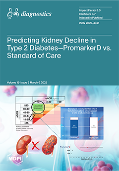
Chronic kidney disease in diabetes (DKD) affects up to 50% of people with type 2 diabetes (T2D), leading to high morbidity, mortality, and healthcare costs. The standard of care relies on the estimated glomerular filtration rate (eGFR) and urinary albumin/creatinine ratio (uACR), but these measures are highly variable due to factors like diet and exercise, limiting their accuracy. More precise prognostic tools are needed to improve DKD risk assessment in T2D. This study evaluates the biomarker-based PromarkerD test against eGFR and uACR for predicting kidney decline in people with T2D. PromarkerD demonstrated superior predictive accuracy, identifying 84% of cases that were missed by standard tests, with better risk stratification and fewer false positives. View this paper
  • Issues are regarded as officially published after their release is announced to the table of contents alert mailing list.
  • You may sign up for e-mail alerts to receive table of contents of newly released issues.
  • PDF is the official format for papers published in both, html and pdf forms. To view the papers in pdf format, click on the "PDF Full-text" link, and use the free Adobe Reader to open them.
Order results
Result details
Section
Select all
Export citation of selected articles as:
15 pages, 1017 KiB  
Systematic Review
Clinicopathological Comparison Between GREB1- and ESR1-Rearranged Uterine Tumors Resembling Ovarian Sex Cord Tumors (UTROSCTs): A Systematic Review
by Livia Maccio, Damiano Arciuolo, Angela Santoro, Antonio Raffone, Diego Raimondo, Susanna Ronchi, Nicoletta D’Alessandris, Giulia Scaglione, Michele Valente, Belen Padial Urtueta, Francesca Addante, Nadine Narducci, Emma Bragantini, Jvan Casarin, Giuseppe Angelico, Stefano La Rosa, Gian Franco Zannoni and Antonio Travaglino
Diagnostics 2025, 15(6), 792; https://doi.org/10.3390/diagnostics15060792 - 20 Mar 2025
Viewed by 361
Abstract
Introduction: Among uterine tumors resembling ovarian sex cord tumors (UTROSCTs), it has been suggested that GREB1-rearranged cases are biologically distinct from ESR1-rearranged cases and might be considered as a separate entity. Objectives: The aim of this systematic review was to assess [...] Read more.
Introduction: Among uterine tumors resembling ovarian sex cord tumors (UTROSCTs), it has been suggested that GREB1-rearranged cases are biologically distinct from ESR1-rearranged cases and might be considered as a separate entity. Objectives: The aim of this systematic review was to assess the difference between GREB1- and ESR1-rearranged UTROSCTs with regard to several clinico-pathological parameters. Methods: Three electronic databases were searched from their inception to February 2025 for all studies assessing the presence of GREB1 and ESR1 rearrangements in UTROSCTs. Exclusion criteria comprised overlapping patient data, case reports, and reviews. Statistical analysis was performed to compare clinicopathological variables between GREB1- and ESR1-rearranged UTROSCTs. Dichotomous variables were compared by using Fisher’s exact test; continuous variables were compared by using Student’s t-test. A p-value < 0.05 was considered significant. Results: Six studies with 88 molecularly classified UTROSCTs were included. A total of 36 cases were GREB1-rearranged, and 52 cases were ESR1-rearranged. GREB1-rearranged UTROSCTs showed a significantly older age (p < 0.001), larger tumor size (p = 0.002), less common submucosal/polypoid growth (p = 0.005), higher mitotic index (p = 0.010), more common LVSI (p = 0.049), and higher likelihood to undergo hysterectomy (p = 0.008) compared to ESR1-rearranged cases. No significant differences were detected with regard to margins, cytological atypia, necrosis, retiform pattern, and rhabdoid cells. No significant differences were found in the immunohistochemical expression of any of the assessed markers (wide-spectrum cytokeratins, α-inhibin, calretinin, WT1, CD10, CD56, CD99, smooth muscle actin, desmin, h-caldesmon, Melan-A/MART1, SF1, or Ki67). GREB1-rearranged UTROSCTs showed significantly lower disease-free survival compared to ESR1-rearranged UTROSTCs (p = 0.049). Conclusions: In conclusion, GREB1-rearranged UTROSCTs occur at an older age, are less likely to display a submucosal/polypoid growth, and exhibit larger size, a higher mitotic index, more common lymphovascular space invasion, and lower disease-free survival compared to ESR1-rearranged UTROSCTs. Nonetheless, the similar immunophenotype suggests that they belong to the same tumor family. Further studies are necessary to confirm this point. Full article
Show Figures

Figure 1

19 pages, 2305 KiB  
Article
Comparison of 46 Cytokines in Peripheral Blood Between Patients with Papillary Thyroid Cancer and Healthy Individuals with AI-Driven Analysis to Distinguish Between the Two Groups
by Kyung-Jin Bae, Jun-Hyung Bae, Ae-Chin Oh and Chi-Hyun Cho
Diagnostics 2025, 15(6), 791; https://doi.org/10.3390/diagnostics15060791 - 20 Mar 2025
Viewed by 400
Abstract
Background: Recent studies have analyzed some cytokines in patients with papillary thyroid carcinoma (PTC), but simultaneous analysis of multiple cytokines remains rare. Nonetheless, the simultaneous assessment of multiple cytokines is increasingly recognized as crucial for understanding the cytokine characteristics and developmental mechanisms [...] Read more.
Background: Recent studies have analyzed some cytokines in patients with papillary thyroid carcinoma (PTC), but simultaneous analysis of multiple cytokines remains rare. Nonetheless, the simultaneous assessment of multiple cytokines is increasingly recognized as crucial for understanding the cytokine characteristics and developmental mechanisms in PTC. In addition, studies applying artificial intelligence (AI) to discriminate patients with PTC based on serum multiple cytokine data have been performed rarely. Here, we measured and compared 46 cytokines in patients with PTC and healthy individuals, applying AI algorithms to classify the two groups. Methods: Blood serum was isolated from 63 patients with PTC and 63 control individuals. Forty-six cytokines were analyzed simultaneously using Luminex assay Human XL Cytokine Panel. Several laboratory findings were identified from electronic medical records. Student’s t-test or the Mann–Whitney U test were performed to analyze the difference between the two groups. As AI classification algorithms to categorize patients with PTC, K-nearest neighbor function, Naïve Bayes classifier, logistic regression, support vector machine, and eXtreme Gradient Boosting (XGBoost) were employed. The SHAP analysis assessed how individual parameters influence the classification of patients with PTC. Results: Cytokine levels, including GM-CSF, IFN-γ, IL-1ra, IL-7, IL-10, IL-12p40, IL-15, CCL20/MIP-α, CCL5/RANTES, and TNF-α, were significantly higher in PTC than in controls. Conversely, CD40 Ligand, EGF, IL-1β, PDGF-AA, and TGF-α exhibited significantly lower concentrations in PTC compared to controls. Among the five classification algorithms evaluated, XGBoost demonstrated superior performance in terms of accuracy, precision, sensitivity (recall), specificity, F1-score, and ROC-AUC score. Notably, EGF and IL-10 were identified as critical cytokines that significantly contributed to the differentiation of patients with PTC. Conclusions: A total of 5 cytokines showed lower levels in the PTC group than in the control, while 10 cytokines showed higher levels. While XGBoost demonstrated the best performance in discriminating between the PTC group and the control group, EGF and IL-10 were considered to be closely associated with PTC. Full article
(This article belongs to the Section Clinical Laboratory Medicine)
Show Figures

Figure 1

14 pages, 3652 KiB  
Article
Improving Diagnostic Performance for Head and Neck Tumors with Simple Diffusion Kurtosis Imaging and Machine Learning Bi-Parameter Analysis
by Suzuka Yoshida, Masahiro Kuroda, Yoshihide Nakamura, Yuka Fukumura, Yuki Nakamitsu, Wlla E. Al-Hammad, Kazuhiro Kuroda, Yudai Shimizu, Yoshinori Tanabe, Masataka Oita, Irfan Sugianto, Majd Barham, Nouha Tekiki, Nurul N. Kamaruddin, Miki Hisatomi, Yoshinobu Yanagi and Junichi Asaumi
Diagnostics 2025, 15(6), 790; https://doi.org/10.3390/diagnostics15060790 - 20 Mar 2025
Viewed by 309
Abstract
Background/Objectives: Mean kurtosis (MK) values in simple diffusion kurtosis imaging (SDI)—a type of diffusion kurtosis imaging (DKI)—have been reported to be useful in the diagnosis of head and neck malignancies, for which pre-processing with smoothing filters has been reported to improve the diagnostic [...] Read more.
Background/Objectives: Mean kurtosis (MK) values in simple diffusion kurtosis imaging (SDI)—a type of diffusion kurtosis imaging (DKI)—have been reported to be useful in the diagnosis of head and neck malignancies, for which pre-processing with smoothing filters has been reported to improve the diagnostic accuracy. Multi-parameter analysis using DKI in combination with other image types has recently been reported to improve the diagnostic performance. The purpose of this study was to evaluate the usefulness of machine learning (ML)-based multi-parameter analysis using the MK and apparent diffusion coefficient (ADC) values—which can be acquired simultaneously through SDI—for the differential diagnosis of benign and malignant head and neck tumors, which is important for determining the treatment strategy, as well as examining the usefulness of filter pre-processing. Methods: A total of 32 pathologically diagnosed head and neck tumors were included in the study, and a Gaussian filter was used for image pre-processing. MK and ADC values were extracted from pixels within the tumor area and used as explanatory variables. Five ML algorithms were used to create models for the prediction of tumor status (benign or malignant), which were evaluated through ROC analysis. Results: Bi-parameter analysis with gradient boosting achieved the best diagnostic performance, with an AUC of 0.81. Conclusions: The usefulness of bi-parameter analysis with ML methods for the differential diagnosis of benign and malignant head and neck tumors using SDI data were demonstrated. Full article
(This article belongs to the Section Medical Imaging and Theranostics)
Show Figures

Figure 1

36 pages, 10750 KiB  
Article
A Novel Diagnostic Framework with an Optimized Ensemble of Vision Transformers and Convolutional Neural Networks for Enhanced Alzheimer’s Disease Detection in Medical Imaging
by Joy Chakra Bortty, Gouri Shankar Chakraborty, Inshad Rahman Noman, Salil Batra, Joy Das, Kanchon Kumar Bishnu, Md Tanvir Rahman Tarafder and Araf Islam
Diagnostics 2025, 15(6), 789; https://doi.org/10.3390/diagnostics15060789 - 20 Mar 2025
Viewed by 505
Abstract
Background/Objectives: Alzheimer’s disease (AD) is a progressive, neurodegenerative disorder, which causes memory loss and loss of cognitive functioning, along with behavioral changes. Early detection is important to delay disease progression, timely intervention and to increase patients’ and caregivers’ quality of life (QoL). One [...] Read more.
Background/Objectives: Alzheimer’s disease (AD) is a progressive, neurodegenerative disorder, which causes memory loss and loss of cognitive functioning, along with behavioral changes. Early detection is important to delay disease progression, timely intervention and to increase patients’ and caregivers’ quality of life (QoL). One of the major and primary challenges for preventing any disease is to identify the disease at the initial stage through a quick and reliable detection process. Different researchers across the world are still working relentlessly, coming up with significant solutions. Artificial intelligence-based solutions are putting great importance on identifying the disease efficiently, where deep learning with medical imaging is highly being utilized to develop disease detection frameworks. In this work, a novel and optimized detection framework has been proposed that comes with remarkable performance that can classify the level of Alzheimer’s accurately and efficiently. Methods: A powerful vision transformer model (ViT-B16) with three efficient Convolutional Neural Network (CNN) models (VGG19, ResNet152V2, and EfficientNetV2B3) has been trained with a benchmark dataset, ‘OASIS’, that comes with a high volume of brain Magnetic Resonance Images (MRI). Results: A weighted average ensemble technique with a Grasshopper optimization algorithm has been designed and utilized to ensure maximum performance with high accuracy of 97.31%, precision of 97.32, recall of 97.35, and F1 score of 0.97. Conclusions: The work has been compared with other existing state-of-the-art techniques, where it comes with high efficiency, sensitivity, and reliability. The framework can be utilized in IoMT infrastructure where one can access smart and remote diagnosis services. Full article
(This article belongs to the Special Issue Artificial Intelligence in Brain Diseases)
Show Figures

Figure 1

9 pages, 1066 KiB  
Article
MRI Detection of Unknown Primary Tumours in the Head and Neck: What Is the Expected Normal Asymmetry in the Size of the Palatine Tonsils?
by Kaijing Mao, Qi Yong H. Ai, Kuo Feng Hung, Irene O. L. Tse, Ho Sang Leung, Yannis Yan Liang, Yu Chen, Lun M. Wong, W. K. Jacky Lam and Ann D. King
Diagnostics 2025, 15(6), 788; https://doi.org/10.3390/diagnostics15060788 - 20 Mar 2025
Viewed by 292
Abstract
Background/Objectives: The detection of unknown primary tumours in the palatine tonsils (PTs) on imaging relies heavily on asymmetry in size between the right and left sides, but the expected normal range in asymmetry is not well documented. This study aimed to document the [...] Read more.
Background/Objectives: The detection of unknown primary tumours in the palatine tonsils (PTs) on imaging relies heavily on asymmetry in size between the right and left sides, but the expected normal range in asymmetry is not well documented. This study aimed to document the expected range of asymmetry in the size of the PTs in adults without cancer. Methods: This retrospective study evaluated 250 pairs of normal PTs on MRIs of adults without head and neck cancer. The size (volume, V) of the PTs on the left and right sides were measured, and the percentage difference in volume (ΔV%) between the two sides was calculated. An additional analysis of PT volumes in 29 patients with ipsilateral early-stage palatine tonsillar cancer (PTCs) was performed. Results: In patients without PTC, the normal PTs had a mean volume of 3.0 ± 1.7 cm3, and there was a difference in size between the left and right PTs, showing a median ΔV% of 11.6% (range: 0.1–79.0%); most patients had a ΔV% of ≤40% (95%) for PTs. In patients with ipsilateral PTC, the normal PT had a smaller size compared with PTC (p < 0.01), showing a median ΔV% of 132.9% (range: 8.5–863.2%). Compared with patients without PTC, those with PTC showed a greater ΔV% (p < 0.01). An optimal ΔV% threshold of >39.6% achieved the best accuracy of 95% for identifying PTC. Conclusions: PTs are asymmetrical in size in adults without PTC. An additional analysis involving patients with PTC confirmed a threshold of ΔV% of 40% for PTs, which may be clinically valuable to help detect pathology using MRI. Full article
(This article belongs to the Special Issue Advances in Diagnosis and Treatment in Otolaryngology)
Show Figures

Figure 1

28 pages, 1031 KiB  
Review
AI and Smart Devices in Cardio-Oncology: Advancements in Cardiotoxicity Prediction and Cardiovascular Monitoring
by Luiza Camelia Nechita, Dana Tutunaru, Aurel Nechita, Andreea Elena Voipan, Daniel Voipan, Ancuta Elena Tupu and Carmina Liana Musat
Diagnostics 2025, 15(6), 787; https://doi.org/10.3390/diagnostics15060787 - 20 Mar 2025
Viewed by 572
Abstract
The increasing prevalence of cardiovascular complications in cancer patients due to cardiotoxic treatments has necessitated advanced monitoring and predictive solutions. Cardio-oncology is an evolving interdisciplinary field that addresses these challenges by integrating artificial intelligence (AI) and smart cardiac devices. This comprehensive review explores [...] Read more.
The increasing prevalence of cardiovascular complications in cancer patients due to cardiotoxic treatments has necessitated advanced monitoring and predictive solutions. Cardio-oncology is an evolving interdisciplinary field that addresses these challenges by integrating artificial intelligence (AI) and smart cardiac devices. This comprehensive review explores the integration of artificial intelligence (AI) and smart cardiac devices in cardio-oncology, highlighting their role in improving cardiovascular risk assessment and the early detection and real-time monitoring of cardiotoxicity. AI-driven techniques, including machine learning (ML) and deep learning (DL), enhance risk stratification, optimize treatment decisions, and support personalized care for oncology patients at cardiovascular risk. Wearable ECG patches, biosensors, and AI-integrated implantable devices enable continuous cardiac surveillance and predictive analytics. While these advancements offer significant potential, challenges such as data standardization, regulatory approvals, and equitable access must be addressed. Further research, clinical validation, and multidisciplinary collaboration are essential to fully integrate AI-driven solutions into cardio-oncology practices and improve patient outcomes. Full article
(This article belongs to the Special Issue AI-Assisted Diagnostics in Telemedicine and Digital Health)
Show Figures

Figure 1

10 pages, 1842 KiB  
Article
Do We Need to Add the Type of Treatment Planning System, Dose Calculation Grid Size, and CT Density Curve to Predictive Models?
by Reza Reiazi, Surendra Prajapati, Leonardo Che Fru, Dongyeon Lee and Mohammad Salehpour
Diagnostics 2025, 15(6), 786; https://doi.org/10.3390/diagnostics15060786 - 20 Mar 2025
Viewed by 309
Abstract
Background: Generalizability and domain dependency are critical challenges in developing predictive models for healthcare, particularly in medical diagnostics and radiation oncology. Predictive models designed to assess tumor recurrence rely on comprehensive and high-quality datasets, encompassing treatment planning parameters, imaging protocols, and patient-specific data. [...] Read more.
Background: Generalizability and domain dependency are critical challenges in developing predictive models for healthcare, particularly in medical diagnostics and radiation oncology. Predictive models designed to assess tumor recurrence rely on comprehensive and high-quality datasets, encompassing treatment planning parameters, imaging protocols, and patient-specific data. However, domain dependency, arising from variations in dose calculation algorithms, computed tomography (CT) density conversion curves, imaging modalities, and institutional protocols, can significantly undermine model reliability and clinical utility. Methods: This study evaluated dose calculation differences in the head and neck cancer treatment plans of 19 patients using two treatment planning systems, Pinnacle 9.10 and RayStation 11, with similar dose calculation algorithms. Variations in the dose grid size and CT density conversion curves were assessed for their impact on domain dependency. Results: Results showed that dose grid size differences had a more significant influence within RayStation than Pinnacle, while CT curve variations introduced potential domain discrepancies. The findings underscore the critical role of precise and standardized treatment planning in enhancing the reliability of predictive modeling for tumor recurrence assessment. Conclusions: Incorporating treatment planning parameters, such as dose distribution and target volumes, as explicit features in model training can mitigate the impact of domain dependency and enhance prediction accuracy. Solutions such as multi-institutional data harmonization and domain adaptation techniques are essential to improve model generalizability and robustness. These strategies support the better integration of predictive modeling into clinical workflows, ultimately optimizing patient outcomes and personalized treatment strategies. Full article
(This article belongs to the Special Issue Artificial Intelligence in Clinical Decision Support—2nd Edition)
Show Figures

Figure 1

22 pages, 7900 KiB  
Systematic Review
What the Radiologist Needs to Know About Sport Hernias: A Systematic Review of the Current Literature
by Gian Nicola Bisciotti, Andrea Bisciotti, Alessandro Bisciotti and Alessio Auci
Diagnostics 2025, 15(6), 785; https://doi.org/10.3390/diagnostics15060785 - 20 Mar 2025
Viewed by 379
Abstract
Introduction: The sports hernia (SH) is one of the most important causes of groin pain syndrome (GPS). However, despite its importance in GPS etiopathogenesis, SH is one of the least understood and poorly defined clinical conditions in sports medicine. The aim of this [...] Read more.
Introduction: The sports hernia (SH) is one of the most important causes of groin pain syndrome (GPS). However, despite its importance in GPS etiopathogenesis, SH is one of the least understood and poorly defined clinical conditions in sports medicine. The aim of this systematic review is to clearly define SH from a radiological point of view and to clarify the relationship between the radiological presentation of SH and its clinical manifestation. Methods: The PubMed/MEDLINE, Scopus, ISI, Cochrane Database of Systematic Reviews, and PEDro databases were consulted for systematic reviews on the role of SH in the onset of GPS. The inclusion and exclusion criteria were based on PICO tool. Results: After screening 560 articles, 81 studies were included and summarized in this systematic review. All studies were checked to identify any potential conflict of interest. The quality assessment of each individual study considered was performed in agreement with the Joanna Briggs Institute quantitative critical appraisal tools. Conclusions: The correct definition of SH is “weakness of the posterior wall of the inguinal canal”, which, in response to a Valsalva maneuver, forms a bulging that compresses the nerves passing along the inguinal canal. Thus, from an anatomical point of view, SH represents a direct inguinal hernia “in fieri”. Furthermore, an excessive dilation of the external inguinal ring represents an indirect sign of possible posterior inguinal canal wall weakness. Full article
(This article belongs to the Special Issue Imaging Diagnosis in Abdomen, 2nd Edition)
Show Figures

Figure 1

13 pages, 2033 KiB  
Article
Relationship Between Sigmoid Volvulus Subtypes, Clinical Course, and Imaging Findings
by Kemal Bugra Memis and Sonay Aydin
Diagnostics 2025, 15(6), 784; https://doi.org/10.3390/diagnostics15060784 - 20 Mar 2025
Viewed by 389
Abstract
Background: Recent studies indicate that the organo-axial subtype of a sigmoid volvulus is more prevalent than the conventional mesentero-axial subtype. Our study aimed to assess the clinical and radiological findings that differentiate between these two subtypes, as well as to ascertain treatment outcomes [...] Read more.
Background: Recent studies indicate that the organo-axial subtype of a sigmoid volvulus is more prevalent than the conventional mesentero-axial subtype. Our study aimed to assess the clinical and radiological findings that differentiate between these two subtypes, as well as to ascertain treatment outcomes and prognostic characteristics. Methods: A retrospective review included 54 patients, during which abdominal plain radiographs and computed tomography images were analyzed by two radiologists, and data on recurrence, mortality, and treatment outcomes were documented. Results: The mesentero-axial subtype comprised 40 cases (74%). No distinct radiographic findings were observed to differentiate between the two groups. In computed tomography, the sole significant parameter for differentiation was the number of transition zones. The diameter of the segment exhibiting a volvulus was greater in instances of the mesentero-axial subtype. The endoscopic detorsion treatment proved ineffective in five patients within the mesentero-axial sigmoid volvulus cohort. Conclusions: Identifying these two types of SV on CT images is essential because of their distinct prognoses and therapeutic results. Full article
(This article belongs to the Special Issue Diagnostic Imaging in Gastrointestinal and Liver Diseases)
Show Figures

Figure 1

16 pages, 2180 KiB  
Article
Machine Learning Based Multi-Parameter Modeling for Prediction of Post-Inflammatory Lung Changes
by Gerlig Widmann, Anna Katharina Luger, Thomas Sonnweber, Christoph Schwabl, Katharina Cima, Anna Katharina Gerstner, Alex Pizzini, Sabina Sahanic, Anna Boehm, Maxmilian Coen, Ewald Wöll, Günter Weiss, Rudolf Kirchmair, Leonhard Gruber, Gudrun M. Feuchtner, Ivan Tancevski, Judith Löffler-Ragg and Piotr Tymoszuk
Diagnostics 2025, 15(6), 783; https://doi.org/10.3390/diagnostics15060783 - 20 Mar 2025
Viewed by 301
Abstract
Objectives: Prediction of lung function deficits following pulmonary infection is challenging and suffers from inaccuracy. We sought to develop machine-learning models for prediction of post-inflammatory lung changes based on COVID-19 recovery data. Methods: In the prospective CovILD study (n = [...] Read more.
Objectives: Prediction of lung function deficits following pulmonary infection is challenging and suffers from inaccuracy. We sought to develop machine-learning models for prediction of post-inflammatory lung changes based on COVID-19 recovery data. Methods: In the prospective CovILD study (n = 420 longitudinal observations from n = 140 COVID-19 survivors), data on lung function testing (LFT), chest CT including severity scoring by a human radiologist and density measurement by artificial intelligence, demography, and persistent symptoms were collected. This information was used to develop models of numeric readouts and abnormalities of LFT with four machine learning algorithms (Random Forest, gradient boosted machines, neural network, and support vector machines). Results: Reduced DLCO (diffusion capacity for carbon monoxide <80% of reference) was found in 94 (22%) observations. Those observations were modeled with a cross-validated accuracy of 82–85%, AUC of 0.87–0.9, and Cohen’s κ of 0.45–0.5. No reliable models could be established for FEV1 or FVC. For DLCO as a continuous variable, three machine learning algorithms yielded meaningful models with cross-validated mean absolute errors of 11.6–12.5% and R2 of 0.26–0.34. CT-derived features such as opacity, high opacity, and CT severity score were among the most influential predictors of DLCO impairment. Conclusions: Multi-parameter machine learning trained with demographic, clinical, and artificial intelligence chest CT data reliably and reproducibly predicts LFT deficits and outperforms single markers of lung pathology and human radiologist’s assessment. It may improve diagnostic and foster personalized treatment. Full article
(This article belongs to the Special Issue Artificial Intelligence in Lung Diseases: 3rd Edition)
Show Figures

Figure 1

14 pages, 5303 KiB  
Article
The Zucker Diabetic Fatty Rat as a Model for Vascular Changes in Diabetic Kidney Disease: Characterising Hydronephrosis
by Amy McDermott, Nathalie Sarup Panduro, Iman Taghavi, Hans Martin Kjer, Stinne Byrholdt Søgaard, Michael Bachmann Nielsen, Jørgen Arendt Jensen and Charlotte Mehlin Sørensen
Diagnostics 2025, 15(6), 782; https://doi.org/10.3390/diagnostics15060782 - 20 Mar 2025
Viewed by 316
Abstract
Background/Objectives: Diabetic kidney disease (DKD) is a significant concern for global healthcare, particularly in individuals with diabetes. The Zucker rat strain is a commonly used model of type 2 diabetes, despite awareness that this animal can develop hydronephrosis. In this study, we present [...] Read more.
Background/Objectives: Diabetic kidney disease (DKD) is a significant concern for global healthcare, particularly in individuals with diabetes. The Zucker rat strain is a commonly used model of type 2 diabetes, despite awareness that this animal can develop hydronephrosis. In this study, we present novel imaging data evaluating the accuracy of this animal model in replicating the vascular aspects of human DKD while examining the impact of hydronephrosis on its validity as a disease model. Methods: This study reused data from a population of male Zucker Diabetic Fatty (ZDF; n = 22) rats and Zucker Lean (ZL) rats (n = 22) aged 12 to approximately 40 weeks. Vascular casting was performed to enable visualisation of the renal vasculature. Anatomical regional volumes and vascular density data were obtained from μCT scans using image thresholding and manual analysis. The effects of hydronephrosis were evaluated using renal functional parameters and histological examination. Results: A significantly lower cortical vascular density, as well as lower total renal vascular density, was seen in ZDF rats compared to ZL rats, independent of age. We identified that hydronephrosis affected 92% of ZDF rats and 69% of ZL rats. Hydronephrosis cavity size was significantly correlated with the degree of hyperglycaemia and rate of diuresis but had no other detected impact on renal function, vascularity, or tissue histological architecture. Conclusions: These findings support using the Zucker rat strain as a model for vascular changes in DKD. Despite identifying severe hydronephrosis in this population, it had minimal quantifiable impact on renal function or diabetes modelling. Full article
(This article belongs to the Special Issue Current Issues on Kidney Diseases Diagnosis and Management 2025)
Show Figures

Figure 1

11 pages, 6299 KiB  
Case Report
Cladosporium species novum Invasive Pulmonary Infection in a Patient with Post-COVID-19 Syndrome and AIDS
by Milorad Bijelović, Nikola Gardić, Aleksandra Lovrenski, Danijela Petrović, Gordana Kozoderović, Vesna Lalošević, Vuk Vračar and Dušan Lalošević
Diagnostics 2025, 15(6), 781; https://doi.org/10.3390/diagnostics15060781 - 20 Mar 2025
Viewed by 1189
Abstract
Background and Clinical Significance: Since the prevalence of fungal lung infections is increasing, certain agents, such as Cladosporium spp., have emerged as unexpected causes. Cladosporium spp. fungi are ubiquitous in environments such as soil, fruits, and wine corks; they are a part of [...] Read more.
Background and Clinical Significance: Since the prevalence of fungal lung infections is increasing, certain agents, such as Cladosporium spp., have emerged as unexpected causes. Cladosporium spp. fungi are ubiquitous in environments such as soil, fruits, and wine corks; they are a part of the normal human skin flora; and they are known respiratory allergens. Case Presentation: A patient with a history of post-COVID-19 syndrome and AIDS presented with lung pathology indicative of an invasive fungal infection. The initial histopathological examination revealed numerous yeast-like cells with narrow-based budding, which led to a mistaken diagnosis of cryptococcosis. However, further detailed examination revealed sparse hyphae in the lung tissue, suggesting a more complex fungal infection. Molecular analyses and sequence BLAST alignment were performed, ultimately identifying the infectious agent as “Cladosporium species novum”, a rare cause of invasive pulmonary cladosporiasis. Conclusions: Invasive pulmonary cladosporiasis is a rare condition, and the morphological features of the fungus alone were insufficient to establish a correct diagnosis. A comprehensive pathohistological and molecular approach with bioinformatics tools is essential for the correct identification of rare and potentially life-threatening fungal pathogens in immunocompromised patients. Full article
(This article belongs to the Section Pathology and Molecular Diagnostics)
Show Figures

Figure 1

4 pages, 4209 KiB  
Interesting Images
Orbital Neurolymphomatosis in Patient with CNS Lymphoma
by Tara Shooshani, Michael Han, Jeremiah P. Tao, Samuel J. Spiegel and Maria Del Valle Estopinal
Diagnostics 2025, 15(6), 780; https://doi.org/10.3390/diagnostics15060780 - 20 Mar 2025
Viewed by 317
Abstract
Neurolymphomatosis (NL) is a rare manifestation of hematologic malignancies, characterized by a neoplastic infiltration of the peripheral nervous system and cranial nerves (CNs). Non-Hodgkin lymphomas (NHLs) account for 90% of NL cases, while acute leukemia represents 10% of the cases. NL can occur [...] Read more.
Neurolymphomatosis (NL) is a rare manifestation of hematologic malignancies, characterized by a neoplastic infiltration of the peripheral nervous system and cranial nerves (CNs). Non-Hodgkin lymphomas (NHLs) account for 90% of NL cases, while acute leukemia represents 10% of the cases. NL can occur as the first manifestation of a malignancy (primary), or as a relapse or progression of a previously treated disease (secondary). Herein, we report a unique case of NL involving the left orbit and CNs in a 74-year-old female with primary central nervous system (CNS) diffuse large B-cell lymphoma (DLBCL). Our patient developed secondary neurolymphomatosis involving the left orbit and CNs II, III, V, and VI, supported by clinical, radiologic, and histologic findings. The lacrimal gland enhancement was histopathologically proven to be caused by the direct spread of CNS DLBCL to the lacrimal nerve, a branch of CN V, identifying NL as one of the conditions that can affect this organ. The lacrimal gland could be considered as a more accessible biopsy site when the involvement of CN V is suspected. Full article
(This article belongs to the Special Issue Pathology and Diagnosis of Head and Neck Diseases)
Show Figures

Figure 1

14 pages, 2871 KiB  
Article
The Association of Heart Failure and Liver T1 Mapping in Cardiac Magnetic Resonance Imaging
by Adrian T. Huber, Joanna Bartkowiak, Robin Seitz, Benedikt Bernhard, Martina Boscolo Berto, Giancarlo Spano, Benedikt Wagner, Verena C. Obmann, Lukas Ebner, Inga A. S. Todorski, Michael P. Brönnimann, Kady Fischer, Dominik P. Guensch, Andreas Christe, Annalisa Berzigotti, Lorenz Räber, Tobias Reichlin, Thomas Pilgrim, Fabien Praz, Christoph Gräni, Nicholas Brugger and Alan A. Petersadd Show full author list remove Hide full author list
Diagnostics 2025, 15(6), 779; https://doi.org/10.3390/diagnostics15060779 - 20 Mar 2025
Viewed by 348
Abstract
Background/Objectives: The objective of this study was to investigate the association between congestive heart failure (CHF) and T1 mapping in both liver lobes using cardiac MRI. Methods: This retrospective study included patients who underwent cardiac MRI with T1 mapping sequences on a 1.5 [...] Read more.
Background/Objectives: The objective of this study was to investigate the association between congestive heart failure (CHF) and T1 mapping in both liver lobes using cardiac MRI. Methods: This retrospective study included patients who underwent cardiac MRI with T1 mapping sequences on a 1.5 T scanner. The liver T1 values were measured in four hepatic regions, utilizing cardiac short axis and four-chamber views. Echocardiographic and laboratory data were collected within 90 days of the cardiac MRI. Comparisons of the liver T1 values and echocardiographic parameters between patients with and without elevated NT-proBNP levels (>125 pg/mL) were conducted using the Mann–Whitney U test. Logistic regression models were employed to adjust for confounding factors. Results: A total of 397 patients were included (with a median age of 56 years; 127 females), of whom 35% (n = 138) exhibited elevated NT-proBNP levels. The patients with elevated NT-proBNP levels showed a larger end-diastolic volume (EDV: 92 vs. 81 mL/m2, p < 0.001) and a lower LVEF level (50% vs. 60%, p < 0.001). The liver T1 was significantly higher in the right liver lobe (670 vs. 596 ms, p < 0.001) and the caudate lobe (664 vs. 598 ms, p < 0.001), but not in the left lobe (571 vs. 568 ms, p = 0.068) or the dome (590 vs. 560 ms, p = 0.1). T1 mapping in the caudate (OR 1.013, 95% CI 1.004–1.023, p = 0.005) and right liver lobes (OR 1.012, 95% CI 1.003–1.021, p = 0.009) remained independently predictive in the logistic regression analysis. Conclusions: Elevated T1 values in the caudate and right liver lobes assessed by cardiac MRI were independently associated with CHF and outperformed T1 measurements in the left liver lobe in predicting disease. Full article
(This article belongs to the Section Medical Imaging and Theranostics)
Show Figures

Figure 1

18 pages, 9445 KiB  
Review
The Role of Autopsy in Diagnosing Fatal Chest Injuries in Road Traffic Accidents: A Literature Review
by Matteo Antonio Sacco, Maria Cristina Verrina, Saverio Gualtieri, Agostinho Santos, Bárbara Ferreira Mendes, Alessandro Pasquale Tarallo, Aurora Princi, Stefano Lombardo, Pietrantonio Ricci and Isabella Aquila
Diagnostics 2025, 15(6), 778; https://doi.org/10.3390/diagnostics15060778 - 19 Mar 2025
Viewed by 471
Abstract
Road accidents are one of the leading causes of death worldwide, with significant repercussions on public health and the global economy. Fatal accidents can cause injuries in various anatomical areas with different dynamics. The thorax is one of the main sites involved in [...] Read more.
Road accidents are one of the leading causes of death worldwide, with significant repercussions on public health and the global economy. Fatal accidents can cause injuries in various anatomical areas with different dynamics. The thorax is one of the main sites involved in fatal accidents, due to the presence of vital organs such as the heart and lungs. Protective devices, such as seatbelts and airbags, also play a fundamental role in preventing chest injuries. However, external examination is often insufficient to determine the extent of internal trauma, resulting in significant difficulties in reconstructing the accident dynamics. In particular, in the absence of an autopsy, it is difficult to determine whether the driver or passengers were wearing protective devices, such as seatbelts, at the time of the accident. Diagnosing injuries secondary to protective devices, such as airbags, can also be complex without this assessment. Through a review of the literature, this work analyzes the different types of thoracic trauma that can be found at autopsy, providing indications to the forensic pathologist for the examination of these injuries. This review highlights the importance of the autopsy examination as a gold-standard investigation in the analysis of thoracic trauma from road accidents, in order to evaluate with certainty the injuries that caused death, and to facilitate the reconstruction of the dynamics for judicial purposes. Finally, an analysis of postmortem radiological investigations and of the role of protective measures in these events, such as the seatbelt and airbag, is provided. Full article
(This article belongs to the Special Issue Advances in Forensic Medical Diagnosis)
Show Figures

Figure 1

12 pages, 1547 KiB  
Article
Impact of Radiologist Experience on AI Annotation Quality in Chest Radiographs: A Comparative Analysis
by Malte Michel Multusch, Lasse Hansen, Mattias Paul Heinrich, Lennart Berkel, Axel Saalbach, Heinrich Schulz, Franz Wegner, Joerg Barkhausen and Malte Maria Sieren
Diagnostics 2025, 15(6), 777; https://doi.org/10.3390/diagnostics15060777 - 19 Mar 2025
Viewed by 312
Abstract
Background/Objectives: In the burgeoning field of medical imaging and Artificial Intelligence (AI), high-quality annotations for training AI-models are crucial. However, there are still only a few large datasets, as segmentation is time-consuming, experts have limited time. This study investigates how the experience [...] Read more.
Background/Objectives: In the burgeoning field of medical imaging and Artificial Intelligence (AI), high-quality annotations for training AI-models are crucial. However, there are still only a few large datasets, as segmentation is time-consuming, experts have limited time. This study investigates how the experience of radiologists affects the quality of annotations. Methods: We randomly collected 53 anonymized chest radiographs. Fifteen readers with varying levels of expertise annotated the anatomical structures of different complexity, pneumonic opacities and central venous catheters (CVC) as examples of pathologies and foreign material. The readers were divided into three groups of five. The groups consisted of medical students (MS), junior professionals (JP) with less than five years of working experience and senior professionals (SP) with more than five years of experience. Each annotation was compared to a gold standard consisting of a consensus annotation of three senior board-certified radiologists. We calculated the Dice coefficient (DSC) and Hausdorff distance (HD) to evaluate annotation quality. Inter- and intrareader variability and time dependencies were investigated using Intraclass Correlation Coefficient (ICC) and Ordinary Least Squares (OLS). Results: Senior professionals generally showed better performance, while medical students had higher variability in their annotations. Significant differences were noted, especially for complex structures (DSC Pneumonic Opacities as mean [standard deviation]: MS: 0.516 [0.246]; SP: 0.631 [0.211]). However, it should be noted that overall deviation and intraclass variance was higher for these structures even for seniors, highlighting the inherent limitations of conventional radiography. Experience showed a positive relationship with annotation quality for VCS and lung but was not a significant factor for other structures. Conclusions: Experience level significantly impacts annotation quality. Senior radiologists provided higher-quality annotations for complex structures, while less experienced readers could still annotate simpler structures with satisfying accuracy. We suggest a mixed-expertise approach, enabling the highly experienced to utilize their knowledge most effectively. With the increase in numbers of examinations, radiology will rely on AI support tools in the future. Therefore, economizing the process of data acquisition and AI-training; for example, by integrating less experienced radiologists, will help to meet the coming challenges. Full article
(This article belongs to the Section Machine Learning and Artificial Intelligence in Diagnostics)
Show Figures

Figure 1

16 pages, 1219 KiB  
Review
Deep Learning Models to Detect Anterior Cruciate Ligament Injury on MRI: A Comprehensive Review
by Michele Mercurio, Federica Denami, Dimitra Melissaridou, Katia Corona, Simone Cerciello, Domenico Laganà, Giorgio Gasparini and Roberto Minici
Diagnostics 2025, 15(6), 776; https://doi.org/10.3390/diagnostics15060776 - 19 Mar 2025
Viewed by 538
Abstract
Magnetic resonance imaging (MRI) is routinely used to confirm the suspected diagnosis of anterior cruciate ligament (ACL) injury. Recently, many studies explored the role of artificial intelligence (AI) and deep learning (DL), a sub-category of AI, in the musculoskeletal field and medical imaging. [...] Read more.
Magnetic resonance imaging (MRI) is routinely used to confirm the suspected diagnosis of anterior cruciate ligament (ACL) injury. Recently, many studies explored the role of artificial intelligence (AI) and deep learning (DL), a sub-category of AI, in the musculoskeletal field and medical imaging. The aim of this study was to review the current applications of DL models to detect ACL injury on MRI, thus providing an updated and critical synthesis of the existing literature and identifying emerging trends and challenges in the field. A total of 23 relevant articles were identified and included in the review. Articles originated from 10 countries, with China having the most contributions (n = 9), followed by the United State of America (n = 4). Throughout the article, we analyzed the concept of DL in ACL tears and provided examples of how these tools can impact clinical practice and patient care. DL models for MRI detection of ACL injury reported high values of accuracy, especially helpful for less experienced clinicians. Time efficiency was also demonstrated. Overall, the deep learning models have proven to be a valid resource, although still requiring technological developments for implementation in daily practice. Full article
(This article belongs to the Special Issue Artificial Intelligence in Clinical Medical Imaging: 2nd Edition)
Show Figures

Figure 1

14 pages, 3375 KiB  
Case Report
Large-Cell Neuroendocrine Carcinoma of the Cervix: Case Report and Literature Review
by Wing Yu Sharon Siu, Chiu-Hsuan Cheng and Dah-Ching Ding
Diagnostics 2025, 15(6), 775; https://doi.org/10.3390/diagnostics15060775 - 19 Mar 2025
Viewed by 487
Abstract
Background and clinical significance: Large-cell neuroendocrine carcinoma (LCNEC) of the cervix is considered a rare type of cancer: it represents <1% of invasive cervical cancers. The optimal treatment protocol is not fully established because of its rarity and diagnostic challenges. Case Presentation [...] Read more.
Background and clinical significance: Large-cell neuroendocrine carcinoma (LCNEC) of the cervix is considered a rare type of cancer: it represents <1% of invasive cervical cancers. The optimal treatment protocol is not fully established because of its rarity and diagnostic challenges. Case Presentation: A 72-year-old Asian female presented to our outpatient clinic with postmenopausal vaginal spotting for 1 month. Vaginal sonography revealed a cervical tumor of 2.7 cm in diameter with hypervascularity. Tumor markers such as CA 125, CA 19-9, carcinoembryonic antigen, and squamous cell carcinoma antigen all showed no abnormality. Due to high suspicion of cervical cancer, a pap smear and endocervical curettage were performed and confirmed the diagnosis of LCNEC. A positron emission tomography–computed tomography scan demonstrated a glucose hypermetabolic lesion in the mid-pelvic region, localized to the uterus, consistent with LCNEC. Surgery with radical hysterectomy, bilateral salpingo-oophorectomy, and bilateral pelvic lymph node dissection was performed. The patient was finally diagnosed with pT1b2N1mi, FIGO IIIC1. Immunohistochemical stain shows that the neoplastic cells were CK (+), p63 (−), p16 (−), CEA (−), vimentin (−), ER (−), WT-1 (−), p53 (−), and CD56 (+), with a high Ki67 index (75%). Concurrent chemotherapy with cisplatin and radiotherapy was performed. Four cycles of etoposide and cisplatin were planned. A 3-month follow-up of this patient revealed stable tumor marker levels. Conclusions: This case highlights the diagnostic challenges and aggressive nature of LCNEC of the cervix, emphasizing the need for a standardized treatment approach to improve patient outcomes. Full article
(This article belongs to the Section Clinical Diagnosis and Prognosis)
Show Figures

Figure 1

10 pages, 2624 KiB  
Case Report
A Silent Threat: Internal Carotid Artery Hypoplasia and Its Role in Basilar Artery Aneurysm Formation—A Case Study
by Paula Mežvinska, Artis Brokāns, Sergejs Pavlovičs, Matīss Dravnieks, Ardis Platkājis and Arturs Balodis
Diagnostics 2025, 15(6), 774; https://doi.org/10.3390/diagnostics15060774 - 19 Mar 2025
Viewed by 311
Abstract
Background and Clinical Significance: Hypoplasia of the internal carotid artery (ICA) is a rare vascular anomaly, with an estimated incidence of less than 0.01%. This condition can result in reduced blood flow to the anterior circulation, often compensated by collateral circulation. Radiological imaging, [...] Read more.
Background and Clinical Significance: Hypoplasia of the internal carotid artery (ICA) is a rare vascular anomaly, with an estimated incidence of less than 0.01%. This condition can result in reduced blood flow to the anterior circulation, often compensated by collateral circulation. Radiological imaging, particularly computed tomography angiography (CTA), digital subtraction angiography (DSA), magnetic resonance angiography (MRA), and ultrasound, plays a crucial role in diagnosing this condition, revealing structural abnormalities in the arterial system. Case Presentation: This case is about a 75-year-old woman who lived her entire life unaware of ICAH until a basilar artery aneurysm ruptured, leading to a large, centrally localized SAH. Further diagnostic workup, including CTA and DSA, confirmed left ICA hypoplasia, with the artery terminating as the ophthalmic artery, along with a developmental anomaly of the left middle cerebral artery from posterior circulation territory. Due to the high risk associated with surgical and endovascular intervention, conservative management was pursued, and the patient’s condition stabilized, though she continued to have significant neurological deficits. Conclusions: This case report supports the hypothesis that ICAH may be associated with aneurysm formation. This case demonstrates that if ICAH is not diagnosed early, it can lead to severe complications with permanent neurological deficits. Additionally, it highlights the critical importance of advanced imaging techniques, such as CTA and DSA, in diagnosing complex vascular conditions. Full article
(This article belongs to the Special Issue Diagnostic Imaging of Brain Disease, Second Edition)
Show Figures

Figure 1

20 pages, 4694 KiB  
Article
A Comparative Study of Machine Learning and Deep Learning Models for Automatic Parkinson’s Disease Detection from Electroencephalogram Signals
by Sankhadip Bera, Zong Woo Geem, Young-Im Cho and Pawan Kumar Singh
Diagnostics 2025, 15(6), 773; https://doi.org/10.3390/diagnostics15060773 - 19 Mar 2025
Viewed by 629
Abstract
Background: Parkinson’s disease (PD) is one of the most prevalent, widespread, and intricate neurodegenerative disorders. According to the experts, at least 1% of people over the age of 60 are affected worldwide. In the present time, the early detection of PD remains difficult [...] Read more.
Background: Parkinson’s disease (PD) is one of the most prevalent, widespread, and intricate neurodegenerative disorders. According to the experts, at least 1% of people over the age of 60 are affected worldwide. In the present time, the early detection of PD remains difficult due to the absence of a clear consensus on its brain characterization. Therefore, there is an urgent need for a more reliable and efficient technique for early detection of PD. Using the potential of electroencephalogram (EEG) signals, this study introduces an innovative method for the detection or classification of PD patients through machine learning, as well as a more accurate deep learning approach. Methods: We propose an innovative EEG-based PD detection approach by integrating advanced spectral feature engineering with machine learning and deep learning models. Using (a) the UC San Diego Resting State EEG dataset and (b) IOWA dataset, we extract a standardized EEG feature from five key frequency bands—alpha, beta, theta, gamma, delta (α,β,θ,γ,δ) and employ an SVM (Support Vector Machine) classifier as a baseline, achieving a notable accuracy. Furthermore, we implement a deep learning classifier (CNN) with a complex multi-dimensional feature set by combining power values from all frequency bands, which gives superior performance in distinguishing PD patients (both with medication and without medication states) from healthy patients. Results: With the five-fold cross-validation on these two datasets, our approaches successfully achieve promising results in a subject dependent scenario. The SVM classifier achieves competitive accuracies of 82% and 94% in the UC San Diego Resting State EEG dataset (using gamma band) and IOWA dataset, respectively in distinguishing PD patients from non-PD patients in subject. With the CNN classifier, our model is able to capture major cross-frequency dependencies of EEG; therefore, the classification accuracies reach beyond 96% and 99% with those two datasets, respectively. We also perform our experiments in a subject independent environment, where the SVM generates 68.09% accuracy. Conclusions: Our findings, coupled with advanced feature extraction and deep learning, have the potential to provide a non-invasive, efficient, and reliable approach for diagnosing PD, with further work aimed at enhancing feature sets, inclusion of a large number of subjects, and improving model generalizability across more diverse environments. Full article
Show Figures

Figure 1

10 pages, 1196 KiB  
Article
Comprehensive Analysis of the Aberrant Right Subclavian Artery: A Perspective from a Single Institute
by Rou Jiun Lin, Kim-Seng Law and Pei-Jhen Wu
Diagnostics 2025, 15(6), 772; https://doi.org/10.3390/diagnostics15060772 - 19 Mar 2025
Viewed by 397
Abstract
Background/Objectives: This study aimed to provide a descriptive review of fetal aberrant right subclavian artery (ARSA), with a discussion of the genomic and structural anatomy and perinatal prognosis in our hospital’s obstetric department. Methods: In total, 3266 fetal level II sonographies [...] Read more.
Background/Objectives: This study aimed to provide a descriptive review of fetal aberrant right subclavian artery (ARSA), with a discussion of the genomic and structural anatomy and perinatal prognosis in our hospital’s obstetric department. Methods: In total, 3266 fetal level II sonographies were performed between January 2020 and June 2023. The 21 cases diagnosed with ARSA were included in this study. Obstetric ultrasound screening, noninvasive prenatal screening, and fetal karyotyping were performed. Fetal echocardiograms, postnatal information, and follow-up data were recorded. Results: In our dataset of 3266 cases, the overall incidence rate of ARSA was 0.6%. Of the 21 fetuses with ARSA, no abnormalities were detected in either prenatal or genetic tests, and no chromosomal anomalies were identified. Conclusions: Our study provides informative insights into ARSA, emphasizing the need for a comprehensive evaluation of its structural and genetic aspects. The findings of this study prompt further exploration, especially regarding the increasing incidence of ARSA and the potential role of advanced genetic analyses in enhancing diagnostic precision and fetal prognostic evaluation. Full article
(This article belongs to the Special Issue Advancements in Maternal–Fetal Medicine)
Show Figures

Figure 1

12 pages, 2423 KiB  
Article
Predictors of Diagnostic Inaccuracy of Detecting Coronary Artery Stenosis by Preprocedural CT Angiography in Patients Prior to Transcatheter Aortic Valve Implantation
by Matthias Renker, Steffen D. Kriechbaum, Stefan Baumann, Christian Tesche, Grigorios Korosoglou, Efstratios I. Charitos, Birgid Gonska, Tim Seidler, Yeong-Hoon Choi, Andreas Rolf, Won-Keun Kim and Samuel T. Sossalla
Diagnostics 2025, 15(6), 771; https://doi.org/10.3390/diagnostics15060771 - 19 Mar 2025
Viewed by 380
Abstract
Background: The diagnostic performance of preprocedural CT angiography in detecting coronary artery disease (CAD) in patients scheduled for transcatheter aortic valve implantation (TAVI) has been reported. However, data on predictors of diagnostic inaccuracy are sparse. We sought to investigate clinical characteristics and imaging [...] Read more.
Background: The diagnostic performance of preprocedural CT angiography in detecting coronary artery disease (CAD) in patients scheduled for transcatheter aortic valve implantation (TAVI) has been reported. However, data on predictors of diagnostic inaccuracy are sparse. We sought to investigate clinical characteristics and imaging criteria that predict the inaccurate assessment of coronary artery stenosis based on pre-TAVI-CT. Methods: The patient- and vessel-level analysis of all CT datasets from 192 patients (mean age 82.1 ± 4.8 years; 63.5% female) without known CAD or severe renal dysfunction was performed retrospectively in a blinded fashion. Significant CAD was defined as a CAD-RADS™ 2.0 category ≥ 4 by CT. Invasive coronary angiography (ICA) served as the reference standard for relevant CAD (≥70% luminal diameter stenosis or fractional flow reserve ≤ 0.80). Pertinent clinical characteristics and imaging criteria of all true-positive (n = 71), false-positive (n = 30), false-negative (n = 4), and true-negative patient-level CT diagnoses (n = 87) for relevant stenosis according to ICA were assessed. Results: In the univariate per-patient analysis, the following parameters yielded discriminative power (p < 0.10) regarding inaccurate CAD assessment by pre-TAVI-CT: age, atrial fibrillation, scanner generation, and image quality. Factors independently associated with CT diagnostic inaccuracy were determined using multivariable logistic regression analysis: a younger age (odds ratio [OR] 0.87; 95% confidence interval [CI] 0.80 to 0.94; p < 0.01) and insufficient CT image quality (OR 0.6; CI 0.41 to 0.89; p < 0.01). Conclusions: Our results demonstrate younger age and poor CT image quality to predict less accurate CAD assessments by pre-TAVI-CT in comparison with ICA. Knowledge of these predictors may aid in more efficient coronary artery interpretations based on pre-TAVI-CT. Full article
(This article belongs to the Special Issue Novelty and Challenge in CT Angiography)
Show Figures

Figure 1

14 pages, 2670 KiB  
Systematic Review
Evidence Report on the Safety of Gastrointestinal Endoscopy in Patients on Glucagon-like Peptide-1 Receptor Agonists: A Systematic Review and Meta-Analysis
by Zahid Ijaz Tarar, Umer Farooq, Ahtshamullah Chaudhry, Mustafa Gandhi, Abdallah El Alayli, Mark Ayoub, Baltej Singh, Ebubekir Daglilar and Nirav Thosani
Diagnostics 2025, 15(6), 770; https://doi.org/10.3390/diagnostics15060770 - 19 Mar 2025
Viewed by 515
Abstract
Background/Objectives: Glucagon-like peptide-1 receptor agonists are increasingly used worldwide for weight and hyperglycemia management. There is an ongoing debate on the presence of increased gastric residue, leading to complications such as aspiration and overall safety in patients receiving upper gastrointestinal endoscopy. We [...] Read more.
Background/Objectives: Glucagon-like peptide-1 receptor agonists are increasingly used worldwide for weight and hyperglycemia management. There is an ongoing debate on the presence of increased gastric residue, leading to complications such as aspiration and overall safety in patients receiving upper gastrointestinal endoscopy. We aimed to study the effect of GLP-RAs on endoscopy outcomes. Methods: We conducted a detailed search of online databases to select the studies which provided details of the effects of GLP-RAs on patients undergoing endoscopy. The outcomes of interest were odds of retained gastric content (RGC), aspiration risk, and aborted and repeated procedures. A random effect model was used to calculate the pooled odds of outcomes with a 95% CI. We further calculated the pooled odds of predictive factors associated with an increased rate of retained gastric residues in the study population. Results: We included 12 studies with a total of 105,515 patients, of which 32,144 were on GLP-1 RAs and 73,273 were in the control group. A total of 234 (0.73%) aspiration events in GLP-RA users were noted compared to 257 (0.35%) events in the control group. No increased odds (1.26, 95% CI 0.86–1.87, I2 34%) of aspiration were found in GLP-1 users compared to the non-GLP-1 group. Patients on GLP-1 RA had increased RGC compared to the control group (OR 6.30, 95% CI 5.30–7.49, I2 0%). The pooled odds of aborted (OR 5.50, 95% CI 3.25–9.32, I2 0%) and repeated procedures (OR 2.19, 95% CI 1.42–3.38, I2 0%) were significantly higher in GLP-1 RA users. Patients taking Tirazepatide had the highest percentage of RGC (18.9%), while exenatide users had the lowest rate (6.2%) of food retention. Patients undergoing concomitant colonoscopy were found to have significantly low pooled odds of RGC (OR 0.26, 95% CI 0.04–0.48). GLP-1 RAs use was independently associated with increased odds of RGC (3.91, 95% CI 3.21–4.62, I2 0%). The results were homogenous and stayed consistent in the sensitivity analysis. Conclusions: Although the odds of RGC and aborted procedures are high in the GLP-1 RAs group compared to the control, no significant difference in the odds of aspiration was found between the two groups. Simple measures such as a clear liquid diet for 24 h, as routinely set for patients undergoing colonoscopy, may reduce the risk of retaining gastric residue in these patient populations. Full article
(This article belongs to the Special Issue Endoscopy in Diagnosis of Gastrointestinal Disorders—2nd Edition)
Show Figures

Figure 1

11 pages, 9995 KiB  
Article
Ultrasound Screening in the First and Second Trimester of Pregnancy for the Detection of Fetal Cardiac Anomalies in a Low-Risk Population
by Aura Iuliana Popa, Nicolae Cernea, Marius Cristian Marinaș, Maria Cristina Comănescu, Ovidiu Costinel Sîrbu, Dragoș George Popa, Larisa Pătru, Vlad Pădureanu and Ciprian Laurențiu Pătru
Diagnostics 2025, 15(6), 769; https://doi.org/10.3390/diagnostics15060769 - 19 Mar 2025
Viewed by 502
Abstract
Background/Objectives: Congenital heart disease (CHD) is the most common birth defect, an important cause of morbidity and mortality, with a reported prevalence of 5–12 per 1000 live births. The aim of our study was to identify the role of fetal morphological ultrasound examination [...] Read more.
Background/Objectives: Congenital heart disease (CHD) is the most common birth defect, an important cause of morbidity and mortality, with a reported prevalence of 5–12 per 1000 live births. The aim of our study was to identify the role of fetal morphological ultrasound examination in the first and second trimester of pregnancy in the detection of fetal congenital cardiac anomalies in a low-risk population. Methods: We performed a retrospective study in a tertiary fetal medicine center in Emergency Hospital Craiova, Romania. The longitudinal analysis combined first- and second-trimester screening using improved ultrasound protocols. Our study evaluated 8944 pregnant women with singleton pregnancies in a 6-year period between January 2018 and December 2023. All ultrasound examinations were performed using a standard extended protocol according to the main guidelines’ recommendations for the detection of fetal anomalies. Results: In the first trimester of pregnancy, 37 cases with cardiac anomalies were diagnosed. Thirteen of these cases were associated with genetic anomalies (Down syndrome—eight cases, Edwards syndrome—four cases, Turner syndrome—one case). Some of these pregnancies were associated with at least one of the minor ultrasound markers (inverted ductus venosus, abnormal flow in the tricuspid valve, presence of choroid plexus cysts, absent/hypoplastic nasal bone). In the second trimester of pregnancy, 17 cases of cardiac anomalies were diagnosed. From these cases, one was associated with genetic anomalies (DiGeorge Syndrome), and one case developed hydrops and delivered prematurely in the early third trimester. Conclusions: Ultrasound screening for the detection of congenital heart disease is feasible early in pregnancy, but some anomalies would be obvious later in pregnancy. An early diagnosis using an extended ultrasound protocol, genetic testing, and a multidisciplinary evaluation would improve the prognosis and the overall survival rate by delivering in a tertiary center that allows for rapid cardiac surgery in dedicated cases. Full article
(This article belongs to the Special Issue Echocardiography Applications in Cardiovascular Diseases)
Show Figures

Figure 1

10 pages, 886 KiB  
Article
Correlation Between Condylar Shape and Malocclusion: CBCT Analysis
by Neamat Hassan Abubakr, Tanya Al-Talib, Nastaran Bahar, Arshia Badani, Stanely Nelson and Jyoti Mago
Diagnostics 2025, 15(6), 768; https://doi.org/10.3390/diagnostics15060768 - 19 Mar 2025
Viewed by 295
Abstract
Background/Objectives: This study introduces a novel classification system using cone-beam computed tomography (CBCT) to assess condylar morphology and its correlation with different skeletal classifications. Methods: A retrospective CBCT analysis of 288 subjects evaluated condylar shape, flattening at the medial and lateral poles, and [...] Read more.
Background/Objectives: This study introduces a novel classification system using cone-beam computed tomography (CBCT) to assess condylar morphology and its correlation with different skeletal classifications. Methods: A retrospective CBCT analysis of 288 subjects evaluated condylar shape, flattening at the medial and lateral poles, and the presence of degenerative changes. Statistical analyses identified significant associations. Results: Class II skeletal malocclusion was the most prevalent (63.5% females and 36.4% males). Females exhibited a significantly higher prevalence of degenerative changes (p < 0.001), with notable lateral pole flattening. The most common condylar morphology was convex (52.43% left and 51% right), followed by angled, round, and flat. Degenerative changes were more frequent on the left side, particularly in Class II Division 1 cases (37%). Conclusions: This classification system enhances temporomandibular joint (TMJ) evaluation in orthodontic diagnosis and treatment planning, allowing for the early detection of morphological changes to optimize patient care. Full article
(This article belongs to the Special Issue Advances in Oral and Maxillofacial Radiology)
Show Figures

Figure 1

17 pages, 5010 KiB  
Review
Radiological Assessment of Charcot Neuro-Osteoarthropathy in Diabetic Foot: A Narrative Review
by Antonio Mascio, Chiara Comisi, Virginia Cinelli, Dario Pitocco, Tommaso Greco, Giulio Maccauro and Carlo Perisano
Diagnostics 2025, 15(6), 767; https://doi.org/10.3390/diagnostics15060767 - 19 Mar 2025
Viewed by 603
Abstract
Charcot Neuro-Osteoarthropathy (CNO) is a debilitating complication predominantly affecting individuals with diabetes and peripheral neuropathy. Radiological assessment plays a central role in the diagnosis, staging, and management of CNO. While plain radiographs remain the cornerstone of initial imaging, advanced modalities such as Magnetic [...] Read more.
Charcot Neuro-Osteoarthropathy (CNO) is a debilitating complication predominantly affecting individuals with diabetes and peripheral neuropathy. Radiological assessment plays a central role in the diagnosis, staging, and management of CNO. While plain radiographs remain the cornerstone of initial imaging, advanced modalities such as Magnetic Resonance Imaging (MRI) and Computed Tomography (CT) have significantly enhanced diagnostic accuracy. Nuclear imaging, including bone scintigraphy, radiolabeled leukocyte scans, and FDG-PET/CT, offers additional diagnostic precision in complex cases, especially when differentiating CNO from infections or evaluating patients with metal implants. This review underscores the importance of a multimodal imaging approach suited to the clinical stage and specific diagnostic challenges of CNO. It highlights the critical need for standardized imaging protocols and integrated diagnostic algorithms that combine radiological, clinical, and laboratory findings. Advances in imaging biomarkers and novel techniques such as diffusion-weighted MRI hold promise for improving early detection and monitoring treatment efficacy. In conclusion, the effective management of CNO in diabetic foot patients requires a multidisciplinary approach that integrates advanced imaging technologies with clinical expertise. Timely and accurate diagnosis not only prevents debilitating complications but also facilitates the development of personalized therapeutic strategies, ultimately improving patient outcomes. Full article
(This article belongs to the Special Issue Recent Advances in Bone and Joint Imaging—2nd Edition)
Show Figures

Figure 1

16 pages, 940 KiB  
Article
Contact Allergy in Atopic Dermatitis and Psoriasis: A Retrospective Study
by Domenico Bonamonte, Aurora De Marco, Giulia Ciccarese, Paolo Romita, Giulio Giancaspro, Francesca Ambrogio and Caterina Foti
Diagnostics 2025, 15(6), 766; https://doi.org/10.3390/diagnostics15060766 - 19 Mar 2025
Viewed by 511
Abstract
Background/Objectives: The correlation between contact allergy (CA), atopic dermatitis (AD) and psoriasis is still debated. Therefore, the present study aims to retrospectively analyze the frequency of contact sensitization among patients with psoriasis and AD compared to controls, in order to further investigate [...] Read more.
Background/Objectives: The correlation between contact allergy (CA), atopic dermatitis (AD) and psoriasis is still debated. Therefore, the present study aims to retrospectively analyze the frequency of contact sensitization among patients with psoriasis and AD compared to controls, in order to further investigate the relationship between CA and the underlying immunological background. Methods: All data concerning patients who underwent patch testing from 2016 to 2022 in the dermatology clinic of a tertiary center in Southern Italy have been retrospectively collected. Only patients who underwent patch testing with the S.I.D.A.PA. standard series have been selected and divided into three groups: AD group, psoriasis group and control group. Acquired data were organized into database and underwent statistical examination. Results: A total of 2287 patients have been enrolled, including 377 AD patients, 127 psoriatic patients and 1783 controls. The most frequent allergens were nickel and balsam of Peru. Methylisothiazolinone (4.2% vs. 2.2%), paraben mix (0.3% vs. 0%) and neomycin (1.3% vs. 0.4%) significantly provided more positive reactions (PSR) in the AD group compared to the control one, and fragrance mix II displayed a higher rate of positivity in the atopic group compared to the psoriatic one (3.2% vs. 0%). Conclusions: Psoriasis turned out to be a possible protective factor for CA (odds ratio = 0.6), while AD seems to facilitate its development (odds ratio: 1.42). The limitations of this study mainly rely upon its retrospective nature which limited the acquisition of clinical relevance for PSR. Further studies are required to better investigate this topic. Full article
Show Figures

Figure 1

12 pages, 11160 KiB  
Case Report
Implant–Natural Teeth Connection for a Patient with Periodontitis and Malocclusion: A Case Report
by Shogo Ando and Atsutoshi Yoshimura
Diagnostics 2025, 15(6), 765; https://doi.org/10.3390/diagnostics15060765 - 18 Mar 2025
Viewed by 374
Abstract
Background and Clinical Significance: Dental implants are widely used; however, tooth extraction often results in alveolar bone loss and gingival recession, necessitating bone and connective tissue reconstruction, especially in the esthetic anterior regions. To address these issues, implants are occasionally connected to [...] Read more.
Background and Clinical Significance: Dental implants are widely used; however, tooth extraction often results in alveolar bone loss and gingival recession, necessitating bone and connective tissue reconstruction, especially in the esthetic anterior regions. To address these issues, implants are occasionally connected to adjacent teeth, but this remains controversial, as complications (e.g., intrusion of natural teeth) have been observed. This report demonstrates the long-term success of implants replaced after removing maxillary bilateral central incisors and connecting them to lateral incisors with reduced supportive bone due to periodontitis. Case Presentation: A 57-year-old woman with root fractures in maxillary bilateral central incisors, periodontitis, and malocclusion was treated with connecting implants and natural teeth. Bone levels surrounding maxillary bilateral lateral incisors were diminished due to root fractures in adjacent central incisors and periodontitis. After initial periodontal therapy, hopeless maxillary central incisors were extracted, replaced with implants using a digitally simulated surgical guide, and guided bone regeneration and connective tissue grafting were performed. Implants were connected to lateral incisors with provisional restorations, and orthodontic treatment was initiated following digital set-ups incorporating implants into the overall strategy. Final porcelain-fused-to-zirconia restorations were placed after orthodontic treatment. At the 5-year follow-up, gingival morphology, coloration, and position of lateral incisors remained stable. Conclusions: This case demonstrates that connecting implants to natural teeth in the anterior region can effectively maintain periodontal tissues around natural teeth and allow for minimally invasive, short-term, and esthetic treatment. However, careful long-term observation through maintenance is necessary due to limited evidence for this approach in the anterior region. Full article
Show Figures

Figure 1

14 pages, 2680 KiB  
Article
Electroencephalography-Based Neuroinflammation Diagnosis and Its Role in Learning Disabilities
by Günet Eroğlu
Diagnostics 2025, 15(6), 764; https://doi.org/10.3390/diagnostics15060764 - 18 Mar 2025
Cited by 1 | Viewed by 522
Abstract
Background/Objectives: Learning disabilities (LDs) are complex neurodevelopmental conditions influenced by genetic, epigenetic, and environmental factors. Recent research suggests that maternal autoimmune conditions, perinatal stress, and vitamin D deficiency may contribute to neuroinflammation, which, in turn, can disrupt brain development. Chronic neuroinflammation, driven by [...] Read more.
Background/Objectives: Learning disabilities (LDs) are complex neurodevelopmental conditions influenced by genetic, epigenetic, and environmental factors. Recent research suggests that maternal autoimmune conditions, perinatal stress, and vitamin D deficiency may contribute to neuroinflammation, which, in turn, can disrupt brain development. Chronic neuroinflammation, driven by activated microglia and astrocytes, has been associated with synaptic dysfunction and cognitive impairment, potentially impacting learning and memory processes. This study aims to explore the relationship between neuroinflammation and LDs, emphasizing the role of electroencephalography (EEG) biomarkers in early diagnosis and intervention. Methods: A systematic analysis was conducted to examine the prevalence, core symptoms, and typical age of diagnosis of LDs. EEG biomarkers, particularly theta, gamma, and alpha power, were assessed as indicators of neuroinflammatory states. Additionally, artificial neural networks (ANNs) were employed to classify EEG patterns associated with LDs, evaluating their diagnostic accuracy. Results: Findings indicate that EEG biomarkers can serve as potential indicators of neuroinflammatory patterns in children with LDs. ANNs demonstrated high classification accuracy in distinguishing EEG signatures related to LDs, highlighting their potential as a diagnostic tool. Conclusions: EEG-based biomarkers, combined with machine learning approaches, offer a non-invasive and precise method for detecting neuroinflammatory patterns associated with LDs. This integrative approach advances precision medicine by enabling early diagnosis and targeted interventions for neurodevelopmental disorders. Further research is required to validate these findings and establish standardized diagnostic protocols. Full article
(This article belongs to the Special Issue EEG Analysis in Diagnostics)
Show Figures

Figure 1

12 pages, 2402 KiB  
Article
Foveal Hypoplasia Grading with Optical Coherence Tomography: Agreement and Challenges Across Experience Levels
by Riddhi Shenoy, Gail D. E. Maconachie, Swati Parida, Zhanhan Tu, Abdullah Aamir, Chung S. Chean, Ayesha Roked, Michael Taylor, George Garratt, Sohaib Rufai, Basu Dawar, Steven Isherwood, Ryan Ramoutar, Alex Stubbing-Moore, Esha Prakash, Kishan Lakhani, Ethan Maltyn, Jennifer Kwan, Ian DeSilva, Helen J. Kuht, Irene Gottlob and Mervyn G. Thomasadd Show full author list remove Hide full author list
Diagnostics 2025, 15(6), 763; https://doi.org/10.3390/diagnostics15060763 - 18 Mar 2025
Viewed by 474
Abstract
Background/Objectives: The diagnosis and prognosis of arrested foveal development or foveal hypoplasia (FH) can be made using the Leicester grading system for FH and optical coherence tomography (OCT). In clinical practice, ophthalmologists and ophthalmic health professionals with varying experience consult patients with [...] Read more.
Background/Objectives: The diagnosis and prognosis of arrested foveal development or foveal hypoplasia (FH) can be made using the Leicester grading system for FH and optical coherence tomography (OCT). In clinical practice, ophthalmologists and ophthalmic health professionals with varying experience consult patients with FH; however, to date, the FH grading system has only been validated amongst experts. We compare the inter-grader and intra-grade agreement of healthcare professionals against expert consensus across all grades of FH. Methods: Handheld and table-mounted OCT images (n = 341) were graded independently at a single centre by experts (n = 3) with over six years of experience and “novice” medical and allied health professionals (n = 5) with less than three years of experience. Sensitivity, specificity, and Cohen’s kappa scores were calculated for each grader, and expert vs. novice performance was compared. Results: All graders showed high sensitivity (median 97% (IQR: 94–99)) and specificity (median 94% (IQR: 90–95)) in identifying the presence or absence of FH. No significant difference was seen in specificity between expert and novice graders, but experts had significantly greater diagnostic sensitivity (median difference = 5.3%, H = 5.00, p = 0.025). Expert graders had the highest agreement with the ground truth and novice graders showed great variability in grading uncommon grades, such as atypical FH. The proposed causes of misclassification included macular decentring in handheld OCT scans in children. Conclusions: Ophthalmologists of varying experience and allied health professionals can accurately identify FH using handheld and table-mounted OCT images. FH identification and paediatric OCT interpretation can be improved in wider ophthalmic clinical settings through the education of ophthalmic staff. Full article
(This article belongs to the Special Issue New Perspectives in Ophthalmic Imaging)
Show Figures

Figure 1

Previous Issue
Next Issue
Back to TopTop