

Predictors of Diagnostic Inaccuracy of Detecting Coronary Artery Stenosis by Preprocedural CT Angiography in Patients Prior to Transcatheter Aortic Valve Implantation
 , ,
, ,  , , , , and
Abstract
1. Introduction
2. Methods
2.1. Patient and Study Criteria
2.2. CT Protocol
2.3. CT Angiography
2.4. Invasive Reference Standard
2.5. Statistical Analysis
3. Results
3.1. Patient and Vessel Characteristics
3.2. Univariate Analyses
3.3. Multivariate Logistic Regression Analysis
4. Discussion
4.1. Major Findings
4.2. Integration into the Literature
4.3. Clinical Implications and Future Directions
4.4. Study Limitations
4.5. Conclusions
Supplementary Materials
Author Contributions
Funding
Institutional Review Board Statement
Informed Consent Statement
Data Availability Statement
Acknowledgments
Conflicts of Interest
References
- Sabbah, M.; Engstrøm, T.; De Backer, O.; Søndergaard, L.; Lønborg, J. Coronary Assessment and Revascularization Before Transcutaneous Aortic Valve Implantation: An Update on Current Knowledge. Front. Cardiovasc. Med. 2021, 8, 654892. [Google Scholar] [CrossRef] [PubMed]
- Faroux, L.; Guimaraes, L.; Wintzer-Wehekind, J.; Junquera, L.; Ferreira-Neto, A.N.; Del Val, D.; Muntané-Carol, G.; Mohammadi, S.; Paradis, J.M.; Rodés-Cabau, J. Coronary Artery Disease and Transcatheter Aortic Valve Replacement: JACC State-of-the-Art Review. J. Am. Coll. Cardiol. 2019, 74, 362–372. [Google Scholar] [CrossRef]
- Vahanian, A.; Beyersdorf, F.; Praz, F.; Milojevic, M.; Baldus, S.; Bauersachs, J.; Capodanno, D.; Conradi, L.; De Bonis, M.; De Paulis, R.; et al. 2021 ESC/EACTS Guidelines for the management of valvular heart disease. Eur. Heart J. 2022, 43, 561–632. [Google Scholar] [CrossRef]
- Francone, M.; Budde, R.P.J.; Bremerich, J.; Dacher, J.N.; Loewe, C.; Wolf, F.; Natale, L.; Pontone, G.; Redheuil, A.; Vliegenthart, R.; et al. CT and MR imaging prior to transcatheter aortic valve implantation: Standardisation of scanning protocols, measurements and reporting-a consensus document by the European Society of Cardiovascular Radiology (ESCR). Eur. Radiol. 2020, 30, 2627–2650. [Google Scholar] [CrossRef]
- Blanke, P.; Weir-McCall, J.R.; Achenbach, S.; Delgado, V.; Hausleiter, J.; Jilaihawi, H.; Marwan, M.; Norgaard, B.L.; Piazza, N.; Schoenhagen, P.; et al. Computed tomography imaging in the context of transcatheter aortic valve implantation (TAVI)/transcatheter aortic valve replacement (TAVR): An expert consensus document of the Society of Cardiovascular Computed Tomography. J. Cardiovasc. Comput. Tomogr. 2019, 12, 1–24. [Google Scholar] [CrossRef] [PubMed]
- Korosoglou, G.; Gitsioudis, G.; Waechter-Stehle, I.; Weese, J.; Krumsdorf, U.; Chorianopoulos, E.; Hosch, W.; Kauczor, H.U.; Katus, H.A.; Bekeredjian, R. Objective quantification of aortic valvular structures by cardiac computed tomography angiography in patients considered for transcatheter aortic valve implantation. Catheter. Cardiovasc. Interv. 2013, 81, 148–159. [Google Scholar] [CrossRef]
- Renker, M.; Schoepf, U.J.; Kim, W.K. Combined CT Coronary Artery Assessment and TAVI Planning. Diagnostics 2023, 13, 1327. [Google Scholar] [CrossRef] [PubMed]
- Chaikriangkrai, K.; Jhun, H.Y.; Shantha, G.P.S.; Abdulhak, A.B.; Tandon, R.; Alqasrawi, M.; Klappa, A.; Pancholy, S.; Deshmukh, A.; Bhama, J.; et al. Diagnostic Accuracy of Coronary Computed Tomography Before Aortic Valve Replacement: Systematic Review and Meta-Analysis. J. Thorac. Imaging 2018, 33, 207–216. [Google Scholar] [CrossRef]
- van den Boogert, T.P.W.; Vendrik, J.; Claessen, B.E.P.M.; Baan, J.; Beijk, M.A.; Limpens, J.; Boekholdt, S.A.M.; Hoek, R.; Planken, R.N.; Henriques, J.P. CTCA for detection of significant coronary artery disease in routine TAVI work-up: A systematic review and meta-analysis. Neth. Heart J. 2018, 26, 591–599. [Google Scholar] [CrossRef]
- Becker, L.M.; Peper, J.; van Ginkel, D.J.; Overduin, D.C.; van Es, H.W.; Rensing, B.J.M.W.; Timmers, L.; Ten Berg, J.M.; Mohamed Hoesein, F.A.A.; Leiner, T.; et al. Coronary CTA and CT-FFR in trans-catheter aortic valve implantation candidates: A systematic review and meta-analysis. Eur. Radiol. 2024; ahead of print. [Google Scholar] [CrossRef]
- Gatti, M.; Gallone, G.; Poggi, V.; Bruno, F.; Serafini, A.; Depaoli, A.; De Filippo, O.; Conrotto, F.; Darvizeh, F.; Faletti, R.; et al. Diagnostic accuracy of coronary computed tomography angiography for the evaluation of obstructive coronary artery disease in patients referred for transcatheter aortic valve implantation: A systematic review and meta-analysis. Eur. Radiol. 2022, 32, 5189–5200. [Google Scholar] [CrossRef]
- Renker, M.; Korosoglou, G. The role of computed tomography prior to transcatheter aortic valve implantation: Preprocedural planning and simultaneous coronary artery assessment. J. Thorac. Dis. 2024, 16, 833–838. [Google Scholar] [CrossRef] [PubMed]
- Renker, M.; Steinbach, R.; Schoepf, U.J.; Fischer-Rasokat, U.; Choi, Y.H.; Hamm, C.W.; Rolf, A.; Kim, W.K. Comparison of First-generation and Third-generation Dual-source Computed Tomography for Detecting Coronary Artery Disease in Patients Evaluated for Transcatheter Aortic Valve Replacement. J. Thorac. Imaging 2023, 38, 165–173. [Google Scholar] [CrossRef]
- Narula, J.; Chandrashekhar, Y.; Ahmadi, A.; Abbara, S.; Berman, D.S.; Blankstein, R.; Leipsic, J.; Newby, D.; Nicol, E.D.; Nieman, K.; et al. SCCT 2021 Expert Consensus Document on Coronary Computed Tomographic Angiography: A Report of the Society of Cardiovascular Computed Tomography. J. Cardiovasc. Comput. Tomogr. 2021, 15, 192–217. [Google Scholar] [CrossRef] [PubMed]
- Cury, R.C.; Leipsic, J.; Abbara, S.; Achenbach, S.; Berman, D.; Bittencourt, M.; Budoff, M.; Chinnaiyan, K.; Choi, A.D.; Ghoshhajra, B.; et al. CAD-RADS™ 2.0—2022 Coronary Artery Disease—Reporting and Data System.: An expert consensus document of the Society of Cardiovascular Computed Tomography (SCCT), the American College of Cardiology (ACC), the American College of Radiology (ACR) and the North America Society of Cardiovascular Imaging (NASCI). J. Am. Coll. Radiol. 2022, 19, 1185–1212. [Google Scholar] [CrossRef] [PubMed]
- Yan, R.T.; Miller, J.M.; Rochitte, C.E.; Dewey, M.; Niinuma, H.; Clouse, M.E.; Vavere, A.L.; Brinker, J.; Lima, J.A.; Arbab-Zadeh, A. Predictors of inaccurate coronary arterial stenosis assessment by CT angiography. JACC Cardiovasc. Imaging 2013, 6, 963–972. [Google Scholar] [CrossRef]
- Budoff, M.J.; Dowe, D.; Jollis, J.G.; Gitter, M.; Sutherland, J.; Halamert, E.; Scherer, M.; Bellinger, R.; Martin, A.; Benton, R.; et al. Diagnostic performance of 64-multidetector row coronary computed tomographic angiography for evaluation of coronary artery stenosis in individuals without known coronary artery disease: Results from the prospective multicenter ACCURACY (Assessment by Coronary Computed Tomographic Angiography of Individuals Undergoing Invasive Coronary Angiography). trial. J. Am. Coll. Cardiol. 2008, 52, 1724–1732. [Google Scholar] [CrossRef]
- Alkadhi, H.; Scheffel, H.; Desbiolles, L.; Gaemperli, O.; Stolzmann, P.; Plass, A.; Goerres, G.W.; Luescher, T.F.; Genoni, M.; Marincek, B.; et al. Dual-source computed tomography coronary angiography: Influence of obesity, calcium load, and heart rate on diagnostic accuracy. Eur. Heart J. 2008, 29, 766–776. [Google Scholar] [CrossRef]
- Achenbach, S.; Moselewski, F.; Ropers, D.; Ferencik, M.; Hoffmann, U.; MacNeill, B.; Pohle, K.; Baum, U.; Anders, K.; Jang, I.K.; et al. Detection of calcified and noncalcified coronary atherosclerotic plaque by contrast-enhanced, submillimeter multidetector spiral computed tomography: A segment-based comparison with intravascular ultrasound. Circulation 2004, 109, 14–17. [Google Scholar] [CrossRef]
- Arbab-Zadeh, A.; Miller, J.M.; Rochitte, C.E.; Dewey, M.; Niinuma, H.; Gottlieb, I.; Paul, N.; Clouse, M.E.; Shapiro, E.P.; Hoe, J.; et al. Diagnostic accuracy of computed tomography coronary angiography according to pre-test probability of coronary artery disease and severity of coronary arterial calcification. The CORE-64 (Coronary Artery Evaluation Using 64-Row Multidetector Computed Tomography Angiography) International Multicenter Study. J. Am. Coll. Cardiol. 2012, 59, 379–387. [Google Scholar] [CrossRef]
- Mancini, G.B.J.; Leipsic, J.; Budoff, M.J.; Hague, C.J.; Min, J.K.; Stevens, S.R.; Reynolds, H.R.; O’Brien, S.M.; Shaw, L.J.; Manjunath, C.N.; et al. CT Angiography Followed by Invasive Angiography in Patients With Moderate or Severe Ischemia-Insights From the ISCHEMIA Trial. JACC Cardiovasc. Imaging 2021, 14, 1384–1393, Erratum in: JACC Cardiovasc. Imaging 2021, 14, 1296. https://doi.org/10.1016/j.jcmg.2021.04.003. [Google Scholar] [CrossRef]
- Dewey, M.; Vavere, A.L.; Arbab-Zadeh, A.; Miller, J.M.; Sara, L.; Cox, C.; Gottlieb, I.; Yoshioka, K.; Paul, N.; Hoe, J.; et al. Patient characteristics as predictors of image quality and diagnostic accuracy of MDCT compared with conventional coronary angiography for detecting coronary artery stenoses: CORE-64 Multicenter International Trial. AJR Am. J. Roentgenol. 2010, 194, 93–102. [Google Scholar] [CrossRef] [PubMed]
- Michail, M.; Ihdayhid, A.R.; Comella, A.; Thakur, U.; Cameron, J.D.; McCormick, L.M.; Gooley, R.P.; Nicholls, S.J.; Mathur, A.; Hughes, A.D.; et al. Feasibility and Validity of Computed Tomography-Derived Fractional Flow Reserve in Patients With Severe Aortic Stenosis: The CAST-FFR Study. Circ. Cardiovasc. Interv. 2021, 14, e009586. [Google Scholar] [CrossRef] [PubMed]
- Thribhuvan Reddy, D.; Grewal, I.; García Pinzon, L.F.; Latchireddy, B.; Goraya, S.; Ali Alansari, B.; Gadwal, A. The Role of Artificial Intelligence in Healthcare: Enhancing Coronary Computed Tomography Angiography for Coronary Artery Disease Management. Cureus 2024, 16, e61523. [Google Scholar] [CrossRef] [PubMed]
- Lunardi, M.; Scarsini, R.; Venturi, G.; Pesarini, G.; Pighi, M.; Gratta, A.; Gottin, L.; Barbierato, M.; Caprioglio, F.; Piccoli, A.; et al. Physiological Versus Angiographic Guidance for Myocardial Revascularization in Patients Undergoing Transcatheter Aortic Valve Implantation. J. Am. Heart Assoc. 2019, 8, e012618. [Google Scholar] [CrossRef]
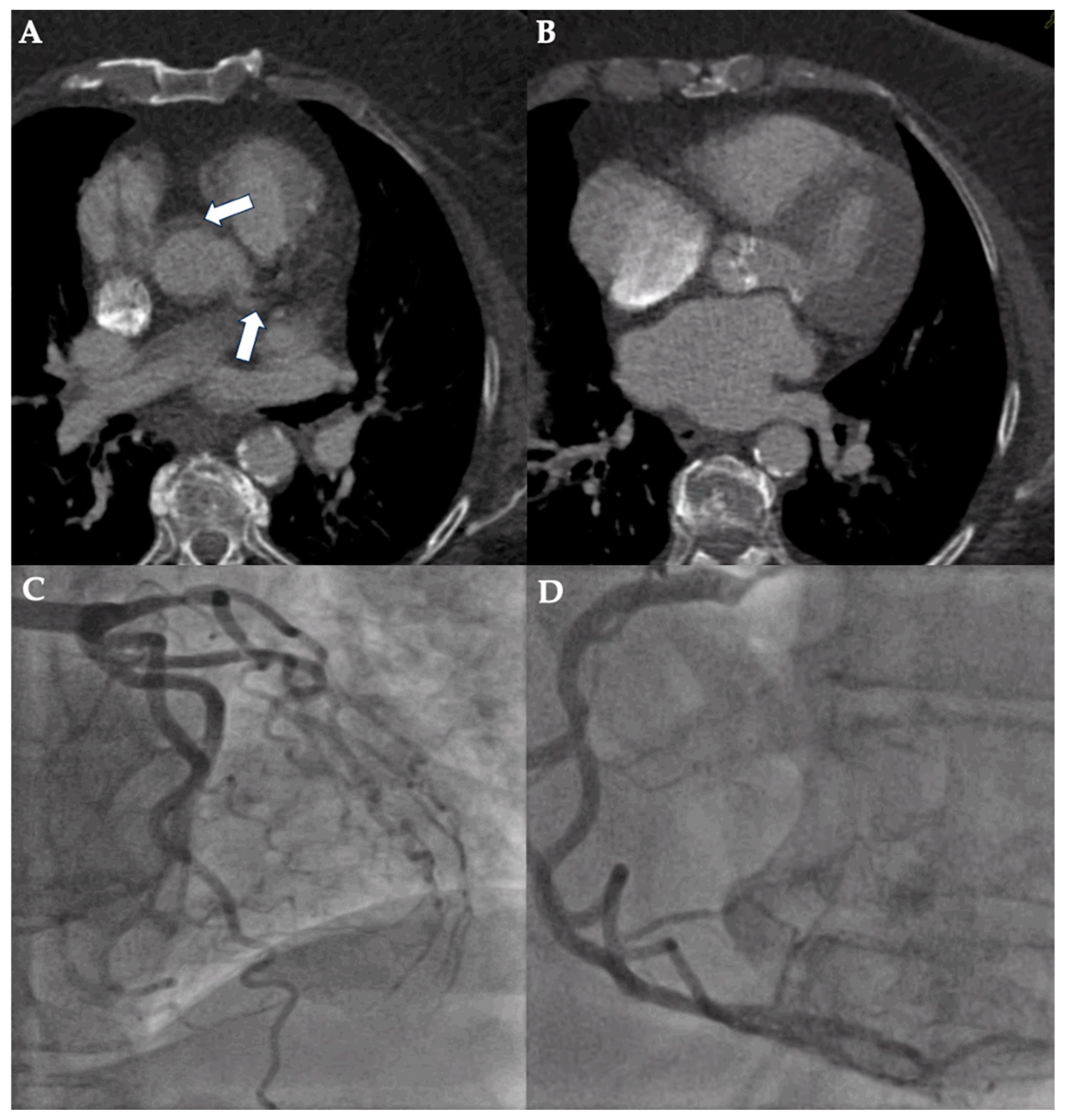
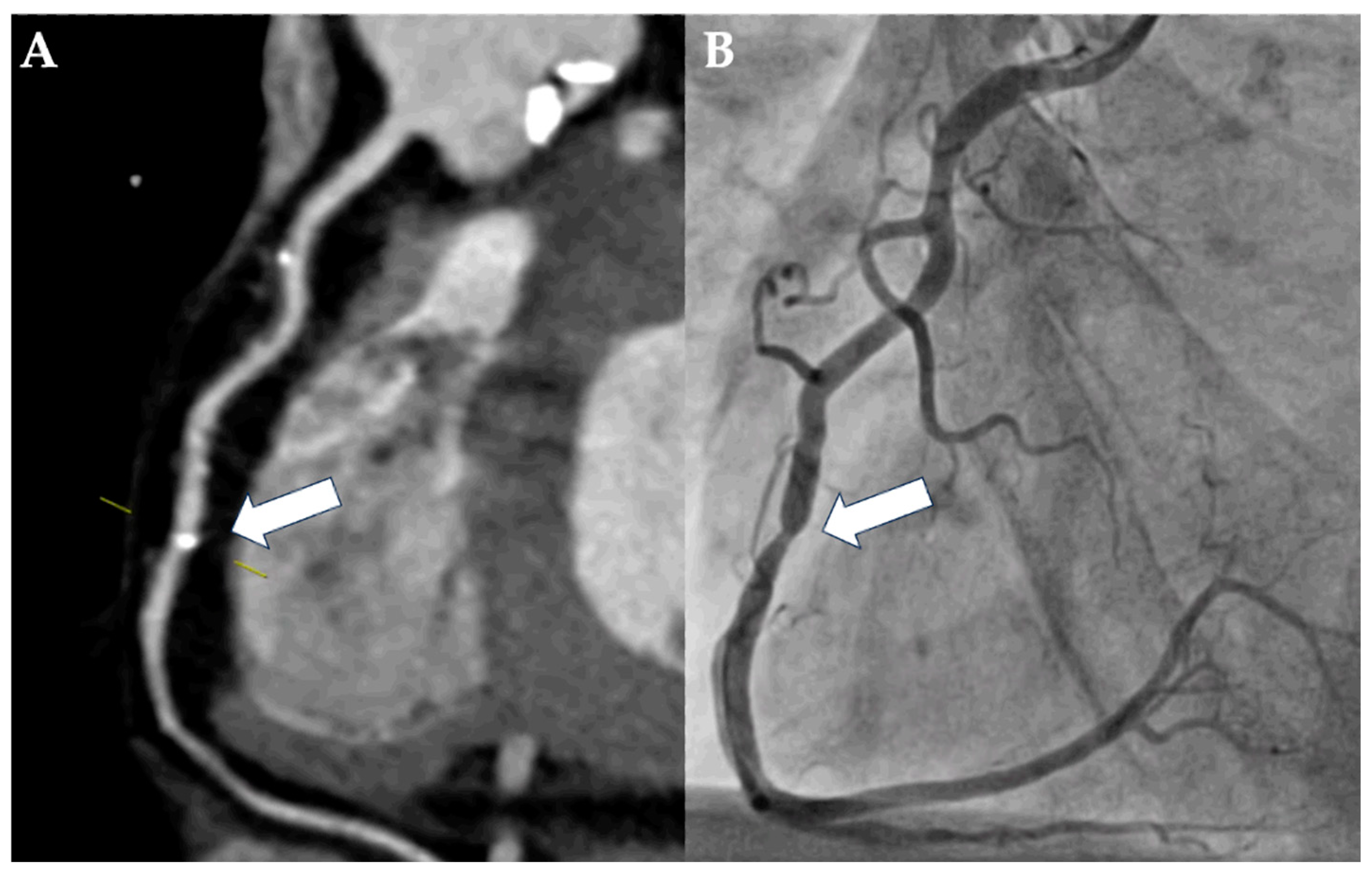

| Diagnostic Concordance of CT Angiography (Accuracy; TP and TN) n = 158 | Diagnostic Discordance of CT Angiography (Inaccuracy; FP and FN) n = 34 | p-Value | |
|---|---|---|---|
| Patient characteristics | |||
| Sex (female) | 103 (65.2) | 19 (55.9) | 0.31 |
| Age, years | 82.4 [80.1–85.5] | 79.8 [76.8–84.2] | 0.01 |
| Body mass index, kg/m2 | 26.3 [24.0–30.4] | 27.5 [24.9–32.8] | 0.13 |
| Diabetes | 40 (25.3) | 11 (32.4) | 0.39 |
| Hypertension | 148 (93.7) | 34 (100) | 0.13 |
| Dyslipidemia | 41 (25.9) | 7 (20.6) | 0.52 |
| Smoking | 18 (11.4) | 5 (14.7) | 0.59 |
| Family history of CAD | 8 (5.1) | 1 (2.9) | 0.58 |
| Atrial fibrillation | 62 (39.2) | 19 (55.9) | 0.07 |
| NYHA class | 3 [3–3] | 3 [2–3] | 0.64 |
| eGFR, mL/min/1.73 m2 | 70.0 [52.0–84.8] | 67.5 [51.3–83.8] | 0.64 |
| Results from echocardiography | |||
| LVEF, % | 62.0 [55.0–65.0] | 60.0 [60.0–65.0] | 0.22 |
| AVA, cm2 | 0.7 [0.5–0.9] | 0.7 [0.5–0.8] | 0.77 |
| CT imaging parameters | |||
| CT system (not capable of single-heartbeat acquisition) | 88 (55.7) | 26 (76.5) | 0.03 |
| Heart rate during CT, bpm | 71.0 [63.0–80.0] | 77.5 [66.5–80.0] | 0.12 |
| Image quality (1 = non-diagnostic to 5 = excellent) | 3.5 [3–4] | 3 [1–3] | <0.01 |
| Agatston score, HU | 468.5 [147.2–1202.5] | 679.1 [242.2–1179.3] | 0.69 |
| Diagnostic Concordance of CT Angiography (Accuracy; TP and TN) n = 506 | Diagnostic Discordance of CT Angiography (Inaccuracy; FP and FN) n = 70 | p-Value | |
|---|---|---|---|
| Vessel-specific signal-to-noise ratio | 15.3 [12.0–18.9] | 13.7 [10.2–19.9] | 0.14 |
Lesion location *
| 9 (1.8) 168 (33.4) 176 (34.8) 162 (32.0) | 1 (1.4) 24 (34.3) 16 (22.9) 30 (42.9) | 0.83 0.81 0.04 0.07 |
| Vessel-specific Agatston score, HU | 95.8 [11.9–290.9] | 200.2 [54.2–573.2] | <0.01 |
| Plaque composition (calcified vs. non-calcified) ** | 366/397 (92.2) vs. 31/397 (7.8) ** | 60/64 (93.8) vs. 4/64 (6.2) ** | 0.66 |
| Overall Misdiagnosis n = 34 | ||
|---|---|---|
| Odds Ratio (95% Confidence Interval) | p-Value | |
| Age, per year | 0.87 (0.80–0.94) | <0.01 |
| Atrial fibrillation | 1.81 (0.79–4.16) | 0.16 |
| CT system (not capable of single-heartbeat acquisition) | 2.51 (0.96–6.56) | 0.06 |
| CT image quality (1 = non-diagnostic–5 = excellent) | 0.60 (0.41–0.89) | <0.01 |
| Overall Misdiagnosis n = 70 | ||
|---|---|---|
| Odds Ratio (95% Confidence Interval) | p-Value | |
| LCx | 0.70 (0.35–1.37) | 0.30 |
| RCA | 1.28 (0.71–1.37) | 0.40 |
| Vessel-specific Agatston score, HU | 1.00 (1.00–1.00) | 0.11 |
Disclaimer/Publisher’s Note: The statements, opinions and data contained in all publications are solely those of the individual author(s) and contributor(s) and not of MDPI and/or the editor(s). MDPI and/or the editor(s) disclaim responsibility for any injury to people or property resulting from any ideas, methods, instructions or products referred to in the content. |
© 2025 by the authors. Licensee MDPI, Basel, Switzerland. This article is an open access article distributed under the terms and conditions of the Creative Commons Attribution (CC BY) license (https://creativecommons.org/licenses/by/4.0/).
Share and Cite
Renker, M.; Kriechbaum, S.D.; Baumann, S.; Tesche, C.; Korosoglou, G.; Charitos, E.I.; Gonska, B.; Seidler, T.; Choi, Y.-H.; Rolf, A.; et al. Predictors of Diagnostic Inaccuracy of Detecting Coronary Artery Stenosis by Preprocedural CT Angiography in Patients Prior to Transcatheter Aortic Valve Implantation. Diagnostics 2025, 15, 771. https://doi.org/10.3390/diagnostics15060771
Renker M, Kriechbaum SD, Baumann S, Tesche C, Korosoglou G, Charitos EI, Gonska B, Seidler T, Choi Y-H, Rolf A, et al. Predictors of Diagnostic Inaccuracy of Detecting Coronary Artery Stenosis by Preprocedural CT Angiography in Patients Prior to Transcatheter Aortic Valve Implantation. Diagnostics. 2025; 15(6):771. https://doi.org/10.3390/diagnostics15060771
Chicago/Turabian StyleRenker, Matthias, Steffen D. Kriechbaum, Stefan Baumann, Christian Tesche, Grigorios Korosoglou, Efstratios I. Charitos, Birgid Gonska, Tim Seidler, Yeong-Hoon Choi, Andreas Rolf, and et al. 2025. "Predictors of Diagnostic Inaccuracy of Detecting Coronary Artery Stenosis by Preprocedural CT Angiography in Patients Prior to Transcatheter Aortic Valve Implantation" Diagnostics 15, no. 6: 771. https://doi.org/10.3390/diagnostics15060771
APA StyleRenker, M., Kriechbaum, S. D., Baumann, S., Tesche, C., Korosoglou, G., Charitos, E. I., Gonska, B., Seidler, T., Choi, Y.-H., Rolf, A., Kim, W.-K., & Sossalla, S. T. (2025). Predictors of Diagnostic Inaccuracy of Detecting Coronary Artery Stenosis by Preprocedural CT Angiography in Patients Prior to Transcatheter Aortic Valve Implantation. Diagnostics, 15(6), 771. https://doi.org/10.3390/diagnostics15060771