Review

TREM2 in Neurodegenerative Diseases: Mechanisms and Therapeutic Potential

1
Department of Neurology, Beijing Tsinghua Changgung Hospital, School of Clinical Medicine, Tsinghua Medicine, Tsinghua University, Beijing 102218, China
2
Key Laboratory of New Ceramics and Fine Processing, School of Materials Science and Engineering, Tsinghua University, Beijing 100084, China
3
Key Laboratory of Advanced Materials of Ministry of Education of China, School of Materials Science and Engineering, Tsinghua University, Beijing 100084, China
*
Author to whom correspondence should be addressed.
Cells 2025, 14(17), 1387; https://doi.org/10.3390/cells14171387
Submission received: 29 July 2025 / Revised: 29 August 2025 / Accepted: 1 September 2025 / Published: 5 September 2025

Abstract

Neurodegenerative diseases, including Alzheimer’s disease (AD), Parkinson’s disease (PD), and amyotrophic lateral sclerosis (ALS), represent significant global health challenges, affecting millions and straining healthcare systems. These disorders involve progressive neuronal loss and cognitive decline, with incompletely elucidated underlying mechanisms. Chronic neuroinflammation is increasingly recognized as a critical contributor to disease progression. The brain’s resident immune cells, microglia, are central to this inflammatory response. When overactivated, microglia and other immune cells, such as peripheral macrophages, can exacerbate inflammation and accelerate disease development. Triggering Receptor Expressed on Myeloid Cells 2 (TREM2) is a transmembrane receptor of the immunoglobulin superfamily that demonstrates high expression on microglia in the central nervous system. TREM2 serves a vital role in regulating phagocytosis, synaptic pruning, and energy metabolism. This review examines the functions of TREM2 in neurodegenerative diseases and its potential as a therapeutic target, aiming to inform future treatment strategies.

1. Introduction

Neurodegenerative diseases, including Alzheimer’s disease (AD), Parkinson’s disease (PD), and amyotrophic lateral sclerosis (ALS), constitute a critical global public health challenge. These disorders are characterized by progressive neuronal loss and cognitive or motor decline—sometimes simultaneously, as in Parkinson’s disease dementia (PDD)—leading to severe disability, substantial caregiver burdens, and ever-increasing socioeconomic and healthcare costs [1]. AD alone accounts for 60–70% of global dementia cases, with projections indicating there will be over 150 million affected individuals by 2050. Despite extensive research, the pathogenic mechanisms driving neurodegeneration remain incompletely understood. While hallmark pathologies such as amyloid plaques, neurofibrillary tangles, and α-synuclein aggregates have been identified, multiple hypotheses exist, including the amyloid cascade, neuroinflammation, vascular and infection-related mechanisms, and the recently proposed multi-sensor theory [2]. Current therapies primarily address symptoms without halting disease progression, highlighting the urgent need for novel therapeutic targets [3].
Emerging evidence indicates chronic neuroinflammation is a fundamental driver of neurodegenerative processes [4]. Microglia, the brain’s resident immune cells, serve dual roles in maintaining homeostasis by phagocytizing pathological debris, regulating synaptic plasticity, and modulating inflammatory responses [5]. However, under pathological conditions, sustained microglial activation induces excessive release of pro-inflammatory cytokines (e.g., Interleukin-1 beta (IL-1β) and TNF-α), reactive oxygen species, and complement system proteins, establishing a self-perpetuating inflammatory cascade. This dysregulation is further amplified by peripheral macrophage infiltration and astrocytic reactivity, collectively creating a neurotoxic microenvironment that accelerates synaptic loss and neuronal death. Such immune–neural network imbalances not only propagate amyloid-β (Aβ) and tau pathologies but also intersect with metabolic dysfunction and impaired autophagy, forming a multifaceted pathogenic framework [6,7].
Within this context, Triggering Receptor Expressed on Myeloid Cells 2 (TREM2), a key regulator of microglial function, has emerged as a significant focus [8]. As a member of the immunoglobulin superfamily, TREM2 activates downstream DNAX-activating protein 12 (DAP12)/Spleen Tyrosine Kinase (SYK) signaling upon binding ligands, such as lipids, apoptotic cells, and Aβ aggregates, thereby modulating phagocytosis, inflammatory responses, and metabolic adaptation [9]. Genome-wide association studies (GWASs) have identified loss-of-function TREM2 variants (e.g., R47H) as significant genetic risk factors for AD, frontotemporal dementia, and Nasu–Hakola disease [10]. Additionally, elevated cerebrospinal fluid levels of soluble TREM2 (sTREM2) correlate with neurodegenerative biomarkers (e.g., tau), suggesting its potential as a dynamic indicator of disease activity [11]. Preclinical studies have demonstrated that TREM2 agonists enhance microglial clearance of pathological proteins and promote metabolic resilience, reducing amyloid burden and cognitive deficits [12]. This review comprehensively examines the multifaceted roles of TREM2 in neurodegenerative diseases, evaluates its therapeutic potential, and provides direction for developing innovative immunometabolic interventions.

2. Materials and Methods

We conducted a systematic literature search of the PubMed and Web of Science databases to identify studies addressing TREM2 in neurodegenerative diseases (search period: January 2000–May 2025). The following keywords were used in different combinations: “TREM2”, “triggering receptor expressed on myeloid cells 2”, “neurodegenerative diseases”, “Alzheimer’s disease”, “Parkinson’s disease”, “frontotemporal dementia”, and “ALS”. Boolean operators (AND and OR) and database-specific filters were applied.

2.1. Inclusion Criteria

Peer-reviewed original research or review articles;
Articles written in English;
Studies focusing on TREM2 in the context of neurodegenerative diseases (mechanisms, pathology, or therapeutic potential).

2.2. Exclusion Criteria

Duplicates across databases;
Conference abstracts, editorials, book chapters, and non-peer-reviewed articles;
Articles lacking experimental or mechanistic insights (e.g., commentary-only articles).

2.3. Screening Process

The literature search yielded 3384 articles (PubMed: 1402; Web of Science: 1982). After duplicate removal, 1542 articles remained. Title and abstract screening excluded 1026 articles, leaving 516 for full-text review. Following comprehensive evaluation, 170 articles met the inclusion criteria and were incorporated into this review (128 original research studies and 42 review articles).

3. Structure of TREM2

TREM2 is a 26 kDa transmembrane immunoregulatory receptor encoded by the TREM2 gene, located on human chromosome 6p21.1 and mouse chromosome 17C3. The TREM2 protein comprises five exons, encoding a full-length protein consisting of 230 amino acids [13,14]. TREM2 functions as a single-pass transmembrane receptor protein within the immunoglobulin superfamily. Its structure includes an extracellular immunoglobulin-like V-type domain, a stalk region, a helical transmembrane domain, and a short cytoplasmic tail lacking signaling and trafficking motifs [15,16]. Figure 1 illustrates the structure and pathogenic mutations of TREM2.
The extracellular domain of TREM2 consists of two primary structural components: an immunoglobulin (Ig)-like ligand-binding module composed of nine antiparallel β-strands and a proximal stalk region [17]. This ligand-binding domain contains essential post-translational modification sites, including N-linked glycosylation, disulfide bonds, and phosphorylation motifs. The transmembrane segment incorporates conserved lysine residues that facilitate interaction with adaptor proteins DAP12 and DAP10 through electrostatic coupling [18]. These positively charged residues establish salt bridges with aspartic acid residues in the transmembrane helices of DAP12/DAP10, forming a stable heterodimeric signaling complex essential for signal transduction [16]. Although TREM2’s short intracellular C-terminal domain lacks inherent signaling capacity, the transmembrane lysine residues are essential for maintaining the DAP12/DAP10 interaction [19]. Biochemical analyses have demonstrated that TREM2 undergoes constitutive membrane shedding with a brief half-life (<1 h), producing soluble TREM2 (sTREM2) fragments under normal conditions [20]. Within the central nervous system (CNS), TREM2 exhibits widespread neuroanatomical distribution, with notable expression in the basal ganglia, corpus callosum, and spinal cord [21,22]. Cellular expression studies have identified myeloid lineage cells, particularly immature dendritic cells and tissue-resident macrophages, as primary expressors [23].
The Ig-like domain facilitates diverse ligand interactions through three primary mechanisms: (1) pattern recognition of bacterial components (e.g., lipopolysaccharide); (2) phospholipid sensing via phosphatidylserine binding; and (3) molecular chaperone interactions with apolipoproteins, including Apolipoprotein E (APOE) [24,25]. In the pathogenesis of AD, the extracellular domain specifically recognizes β-amyloid aggregates, initiating DAP12/DAP10-dependent signal transduction cascades upon ligand engagement [26].

4. TREM2 Signaling Pathways

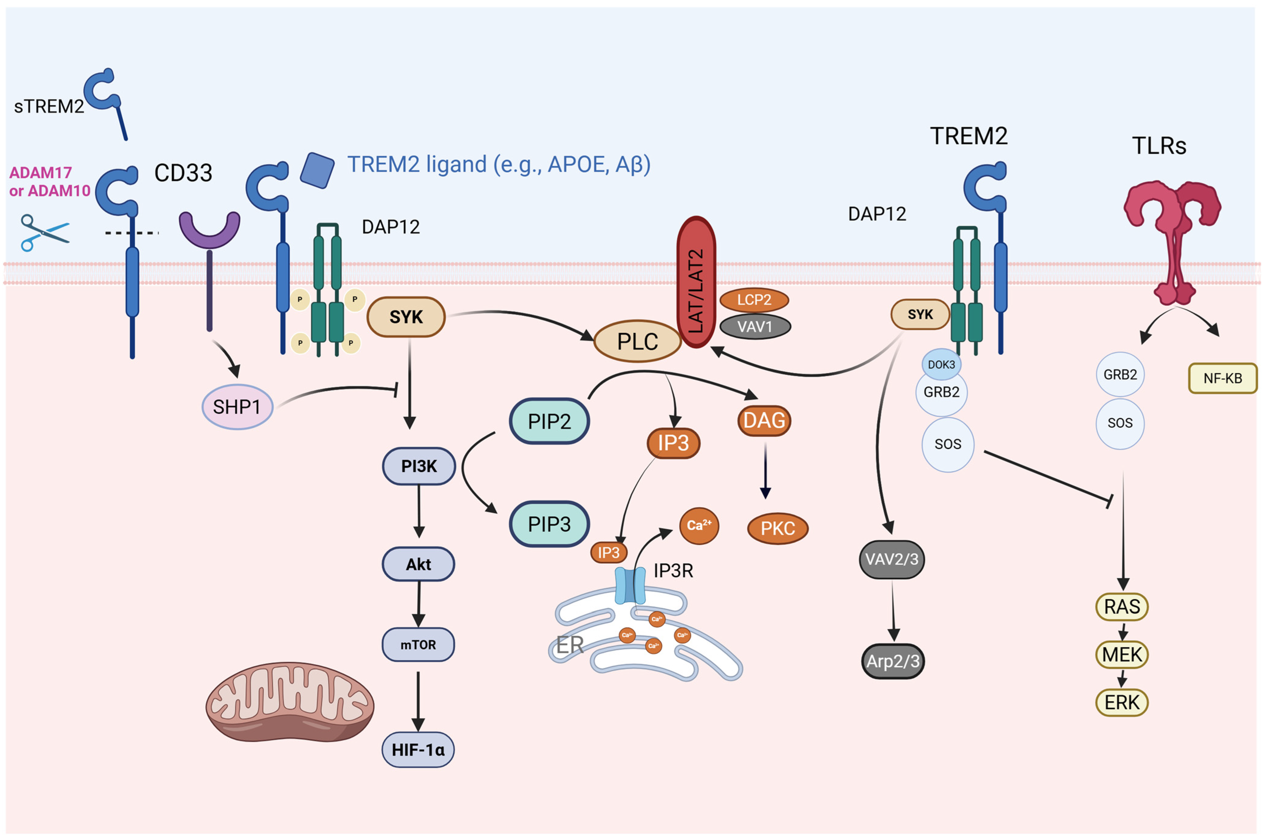
DAP12 contains an immunoreceptor tyrosine-based activation motif (ITAM). When TREM2 is activated, the tyrosine residues within the ITAM of DAP12 undergo phosphorylation, triggering the recruitment and activation of tyrosine protein kinase SYK. SYK subsequently activates downstream signaling molecules, including Vav guanine nucleotide exchange factors, non-receptor tyrosine kinase Pyk2, phosphatidylinositol 3-kinase (PI3K), phospholipase Cγ (PLCγ), and membrane-bound adaptors such as LAT1/2. PI3K, in an action facilitated by DAP10, localizes to the membrane, where it catalyzes the conversion of phosphatidylinositol-4,5-bisphosphate (PIP2) into phosphatidylinositol-3,4,5-trisphosphate (PIP3). This PI3K-mediated conversion activates PLCγ, which subsequently converts PIP2 into inositol trisphosphate (IP3) and diacylglycerol (DAG). The TREM2–DAP12–DAP10 signaling cascade initiates a series of protein and lipid phosphorylation events, resulting in Ca2+ mobilization, integrin activation, cytoskeletal rearrangement, activation of the mechanistic target of rapamycin (mTOR) and mitogen-activated protein kinase (MAPK) signaling, and enhanced energy metabolism [18,27]. DAP10’s intracellular domain contains a tyrosine–isoleucine–asparagine–methionine motif (YINM), which enables the recruitment of p85, a regulatory subunit of PI3K, promoting PIP2-to-PIP3 conversion. Furthermore, activated TREM2 partially phosphorylates DAP12’s ITAM region, creating binding sites for the Src homology 2 (SH2) domains of SYK and Zeta-chain-associated protein kinase 70 (ZAP70) kinases. Simultaneously, inositol phosphatase-1 (SHIP1) competes with SYK for ITAM binding, thereby reducing DAP12-SYK and DAP10-PI3K interactions, ultimately suppressing downstream immune responses [28,29]. Figure 2 illustrates the TREM2-DAP12 pathway and the downstream regulatory network.

4.1. TREM2-Toll-like Receptor 4 (TLR4)/MAPK Axis in Neuroinflammatory Regulation

Recent evidence has demonstrated the critical role of TREM2 dysregulation in the pathogenesis of neuroinflammation. Studies using APP/PS1 transgenic mice and BV2 microglial models have revealed that TREM2 deficiency aggravates AD pathology through TLR4-dependent MAPK pathway activation, resulting in accelerated Aβ deposition, enhanced gliosis around amyloid plaques, and substantial neuronal/synaptic protein loss alongside cognitive decline. At the molecular level, TREM2 downregulation initiates a neuroinflammatory cascade by promoting TLR4/MAPK-mediated production of pro-inflammatory cytokines in both cellular and organismal systems [30]. The neuroinflammatory responses triggered by TREM2 reduction may contribute to the pathological mechanisms of AD. Significantly, TREM2 overexpression studies in intracerebral hemorrhage models have indicated therapeutic potential: elevated TREM2 expression provided neuroprotection through multiple mechanisms, namely, reducing cerebral edema, inhibiting neuroinflammatory responses via TLR4/NF-κB/MAPK pathway suppression, and reducing apoptotic processes [31]. This variation may result from differential activation of TLR4 by damage-associated molecular patterns (DAMPs) in the pathological microenvironment.

4.2. TREM2 and Calcium-Modulated Signaling Pathways

TREM2 activates downstream signaling pathways, including calcium signaling, through its association with DAP12. TREM2 transmits signals via its co-receptor DAP12, activating downstream signaling molecules, including PLCγ2. PLCγ2 activation leads to PIP2 hydrolysis, generating IP3 and DAG. IP3 binds to IP3 receptors on the endoplasmic reticulum (ER), triggering the release of stored calcium ions into the cytoplasm, thus increasing intracellular calcium ion concentrations. The elevated calcium ions can facilitate the assembly and activation of the NOD-like receptor family pyrin domain containing 3 (NLRP3) inflammasome, a multi-protein complex involved in IL-1β maturation and secretion [32]. These alterations are essential for microglial function [25]. Obst et al. demonstrated that PLCγ2 deficiency results in cell adhesion and migration defects in human-induced Pluripotent Stem Cell (iPSC)-derived macrophages, likely through integrin-dependent mechanisms. These findings suggest PLCγ2 is crucial for various cellular responses, supporting the hypothesis that PLCγ2 modulation could serve as a potential therapeutic strategy for microglial function in AD [33]. Jairaman et al. discovered that iPSC-derived microglia lacking TREM2 exhibit hyperactive Ca2+ signaling in response to adenosine agonists, such as ADP, affecting microglial injury responses. This ADP hypersensitivity results from increased P2Y12 and P2Y13 receptor expression, leading to increased release of stored Ca2+ from the ER, triggering sustained Ca2+ influx through Orai channels and altering TREM2 knockout (KO) microglia motility. P2Y12 receptor antagonists can restore the chemotaxis defect in TREM2 KO microglia by reducing cytosolic Ca2+ [34]. These findings indicate that the absence of TREM2 leads to defective Ca2+ responses to adenosine signals in microglia, suggesting there is an optimal Ca2+ signaling window for microglial motility [34].

4.3. TREM2 and SHIP1-Related Signaling Pathways

Upon activation, TREM2 signals through the adaptor molecule DAP12, which contains an ITAM. When TREM2 is activated, the ITAM of DAP12 recruits and activates the tyrosine protein kinase SYK. SHIP1 regulates microglial function and activation by modulating different but converging signaling pathways. SHIP1 inhibits TREM2 signaling by binding to the ITAM of DAP12 in osteoclasts, preventing the p85 regulatory subunit of PI3K from binding to the ITAM of DAP12. This indicates that SHIP1 inhibits PI3K-mediated signaling events downstream of TREM2/DAP12 [29,35]. Research using human microglia-like HMC3 cells expressing wild-type TREM2, the R47H variant, and TREM2-deficient cells demonstrated that functional TREM2 is essential for Aβ phagocytosis, lysosomal capacity, and mitochondrial activity. SHIP1, which regulates TREM2 signaling in other cells, functions as a negative regulator of these TREM2-mediated processes. Moreover, pharmacological inhibition of SHIP1 and its isoform Src Homology 2-containing Inositol Phosphatase 2 (SHIP2) enhances Aβ phagocytosis and lysosomal capacity, independently of TREM2 or SHIP1 expression, suggesting SHIP2’s regulatory role in these functions [36].

4.4. Crosstalk Between TREM2 and CD33

Research has demonstrated that the microglial receptors CD33 and TREM2 are associated with the risk of developing AD. In 5xFAD mouse models, CD33 KO mitigated Aβ pathology and enhanced cognitive function, though these effects diminished when TREM2 was also knocked out. TREM2 knockout exacerbates Aβ pathology and neurodegeneration while reducing Iba1+ cell numbers, effects that persist despite additional CD33 KO. RNA-seq analysis revealed upregulation of genes related to phagocytosis and signaling, including Interleukin-6 (IL-6), IL-8, and acute-phase response in 5xFAD; CD33−/− mice, while these genes were downregulated in 5xFAD; TREM2−/− mice, indicating TREM2’s downstream position relative to CD33. The CD33-TREM2 crosstalk encompasses regulation of the IL-1β/IL-1RN axis and the “receptor activity chemokine” gene set. These insights contribute to the development of AD therapies targeting these receptors [37]. Studies have indicated that CD33 inhibits Syk activation through SHIP1 interaction, thereby suppressing PI3K activation, autophagy, and microglial metabolic homeostasis [38,39]. Additionally, CD33 and TREM2 exhibit complex interactions that may influence microglial Aβ uptake and degradation.
TREM2 operates within a complex network. Its interactions with CD33- and SYK-dependent signaling potentially determine microglial functional states. These relationships are crucial for developing combinatorial therapies but remain oversimplified in the literature. Further investigation of the intricate interactions between TREM2-, CD33-, and SYK-dependent signaling is necessary.

5. Soluble TREM2 (sTREM2)

Soluble TREM2 (sTREM2), generated through proteolytic cleavage of the full-length protein by metalloproteinase 10 (ADAM10) and ADAM17, is a biologically active molecule capable of ligand binding, microglial activation, and immune response modulation in the AD continuum [40]. In clinical settings, cerebrospinal fluid (CSF) sTREM2 functions as a dynamic biomarker indicating microglial activation intensity, with elevated levels in AD patients correlating strongly with neurodegeneration markers (T-tau and P-tau181) [11]. Mendelian randomization analyses have established causal associations between genetically elevated CSF sTREM2 and the risk of developing multiple sclerosis [41], while case–control studies have revealed its diagnostic potential in ALS through elevated serum/CSF levels [42]. These observations establish sTREM2 as a pan-neuroinflammatory biomarker valuable for disease stratification and therapeutic monitoring. Mechanistically, Zhong et al. demonstrated sTREM2’s therapeutic efficacy in AD models, where it mitigates amyloid pathology through microglial reprogramming, enhancing plaque surveillance and Aβ clearance, subsequently improving synaptic plasticity and cognitive function. Notably, microglial depletion nullifies these benefits, confirming the necessity of intact microglial responses for sTREM2-mediated functional restoration [43]. Belsare et al. demonstrated that sTREM2 specifically interacts with fibrillar Aβ40 and Aβ42 with micromolar affinity rather than monomeric Aβ, selectively inhibiting secondary nucleation in fibrillization. Additionally, sTREM2 enhances cellular uptake of Aβ fibrils, a function compromised by the AD-associated R47H mutation, indicating a structural basis for its activity [44]. The dual functionality of sTREM2 may relate to the ADAM10/17 cleavage site and disease progression stages [40,45]. While there is an increasing amount of evidence supporting sTREM2’s critical role in neurological disorders, further research is needed to fully understand its mechanisms in modulating microglial function across different pathological contexts and disease stages as well as its interaction with membrane-bound TREM2 [46].

6. TREM2 Variants

Genetic variants in TREM2 constitute established risk factors for multiple neurodegenerative diseases, including AD, PD, ALS, and frontotemporal dementia (FTD) (Table 1). The significance of TREM2 was initially revealed through the discovery of the fact that biallelic loss-of-function mutations cause Nasu–Hakola disease (NHD), a rare autosomal recessive disorder characterized by early-onset dementia and bone lesions [47,48,49]. Research on NHD-associated mutations has established that complete loss of TREM2 function results in severe microglial dysfunction, including impaired lysosomal activity [50].
The heterozygous R47H variant has emerged as a significant risk factor for several common neurodegenerative diseases. This variant substantially increases the risk of late-onset AD, associated with decreased microglial coverage of amyloid plaques, enhanced neuritic dystrophy, and compromised phagocytosis [51,52]. The R47H variant’s risk effect extends to FTD, PD, and ALS [53,54], demonstrating its broad impact on neuroimmune homeostasis.
Table 1. Summary of TREM2 genetic variants in neurodegenerative diseases.
VariantMutation TypeAssociated DiseasesDomain/LocationFunctional ImpactClinical SignificanceReferences
R47HMissenseAD, ALSCoding Exon 2Reduces binding to APOE, Aβ, and lipoproteins; impairs SYK/PI3K signaling and phagocytosis.Increases AD risk (OR ≈ 2–3); linked to diffuse amyloid plaques and axonal dystrophy.[54,55]
R62HMissenseAD, FTDCoding Exon 2Disrupts dimerization and ligand binding; reduces signaling efficiency.Moderately increases AD risk (OR ≈ 1.5).[56,57]
T66MMissenseFTD/FTLDNHDCoding Exon 2Results in misfolding of the TREM2 protein and loss of function.Results in early-onset dementia and bone cysts.[58,59,60,61]
Y38CFrameshift mutationFTD/FTLDNHDCoding Exon 2Alters the flanking sequence of a cysteine for the Ig V-fold interchain disulfide bond; disrupts protein function.Severe early-onset NHD with dementia and bone lesions.[58,62,63]
Q33XNonsenseNHD, ADCoding Exon 2Produces truncated proteins, resulting in a complete loss of protein function.Homozygous: NHD; heterozygous: increases AD risk.[58,64]
D87NMissenseADCoding Exon 2Reduces APOE binding but enhances signaling in some contexts.Increases AD risk in controversy.[55]
T96KMissenseAD, FTDCoding Exon 2-A significant genetic risk factor for late-onset AD (LOAD) in the Tunisian population.[65,66]
H157YMissenseADCoding Exon 3Increases shedding of sTREM2.
Reduces activation in response to
phospholipid ligands and decreases
phagocytosis.
Increases risk of AD.[67,68]
L211PMissenseFTDCoding Exon 4-Increases risk of FTD, especially bvFTD.[65]
W44XNonsenseNHDCoding Exon 2Produces non-functional truncated proteins that fail to activate microglia.Strongly associated with NHD phenotype.[47]
W191XNonsenseADCoding Exon 4 Stop-gain mutation.Suggestive association with AD.[69]
Analysis of these variants, particularly R47H, provides essential genetic evidence that even a partial reduction in TREM2 function—typically through impaired ligand binding—sufficiently disrupts key microglial signaling pathways. This disruption compromises critical functions, including lipid metabolism, neuronal damage response, and plaque compaction. The subsequent sections examine these downstream signaling pathways (e.g., SYK and PI3K-AKT), their mechanistic roles in disease, and the resulting therapeutic implications.

7. Impact of TREM2 on Microglial Function

7.1. Influence on Microglial Homeostasis and Activation

Ligand binding initiates downstream microglial signaling through transcriptional changes, transitioning these cells from a homeostatic state to a disease-associated microglia (DAM) state in neurodegenerative mouse models [70]. TREM2 plays a critical role in guiding microglia in the transition from a homeostatic state to an activated state in specific contexts, a transition that is essential for responding to neuroinflammation and demyelination. Single-cell analysis of DAM and TREM2 in transgenic Alzheimer’s disease (Tg-AD) models has demonstrated that the DAM program is activated in a two-step process. The initial activation occurs in a TREM2-independent manner, involving the downregulation of microglial checkpoints, followed by the activation of a TREM2-dependent program. This distinct microglial phenotype exhibits the potential to limit neurodegeneration, which may have significant implications for future treatments of AD and other neurodegenerative diseases [70]. TREM2 is fundamental for maintaining microglial metabolic adaptability during stress events, enabling microglia to develop into fully mature DAM and sustain their response to Aβ plaque-induced pathology [18]. Microglia adhering to Aβ plaques acquire a transcriptional signature known as “disease-associated microglia”, which primarily derives from the TREM2-DAP12 receptor complex. This complex transmits intracellular signals via the protein tyrosine kinase SYK. The human TREM2 R47H variant, associated with a high risk of AD development, cannot activate microglia via SYK. SYK-deficient microglia cannot envelop Aβ plaques, resulting in accelerated brain pathology and behavioral deficits. SYK deficiency impairs the PI3K-AKT-GSK-3β-mTOR pathway, leading to a loss of the anabolic support required for acquiring the DAM profile [71]. Single-nucleus transcriptomics studies have revealed that in 5xFAD mice, compared with wild-type (WT) mice, genes such as Cst7, Csf1, Apoe, Trem2, Lpl, Lilrb4a, MHC-I (H2-d1), MHC-II (Cd74), and various cathepsin genes are significantly upregulated. Conversely, homeostatic genes including P2ry12, Selplg, Tmem119, and Cx3cr1 are downregulated. Furthermore, genes such as Cst7, Csf1, and MHC-I (H2-K and b2m) are significantly more expressed in 5xFAD microglia relative to TREM2−/− 5xFAD microglia, highlighting the TREM2-dependent upregulation of these genes [72].

7.2. TREM2 and Microglial Metabolism

Metabolic flexibility or plasticity describes cells’ capacity to modify their nutrient metabolism in response to environmental changes. This adaptability is essential for maintaining cellular function. The polarization of immune cells following the activation of inflammatory responses requires the reprogramming of cellular metabolic pathways. This metabolic reprogramming is characterized by increased glycolytic flux, the accumulation of specific TCA cycle metabolites, reduced mitochondrial respiration, and enhanced transcriptional control by HIF-1α and mTOR. These alterations regulate microglial immune functions, including cytokine production and phagocytosis, contributing to neuroinflammation in the brain [73]. Research has demonstrated that TREM2 signaling regulates microglial energetics through the mTOR pathway in AD-like mouse models. Microglia from 5xFAD mice lacking TREM2 exhibit more autophagic vesicles compared to those from 5xFAD mice with functional TREM2. Similarly, microglia from AD patients with the TREM2 R47H display more autophagic vesicles than those with the common TREM2 variant. Autophagy constitutes a crucial intracellular degradation pathway for cellular and energy homeostasis [74]. Metabolomic and RNA-seq analyses have revealed deficiencies in metabolites and enzymes involved in glycolysis, the TCA cycle, and the pentose phosphate pathway in TREM2−/− bone-marrow-derived macrophages (BMDMs). These deficiencies can be counteracted in vitro by Dectin-1 and creatine. Dietary creatine modulates autophagy, enhances microglial clustering around plaques, and reduces plaque-associated neuritic dystrophy in Trem2-deficient mice with amyloid-β pathology. Thus, TREM2 facilitates microglial responses during AD by maintaining cellular energy and biosynthetic metabolism [27]. Treatment of BMDMs with a TREM2 agonist enhances mitochondrial potential [75]. During development, TREM2 expression in microglia maintains normal neuronal bioenergetics. Mice lacking TREM2 demonstrate impaired hippocampal neuronal bioenergetics during development, with developing neurons in the hippocampal CA1, but not CA3, subregion exhibiting impaired energy metabolism, reduced mitochondrial mass, and organelle ultrastructural abnormalities. These findings indicate that TREM2 regulates neuronal development by controlling metabolic adaptability in a region-specific manner [76]. When APP/PS1 mice, an AD model, consume a Western diet, microgliosis and increased TREM2 expression occur. This results in increased plaque burden, suggesting that altered microglial phagocytosis may explain how certain nutritional factors increase the risk of the progression of AD [77]. Long-term running exercises enhance cognitive function in the hippocampus in APP/PS1 mice, inhibit TREM2 shedding, and maintain TREM2 protein levels. Running exercises also increase FDG uptake and protein expression of GLUT5, TREM2, SPP1, and p-SYK in the hippocampus in APP/PS1 mice, correlating with improved brain glucose metabolism, microglial glucose metabolism, and hippocampal microglial morphological plasticity. These findings suggest microglia may be structural targets responsible for the benefits of running exercise in relation to AD. Therefore, enhancing TREM2-regulated microglial glucose metabolism and morphological plasticity could represent a novel AD treatment strategy [78].
TREM2 functions as a receptor for specific extracellular lipids [79]. Research has indicated that TREM2, as a lipid receptor, significantly influences adipose tissue macrophage responses in obese individuals. Single-cell genomic analysis has revealed that TREM2+ LAM (lipid-associated macrophage) cells represent the most expanded immune cell population in adipose tissue across multiple mouse models of obesity. The signature of TREM2 and associated genes involved in phagocytosis and lipid catabolism appears in macrophages across multiple organs in both mice and humans, suggesting this pathway represents a conserved response to irregular lipid composition, levels, and distribution. Furthermore, TREM2 signaling facilitates the formation of lipid-associated macrophages in crown structures within adipose tissue, preventing adipocyte hypertrophy and maintaining systemic lipid homeostasis under obese conditions. Complete removal of Trem2 in mice inhibits the downstream LAM program, resulting in adipocyte hypertrophy, systemic hypercholesterolemia, fat accumulation, and glucose intolerance [80]. Recent evidence indicates that extracellular lipid–TREM2 interactions may regulate intracellular lipid metabolic programs at the transcriptional level, potentially maintaining lipid homeostasis by preventing pathological accumulation of phagocytosis-derived toxic metabolites. While TREM2-deficient microglia demonstrate intact myelin phagocytosis, they show impaired clearance of myelin-derived cholesterol, leading to cholesterol ester (CE) overload—a phenotype observed in APOE-deficient glia and replicated across TREM2−/− murine macrophages, human iPSC-derived microglia, and foam cell models. Notably, pharmacological inhibition of Acyl-CoA Cholesterol Acyltransferase 1 (ACAT1) (blocking CE synthesis) or Liver X Receptor (LXR) agonism (enhancing cholesterol efflux) corrects this metabolic defect, establishing TREM2 as a key transcriptional regulator of cholesterol flux during chronic phagocytic challenges [26]. Studies using atherosclerosis models have demonstrated TREM2’s diverse roles: myeloid-specific TREM2 deletion reduces oxLDL uptake in foam cells and diminishes plaque progression—even in established lesions—independent of systemic inflammation or lipidemia. Mechanistic analyses have shown that TREM2 deficiency disrupts cholesterol efflux pathways (e.g., A member 1 (ABCA1) downregulation), compromising macrophage proliferative capacity and survival [81]. Conversely, therapeutic administration of TREM2 agonists increases plaque macrophage populations through enhanced survival/proliferation while improving plaque stability by augmenting cholesterol efflux and collagen deposition [82].

7.3. TREM2 and Microglial Regulation of Inflammation

While earlier studies characterized TREM2 primarily as an anti-inflammatory agent, recent research demonstrates its dual nature in regulating inflammation, capable of both promoting and inhibiting inflammatory responses. This binary role appears dependent on the specific pathological context and TREM2 expression levels. Studies of the senescence-accelerated mouse prone 8 (SAMP8) model have demonstrated increased TREM2 protein levels during aging. Trem2 knockdown in SAMP8 mouse brains leads to significant increases in pro-inflammatory cytokines, including TNF-α and IL-6, while decreasing IL-10 levels [83]. Research utilizing postoperative cognitive dysfunction (POCD) mouse models and in vitro systems has indicated that TREM2 activation mitigates neuroinflammation through modulation of the PI3K/protein kinase B (Akt) signaling pathway, thereby improving postoperative learning and memory deficits [84]. In PD, TREM2 delivers substantial neuroprotection through regulation of microglial phenotypes. TREM2 knockdown enhances NLRP3 inflammasome activation and subsequent inflammatory responses, intensifying pyroptosis and dopaminergic neuron loss. Additionally, TREM2 demonstrates anti-inflammatory effects in PD through the TLR4/MyD88/NF-κB pathway [85].
Network analysis of TREM2-related gene expression in the human brain has revealed distinct clusters of both anti- and pro-inflammatory genes [86]. In patients with polycystic lipomembranous osteodysplasia with sclerosing leukoencephalopathy (PLOSL), microarray analysis of peripheral blood mononuclear cells (PBMCs) has demonstrated an upregulation of inflammation-related genes and a downregulation of innate immune response genes relative to healthy controls. Under non-disease conditions, the transcriptional profiles of bone marrow cells from Trem2-deficient [79] or Trem2-overexpressing [87] mice remain largely similar to controls [13]. However, in disease contexts, TREM2 significantly influences inflammation-related pathways. In a spinal nerve transection model, pharmacological activation of TREM2 led to elevated TNFα and IL1β levels. Notably, intrathecal administration of a TREM2 agonistic antibody in mice without nerve injuries induced pro-inflammatory cytokine expression and neuropathic pain [88]. Studies have shown [45] that soluble TREM2 (sTREM2) promotes microglial survival through PI3K/Akt-dependent mechanisms and stimulates inflammatory cytokine production. Variants of sTREM2 associated with AD risk mutations demonstrate reduced effectiveness in inhibiting apoptosis and triggering inflammatory responses. When administered to the hippocampus of wild-type and Trem2 knockout mice, sTREM2 increases inflammatory cytokine expression and induces microglial morphological changes. These findings indicate that sTREM2 activates microglia, induces inflammatory responses, and promotes survival. Although TREM2 signaling typically exhibits anti-inflammatory effects in myeloid cells, including microglia, evidence suggests that sTREM2 may function independently of full-length TREM2 and potentially exert opposing effects on inflammation. Research has indicated that acute inflammation may initially suppress TREM2 expression, while chronic inflammation, as observed in conditions such as AD, increases TREM2 expression [89].

7.4. Impact of TREM2 on Microglial Phagocytosis

Recent studies have demonstrated TREM2’s crucial role in regulating microglial phagocytic capacity. TREM2 enhances microglial recognition and clearance of pathological substances through various ligand interactions. For instance, TREM2 recognizes phosphatidylserine, a signal typically exposed on apoptotic cell surfaces, initiating microglial phagocytosis [90]. TREM2 activation significantly enhances microglial phagocytic ability for Aβ plaques and synapses, thereby reducing AD-related neuronal damage [91]. In regard to PD, characterized by dopaminergic neuron loss, abnormal α-synuclein (α-syn) accumulation, and microglial activation, Yin et al. found that Trem2 knockout mice exhibited increased pathological α-syn spread, reduced microglial reactivity, and enhanced loss of TH-positive neurons compared to wild-type mice. TREM2 overexpression enhanced reactive microglial clustering at pathological sites, suggesting TREM2 signaling maintains microglial phagocytosis, proliferation, and reactivity in PD [92]. Xie et al. discovered that TREM2 interacts with TAR-DNA-binding protein 43 kDa (TDP-43) and mediates microglial neuroprotection against TDP-43-related neurodegeneration. TREM2 deficiency impairs pathological TDP-43 clearance by microglia and enhances neuronal damage and motor deficits. Mass cytometry analysis has revealed that human TDP-43 (hTDP-43) induces a TREM2-dependent microglial subpopulation with high CD11c expression and phagocytic capacity [93]. Pang et al. demonstrated that chronic hypoperfusion induced white matter demyelination, microglial phagocytosis, and autophagy–lysosomal pathway activation, accompanied by increased TREM2 expression. TREM2 knockout attenuated white matter lesions and microglial responses, inhibited microglial phagocytosis, reduced autophagy–lysosomal pathway activation, and shifted microglial polarization toward an anti-inflammatory and homeostatic phenotype [94]. Xue et al. found that sphingosine-1-phosphate (S1P) or FTY720 (an S1P analog) promotes microglial phagocytosis in stroke. Their study confirmed that S1P has a pro-phagocytic effect on TREM2-DAP12-transfected CHO cells and TREM2 knockdown microglia [95]. In a multiple sclerosis model, Cignarella et al. demonstrated that microglial TREM2 activation promotes myelin debris clearance and regeneration [96].

8. TREM2 in AD

AD is the most common neurodegenerative disorder. It is primarily characterized by a progressive decline in cognitive and memory functions. As the disease advances, patients exhibit clinical symptoms including memory loss, cognitive impairment, behavioral abnormalities, and social difficulties. The two principal pathological hallmarks of AD are extracellular amyloid plaques composed of Aβ peptides and intraneuronal neurofibrillary tangles (NFTs) formed from abnormally aggregated and hyperphosphorylated tau protein. Despite extensive research spanning over a century, the precise etiology of AD remains incompletely understood, and current therapeutic approaches are limited to symptom management, with no effective interventions capable of halting or reversing the progression of the disease.
Recent progress in whole-genome sequencing and GWASs has led to the identification of multiple genetic variants that increase the risk of developing AD. The most prevalent and extensively studied TREM2 variant associated with an elevated the risk of developing AD is rs75932628. This single-nucleotide polymorphism (SNP) encodes a missense substitution of arginine to histidine at amino acid 47 (R47H) [89]. Jonsson et al. identified this novel risk variant, rs75932628-T, in the context of AD. Although this variant occurs less frequently than the APOE ε4 allele, it confers a comparable risk of AD development. Since TREM2 facilitates microglial phagocytosis of amyloid plaques, reduced TREM2 activity due to the R47H substitution may result in brain damage through impaired clearance of these toxic products. Furthermore, 46 TREM2 mutations associated with AD have been identified, including R62H (rs143332484), D87N (rs142232675), T96K (rs2234353), L211P (rs2234256), R136Q (rs149622783), and p.H157Y (rs2234255) [55,97,98,99,100,101,102], all of which increase the risk of AD development [103]. Most investigations have examined TREM2 variant effects on AD in vitro, necessitating additional animal studies to elucidate their precise pathogenic mechanisms.
In P301S tau models and senescence-accelerated SAMP8 models, transient knockdown of TREM2 increases inflammatory cytokine production [83,104]. In 5xFAD mice, TREM2 knockdown significantly elevates levels of pro-inflammatory cytokines such as IL-1β and TNF-α, exacerbating cognitive deficits [105]. Conversely, TREM2 overexpression in primary mouse glial cells and P301S mice substantially reduces inflammatory factor expression and improves neuroinflammatory responses [83,104,106]. Additionally, lipopolysaccharide (LPS) stimulation of cultured TREM2 KO microglia results in a significant increase in inflammatory gene expression. In vitro studies have demonstrated that TREM2 overexpression can attenuate LPS-induced inflammatory responses and induce microglial polarization towards an M2 phenotype. These findings indicate that TREM2 functions in regulating both microglial inflammatory responses and phenotype modulation [105]. Some studies have also reported on TREM2’s role in promoting pro-inflammatory signaling [88,107]. Analysis of genes most strongly associated with TREM2 revealed clusters of both anti-inflammatory and pro-inflammatory genes in the brain [86]. These recent findings indicate that TREM2’s role in inflammatory processes is more complex than initially understood.
Microglia are essential immune cells in the brain that facilitate phagocytosis of Aβ plaques, and TREM2 is highly expressed in these cells. However, studies examining the impact of TREM2 deficiency on Aβ accumulation have yielded inconsistent results. Jay et al. observed that in the APPPS1-21 mouse model of AD, TREM2 deficiency improved amyloid pathology during early disease stages but worsened it in later stages [108]. In the 5xFAD mouse model, Wang et al. [79] demonstrated that TREM2 deficiency and haploinsufficiency increase Aβ accumulation due to microglial dysfunction, characterized by impaired clustering around Aβ plaques and increased apoptotic tendency. Early studies on 4-month-old 5xFAD mice revealed similar levels of microgliosis and Aβ deposition between Trem2-deficient and Trem2-sufficient mice [109]. However, TREM2 deficiency affected microglial clustering and Aβ plaque structure at early time points. Trem2-deficient 5xFAD mice exhibited more diffuse Aβ plaques with altered Aβ sub-species compositions, associated with increased neuroinflammation and dystrophy. Meilandt et al. [110] compared PS2APP;Trem2 KO mice with PS2APP transgenic mice at various ages, finding increased Aβ plaque abundance at 6–7 months and a significant reduction at 12 months or 19–22 months in Trem2-deficient mice. These mice showed more diffuse Aβ plaques with elevated Aβ42:Aβ40 ratios and higher soluble and fibrillar Aβ oligomer levels. These studies suggest TREM2’s impact on Aβ plaque load varies with the model, age, and brain region analyzed [13,111]. Thus, TREM2 plays a dynamic and complex role in AD through its influence on microglial responses to Aβ and regulation of Aβ plaque aggregation and morphology.
In pure-tauopathy mouse models, the relationship between TREM2 and tau pathology is complex. Research has indicated that partial or normal TREM2 function can contribute to tau pathology and tau-mediated damage, while complete loss of function may reduce tau-mediated brain injuries. Leyns et al. observed no differences in tau phosphorylation and insolubility in PS19 mice with or without TREM2. Their findings suggested that in pure tauopathy, impaired microglial TREM2 signaling can decrease neuroinflammation and prevent neurodegeneration [112]. Similarly, Gratuze et al. investigated [113] the effect of the AD-associated TREM2 variant (R47H) on tau-mediated neuropathology in the PS19 tauopathy mouse model. In their evaluation of PS19 mice expressing human TREM2CV (the common variant) or human TREM2R47H, PS19-TREM2R47H mice exhibited significantly reduced brain atrophy and synapse loss compared to PS19-TREM2CV mice. Gene expression analysis and CD68 immunostaining indicated reduced microglial reactivity in PS19-TREM2R47H mice. Furthermore, microglia expressing TREM2R47H in PS19 mouse and human AD brains exhibited reduced phagocytosis of postsynaptic elements. These findings suggest that impaired TREM2 signaling reduces microglia-mediated neurodegeneration in tauopathy. In contrast, Lee et al. examined [114] Trem2 deficiency effects in pR5-183 and TauPS2APP mouse models, yielding different results. In these models, Aβ pathology intensified tau pathology and neurodegeneration. Single-cell RNA sequencing revealed distinct, Aβ- and TREM2-dependent activation of DAM in TauPS2APP mice. With Aβ pathology present, TREM2 deficiency further increased tau accumulation and spread and accelerated brain atrophy. Without Aβ pathology, TREM2 deficiency had no significant effect on these pathological processes. Thus, TREM2 appears to play a dynamic and complex role in AD progression, potentially modulating tau-driven neurodegeneration by limiting Aβ-promoted pathogenic tau propagation. This TREM2-dependent microglial function proves particularly crucial in preventing tau accumulation and spread, especially in the context of Aβ pathology.
TREM2 maintains a close association with lipid metabolism in microglia [79,115]. TREM2 binds to lipid-related ligands, including phospholipids, high-density lipoprotein (HDL), LDL, and APOE [116,117,118]. APOE, the primary cholesterol transporter in the CNS, is expressed in both microglia and astrocytes. TREM2 regulates APOE expression on microglia, influencing lipid uptake and metabolism, which may explain cholesterol ester accumulation in TREM2-deficient microglia. TREM2 function loss reduces plaque-associated APOE [111,119]. Moreover, Trem2 deficiency exacerbates neurodegeneration in tau mutant mice expressing human APOE4 [14].
Changes in the levels of AD-related proteins, such as Aβ42 and tau, in CSF reflect the presence and progression of AD in the brain. A reduction in CSF Aβ42 levels represents one of the earliest detectable fluid biomarkers of AD pathology. Although low CSF Aβ42 levels predict future cognitive impairment, individuals with reduced CSF Aβ42 levels may remain asymptomatic for years [120]. Following the identification of TREM2 variants associated with AD, several studies have demonstrated that CSF sTREM2 level changes may indicate inflammatory processes associated with the transition from preclinical to clinical AD [20,121]. Studies on late-onset AD and autosomal dominant AD populations have revealed elevated sTREM2 levels in the CSF of AD patients, showing positive correlation with tau and p-tau levels. However, researchers observed no significant correlation between sTREM2 and Aβ42 levels [10,11,122,123].

9. TREM2 in PD

PD is the second most common neurodegenerative disorder after AD [124]. Research has demonstrated that immune pathway dysregulation, including alterations in cytokine signaling, immune cell proliferation and migration, and phagocytosis, is closely linked to dopaminergic (DA) neurodegeneration [125]. During DA neurodegeneration, microglial activation increases alongside inflammatory mediator accumulation [126]. Consequently, microglia-mediated neuroinflammation plays a crucial role in the pathogenesis of PD [127]. The aggregation of α-synuclein (α-syn) monomers into amyloid fibrils through oligomeric intermediates is a toxic mechanism leading to PD. Yin et al. demonstrated that microglia phagocytose pre-formed α-syn fibrils in a concentration- and time-dependent manner, showing a capacity to degrade α-syn aggregates. In PD studies, Trem2 knockout mice displayed enhanced pathological α-syn spread, decreased microglial reactivity, and increased loss of TH-positive neurons compared with wild-type mice, indicating that TREM2 modulation may benefit PD treatment [92]. Similarly, Guo et al. utilized a PD model to demonstrate that TREM2 deficiency both in vitro and in vivo triggered a shift from anti-inflammatory to pro-inflammatory microglial activation states, compromising TREM2 signaling and intensifying the pro-inflammatory response to α-syn, thus aggravating α-syn-induced neurodegeneration [128]. Multiple studies have identified TREM2 as a significant risk factor for PD, suggesting its potential as a therapeutic target for PD and other neurodegenerative diseases. The TREM2 variant rs75932628 (p.R47H) has been associated with increased PD risk [53,129]. However, studies from Greece and China indicate that R47H may not represent a major genetic risk factor for PD [130,131]. A case–control study involving a Chinese population revealed significantly elevated sTREM2 levels in the CSF of PD patients compared to that in healthy controls, though no significant plasma upregulation was observed [132]. Furthermore, Zhang et al. found higher CSF sTREM2 levels in late-onset versus early-onset PD patients, with baseline sTREM2 levels predicting motor progression over a 4-year follow-up [133]. Essential tremor (ET) represents the most common movement disorder in adults, with an unclear pathophysiology [134]. Evidence suggests ET shares common etiologies with other neurodegenerative diseases such as AD and PD [85,135]. A cross-sectional, multicenter international study identified a significant association between the TREM2 p.R47H variant and ET in a Spanish cohort, though this association was not replicated in other studied populations.

10. TREM2 in ALS

ALS is a fatal neurodegenerative disease characterized by progressive, painless muscle weakness resulting from motor neuron degeneration in the brain and spinal cord. The condition manifests clinically as progressive loss of motor function, ultimately leading to death, typically via respiratory failure [136]. Our current understanding indicates that ALS not only affects motor neurons but also involves microglial activation and enhanced inflammatory responses from peripheral lymphocytes and macrophages, which intensify disease progression [137]. TDP-43, essential for gene expression regulation, constitutes a primary component of insoluble and ubiquitinated protein aggregates present in most ALS patients [138]. Research utilizing viral-mediated and transgenic mouse models has examined microglial TREM2’s role in TDP-43-related neurodegeneration. These investigations have revealed that TREM2 deficiency compromises the capacity of microglia to phagocytose and eliminate pathological TDP-43, resulting in increased neuronal damage and motor dysfunction. Additional studies have demonstrated that microglial TREM2 interacts with pathological human TDP-43 (hTDP-43), suggesting a molecular mechanism through which microglia provide neuroprotection in TDP-43-mediated neurodegeneration [93]. A case–control study demonstrated elevated overall and transcript-specific TREM2 mRNA levels in ALS patients’ spinal cords compared to controls, with corresponding changes in TREM2 protein levels. Furthermore, sTREM2 levels in both CSF and serum were markedly elevated in ALS patients versus controls, suggesting potential non-invasive biomarkers for ALS [42]. The GGGGCC hexanucleotide repeat expansion in the C9orf72 gene represents the most prevalent genetic factor in ALS and FTD. Research has revealed interaction between NLRP3 and TREM2 signaling, indicating that modulating the NLRP3 inflammasome to maintain TREM2 function could offer therapeutic potential for C9orf72-ALS/FTD [139]. Janet Cady et al. identified the TREM2 p.R47H variant as a significant risk factor for sporadic ALS [54]. However, Vasileios Siokas et al. found no significant association between the TREM2 rs75932628-T variant and ALS, suggesting limited involvement in ALS pathophysiology [140]. Similarly, a large-scale Chinese study detected no TREM2 rs75932628-T variant in sporadic ALS patients or controls, indicating a minimal contribution to ALS pathogenesis in this population [141]. Data-driven analysis has indicated that CSF soluble TREM2 protein levels may predict survival in late-stage ALS patients, suggesting TREM2’s potential as a therapeutic target [142]. While genetic evidence connects TREM2 to ALS, its precise role in pathogenesis and therapeutic development requires further investigation.

11. TREM2 in NHD

NHD, also known as polycystic lipomembranous osteodysplasia with sclerosing leukoencephalopathy (PLOSL; OMIM 221770, OMIM 605086), is a rare autosomal recessive disorder characterized by rapid psychiatric symptom development progressing to presenile dementia and bone cysts confined to the wrists and ankles. NHD results from homozygous pathogenic mutations in TREM2 or TYRO protein tyrosine kinase-binding protein (TYROBP, also known as DAP12 genes) [143]. Established mutations in TREM2’s extracellular domain associated with NHD include Y38C, W50C, T66M and V126G [58,60,144,145]. Fabia Filipello et al. demonstrated that the TREM2 p.Q33X mutation in microglia causes lysosomal dysfunction, while compounds targeting lysosomal biogenesis can ameliorate numerous NHD microglial defects. Efthimios Dardiotis et al. [50] documented a 33-year-old Greek woman presenting with an NHD-suggestive phenotype. Whole-genome sequencing identified a novel mutation in TREM2 gene exon 2, c.244G>T (p.W50C), in a heterozygous state in the patient and family members, expanding the spectrum of TREM2 mutations associated with the NHD phenotype [145]. Additionally, Y. Numasawa described a Japanese NHD family affected by a TREM2 splicing mutation. A homozygous T-to-C conversion at intron 3’s second position (c.482+2T>C) in the splice donor consensus site was identified, causing exon 3 skipping and truncated protein expression. This mutation triggers neuroinflammation and neurodegeneration [146].

12. TREM2 in Other Neurological Disorders

12.1. Multiple Sclerosis (MS)

MS is a CNS demyelinating disorder characterized by combined inflammatory and neurodegenerative mechanisms. While current immunomodulatory therapies address neuroinflammation, effective strategies for myelin regeneration and disease progression prevention have not yet been established [147]. Microglia play a crucial role in clearing myelin debris from demyelinated areas, an essential step enabling remyelination [148]. Brain pathology observed in NHD patients indicates that disruption of the TREM-2/DAP12 pathway leads to neurodegeneration accompanied by demyelination and axonal loss [49]. Laura Piccio et al. demonstrated that TREM2 expression increases in the spinal cord during both early inflammatory and chronic phases of experimental autoimmune encephalomyelitis (EAE) induced by myelin oligodendrocyte glycoprotein (MOG) peptide. Their research showed that TREM2 exhibits high expression on microglia in the CNS during EAE, and TREM2 blockade during the effector phase of EAE exacerbates disease progression, diffuse CNS inflammatory infiltration, and demyelination, indicating TREM2’s critical role in CNS inflammatory response [149]. Cignarella et al. identified that TREM2 shows high expression on microglia/macrophages in active MS lesions. In cuprizone (CPZ)-induced demyelination, TREM2 deficiency impaired myelin clearance, while in vivo treatment with AL002a enhanced efficient myelin debris clearance, improving myelin phagocytosis and intracellular degradation after CPZ-induced demyelination [96]. MS is mediated by pathogenic helper T cell 17 (Th17) cells. Research has indicated that TREM2 exhibits high expression on pathogenic CD4-positive T lymphocytes (CD4 T cells) in MS patients and in the EAE mouse model. Further mechanistic studies have revealed that the TREM-2/ZAP70 signal transducer and activator of transcription 3 (STAT3) signaling axis is essential for the activation and differentiation of Th17 cells during EAE progression [150].

12.2. Neuromyelitis Optica Spectrum Disorder (NMOSD)

NMOSD is an inflammatory demyelinating disease of the CNS triggered by autoimmune mechanisms. Microglia become activated and play a critical role in responding to tissue damage. In an NMOSD mouse model induced by Aquaporin-4 (AQP4)-IgG and complement, TREM2 deficiency intensified brain injury, decreased microglial clearance of myelin debris, and impaired the differentiation of oligodendrocyte precursor cells (OPCs), ultimately resulting in failed remyelination [151]. Chuan Qin et al. employed Mendelian randomization analysis to demonstrate that elevated CSF sTREM2 levels are genetically responsible for the risk of AQP4-IgG seropositive NMOSD. Increased CSF sTREM2 levels correlated positively with microglia/macrophage activation, neuroinflammation, and subsequent myelin damage in patients and NMOSD mouse models. Additional mechanistic studies revealed that sTREM2 exerts pathological effects on microglia/macrophages through the HSP70-NF-κB signaling pathway [152].

12.3. Cerebral Small-Vessel Disease (SVD)

Cerebral SVD constitutes a major cause of stroke and vascular cognitive impairment, characterized by lesions in the small and deep penetrating arteries and arterioles of the brain [153]. Cerebral amyloid angiopathy (CAA) and hypertensive SVD (SVD-HTN) represent the most common sporadic forms of SVD. Preclinical evidence emphasizes the significant contribution of neuroinflammation to CAA progression, with activated microglia specifically clustering around CAA vessels with total amyloid deposits [154]. Targeting inflammation emerges as a novel therapeutic approach for SVD. In a cross-sectional study comparing plasma soluble TREM2 levels in 10 AD patients and 66 survivors of spontaneous intracerebral hemorrhage with cerebral amyloid angiopathy or hypertensive small-vessel disease, plasma-soluble TREM2 demonstrated an association with white matter hyperintensities, independently of amyloid and tau pathology. These findings highlight plasma soluble TREM2’s potential utility as a predictive marker for white matter injury in SVD and the clinical significance of targeting innate immune responses in treatment [153]. In a bilateral carotid artery stenosis (BCAS) mouse model of chronic cerebral hypoperfusion, chronic hypoperfusion-induced white matter demyelination, microglial phagocytosis, and activation of the microglial autophagy–lysosomal pathway, accompanied by an increase in TREM2 expression. TREM2 knockout resulted in attenuated white matter lesions and microglial responses [94]. In a traumatic brain injury (TBI) mouse model, TREM2 expression peaked 3 days post-TBI, primarily localized to microglia within white matter. TREM2 depletion worsened abnormal neurobehavioral outcomes, while TREM2 upregulation mitigated white matter injury, promoted oligodendrocyte regeneration, and facilitated neurobehavioral recovery after TBI [155].

13. Therapeutic Potential of TREM2 in Future Applications

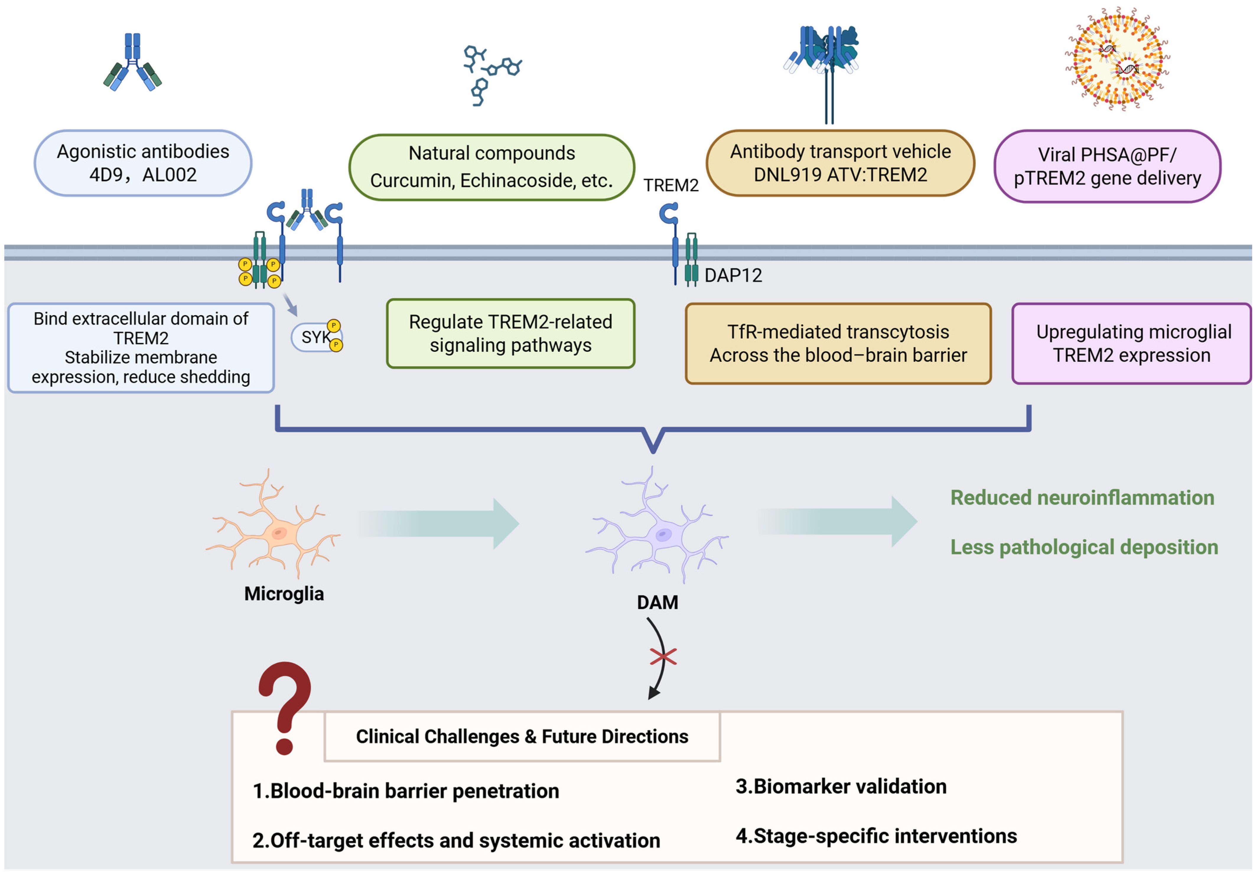
TREM2 holds significant promise as a therapeutic target for neurodegenerative diseases, demonstrating considerable potential in drug development and treatment strategies. The recent progress of TREM2-targeted therapies is presented in Table 2.
The first agonistic antibody targeting TREM2, the monoclonal antibody 4D9, demonstrates reduced amyloid plaque burden in AD mouse models [156]. The epitope of 4D9 localizes to the stalk region of TREM2 near the cleavage site, enabling a dual mechanism of action: stabilization of TREM2 on the cell surface to reduce shedding and activation of downstream phosphorylated SYK signaling. In mouse models of AD-related pathology, 4D9 attenuated amyloid deposition, increased TREM2 expression in microglia, and reduced levels of a homeostatic marker, indicating a protective role through driving microglia toward a disease-associated state.
AL002 is an investigational, engineered, humanized monoclonal immunoglobulin G1 (IgG1) antibody designed to target TREM2. Studies on its variant, AL002c, revealed that acute systemic administration of AL002c induced microglial proliferation in both CV and R47H transgenic mice, as demonstrated by single-cell RNA sequencing. Chronic administration of AL002c reduced filamentous plaques and neuritic dystrophy, modulated behavioral outcomes, and attenuated microglial inflammatory responses [157]. In a preclinical and first-in-human Phase II, randomized, placebo-controlled, double-blind study, researchers evaluated the safety, tolerability, pharmacokinetics, and pharmacodynamics of AL002 in healthy volunteers following single ascending dose (SAD) administration. The findings support the continued clinical development of AL002 for AD and other neurodegenerative disorders [158]. Currently, a Phase II, randomized, double-blind, placebo-controlled study targeting early-stage AD is underway. Another promising antibody, DNL919 (ATV:TREM2), is a high-affinity human TREM2-activating antibody featuring a monovalent transferrin receptor (TfR) binding site, termed an antibody transport vehicle (ATV), which facilitates transcytosis across the blood–brain barrier. Compared with standard anti-TREM2 antibodies, ATV:TREM2 demonstrated improved brain biodistribution and enhanced signaling following peripheral delivery in mice. In human iPSC-derived microglia, ATV:TREM2 induced proliferation and improved mitochondrial metabolism. Single-cell RNA sequencing and morphometric analyses revealed that ATV:TREM2 shifted microglia into a metabolically responsive state distinct from that induced by amyloid pathology. In AD mouse models, ATV:TREM2 enhanced cerebral microglial activity and glucose metabolism [12]. Michael Fassler and colleagues developed a novel monoclonal antibody targeting both membrane-bound and soluble TREM2. The selected antibody promoted microglial proliferation, uptake of oligomeric β-amyloid/apoptotic neurons, and activation in a SYK- and AKT-dependent manner through binding to membrane-bound TREM2. The antibody also demonstrated strong binding to soluble TREM2 in the cerebrospinal fluid of AD patients and attenuated pro-inflammatory programs triggered by intracerebral antibody injection. In vivo therapeutic administration improved cognitive function in experimental amyloidosis models and enhanced plaque-associated microglial coverage and activation [159].
Recent evidence indicates that certain natural compounds exhibit therapeutic potential for neurodegenerative diseases such as AD by modulating TREM2-related molecular pathways. Curcumin exhibits anti-inflammatory and neuroprotective properties. In animal models of cognitive impairment induced by diabetes mellitus and chronic cerebral hypoperfusion (DM/CCH), curcumin exhibited protective effects by inhibiting microglial activation-induced neuroinflammation, modulating the TREM2/TLR4/NF-κB pathway, attenuating apoptosis, and reducing NLRP3-dependent pyroptosis [160]. In APPsw transgenic mice, curcumin enhanced amyloid phagocytic clearance by downregulating CD33 and upregulating TREM2 and TyroBP while restoring neuroinflammatory networks associated with neurodegeneration [161]. Echinacoside (EDB) alleviates hippocampal inflammatory responses in APP/PS1 mice, an effect linked to the TREM2/TLR4/MAPK signaling pathway. Additional in vitro studies revealed that EDB suppresses neuroinflammation in LPS-stimulated BV2 cells by inhibiting the TLR4/MAPK signaling pathway and upregulating TREM2 expression [162]. Wang et al. demonstrated through in vivo and in vitro experiments that Yishen Huazhuo decoction modulates microglial polarization and reduces AD-associated neuroinflammation by regulating the TREM2/NF-κB signaling pathway [163]. Similarly, Hydroxysafflor Yellow A (HSYA) regulates microglial inflammatory phenotypes by modulating Aβ1-42-induced polarization of BV2 microglia (M1/M2), a process potentially mediated through the TREM2/TLR4/NF-κB pathway [164].
Clinically, TREM2-targeted therapies are hindered by significant challenges, including limited blood–brain barrier penetration and unintended systemic effects (such as increased atherosclerosis). Although CSF-soluble TREM2 (sTREM2) demonstrates correlations with tau burden in AD, its variable expression in PD and ALS, combined with confounding factors like the H157Y mutation that increases sTREM2 shedding, highlights the necessity of comprehensive longitudinal biomarker validation. Future research priorities include utilizing single-cell omics to characterize TREM2-dependent microglial subpopulations (particularly disease-associated microglia), implementing conditional modulation models to examine temporal relationships in Aβ/tau dynamics, and integrating multi-omics approaches to identify novel therapeutic targets (especially in regard to lipid metabolism). Successfully addressing these complexities requires careful consideration of pathway interactions, stage-specific biological processes, and translational viability to effectively utilize TREM2’s dual functions. Figure 3 provides an overview of key therapeutic approaches for enhancing TREM2 function in microglia while also addressing major clinical challenges currently encountered.
Table 2. Research progress regarding TREM2-targeted therapies.
Drug NameDrug TypeMechanism of ActionIndicationDevelopment StageNotesReferencesRegistration Number
4D9Monoclonal antibody (agonist)Stabilizes TREM2 on the cell surface and inhibits shedding; activates TREM2-DAP12/SYK signaling through phospho-SYK.AD_Reduces amyloidogenesis and homeostatic markers.
Enhances microglial TREM2 expression in AD models.
[156,165]_
AL002Humanized mAb (agonist)Activates TREM2 signaling.ADPhase II completed (2024)Enhances microglial activity to reduce Aβ plaques and neuroinflammation.[157,158]NCT04592874
Iluzanebart (VGL101)Humanized mAb (agonist)Enhances the TREM2-DAP12/SYK pathway.AD, adult-onset leukoencephalopathy (ALSP)Phase IIWell-tolerated; monthly dosing reduces CSF sTREM2 levels.[166]NCT05677659
PY314Humanized mAb (antagonist)Blocks TREM2 and depletes tumor-associated macrophages.Solid tumors (e.g., ovarian cancer)Phase IbCombined with PD-1 inhibitors for platinum-resistant ovarian cancer.[167,168]NCT04691375
VHB937Humanized mAb (agonist)Activates TREM2 and reduces shedding.Neurodegeneration, metabolic syndromePreclinical/Phase I (2024 filing)Enhances microglial phagocytosis; neuroprotective in animal models.[169]NCT06643481
DNL919 (TAK-920)Monoclonal antibody (agonist)Activates TREM2 signaling.ADDiscontinued (2023)Phase I showed hematologic toxicity; narrow therapeutic window.[12]NCT05450549
Ab18 TVD-Ig/αTfRTetravalent bispecific antibodyEnhances TREM2 clustering and blood–brain barrier (BBB) penetration.ADPreclinicalEngineered antibody reduces Aβ burden and improves cognition in mice.[170]_

14. Conclusions

This review analyzed the structure and function of TREM2 and its influence on microglia, synthesizing our current understanding of its role in neurodegenerative diseases, particularly AD, with an additional focus on PD and ALS. The field has undergone rapid advancement through mechanistic studies and therapeutic developments, notably with monoclonal antibodies progressing to Phase II clinical trials for AD. A significant challenge remains: TREM2-mediated immune stimulation lacks cellular specificity, potentially resulting in off-target effects that may impact therapeutic efficacy and safety. Future research must address several critical aspects: determining the context-dependent approach of activating versus inhibiting TREM2, establishing the optimal therapeutic window, and further elucidating signaling pathways to develop precise immune modulation strategies. Addressing these challenges is essential for effectively exploiting TREM2’s therapeutic potential for neurodegenerative diseases.

Author Contributions

Conceptualization, L.L. and X.F.; data curation, X.L.; investigation, M.Z. and H.M.; writing—original draft preparation, L.L. and X.Z.; writing—review and editing, X.S. and X.L.; supervision, X.F. and X.S. All authors have read and agreed to the published version of the manuscript.

Funding

This research received no external funding.

Institutional Review Board Statement

Not applicable.

Informed Consent Statement

Not applicable.

Data Availability Statement

Not applicable.

Conflicts of Interest

The authors declare no conflicts of interest.

Abbreviations

The following abbreviations are used in this manuscript:
ADAlzheimer’s Disease
PDParkinson’s Disease
ALSAmyotrophic Lateral Sclerosis
TREM2Triggering Receptor Expressed on Myeloid Cells 2
GWASsGenome-Wide Association Studies
DAMDisease-Associated Microglia
ITAMImmunoreceptor Tyrosine-based Activation Motif
DAP12DNAX-Activating Protein 12
SYKSpleen Tyrosine Kinase
PI3KPhosphatidylinositol 3-Kinase
PLCγPhospholipase C gamma
IP3Inositol Trisphosphate
DAGDiacylglycerol
mTORMechanistic Target of Rapamycin
MAPKMitogen-Activated Protein Kinase
TLR4Toll-Like Receptor 4
NF-κBNuclear Factor kappa B
NLRP3NOD-Like Receptor family Pyrin domain containing 3
IL-1βInterleukin-1 beta
TNF-αTumor Necrosis Factor alpha
IL-6Interleukin-6
IL-8Interleukin-8
NHDNasu–Hakola Disease
FTDFrontotemporal Dementia
sTREM2Soluble TREM2
CSFCerebrospinal Fluid
SNPSingle-Nucleotide Polymorphism
ADAM10/17A Disintegrin and Metalloproteinase 10/17
BBBBlood–Brain Barrier
PETPositron Emission Tomography
APOEApolipoprotein E
Amyloid-beta
NFTsNeurofibrillary Tangles
LPSLipopolysaccharide
MOGMyelin Oligodendrocyte Glycoprotein
EAEExperimental Autoimmune Encephalomyelitis
MSMultiple Sclerosis
NMOSDNeuromyelitis Optica Spectrum Disorder
SVDSmall Vessel Disease
CAACerebral Amyloid Angiopathy
BCASBilateral Carotid Artery Stenosis
CD33Cluster of Differentiation 33
TDP-43TAR DNA-binding protein 43
CNSCentral Nervous System
HDLHigh-Density Lipoprotein
LDLLow-Density Lipoprotein
TfRTransferrin Receptor
TBITraumatic Brain Injury
Th17T helper 17 cells
ZAP70Zeta-chain-Associated Protein Kinase 70
STAT3Signal Transducer and Activator of Transcription 3
AQP4Aquaporin-4
OPCsOligodendrocyte Precursor Cells
SVD-HTNHypertensive Small Vessel Disease
ALSPAdult-Onset Leukoencephalopathy
TREMTriggering Receptor Expressed on Myeloid Cells
TYROBPTYRO Protein Tyrosine Kinase-Binding Protein
PLOSLPolycystic Lipomembranous Osteodysplasia with Sclerosing Leukoencephalopathy
BV2Murine Microglial Cell Line
CHOChinese Hamster Ovary
iPSCInduced Pluripotent Stem Cell
BMDMsBone Marrow-Derived Macrophages
CPZCuprizone
LXRLiver X Receptor
ACAT1Acyl-CoA Cholesterol Acyltransferase 1
oxLDLOxidized Low-Density Lipoprotein
ABCA1ATP-Binding Cassette Sub-family A Member 1
SHIP1Src Homology 2-containing Inositol Phosphatase 1
SHIP2Src Homology 2-containing Inositol Phosphatase 2
S1PSphingosine-1-phosphate
TREM2R47HTREM2 R47H Variant
TREM2CVTREM2 Common Variant
TREM2KOTREM2 Knockout
TREM2−/−TREM2-Deficient
SAMP8Senescence-Accelerated Mouse Prone 8
POCDPostoperative Cognitive Dysfunction
MPTP1-Methyl-4-phenyl-1,2,3,6-tetrahydropyridine
α-synα-Synuclein
THTyrosine Hydroxylase
hTDP-43Human TAR DNA-binding Protein 43
hTREM2Human TREM2
CD11cCluster of Differentiation 11c
CD68Cluster of Differentiation 68
CD4Cluster of Differentiation 4
MHC-IMajor Histocompatibility Complex Class I
MHC-IIMajor Histocompatibility Complex Class II
LAMLipid-Associated Macrophages
ATPAdenosine Triphosphate
HSP70Heat Shock Protein 70
GLUT5Glucose Transporter 5
SPP1Secreted Phosphoprotein 1
p-SYKPhosphorylated SYK
FDGFluorodeoxyglucose

References

  1. Liu, L.; Zhao, Y.; Zhang, F. RNA Methylation Modifications in Neurodegenerative Diseases: Focus on Their Enzyme System. J. Adv. Res. 2025, in press. [Google Scholar] [CrossRef]
  2. Grobler, C.; Van Tongeren, M.; Gettemans, J.; Kell, D.B.; Pretorius, E. Alzheimer’s Disease: A Systems View Provides a Unifying Explanation of Its Development. J. Alzheimer’s Dis. 2023, 91, 43–70. [Google Scholar] [CrossRef]
  3. Kurkinen, M.; Fułek, M.; Fułek, K.; Beszłej, J.A.; Kurpas, D.; Leszek, J. The Amyloid Cascade Hypothesis in Alzheimer’s Disease: Should We Change Our Thinking? Biomolecules 2023, 13, 453. [Google Scholar] [CrossRef] [PubMed]
  4. Balkhi, S.; Di Spirito, A.; Poggi, A.; Mortara, L. Immune Modulation in Alzheimer’s Disease: From Pathogenesis to Immunotherapy. Cells 2025, 14, 264. [Google Scholar] [CrossRef] [PubMed]
  5. He, Y.; Liu, T.; He, Q.; Ke, W.; Li, X.; Du, J.; Deng, S.; Shu, Z.; Wu, J.; Yang, B.; et al. Microglia Facilitate and Stabilize the Response to General Anesthesia via Modulating the Neuronal Network in a Brain Region-Specific Manner. eLife 2023, 12, RP92252. [Google Scholar] [CrossRef] [PubMed]
  6. Li, Q.; Barres, B.A. Microglia and Macrophages in Brain Homeostasis and Disease. Nat. Rev. Immunol. 2018, 18, 225–242. [Google Scholar] [CrossRef]
  7. Ma, H.; Zhu, M.; Chen, M.; Li, X.; Feng, X. The Role of Macrophage Plasticity in Neurodegenerative Diseases. Biomark. Res. 2024, 12, 81. [Google Scholar] [CrossRef]
  8. Yang, R.; Li, D.; Li, X.-X.; Yang, X.-X.; Gao, H.-M.; Zhang, F. Dihydroquercetin Alleviates Dopamine Neuron Loss via Regulating TREM2 Activation. Int. J. Biol. Macromol. 2024, 269, 132179. [Google Scholar] [CrossRef]
  9. Wei, W.; Zhang, L.; Xin, W.; Pan, Y.; Tatenhorst, L.; Hao, Z.; Gerner, S.T.; Huber, S.; Juenemann, M.; Butz, M.; et al. TREM2 Regulates Microglial Lipid Droplet Formation and Represses Post-Ischemic Brain Injury. Biomed. Pharmacother. 2024, 170, 115962. [Google Scholar] [CrossRef]
  10. Piccio, L.; Deming, Y.; Del-Águila, J.L.; Ghezzi, L.; Holtzman, D.M.; Fagan, A.M.; Fenoglio, C.; Galimberti, D.; Borroni, B.; Cruchaga, C. Cerebrospinal Fluid Soluble TREM2 is Higher in Alzheimer Disease and Associated with Mutation Status. Acta Neuropathol. 2016, 131, 925–933. [Google Scholar] [CrossRef]
  11. Heslegrave, A.; Heywood, W.; Paterson, R.; Magdalinou, N.; Svensson, J.; Johansson, P.; Öhrfelt, A.; Blennow, K.; Hardy, J.; Schott, J.; et al. Increased Cerebrospinal Fluid Soluble TREM2 Concentration in Alzheimer’s Disease. Mol. Neurodegener. 2016, 11, 3. [Google Scholar] [CrossRef]
  12. Van Lengerich, B.; Zhan, L.; Xia, D.; Chan, D.; Joy, D.; Park, J.I.; Tatarakis, D.; Calvert, M.; Hummel, S.; Lianoglou, S.; et al. A TREM2-Activating Antibody with a Blood–Brain Barrier Transport Vehicle Enhances Microglial Metabolism in Alzheimer’s Disease Models. Nat. Neurosci. 2023, 26, 416–429. [Google Scholar] [CrossRef]
  13. Jay, T.R.; Von Saucken, V.E.; Landreth, G.E. TREM2 in Neurodegenerative Diseases. Mol. Neurodegener. 2017, 12, 56. [Google Scholar] [CrossRef]
  14. Li, Y.; Xu, H.; Wang, H.; Yang, K.; Luan, J.; Wang, S. TREM2: Potential Therapeutic Targeting of Microglia for Alzheimer’s Disease. Biomed. Pharmacother. 2023, 165, 115218. [Google Scholar] [CrossRef]
  15. Wunderlich, P.; Glebov, K.; Kemmerling, N.; Tien, N.T.; Neumann, H.; Walter, J. Sequential Proteolytic Processing of the Triggering Receptor Expressed on Myeloid Cells-2 (TREM2) Protein by Ectodomain Shedding and γ-Secretase-Dependent Intramembranous Cleavage. J. Biol. Chem. 2013, 288, 33027–33036. [Google Scholar] [CrossRef] [PubMed]
  16. Li, R.-Y.; Qin, Q.; Yang, H.-C.; Wang, Y.-Y.; Mi, Y.-X.; Yin, Y.-S.; Wang, M.; Yu, C.-J.; Tang, Y. TREM2 in the Pathogenesis of AD: A Lipid Metabolism Regulator and Potential Metabolic Therapeutic Target. Mol. Neurodegener. 2022, 17, 40. [Google Scholar] [CrossRef]
  17. George, J. TREM2 as an Evolving Therapeutic Target in Alzheimer’s Disease. Neural Regen. Res. 2023, 18, 2680–2681. [Google Scholar] [CrossRef] [PubMed]
  18. Ulland, T.K.; Colonna, M. TREM2—A Key Player in Microglial Biology and Alzheimer Disease. Nat. Rev. Neurol. 2018, 14, 667–675. [Google Scholar] [CrossRef] [PubMed]
  19. Bharadwaj, S.; Groza, Y.; Mierzwicka, J.M.; Malý, P. Current Understanding on TREM-2 Molecular Biology and Physiopathological Functions. Int. Immunopharmacol. 2024, 134, 112042. [Google Scholar] [CrossRef]
  20. Kleinberger, G.; Yamanishi, Y.; Suárez-Calvet, M.; Czirr, E.; Lohmann, E.; Cuyvers, E.; Struyfs, H.; Pettkus, N.; Wenninger-Weinzierl, A.; Mazaheri, F.; et al. TREM2 Mutations Implicated in Neurodegeneration Impair Cell Surface Transport and Phagocytosis. Sci. Transl. Med. 2014, 6, 243ra86. [Google Scholar] [CrossRef]
  21. Neumann, H.; Takahashi, K. Essential Role of the Microglial Triggering Receptor Expressed on Myeloid Cells-2 (TREM2) for Central Nervous Tissue Immune Homeostasis. J. Neuroimmunol. 2007, 184, 92–99. [Google Scholar] [CrossRef]
  22. Hwang, M.; Savarin, C.; Kim, J.; Powers, J.; Towne, N.; Oh, H.; Bergmann, C.C. Trem2 Deficiency Impairs Recovery and Phagocytosis and Dysregulates Myeloid Gene Expression During Virus-Induced Demyelination. J. Neuroinflamm. 2022, 19, 267. [Google Scholar] [CrossRef]
  23. Yu, M.; Chang, Y.; Zhai, Y.; Pang, B.; Wang, P.; Li, G.; Jiang, T.; Zeng, F. TREM2 Is Associated with Tumor Immunity and Implies Poor Prognosis in Glioma. Front. Immunol. 2023, 13, 1089266. [Google Scholar] [CrossRef] [PubMed]
  24. Kober, D.L.; Brett, T.J. TREM2-Ligand Interactions in Health and Disease. J. Mol. Biol. 2017, 429, 1607–1629. [Google Scholar] [CrossRef]
  25. Colonna, M. The Biology of TREM Receptors. Nat. Rev. Immunol. 2023, 23, 580–594. [Google Scholar] [CrossRef]
  26. Nugent, A.A.; Lin, K.; Van Lengerich, B.; Lianoglou, S.; Przybyla, L.; Davis, S.S.; Llapashtica, C.; Wang, J.; Kim, D.J.; Xia, D.; et al. TREM2 Regulates Microglial Cholesterol Metabolism upon Chronic Phagocytic Challenge. Neuron 2020, 105, 837–854.e9. [Google Scholar] [CrossRef] [PubMed]
  27. Ulland, T.K.; Song, W.M.; Huang, S.C.-C.; Ulrich, J.D.; Sergushichev, A.; Beatty, W.L.; Loboda, A.A.; Zhou, Y.; Cairns, N.J.; Kambal, A.; et al. TREM2 Maintains Microglial Metabolic Fitness in Alzheimer’s Disease. Cell 2017, 170, 649–663.e13. [Google Scholar] [CrossRef] [PubMed]
  28. Huang, Z.-Y.; Hunter, S.; Kim, M.-K.; Indik, Z.K.; Schreiber, A.D. The Effect of Phosphatases SHP-1 and SHIP-1 on Signaling by the ITIM- and ITAM-Containing Fcγ Receptors FcγRIIB and FcγRIIA. J. Leukoc. Biol. 2003, 73, 823–829. [Google Scholar] [CrossRef]
  29. Peng, Q.; Malhotra, S.; Torchia, J.A.; Kerr, W.G.; Coggeshall, K.M.; Humphrey, M.B. TREM2- and DAP12-Dependent Activation of PI3K Requires DAP10 and Is Inhibited by SHIP1. Sci. Signal. 2010, 3, ra38. [Google Scholar] [CrossRef]
  30. Ruganzu, J.B.; Peng, X.; He, Y.; Wu, X.; Zheng, Q.; Ding, B.; Lin, C.; Guo, H.; Yang, Z.; Zhang, X.; et al. Downregulation of TREM2 Expression Exacerbates Neuroinflammatory Responses through TLR4-Mediated MAPK Signaling Pathway in a Transgenic Mouse Model of Alzheimer’s Disease. Mol. Immunol. 2022, 142, 22–36. [Google Scholar] [CrossRef]
  31. Chen, S.; Peng, J.; Sherchan, P.; Ma, Y.; Xiang, S.; Yan, F.; Zhao, H.; Jiang, Y.; Wang, N.; Zhang, J.H.; et al. TREM2 Activation Attenuates Neuroinflammation and Neuronal Apoptosis via PI3K/Akt Pathway After Intracerebral Hemorrhage in Mice. J. Neuroinflamm. 2020, 17, 168. [Google Scholar] [CrossRef]
  32. Rossol, M.; Pierer, M.; Raulien, N.; Quandt, D.; Meusch, U.; Rothe, K.; Schubert, K.; Schöneberg, T.; Schaefer, M.; Krügel, U.; et al. Extracellular Ca2+ Is a Danger Signal Activating the NLRP3 Inflammasome through G Protein-Coupled Calcium Sensing Receptors. Nat. Commun. 2012, 3, 1329. [Google Scholar] [CrossRef] [PubMed]
  33. Obst, J.; Hall-Roberts, H.L.; Smith, T.B.; Kreuzer, M.; Magno, L.; Di Daniel, E.; Davis, J.B.; Mead, E. PLCγ2 Regulates TREM2 Signalling and Integrin-Mediated Adhesion and Migration of Human iPSC-Derived Macrophages. Sci. Rep. 2021, 11, 19842. [Google Scholar] [CrossRef] [PubMed]
  34. Jairaman, A.; McQuade, A.; Granzotto, A.; Kang, Y.J.; Chadarevian, J.P.; Gandhi, S.; Parker, I.; Smith, I.; Cho, H.; Sensi, S.L.; et al. TREM2 Regulates Purinergic Receptor-Mediated Calcium Signaling and Motility in Human iPSC-Derived Microglia. eLife 2022, 11, e73021. [Google Scholar] [CrossRef] [PubMed]
  35. Terzioglu, G.; Young-Pearse, T.L. Microglial Function, INPP5D/SHIP1 Signaling, and NLRP3 Inflammasome Activation: Implications for Alzheimer’s Disease. Mol. Neurodegener. 2023, 18, 89. [Google Scholar] [CrossRef]
  36. Ramakrishnan, G.S.; Berry, W.L.; Pacherille, A.; Kerr, W.G.; Chisholm, J.D.; Pedicone, C.; Humphrey, M.B. SHIP Inhibition Mediates Select TREM2-Induced Microglial Functions. Mol. Immunol. 2024, 170, 35–45. [Google Scholar] [CrossRef]
  37. Griciuc, A.; Patel, S.; Federico, A.N.; Choi, S.H.; Innes, B.J.; Oram, M.K.; Cereghetti, G.; McGinty, D.; Anselmo, A.; Sadreyev, R.I.; et al. TREM2 Acts Downstream of CD33 in Modulating Microglial Pathology in Alzheimer’s Disease. Neuron 2019, 103, 820–835.e7. [Google Scholar] [CrossRef]
  38. Wißfeld, J.; Mathews, M.; Mossad, O.; Picardi, P.; Cinti, A.; Redaelli, L.; Pradier, L.; Brüstle, O.; Neumann, H. Reporter Cell Assay for Human CD33 Validated by Specific Antibodies and Human iPSC-Derived Microglia. Sci. Rep. 2021, 11, 13462. [Google Scholar] [CrossRef] [PubMed]
  39. Wißfeld, J.; Nozaki, I.; Mathews, M.; Raschka, T.; Ebeling, C.; Hornung, V.; Brüstle, O.; Neumann, H. Deletion of Alzheimer’s Disease-associated CD33 Results in an Inflammatory Human Microglia Phenotype. Glia 2021, 69, 1393–1412. [Google Scholar] [CrossRef]
  40. Yang, J.; Fu, Z.; Zhang, X.; Xiong, M.; Meng, L.; Zhang, Z. TREM2 Ectodomain and Its Soluble Form in Alzheimer’s Disease. J. Neuroinflamm. 2020, 17, 204. [Google Scholar] [CrossRef]
  41. Dong, M.-H.; Zhou, L.-Q.; Tang, Y.; Chen, M.; Xiao, J.; Shang, K.; Deng, G.; Qin, C.; Tian, D.-S. CSF sTREM2 in Neurological Diseases: A Two-Sample Mendelian Randomization Study. J. Neuroinflamm. 2022, 19, 79. [Google Scholar] [CrossRef] [PubMed]
  42. Jericó, I.; Vicuña-Urriza, J.; Blanco-Luquin, I.; Macias, M.; Martinez-Merino, L.; Roldán, M.; Rojas-Garcia, R.; Pagola-Lorz, I.; Carbayo, A.; De Luna, N.; et al. Profiling TREM2 Expression in Amyotrophic Lateral Sclerosis. Brain Behav. Immun. 2023, 109, 117–126. [Google Scholar] [CrossRef] [PubMed]
  43. Zhong, L.; Xu, Y.; Zhuo, R.; Wang, T.; Wang, K.; Huang, R.; Wang, D.; Gao, Y.; Zhu, Y.; Sheng, X.; et al. Soluble TREM2 Ameliorates Pathological Phenotypes by Modulating Microglial Functions in an Alzheimer’s Disease Model. Nat. Commun. 2019, 10, 1365. [Google Scholar] [CrossRef]
  44. Belsare, K.D.; Wu, H.; Mondal, D.; Bond, A.; Castillo, E.; Jin, J.; Jo, H.; Roush, A.E.; Pilla, K.B.; Sali, A.; et al. Soluble TREM2 Inhibits Secondary Nucleation of Aβ Fibrillization and Enhances Cellular Uptake of Fibrillar Aβ. Proc. Natl. Acad. Sci. USA 2022, 119, e2114486119. [Google Scholar] [CrossRef]
  45. Zhong, L.; Chen, X.-F.; Wang, T.; Wang, Z.; Liao, C.; Wang, Z.; Huang, R.; Wang, D.; Li, X.; Wu, L.; et al. Soluble TREM2 Induces Inflammatory Responses and Enhances Microglial Survival. J. Exp. Med. 2017, 214, 597–607. [Google Scholar] [CrossRef]
  46. Zhang, L.; Xiang, X.; Li, Y.; Bu, G.; Chen, X.-F. TREM2 and sTREM2 in Alzheimer’s Disease: From Mechanisms to Therapies. Mol. Neurodegener. 2025, 20, 43. [Google Scholar] [CrossRef]
  47. Paloneva, J.; Manninen, T.; Christman, G.; Hovanes, K.; Mandelin, J.; Adolfsson, R.; Bianchin, M.; Bird, T.; Miranda, R.; Salmaggi, A.; et al. Mutations in Two Genes Encoding Different Subunits of a Receptor Signaling Complex Result in an Identical Disease Phenotype. Am. J. Hum. Genet. 2002, 71, 656–662. [Google Scholar] [CrossRef] [PubMed]
  48. Paloneva Bm, J.; Autti, T.; Raininko, R.; Partanen, J.; Salonen, O.; Puranen, M.; Hakola, P.; Haltia, M. CNS Manifestations of Nasu–Hakola Disease: A Frontal Dementia with Bone Cysts. Neurology 2001, 56, 1552–1558. [Google Scholar] [CrossRef]
  49. Klünemann, H.H.; Ridha, B.H.; Magy, L.; Wherrett, J.R.; Hemelsoet, D.M.; Keen, R.W.; De Bleecker, J.L.; Rossor, M.N.; Marienhagen, J.; Klein, H.E.; et al. The Genetic Causes of Basal Ganglia Calcification, Dementia, and Bone Cysts: DAP12 and TREM2. Neurology 2005, 64, 1502–1507. [Google Scholar] [CrossRef]
  50. Filipello, F.; You, S.-F.; Mirfakhar, F.S.; Mahali, S.; Bollman, B.; Acquarone, M.; Korvatska, O.; Marsh, J.A.; Sivaraman, A.; Martinez, R.; et al. Defects in Lysosomal Function and Lipid Metabolism in Human Microglia Harboring a TREM2 Loss of Function Mutation. Acta Neuropathol. 2023, 145, 749–772. [Google Scholar] [CrossRef]
  51. Yuan, P.; Condello, C.; Keene, C.D.; Wang, Y.; Bird, T.D.; Paul, S.M.; Luo, W.; Colonna, M.; Baddeley, D.; Grutzendler, J. TREM2 Haplodeficiency in Mice and Humans Impairs the Microglia Barrier Function Leading to Decreased Amyloid Compaction and Severe Axonal Dystrophy. Neuron 2016, 90, 724–739. [Google Scholar] [CrossRef]
  52. Sayed, F.A.; Kodama, L.; Fan, L.; Carling, G.K.; Udeochu, J.C.; Le, D.; Li, Q.; Zhou, L.; Wong, M.Y.; Horowitz, R.; et al. AD-Linked R47H-TREM2 Mutation Induces Disease-Enhancing Microglial States via AKT Hyperactivation. Sci. Transl. Med. 2021, 13, eabe3947. [Google Scholar] [CrossRef]
  53. Rayaprolu, S.; Mullen, B.; Baker, M.; Lynch, T.; Finger, E.; Seeley, W.W.; Hatanpaa, K.J.; Lomen-Hoerth, C.; Kertesz, A.; Bigio, E.H.; et al. TREM2 in Neurodegeneration: Evidence for Association of the p.R47H Variant with Frontotemporal Dementia and Parkinson’s Disease. Mol. Neurodegener. 2013, 8, 19. [Google Scholar] [CrossRef]
  54. Cady, J.; Koval, E.D.; Benitez, B.A.; Zaidman, C.; Jockel-Balsarotti, J.; Allred, P.; Baloh, R.H.; Ravits, J.; Simpson, E.; Appel, S.H.; et al. TREM2 Variant p.R47H as a Risk Factor for Sporadic Amyotrophic Lateral Sclerosis. JAMA Neurol. 2014, 71, 449. [Google Scholar] [CrossRef]
  55. Guerreiro, R.; Wojtas, A.; Bras, J.; Carrasquillo, M.; Rogaeva, E.; Majounie, E.; Cruchaga, C.; Sassi, C.; Kauwe, J.S.K.; Younkin, S.; et al. TREM2 Variants in Alzheimer’s Disease. N. Engl. J. Med. 2013, 368, 117–127. [Google Scholar] [CrossRef]
  56. Jonsson, T.; Stefansson, H.; Steinberg, S.; Jonsdottir, I.; Jonsson, P.V.; Snaedal, J.; Bjornsson, S.; Huttenlocher, J.; Levey, A.I.; Lah, J.J.; et al. Variant of TREM2 Associated with the Risk of Alzheimer’s Disease. N. Engl. J. Med. 2013, 368, 107–116. [Google Scholar] [CrossRef] [PubMed]
  57. Kober, D.L.; Alexander-Brett, J.M.; Karch, C.M.; Cruchaga, C.; Colonna, M.; Holtzman, M.J.; Brett, T.J. Neurodegenerative Disease Mutations in TREM2 Reveal a Functional Surface and Distinct Loss-of-Function Mechanisms. eLife 2016, 5, e20391. [Google Scholar] [CrossRef]
  58. Guerreiro, R.J.; Lohmann, E.; Brás, J.M.; Gibbs, J.R.; Rohrer, J.D.; Gurunlian, N.; Dursun, B.; Bilgic, B.; Hanagasi, H.; Gurvit, H.; et al. Using Exome Sequencing to Reveal Mutations in TREM2 Presenting as a Frontotemporal Dementia-like Syndrome without Bone Involvement. JAMA Neurol. 2013, 70, 78–84. [Google Scholar] [CrossRef] [PubMed]
  59. Kleinberger, G.; Brendel, M.; Mracsko, E.; Wefers, B.; Groeneweg, L.; Xiang, X.; Focke, C.; Deußing, M.; Suárez-Calvet, M.; Mazaheri, F.; et al. The FTD-like Syndrome Causing TREM 2 T66M Mutation Impairs Microglia Function, Brain Perfusion, and Glucose Metabolism. EMBO J. 2017, 36, 1837–1853. [Google Scholar] [CrossRef]
  60. Le Ber, I.; De Septenville, A.; Guerreiro, R.; Bras, J.; Camuzat, A.; Caroppo, P.; Lattante, S.; Couarch, P.; Kabashi, E.; Bouya-Ahmed, K.; et al. Homozygous TREM2 Mutation in a Family with Atypical Frontotemporal Dementia. Neurobiol. Aging 2014, 35, 2419.e23–2419.e25. [Google Scholar] [CrossRef] [PubMed]
  61. Park, J.; Ji, I.J.; An, H.J.; Kang, M.; Kang, S.; Kim, D.; Yoon, S. Disease-Associated Mutations of TREM2 Alter the Processing of N-Linked Oligosaccharides in the Golgi Apparatus. Traffic 2015, 16, 510–518. [Google Scholar] [CrossRef] [PubMed]
  62. Pillai, J.; Sung, K.; Wu, C. Predicting the Impact of Missense Mutations on an Unresolved Protein’s Stability, Structure, and Function: A Case Study of Alzheimer’s Disease-Associated TREM2 R47H Variant. Comput. Struct. Biotechnol. J. 2025, 27, 564–574. [Google Scholar] [CrossRef] [PubMed]
  63. Jadhav, V.S.; Lin, P.B.C.; Pennington, T.; Di Prisco, G.V.; Jannu, A.J.; Xu, G.; Moutinho, M.; Zhang, J.; Atwood, B.K.; Puntambekar, S.S.; et al. Trem2 Y38C Mutation and Loss of Trem2 Impairs Neuronal Synapses in Adult Mice. Mol. Neurodegener. 2020, 15, 62. [Google Scholar] [CrossRef]
  64. Schuler, O. A Case of Possible Autoimmune Bilateral Vestibulopathy Treated with Steroids. J. Neurol. Neurosurg. Psychiatry 2003, 74, 825. [Google Scholar] [CrossRef]
  65. Thelen, M.; Razquin, C.; Hernández, I.; Gorostidi, A.; Sánchez-Valle, R.; Ortega-Cubero, S.; Wolfsgruber, S.; Drichel, D.; Fliessbach, K.; Duenkel, T.; et al. Investigation of the Role of Rare TREM2 Variants in Frontotemporal Dementia Subtypes. Neurobiol. Aging 2014, 35, 2657.e13–2657.e19. [Google Scholar] [CrossRef]
  66. Achouri-Rassas, A.; Fray, S.; Said, Z.; Ben Sassi, S.; Ben Ali, N.; Baraket, G. Genetic Association Study between Rs2234253 (p.T96K) Variant of TREM2 and Alzheimer’s Disease in a Tunisian Population. Neurol. Res. 2025, 47, 290–295. [Google Scholar] [CrossRef]
  67. Jiang, T.; Tan, L.; Chen, Q.; Tan, M.-S.; Zhou, J.-S.; Zhu, X.-C.; Lu, H.; Wang, H.-F.; Zhang, Y.-D.; Yu, J.-T. A Rare Coding Variant in TREM2 Increases Risk for Alzheimer’s Disease in Han Chinese. Neurobiol. Aging 2016, 42, 217.e1–217.e3. [Google Scholar] [CrossRef]
  68. Qiao, W.; Chen, Y.; Zhong, J.; Madden, B.J.; Charlesworth, C.M.; Martens, Y.A.; Liu, C.-C.; Knight, J.; Ikezu, T.C.; Kurti, A.; et al. Trem2 H157Y Increases Soluble TREM2 Production and Reduces Amyloid Pathology. Mol. Neurodegener. 2023, 18, 8. [Google Scholar] [CrossRef]
  69. Jin, S.C.; Carrasquillo, M.M.; Benitez, B.A.; Skorupa, T.; Carrell, D.; Patel, D.; Lincoln, S.; Krishnan, S.; Kachadoorian, M.; Reitz, C.; et al. TREM2 Is Associated with Increased Risk for Alzheimer’s Disease in African Americans. Mol. Neurodegener. 2015, 10, 19. [Google Scholar] [CrossRef]
  70. Keren-Shaul, H.; Spinrad, A.; Weiner, A.; Matcovitch-Natan, O.; Dvir-Szternfeld, R.; Ulland, T.K.; David, E.; Baruch, K.; Lara-Astaiso, D.; Toth, B.; et al. A Unique Microglia Type Associated with Restricting Development of Alzheimer’s Disease. Cell 2017, 169, 1276–1290.e17. [Google Scholar] [CrossRef] [PubMed]
  71. Wang, S.; Sudan, R.; Peng, V.; Zhou, Y.; Du, S.; Yuede, C.M.; Lei, T.; Hou, J.; Cai, Z.; Cella, M.; et al. TREM2 Drives Microglia Response to Amyloid-β via SYK-Dependent and -Independent Pathways. Cell 2022, 185, 4153–4169.e19. [Google Scholar] [CrossRef]
  72. Zhou, Y.; Song, W.M.; Andhey, P.S.; Swain, A.; Levy, T.; Miller, K.R.; Poliani, P.L.; Cominelli, M.; Grover, S.; Gilfillan, S.; et al. Human and Mouse Single-Nucleus Transcriptomics Reveal TREM2-Dependent and TREM2-Independent Cellular Responses in Alzheimer’s Disease. Nat. Med. 2020, 26, 131–142. [Google Scholar] [CrossRef] [PubMed]
  73. Bernier, L.-P.; York, E.M.; MacVicar, B.A. Immunometabolism in the Brain: How Metabolism Shapes Microglial Function. Trends Neurosci. 2020, 43, 854–869. [Google Scholar] [CrossRef]
  74. Galluzzi, L.; Pietrocola, F.; Levine, B.; Kroemer, G. Metabolic Control of Autophagy. Cell 2014, 159, 1263–1276. [Google Scholar] [CrossRef] [PubMed]
  75. Piollet, M.; Porsch, F.; Rizzo, G.; Kapser, F.; Schulz, D.J.J.; Kiss, M.G.; Schlepckow, K.; Morenas-Rodriguez, E.; Sen, M.O.; Gropper, J.; et al. TREM2 Protects from Atherosclerosis by Limiting Necrotic Core Formation. Nat. Cardiovasc. Res. 2024, 3, 269–282. [Google Scholar] [CrossRef] [PubMed]
  76. Tagliatti, E.; Desiato, G.; Mancinelli, S.; Bizzotto, M.; Gagliani, M.C.; Faggiani, E.; Hernández-Soto, R.; Cugurra, A.; Poliseno, P.; Miotto, M.; et al. Trem2 Expression in Microglia Is Required to Maintain Normal Neuronal Bioenergetics during Development. Immunity 2024, 57, 86–105.e9. [Google Scholar] [CrossRef]
  77. Graham, L.C.; Harder, J.M.; Soto, I.; De Vries, W.N.; John, S.W.M.; Howell, G.R. Chronic Consumption of a Western Diet Induces Robust Glial Activation in Aging Mice and in a Mouse Model of Alzheimer’s Disease. Sci. Rep. 2016, 6, 21568. [Google Scholar] [CrossRef]
  78. Zhang, S.; Zhu, L.; Peng, Y.; Zhang, L.; Chao, F.; Jiang, L.; Xiao, Q.; Liang, X.; Tang, J.; Yang, H.; et al. Long-Term Running Exercise Improves Cognitive Function and Promotes Microglial Glucose Metabolism and Morphological Plasticity in the Hippocampus of APP/PS1 Mice. J. Neuroinflamm. 2022, 19, 34. [Google Scholar] [CrossRef]
  79. Wang, Y.; Cella, M.; Mallinson, K.; Ulrich, J.D.; Young, K.L.; Robinette, M.L.; Gilfillan, S.; Krishnan, G.M.; Sudhakar, S.; Zinselmeyer, B.H.; et al. TREM2 Lipid Sensing Sustains the Microglial Response in an Alzheimer’s Disease Model. Cell 2015, 160, 1061–1071. [Google Scholar] [CrossRef]
  80. Jaitin, D.A.; Adlung, L.; Thaiss, C.A.; Weiner, A.; Li, B.; Descamps, H.; Lundgren, P.; Bleriot, C.; Liu, Z.; Deczkowska, A.; et al. Lipid-Associated Macrophages Control Metabolic Homeostasis in a Trem2-Dependent Manner. Cell 2019, 178, 686–698.e14. [Google Scholar] [CrossRef]
  81. Patterson, M.T.; Firulyova, M.M.; Xu, Y.; Hillman, H.; Bishop, C.; Zhu, A.; Hickok, G.H.; Schrank, P.R.; Ronayne, C.E.; Caillot, Z.; et al. Trem2 Promotes Foamy Macrophage Lipid Uptake and Survival in Atherosclerosis. Nat. Cardiovasc. Res. 2023, 2, 1015–1031. [Google Scholar] [CrossRef]
  82. Patterson, M.T.; Xu, Y.; Hillman, H.; Osinski, V.; Schrank, P.R.; Kennedy, A.E.; Barrow, F.; Zhu, A.; Tollison, S.; Shekhar, S.; et al. Trem2 Agonist Reprograms Foamy Macrophages to Promote Atherosclerotic Plaque Stability—Brief Report. Arterioscler. Thromb. Vasc. Biol. 2024, 44, 1646–1657. [Google Scholar] [CrossRef]
  83. Jiang, T.; Yu, J.-T.; Zhu, X.-C.; Tan, M.-S.; Gu, L.-Z.; Zhang, Y.-D.; Tan, L. Triggering Receptor Expressed on Myeloid Cells 2 Knockdown Exacerbates Aging-Related Neuroinflammation and Cognitive Deficiency in Senescence-Accelerated Mouse Prone 8 Mice. Neurobiol. Aging 2014, 35, 1243–1251. [Google Scholar] [CrossRef] [PubMed]
  84. Han, X.; Cheng, X.; Xu, J.; Liu, Y.; Zhou, J.; Jiang, L.; Gu, X.; Xia, T. Activation of TREM2 Attenuates Neuroinflammation via PI3K/Akt Signaling Pathway to Improve Postoperative Cognitive Dysfunction in Mice. Neuropharmacology 2022, 219, 109231. [Google Scholar] [CrossRef] [PubMed]
  85. Huang, P.; Zhang, Z.; Zhang, P.; Feng, J.; Xie, J.; Zheng, Y.; Liang, X.; Zhu, B.; Chen, Z.; Feng, S.; et al. TREM2 Deficiency Aggravates NLRP3 Inflammasome Activation and Pyroptosis in MPTP-Induced Parkinson’s Disease Mice and LPS-Induced BV2 Cells. Mol. Neurobiol. 2024, 61, 2590–2605. [Google Scholar] [CrossRef]
  86. Forabosco, P.; Ramasamy, A.; Trabzuni, D.; Walker, R.; Smith, C.; Bras, J.; Levine, A.P.; Hardy, J.; Pocock, J.M.; Guerreiro, R.; et al. Insights into TREM2 Biology by Network Analysis of Human Brain Gene Expression Data. Neurobiol. Aging 2013, 34, 2699–2714. [Google Scholar] [CrossRef]
  87. Jiang, T.; Tan, L.; Zhu, X.-C.; Zhang, Q.-Q.; Cao, L.; Tan, M.-S.; Gu, L.-Z.; Wang, H.-F.; Ding, Z.-Z.; Zhang, Y.-D.; et al. Upregulation of TREM2 Ameliorates Neuropathology and Rescues Spatial Cognitive Impairment in a Transgenic Mouse Model of Alzheimer’s Disease. Neuropsychopharmacology 2014, 39, 2949–2962. [Google Scholar] [CrossRef] [PubMed]
  88. Kobayashi, M.; Konishi, H.; Sayo, A.; Takai, T.; Kiyama, H. TREM2/DAP12 Signal Elicits Proinflammatory Response in Microglia and Exacerbates Neuropathic Pain. J. Neurosci. 2016, 36, 11138–11150. [Google Scholar] [CrossRef]
  89. Gratuze, M.; Leyns, C.E.G.; Holtzman, D.M. New Insights into the Role of TREM2 in Alzheimer’s Disease. Mol. Neurodegener. 2018, 13, 66. [Google Scholar] [CrossRef]
  90. Popescu, A.S.; Butler, C.A.; Allendorf, D.H.; Piers, T.M.; Mallach, A.; Roewe, J.; Reinhardt, P.; Cinti, A.; Redaelli, L.; Boudesco, C.; et al. Alzheimer’s Disease-associated R47H TREM2 Increases, but Wild-type TREM2 Decreases, Microglial Phagocytosis of Synaptosomes and Neuronal Loss. Glia 2023, 71, 974–990. [Google Scholar] [CrossRef]
  91. Schoch, K.M.; Ezerskiy, L.A.; Morhaus, M.M.; Bannon, R.N.; Sauerbeck, A.D.; Shabsovich, M.; Jafar-nejad, P.; Rigo, F.; Miller, T.M. Acute Trem2 Reduction Triggers Increased Microglial Phagocytosis, Slowing Amyloid Deposition in Mice. Proc. Natl. Acad. Sci. USA 2021, 118, e2100356118. [Google Scholar] [CrossRef]
  92. Yin, S.; Chi, X.; Wan, F.; Li, Y.; Zhou, Q.; Kou, L.; Sun, Y.; Wu, J.; Zou, W.; Wang, Y.; et al. TREM2 Signaling in Parkinson’s Disease: Regulation of Microglial Function and α-Synuclein Pathology. Int. Immunopharmacol. 2024, 143, 113446. [Google Scholar] [CrossRef] [PubMed]
  93. Xie, M.; Liu, Y.U.; Zhao, S.; Zhang, L.; Bosco, D.B.; Pang, Y.-P.; Zhong, J.; Sheth, U.; Martens, Y.A.; Zhao, N.; et al. TREM2 Interacts with TDP-43 and Mediates Microglial Neuroprotection against TDP-43-Related Neurodegeneration. Nat. Neurosci. 2022, 25, 26–38. [Google Scholar] [CrossRef]
  94. Pang, X.; Chu, Y.; Zhou, L.; Chen, M.; You, Y.; Tang, Y.; Yang, S.; Zhang, H.; Xiao, J.; Deng, G.; et al. Trem2 Deficiency Attenuates Microglial Phagocytosis and Autophagic-lysosomal Activation in White Matter Hypoperfusion. J. Neurochem. 2023, 167, 489–504. [Google Scholar] [CrossRef] [PubMed]
  95. Xue, T.; Ji, J.; Sun, Y.; Huang, X.; Cai, Z.; Yang, J.; Guo, W.; Guo, R.; Cheng, H.; Sun, X. Sphingosine-1-Phosphate, a Novel TREM2 Ligand, Promotes Microglial Phagocytosis to Protect against Ischemic Brain Injury. Acta Pharm. Sin. B 2022, 12, 1885–1898. [Google Scholar] [CrossRef]
  96. Cignarella, F.; Filipello, F.; Bollman, B.; Cantoni, C.; Locca, A.; Mikesell, R.; Manis, M.; Ibrahim, A.; Deng, L.; Benitez, B.A.; et al. TREM2 Activation on Microglia Promotes Myelin Debris Clearance and Remyelination in a Model of Multiple Sclerosis. Acta Neuropathol. 2020, 140, 513–534. [Google Scholar] [CrossRef] [PubMed]
  97. Jiang, T.; Hou, J.-K.; Gao, Q.; Yu, J.-T.; Zhou, J.-S.; Zhao, H.-D.; Zhang, Y.-D. TREM2 p.H157Y Variant and the Risk of Alzheimer’s Disease: A Meta-Analysis Involving 14,510 Subjects. Curr. Neurovascular Res. 2016, 13, 318–320. [Google Scholar] [CrossRef]
  98. Song, W.; Hooli, B.; Mullin, K.; Jin, S.C.; Cella, M.; Ulland, T.K.; Wang, Y.; Tanzi, R.E.; Colonna, M. Alzheimer’s Disease-Associated TREM2 Variants Exhibit Either Decreased or Increased Ligand-Dependent Activation. Alzheimer’s Dement. 2017, 13, 381–387. [Google Scholar] [CrossRef]
  99. Benitez, B.A.; Cooper, B.; Pastor, P.; Jin, S.-C.; Lorenzo, E.; Cervantes, S.; Cruchaga, C. TREM2 Is Associated with the Risk of Alzheimer’s Disease in Spanish Population. Neurobiol. Aging 2013, 34, 1711.e15–1711.e17. [Google Scholar] [CrossRef]
  100. Ghani, M.; Sato, C.; Kakhki, E.G.; Gibbs, J.R.; Traynor, B.; St George-Hyslop, P.; Rogaeva, E. Mutation Analysis of the MS4A and TREM Gene Clusters in a Case-Control Alzheimer’s Disease Data Set. Neurobiol. Aging 2016, 42, 217.e7–217.e13. [Google Scholar] [CrossRef]
  101. Jin, S.C.; Benitez, B.A.; Karch, C.M.; Cooper, B.; Skorupa, T.; Carrell, D.; Norton, J.B.; Hsu, S.; Harari, O.; Cai, Y.; et al. Coding Variants in TREM2 Increase Risk for Alzheimer’s Disease. Hum. Mol. Genet. 2014, 23, 5838–5846. [Google Scholar] [CrossRef] [PubMed]
  102. Roussos, P.; Katsel, P.; Fam, P.; Tan, W.; Purohit, D.P.; Haroutunian, V. The Triggering Receptor Expressed on Myeloid Cells 2 (TREM2) Is Associated with Enhanced Inflammation, Neuropathological Lesions and Increased Risk for Alzheimer’s Dementia. Alzheimer’s Dement. 2015, 11, 1163–1170. [Google Scholar] [CrossRef] [PubMed]
  103. Carmona, S.; Zahs, K.; Wu, E.; Dakin, K.; Bras, J.; Guerreiro, R. The Role of TREM2 in Alzheimer’s Disease and Other Neurodegenerative Disorders. Lancet Neurol. 2018, 17, 721–730. [Google Scholar] [CrossRef] [PubMed]
  104. Jiang, T.; Tan, L.; Zhu, X.-C.; Zhou, J.-S.; Cao, L.; Tan, M.-S.; Wang, H.-F.; Chen, Q.; Zhang, Y.-D.; Yu, J.-T. Silencing of TREM2 Exacerbates Tau Pathology, Neurodegenerative Changes, and Spatial Learning Deficits in P301S Tau Transgenic Mice. Neurobiol. Aging 2015, 36, 3176–3186. [Google Scholar] [CrossRef]
  105. Wang, Y.; Lin, Y.; Wang, L.; Zhan, H.; Luo, X.; Zeng, Y.; Wu, W.; Zhang, X.; Wang, F. TREM2 Ameliorates Neuroinflammatory Response and Cognitive Impairment via PI3K/AKT/FoxO3a Signaling Pathway in Alzheimer’s Disease Mice. Aging 2020, 12, 20862–20879. [Google Scholar] [CrossRef]
  106. Jiang, T.; Zhang, Y.-D.; Chen, Q.; Gao, Q.; Zhu, X.-C.; Zhou, J.-S.; Shi, J.-Q.; Lu, H.; Tan, L.; Yu, J.-T. TREM2 Modifies Microglial Phenotype and Provides Neuroprotection in P301S Tau Transgenic Mice. Neuropharmacology 2016, 105, 196–206. [Google Scholar] [CrossRef]
  107. Bouchon, A.; Hernández-Munain, C.; Cella, M.; Colonna, M. A DAP12-Mediated Pathway Regulates Expression of CC Chemokine Receptor 7 and Maturation of Human Dendritic Cells. J. Exp. Med. 2001, 194, 1111–1122. [Google Scholar] [CrossRef]
  108. Jay, T.R.; Hirsch, A.M.; Broihier, M.L.; Miller, C.M.; Neilson, L.E.; Ransohoff, R.M.; Lamb, B.T.; Landreth, G.E. Disease Progression-Dependent Effects of TREM2 Deficiency in a Mouse Model of Alzheimer’s Disease. J. Neurosci. 2017, 37, 637–647. [Google Scholar] [CrossRef]
  109. Wang, Y.; Ulland, T.K.; Ulrich, J.D.; Song, W.; Tzaferis, J.A.; Hole, J.T.; Yuan, P.; Mahan, T.E.; Shi, Y.; Gilfillan, S.; et al. TREM2-Mediated Early Microglial Response Limits Diffusion and Toxicity of Amyloid Plaques. J. Exp. Med. 2016, 213, 667–675. [Google Scholar] [CrossRef]
  110. Meilandt, W.J.; Ngu, H.; Gogineni, A.; Lalehzadeh, G.; Lee, S.-H.; Srinivasan, K.; Imperio, J.; Wu, T.; Weber, M.; Kruse, A.J.; et al. Trem2 Deletion Reduces Late-Stage Amyloid Plaque Accumulation, Elevates the Aβ42:Aβ40 Ratio, and Exacerbates Axonal Dystrophy and Dendritic Spine Loss in the PS2APP Alzheimer’s Mouse Model. J. Neurosci. 2020, 40, 1956–1974. [Google Scholar] [CrossRef]
  111. Parhizkar, S.; Arzberger, T.; Brendel, M.; Kleinberger, G.; Deussing, M.; Focke, C.; Nuscher, B.; Xiong, M.; Ghasemigharagoz, A.; Katzmarski, N.; et al. Loss of TREM2 Function Increases Amyloid Seeding but Reduces Plaque-Associated ApoE. Nat. Neurosci. 2019, 22, 191–204. [Google Scholar] [CrossRef]
  112. Leyns, C.E.G.; Ulrich, J.D.; Finn, M.B.; Stewart, F.R.; Koscal, L.J.; Remolina Serrano, J.; Robinson, G.O.; Anderson, E.; Colonna, M.; Holtzman, D.M. TREM2 Deficiency Attenuates Neuroinflammation and Protects against Neurodegeneration in a Mouse Model of Tauopathy. Proc. Natl. Acad. Sci. USA 2017, 114, 11524–11529. [Google Scholar] [CrossRef] [PubMed]
  113. Gratuze, M.; Leyns, C.E.G.; Sauerbeck, A.D.; St-Pierre, M.-K.; Xiong, M.; Kim, N.; Serrano, J.R.; Tremblay, M.-È.; Kummer, T.T.; Colonna, M.; et al. Impact of TREM2R47H Variant on Tau Pathology–Induced Gliosis and Neurodegeneration. J. Clin. Investig. 2020, 130, 4954–4968. [Google Scholar] [CrossRef] [PubMed]
  114. Lee, S.-H.; Meilandt, W.J.; Xie, L.; Gandham, V.D.; Ngu, H.; Barck, K.H.; Rezzonico, M.G.; Imperio, J.; Lalehzadeh, G.; Huntley, M.A.; et al. Trem2 Restrains the Enhancement of Tau Accumulation and Neurodegeneration by β-Amyloid Pathology. Neuron 2021, 109, 1283–1301.e6. [Google Scholar] [CrossRef]
  115. Shirotani, K.; Hori, Y.; Yoshizaki, R.; Higuchi, E.; Colonna, M.; Saito, T.; Hashimoto, S.; Saito, T.; Saido, T.C.; Iwata, N. Aminophospholipids Are Signal-Transducing TREM2 Ligands on Apoptotic Cells. Sci. Rep. 2019, 9, 7508. [Google Scholar] [CrossRef]
  116. Zhao, N.; Qiao, W.; Li, F.; Ren, Y.; Zheng, J.; Martens, Y.A.; Wang, X.; Li, L.; Liu, C.-C.; Chen, K.; et al. Elevating Microglia TREM2 Reduces Amyloid Seeding and Suppresses Disease-Associated Microglia. J. Exp. Med. 2022, 219, e20212479. [Google Scholar] [CrossRef]
  117. Liu, T.; Zhu, B.; Liu, Y.; Zhang, X.; Yin, J.; Li, X.; Jiang, L.; Hodges, A.P.; Rosenthal, S.B.; Zhou, L.; et al. Multi-Omic Comparison of Alzheimer’s Variants in Human ESC-Derived Microglia Reveals Convergence at APOE. J. Exp. Med. 2020, 217, e20200474. [Google Scholar] [CrossRef]
  118. Kober, D.L.; Stuchell-Brereton, M.D.; Kluender, C.E.; Dean, H.B.; Strickland, M.R.; Steinberg, D.F.; Nelson, S.S.; Baban, B.; Holtzman, D.M.; Frieden, C.; et al. Functional Insights from Biophysical Study of TREM2 Interactions with apoE and Aβ1-42. Alzheimer’s Dement. 2021, 17, 475–488. [Google Scholar] [CrossRef]
  119. Fitz, N.F.; Wolfe, C.M.; Playso, B.E.; Biedrzycki, R.J.; Lu, Y.; Nam, K.N.; Lefterov, I.; Koldamova, R. Trem2 Deficiency Differentially Affects Phenotype and Transcriptome of Human APOE3 and APOE4 Mice. Mol. Neurodegener. 2020, 15, 41. [Google Scholar] [CrossRef] [PubMed]
  120. Fagan, A.M.; Xiong, C.; Jasielec, M.S.; Bateman, R.J.; Goate, A.M.; Benzinger, T.L.S.; Ghetti, B.; Martins, R.N.; Masters, C.L.; Mayeux, R.; et al. Longitudinal Change in CSF Biomarkers in Autosomal-Dominant Alzheimer’s Disease. Sci. Transl. Med. 2014, 6, 226ra30. [Google Scholar] [CrossRef]
  121. Piccio, L.; Buonsanti, C.; Cella, M.; Tassi, I.; Schmidt, R.E.; Fenoglio, C.; Rinker, J.; Naismith, R.T.; Panina-Bordignon, P.; Passini, N.; et al. Identification of Soluble TREM-2 in the Cerebrospinal Fluid and Its Association with Multiple Sclerosis and CNS Inflammation. Brain 2008, 131, 3081–3091. [Google Scholar] [CrossRef] [PubMed]
  122. Suárez-Calvet, M.; Araque Caballero, M.Á.; Kleinberger, G.; Bateman, R.J.; Fagan, A.M.; Morris, J.C.; Levin, J.; Danek, A.; Ewers, M.; Haass, C.; et al. Early Changes in CSF sTREM2 in Dominantly Inherited Alzheimer’s Disease Occur after Amyloid Deposition and Neuronal Injury. Sci. Transl. Med. 2016, 8, 369ra178. [Google Scholar] [CrossRef] [PubMed]
  123. Suárez-Calvet, M.; Kleinberger, G.; Araque Caballero, M.Á.; Brendel, M.; Rominger, A.; Alcolea, D.; Fortea, J.; Lleó, A.; Blesa, R.; Gispert, J.D.; et al. sTREM 2 Cerebrospinal Fluid Levels Are a Potential Biomarker for Microglia Activity in Early-stage Alzheimer’s Disease and Associate with Neuronal Injury Markers. EMBO Mol. Med. 2016, 8, 466–476. [Google Scholar] [CrossRef]
  124. Simon, D.K.; Tanner, C.M.; Brundin, P. Parkinson Disease Epidemiology, Pathology, Genetics, and Pathophysiology. Clin. Geriatr. Med. 2020, 36, 1–12. [Google Scholar] [CrossRef]
  125. Hammond, T.R.; Marsh, S.E.; Stevens, B. Immune Signaling in Neurodegeneration. Immunity 2019, 50, 955–974. [Google Scholar] [CrossRef]
  126. Ouchi, Y.; Yoshikawa, E.; Sekine, Y.; Futatsubashi, M.; Kanno, T.; Ogusu, T.; Torizuka, T. Microglial Activation and Dopamine Terminal Loss in Early Parkinson’s Disease. Ann. Neurol. 2005, 57, 168–175. [Google Scholar] [CrossRef]
  127. Li, X.; Zhang, F. Targeting TREM2 for Parkinson’s Disease: Where to Go? Front. Immunol. 2021, 12, 795036. [Google Scholar] [CrossRef] [PubMed]
  128. Guo, Y.; Wei, X.; Yan, H.; Qin, Y.; Yan, S.; Liu, J.; Zhao, Y.; Jiang, F.; Lou, H. TREM2 Deficiency Aggravates A-synuclein–Induced Neurodegeneration and Neuroinflammation in Parkinson’s Disease Models. FASEB J. 2019, 33, 12164–12174. [Google Scholar] [CrossRef]
  129. Liu, G.; Liu, Y.; Jiang, Q.; Jiang, Y.; Feng, R.; Zhang, L.; Chen, Z.; Li, K.; Liu, J. Convergent Genetic and Expression Datasets Highlight TREM2 in Parkinson’s Disease Susceptibility. Mol. Neurobiol. 2016, 53, 4931–4938. [Google Scholar] [CrossRef]
  130. Li, Z.; Zhong, L.; Gu, L.; Huang, W.; Shi, X.; Zhang, X.; An, X.; Lin, Q.; Tzeng, C.-M. Association Study of TREM2 Polymorphism Rs75932628 with Leucoaraiosis or Parkinson’s Disease in the Han Chinese Population. BMJ Open 2016, 6, e009499. [Google Scholar] [CrossRef]
  131. Dardiotis, E.; Rikos, D.; Siokas, V.; Aloizou, A.-M.; Tsouris, Z.; Sakalakis, E.; Brotis, A.G.; Bogdanos, D.P.; Hadjigeorgiou, G.M. Assessment of TREM2 Rs75932628 Variant’s Association with Parkinson’s Disease in a Greek Population and Meta-Analysis of Current Data. Int. J. Neurosci. 2021, 131, 544–548. [Google Scholar] [CrossRef]
  132. Peng, G.; Qiu, J.; Liu, H.; Zhou, M.; Huang, S.; Guo, W.; Lin, Y.; Chen, X.; Li, Z.; Li, G.; et al. Analysis of Cerebrospinal Fluid Soluble TREM2 and Polymorphisms in Sporadic Parkinson’s Disease in a Chinese Population. J. Mol. Neurosci. 2020, 70, 294–301. [Google Scholar] [CrossRef]
  133. Zhang, X.; Zhong, X.; Wang, L.; Li, H.; Yang, L.; Li, X.; Yu, X.; Xie, A. Effects of Soluble TREM2 on Motor Progression in Parkinson’s Disease. Neurosci. Lett. 2023, 807, 137277. [Google Scholar] [CrossRef]
  134. Ortega-Cubero, S.; Lorenzo-Betancor, O.; Lorenzo, E.; Agúndez, J.A.G.; Jiménez-Jiménez, F.J.; Ross, O.A.; Wurster, I.; Mielke, C.; Lin, J.-J.; Coria, F.; et al. TREM2 R47H Variant and Risk of Essential Tremor: A Cross-Sectional International Multicenter Study. Park. Relat. Disord. 2015, 21, 306–309. [Google Scholar] [CrossRef] [PubMed]
  135. Ren, M.; Guo, Y.; Wei, X.; Yan, S.; Qin, Y.; Zhang, X.; Jiang, F.; Lou, H. TREM2 Overexpression Attenuates Neuroinflammation and Protects Dopaminergic Neurons in Experimental Models of Parkinson’s Disease. Exp. Neurol. 2018, 302, 205–213. [Google Scholar] [CrossRef]
  136. Taylor, J.P.; Brown, R.H.; Cleveland, D.W. Decoding ALS: From Genes to Mechanism. Nature 2016, 539, 197–206. [Google Scholar] [CrossRef] [PubMed]
  137. Beers, D.R.; Appel, S.H. Immune Dysregulation in Amyotrophic Lateral Sclerosis: Mechanisms and Emerging Therapies. Lancet Neurol. 2019, 18, 211–220. [Google Scholar] [CrossRef] [PubMed]
  138. Neumann, M.; Sampathu, D.M.; Kwong, L.K.; Truax, A.C.; Micsenyi, M.C.; Chou, T.T.; Bruce, J.; Schuck, T.; Grossman, M.; Clark, C.M.; et al. Ubiquitinated TDP-43 in Frontotemporal Lobar Degeneration and Amyotrophic Lateral Sclerosis. Science 2006, 314, 130–133. [Google Scholar] [CrossRef]
  139. Shu, X.; Wei, C.; Tu, W.-Y.; Zhong, K.; Qi, S.; Wang, A.; Bai, L.; Zhang, S.-X.; Luo, B.; Xu, Z.-Z.; et al. Negative Regulation of TREM2-Mediated C9orf72 Poly-GA Clearance by the NLRP3 Inflammasome. Cell Rep. 2023, 42, 112133. [Google Scholar] [CrossRef]
  140. Siokas, V.; Aloizou, A.-M.; Liampas, I.; Tsouris, Z.; Mentis, A.-F.A.; Nasios, G.; Papadimitriou, D.; Bogdanos, D.P.; Hadjigeorgiou, G.M.; Dardiotis, E. Lack of Association between TREM2 Rs75932628 Variant and Amyotrophic Lateral Sclerosis. Mol. Biol. Rep. 2021, 48, 2601–2610. [Google Scholar] [CrossRef]
  141. Chen, X.; Chen, Y.; Wei, Q.; Guo, X.; Cao, B.; Ou, R.; Zhao, B.; Shang, H.-F. Assessment of TREM2 Rs75932628 Association with Amyotrophic Lateral Sclerosis in a Chinese Population. J. Neurol. Sci. 2015, 355, 193–195. [Google Scholar] [CrossRef]
  142. Cooper-Knock, J.; Green, C.; Altschuler, G.; Wei, W.; Bury, J.J.; Heath, P.R.; Wyles, M.; Gelsthorpe, C.; Highley, J.R.; Lorente-Pons, A.; et al. A Data-Driven Approach Links Microglia to Pathology and Prognosis in Amyotrophic Lateral Sclerosis. Acta Neuropathol. Commun. 2017, 5, 23. [Google Scholar] [CrossRef]
  143. Paloneva, J.; Kestilä, M.; Wu, J.; Salminen, A.; Böhling, T.; Ruotsalainen, V.; Hakola, P.; Bakker, A.B.H.; Phillips, J.H.; Pekkarinen, P.; et al. Loss-of-Function Mutations in TYROBP (DAP12) Result in a Presenile Dementia with Bone Cysts. Nat. Genet. 2000, 25, 357–361. [Google Scholar] [CrossRef]
  144. Guerreiro, R.; Bilgic, B.; Guven, G.; Brás, J.; Rohrer, J.; Lohmann, E.; Hanagasi, H.; Gurvit, H.; Emre, M. Novel Compound Heterozygous Mutation in TREM2 Found in a Turkish Frontotemporal Dementia-like Family. Neurobiol. Aging 2013, 34, 2890.e1–2890.e5. [Google Scholar] [CrossRef]
  145. Dardiotis, E.; Siokas, V.; Pantazi, E.; Dardioti, M.; Rikos, D.; Xiromerisiou, G.; Markou, A.; Papadimitriou, D.; Speletas, M.; Hadjigeorgiou, G.M. A Novel Mutation in TREM2 Gene Causing Nasu-Hakola Disease and Review of the Literature. Neurobiol. Aging 2017, 53, 194.e13–194.e22. [Google Scholar] [CrossRef]
  146. Numasawa, Y.; Yamaura, C.; Ishihara, S.; Shintani, S.; Yamazaki, M.; Tabunoki, H.; Satoh, J.-I. Nasu-Hakola Disease with a Splicing Mutation of TREM2 in a Japanese Family: Japanese NHD Family with TREM2 Mutation. Eur. J. Neurol. 2011, 18, 1179–1183. [Google Scholar] [CrossRef] [PubMed]
  147. Lassmann, H.; Brück, W.; Lucchinetti, C.F. The Immunopathology of Multiple Sclerosis: An Overview. Brain Pathol. 2007, 17, 210–218. [Google Scholar] [CrossRef] [PubMed]
  148. Voet, S.; Prinz, M.; Van Loo, G. Microglia in Central Nervous System Inflammation and Multiple Sclerosis Pathology. Trends Mol. Med. 2019, 25, 112–123. [Google Scholar] [CrossRef] [PubMed]
  149. Piccio, L.; Buonsanti, C.; Mariani, M.; Cella, M.; Gilfillan, S.; Cross, A.H.; Colonna, M.; Panina-Bordignon, P. Blockade of TREM-2 Exacerbates Experimental Autoimmune Encephalomyelitis. Eur. J. Immunol. 2007, 37, 1290–1301. [Google Scholar] [CrossRef]
  150. Qu, S.; Hu, S.; Xu, H.; Wu, Y.; Ming, S.; Zhan, X.; Wang, C.; Huang, X. TREM-2 Drives Development of Multiple Sclerosis by Promoting Pathogenic Th17 Polarization. Neurosci. Bull. 2024, 40, 17–34. [Google Scholar] [CrossRef]
  151. You, Y.-F.; Chen, M.; Tang, Y.; Yu, W.-X.; Pang, X.-W.; Chu, Y.-H.; Zhang, H.; Shang, K.; Deng, G.; Zhou, L.-Q.; et al. TREM2 Deficiency Inhibits Microglial Activation and Aggravates Demyelinating Injury in Neuromyelitis Optica Spectrum Disorder. J. Neuroinflamm. 2023, 20, 89. [Google Scholar] [CrossRef]
  152. Qin, C.; Chen, M.; Dong, M.-H.; Yang, S.; Zhang, H.; You, Y.-F.; Zhou, L.-Q.; Chu, Y.-H.; Tang, Y.; Pang, X.-W.; et al. Soluble TREM2 Triggers Microglial Dysfunction in Neuromyelitis Optica Spectrum Disorders. Brain 2024, 147, 163–176. [Google Scholar] [CrossRef]
  153. Tsai, H.-H.; Chen, Y.-F.; Yen, R.-F.; Lo, Y.-L.; Yang, K.-C.; Jeng, J.-S.; Tsai, L.-K.; Chang, C.-F. Plasma Soluble TREM2 Is Associated with White Matter Lesions Independent of Amyloid and Tau. Brain 2021, 144, 3371–3380. [Google Scholar] [CrossRef] [PubMed]
  154. Zhu, X.; Hatfield, J.; Sullivan, J.K.; Xu, F.; Van Nostrand, W.E. Robust Neuroinflammation and Perivascular Pathology in rTg-DI Rats, a Novel Model of Microvascular Cerebral Amyloid Angiopathy. J. Neuroinflamm. 2020, 17, 78. [Google Scholar] [CrossRef]
  155. Li, Z.; Yu, S.; Li, L.; Zhou, C.; Wang, L.; Tang, S.; Gu, N.; Zhang, Z.; Huang, Z.; Chen, H.; et al. TREM2 Alleviates White Matter Injury after Traumatic Brain Injury in Mice Might Be Mediated by Regulation of DHCR24/LXR Pathway in Microglia. Clin. Transl. Med. 2024, 14, e1665. [Google Scholar] [CrossRef]
  156. Schlepckow, K.; Monroe, K.M.; Kleinberger, G.; Cantuti-Castelvetri, L.; Parhizkar, S.; Xia, D.; Willem, M.; Werner, G.; Pettkus, N.; Brunner, B.; et al. Enhancing Protective Microglial Activities with a Dual Function TREM2 Antibody to the Stalk Region. EMBO Mol. Med. 2020, 12, e11227. [Google Scholar] [CrossRef]
  157. Wang, S.; Mustafa, M.; Yuede, C.M.; Salazar, S.V.; Kong, P.; Long, H.; Ward, M.; Siddiqui, O.; Paul, R.; Gilfillan, S.; et al. Anti-Human TREM2 Induces Microglia Proliferation and Reduces Pathology in an Alzheimer’s Disease Model. J. Exp. Med. 2020, 217, e20200785. [Google Scholar] [CrossRef] [PubMed]
  158. Long, H.; Simmons, A.; Mayorga, A.; Burgess, B.; Nguyen, T.; Budda, B.; Rychkova, A.; Rhinn, H.; Tassi, I.; Ward, M.; et al. Preclinical and First-in-Human Evaluation of AL002, a Novel TREM2 Agonistic Antibody for Alzheimer’s Disease. Alzheimer’s Res. Ther. 2024, 16, 235. [Google Scholar] [CrossRef] [PubMed]
  159. Fassler, M.; Rappaport, M.S.; Cuño, C.B.; George, J. Engagement of TREM2 by a Novel Monoclonal Antibody Induces Activation of Microglia and Improves Cognitive Function in Alzheimer’s Disease Models. J. Neuroinflamm. 2021, 18, 19. [Google Scholar] [CrossRef]
  160. Zheng, Y.; Zhang, J.; Zhao, Y.; Zhang, Y.; Zhang, X.; Guan, J.; Liu, Y.; Fu, J. Curcumin Protects against Cognitive Impairments in a Rat Model of Chronic Cerebral Hypoperfusion Combined with Diabetes Mellitus by Suppressing Neuroinflammation, Apoptosis, and Pyroptosis. Int. Immunopharmacol. 2021, 93, 107422. [Google Scholar] [CrossRef]
  161. Teter, B.; Morihara, T.; Lim, G.P.; Chu, T.; Jones, M.R.; Zuo, X.; Paul, R.M.; Frautschy, S.A.; Cole, G.M. Curcumin Restores Innate Immune Alzheimer’s Disease Risk Gene Expression to Ameliorate Alzheimer Pathogenesis. Neurobiol. Dis. 2019, 127, 432–448. [Google Scholar] [CrossRef]
  162. Wang, J.; Du, L.; Zhang, T.; Chu, Y.; Wang, Y.; Wang, Y.; Ji, X.; Kang, Y.; Cui, R.; Zhang, G.; et al. Edaravone Dexborneol Ameliorates the Cognitive Deficits of APP/PS1 Mice by Inhibiting TLR4/MAPK Signaling Pathway via Upregulating TREM2. Neuropharmacology 2024, 255, 110006. [Google Scholar] [CrossRef]
  163. Wang, K.; Zan, S.; Xu, J.; Sun, W.; Li, C.; Zhang, W.; Ni, D.; Cheng, R.; Li, L.; Yu, Z.; et al. Yishen Huazhuo Decoction Regulates Microglial Polarization to Reduce Alzheimer’s Disease-Related Neuroinflammation through TREM2. Heliyon 2024, 10, e35800. [Google Scholar] [CrossRef]
  164. Ren, M.; Zhang, M.; Zhang, X.; Wang, C.; Zheng, Y.; Hu, Y. Hydroxysafflor Yellow A Inhibits Aβ1–42-Induced Neuroinflammation by Modulating the Phenotypic Transformation of Microglia via TREM2/TLR4/NF-κB Pathway in BV-2 Cells. Neurochem. Res. 2022, 47, 748–761. [Google Scholar] [CrossRef] [PubMed]
  165. Shojaei, M.; Schaefer, R.; Schlepckow, K.; Kunze, L.H.; Struebing, F.L.; Brunner, B.; Willem, M.; Bartos, L.M.; Feiten, A.; Palumbo, G.; et al. PET Imaging of Microglia in Alzheimer’s Disease Using Copper-64 Labeled TREM2 Antibodies. Theranostics 2024, 14, 6319–6336. [Google Scholar] [CrossRef] [PubMed]
  166. Larson, K.C.; Gergits, F.W.; Renoux, A.J.; Weisman, E.J.; Dejanovic, B.; Huang, L.; Pandya, B.; McLaren, D.G.; Lynch, B.A.; Fisher, R.; et al. Rescue of in Vitro Models of CSF1R-Related Adult-Onset Leukodystrophy by Iluzanebart: Mechanisms and Therapeutic Implications of TREM2 Agonism. J. Neuroinflamm. 2025, 22, 26. [Google Scholar] [CrossRef]
  167. Beckermann, K.E.; Patnaik, A.; Winer, I.; Tan, W.; Bashir, B.; Kyriakopoulos, C.E.; Sweis, R.F.; Chamberlain, M.; Rini, B.I. A Phase 1b Open-Label Study to Evaluate the Safety, Tolerability, Pharmacokinetics, and Pharmacodynamics of Py314 in Combination with Pembrolizumab in Patients with Advanced Renal Cell Carcinoma. Investig. New Drugs 2024, 42, 179–184. [Google Scholar] [CrossRef]
  168. Yeku, O.O.; Barve, M.; Tan, W.W.; Wang, J.; Patnaik, A.; LoRusso, P.; Richardson, D.L.; Naqash, A.R.; Lynam, S.K.; Fu, S.; et al. Myeloid Targeting Antibodies PY159 and PY314 for Platinum-Resistant Ovarian Cancer. J. Immunother. Cancer 2025, 13, e010959. [Google Scholar] [CrossRef] [PubMed]
  169. Stangel, M.; Feuerbach, D.; Shimshek, D.; Gasparini, F.; Galimberti, I.; George, N.; Peraus, G.; Sovago, J. VHB937, a TREM2 Stabilizing and Activating Antibody Strongly Reduces Pathology After Peripheral Administration in a Broad Range of Animal Models for Neuroinflammation and Neurodegeneration (P4-4.004). Neurology 2024, 102, 5160. [Google Scholar] [CrossRef]
  170. Zhao, P.; Xu, Y.; Jiang, L.; Fan, X.; Li, L.; Li, X.; Arase, H.; Zhao, Y.; Cao, W.; Zheng, H.; et al. A Tetravalent TREM2 Agonistic Antibody Reduced Amyloid Pathology in a Mouse Model of Alzheimer’s Disease. Sci. Transl. Med. 2022, 14, eabq0095. [Google Scholar] [CrossRef]
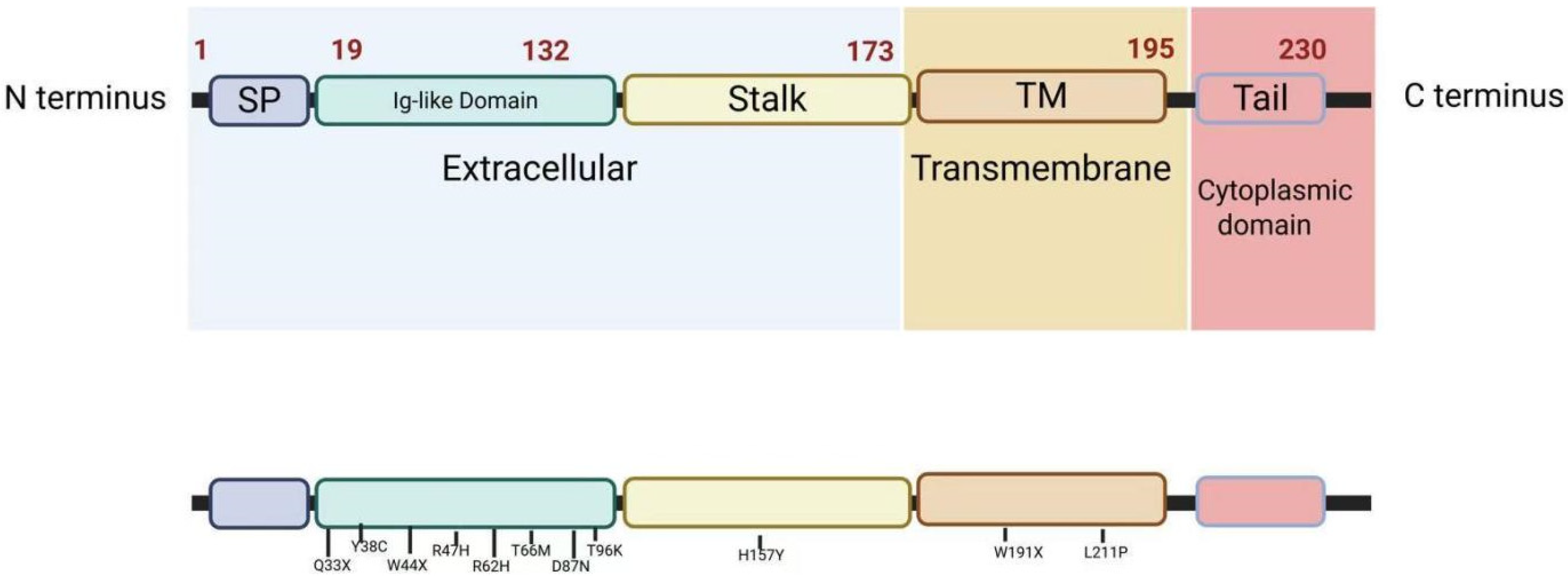
Figure 1. Structural architecture and pathogenic mutations of the TREM2 protein. This schematic illustrates the domain organization and disease-linked mutations of TREM2, a key receptor in microglial function and neurodegenerative disease pathology. TM = transmembrane; SP = signal peptide; and Ig = immunoglobulin-like. The images were created using licensed Biorender online software (https://app.biorender.com/).
Cells 14 01387 g001
Figure 2. TREM2-DAP12 signaling pathway and its downstream regulatory network. This diagram illustrates the immune-related signaling cascade triggered by TREM2 upon binding to its ligands. The pathway involves the following key mechanisms. The images were created using licensed Biorender online software (https://app.biorender.com/).
Cells 14 01387 g002
Figure 3. Therapeutic strategies targeting TREM2. This figure summarizes key therapeutic strategies to enhance TREM2 function in microglia, including agonistic antibodies (e.g., 4D9), natural compounds, antibody transport vehicles (e.g., DNL919), and viral gene delivery systems. These approaches aim to stabilize TREM2 signaling, reduce neuroinflammation, and limit pathological protein deposition. Major clinical challenges such as BBB penetration, off-target effects, biomarker development, and stage-specific treatment are also highlighted. The images were created using licensed Biorender online software (https://app.biorender.com/).
Cells 14 01387 g003
Disclaimer/Publisher’s Note: The statements, opinions and data contained in all publications are solely those of the individual author(s) and contributor(s) and not of MDPI and/or the editor(s). MDPI and/or the editor(s) disclaim responsibility for any injury to people or property resulting from any ideas, methods, instructions or products referred to in the content.