Next Issue
Volume 11, September
Previous Issue
Volume 11, July


Cancers, Volume 11, Issue 8 (August 2019) – 175 articles

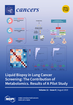
Cover Story (view full-size image): Cancer cells have a fundamentally different metabolism than noncancerous cells, and this difference is manifested in the endogenous metabolites they produce. Metabolomics aims to study metabolic differences in biological systems and has recently been applied to the discovery of tumor biomarkers in solid tumors. Previous studies reported the utility of the enzyme SSAT-1 as a cancer detection tool. SSAT-1 is a key protein involved in the synthesis of the polyamines spermine and spermidine that are involved in physiological cellular processes. This study led by Singhal and Rolfo et al. evaluated a panel of 14 metabolites associated in the SSAT-1/polyamine pathway that correctly discriminated between lung cancer patients from healthy controls, demonstrating the utility of metabolomics for lung cancer detection and adding further evidence on the role of liquid biopsy in cancer interception. View this paper
  • Issues are regarded as officially published after their release is announced to the table of contents alert mailing list.
  • You may sign up for e-mail alerts to receive table of contents of newly released issues.
  • PDF is the official format for papers published in both, html and pdf forms. To view the papers in pdf format, click on the "PDF Full-text" link, and use the free Adobe Reader to open them.
Order results
Result details
Section
Select all
Export citation of selected articles as:
18 pages, 1918 KiB  
Review
Fluorescence-Guided Surgery for Hepatoblastoma with Indocyanine Green
by Yohei Yamada, Michinobu Ohno, Akihiro Fujino, Yutaka Kanamori, Rie Irie, Takako Yoshioka, Osamu Miyazaki, Hajime Uchida, Akinari Fukuda, Seisuke Sakamoto, Mureo Kasahara, Kimikazu Matsumoto, Yasushi Fuchimoto, Ken Hoshino, Tatsuo Kuroda and Tomoro Hishiki
Cancers 2019, 11(8), 1215; https://doi.org/10.3390/cancers11081215 - 20 Aug 2019
Cited by 56 | Viewed by 5103
Abstract
Fluorescence-guided surgery with indocyanine green (ICG) for malignant hepatic tumors has been gaining more attention with technical advancements. Since hepatoblastomas (HBs) possess similar features to hepatocellular carcinoma, fluorescence-guided surgery can be used for HBs, as aggressive surgical resection, even for distant metastases of [...] Read more.
Fluorescence-guided surgery with indocyanine green (ICG) for malignant hepatic tumors has been gaining more attention with technical advancements. Since hepatoblastomas (HBs) possess similar features to hepatocellular carcinoma, fluorescence-guided surgery can be used for HBs, as aggressive surgical resection, even for distant metastases of HBs, often contributes positively to R0 (complete) resection and subsequent patient survival. Despite a few caveats, fluorescence-guided surgery allows for the more sensitive identification of lesions that may go undetected by conventional imaging or be invisible macroscopically. This leads to precise resection of distant metastatic tumors as well as primary liver tumors. Full article
(This article belongs to the Special Issue Hepatoblastoma and Pediatric Liver Tumors)
Show Figures

Figure 1

20 pages, 3361 KiB  
Article
Determination of the Potential Tumor-Suppressive Effects of Gsdme in a Chemically Induced and in a Genetically Modified Intestinal Cancer Mouse Model
by Lieselot Croes, Erik Fransen, Marieke Hylebos, Kimberly Buys, Christophe Hermans, Glenn Broeckx, Marc Peeters, Patrick Pauwels, Ken Op de Beeck and Guy Van Camp
Cancers 2019, 11(8), 1214; https://doi.org/10.3390/cancers11081214 - 20 Aug 2019
Cited by 28 | Viewed by 3702
Abstract
Gasdermin E (GSDME), also known as deafness autosomal dominant 5 (DFNA5) and previously identified to be an inducer of regulated cell death, is frequently epigenetically inactivated in different cancer types, suggesting that GSDME is a tumor suppressor gene. In [...] Read more.
Gasdermin E (GSDME), also known as deafness autosomal dominant 5 (DFNA5) and previously identified to be an inducer of regulated cell death, is frequently epigenetically inactivated in different cancer types, suggesting that GSDME is a tumor suppressor gene. In this study, we aimed to evaluate the tumor-suppressive effects of GSDME in two intestinal cancer mouse models. To mimic the silencing of GSDME by methylation as observed in human cancers, a Gsdme knockout (KO) mouse was developed. The effect of GSDME on tumorigenesis was studied both in a chemically induced and in a genetic intestinal cancer mouse model, as strong evidence shows that GSDME plays a role in human colorectal cancer and representative mouse models for intestinal cancer are available. Azoxymethane (AOM) was used to induce colorectal tumors in the chemically induced intestinal cancer model (n = 100). For the genetic intestinal cancer model, Apc1638N/+ mice were used (n = 37). In both experiments, the number of mice bearing microscopic proliferative lesions, the number and type of lesions per mouse and the histopathological features of the adenocarcinomas were compared between Gsdme KO and wild type (WT) mice. Unfortunately, we found no major differences between Gsdme KO and WT mice, neither for the number of affected mice nor for the multiplicity of proliferative lesions in the mice. However, recent breakthroughs on gasdermin function indicate that GSDME is an executioner of necrotic cell death. Therefore, it is possible that GSDME may be important for creating an inflammatory microenvironment around the tumor. This is in line with the trend towards more severe inflammation in WT compared to Gsdme KO mice, that we observed in our study. We conclude that the effect of GSDME in tumor biology is probably more subtle than previously thought. Full article
Show Figures

Figure 1

16 pages, 913 KiB  
Review
LAG3: The Biological Processes That Motivate Targeting This Immune Checkpoint Molecule in Human Cancer
by Cinzia Solinas, Edoardo Migliori, Pushpamali De Silva and Karen Willard-Gallo
Cancers 2019, 11(8), 1213; https://doi.org/10.3390/cancers11081213 - 20 Aug 2019
Cited by 70 | Viewed by 10457
Abstract
The programmed cell death 1 (PD-1) pathway is an important regulator of immune responses in peripheral tissues, including abnormal situations such as the tumor microenvironment. This pathway is currently the principal target for immunotherapeutic compounds designed to block immune checkpoint pathways, with these [...] Read more.
The programmed cell death 1 (PD-1) pathway is an important regulator of immune responses in peripheral tissues, including abnormal situations such as the tumor microenvironment. This pathway is currently the principal target for immunotherapeutic compounds designed to block immune checkpoint pathways, with these drugs improving clinical outcomes in a number of solid and hematological tumors. Medical oncology is experiencing an immune revolution that has scientists and clinicians looking at alternative, non-redundant inhibitory pathways also involved in regulating immune responses in cancer. A variety of targets have emerged for combinatorial approaches in immune checkpoint blockade. The main purpose of this narrative review is to summarize the biological role of lymphocyte activation gene 3 (LAG3), an emerging targetable inhibitory immune checkpoint molecule. We briefly discuss its role in infection, autoimmune disease and cancer, with a more detailed analysis of current data on LAG3 expression in breast cancer. Current clinical trials testing soluble LAG3 immunoglobulin and LAG3 antagonists are also presented in this work. Full article
(This article belongs to the Special Issue Signaling Pathways and Immune Checkpoint Regulation in Cancer)
Show Figures

Figure 1

14 pages, 1307 KiB  
Article
Circulating Tumor Cell Enumeration and Characterization in Metastatic Castration-Resistant Prostate Cancer Patients Treated with Cabazitaxel
by Ingeborg E. de Kruijff, Anieta M. Sieuwerts, Wendy Onstenk, Jaco Kraan, Marcel Smid, Mai N. Van, Michelle van der Vlugt-Daane, Esther Oomen-de Hoop, Ron H.J. Mathijssen, Martijn P. Lolkema, Ronald de Wit, Paul Hamberg, Hielke J. Meulenbeld, Aart Beeker, Geert-Jan Creemers, John W.M. Martens and Stefan Sleijfer
Cancers 2019, 11(8), 1212; https://doi.org/10.3390/cancers11081212 - 20 Aug 2019
Cited by 18 | Viewed by 3525
Abstract
(1) Background: Markers identifying which patients with metastatic, castration-resistant prostate cancer (mCRPC) will benefit from cabazitaxel therapy are currently lacking. Therefore, the aim of this study was to identify markers associated with outcome to cabazitaxel therapy based on counts and gene expression profiles [...] Read more.
(1) Background: Markers identifying which patients with metastatic, castration-resistant prostate cancer (mCRPC) will benefit from cabazitaxel therapy are currently lacking. Therefore, the aim of this study was to identify markers associated with outcome to cabazitaxel therapy based on counts and gene expression profiles of circulating tumor cells (CTCs). (2) Methods: From 120 mCRPC patients, CellSearch enriched CTCs were obtained at baseline and after 6 weeks of cabazitaxel therapy. Furthermore, 91 genes associated with prostate cancer were measured in mRNA of these CTCs. (3) Results: In 114 mCRPC patients with an evaluable CTC count, the CTC count was independently associated with poor progression-free survival (PFS) and overall survival (OS) in multivariable analysis with other commonly used variables associated with outcome in mCRPC (age, prostate specific antigen (PSA), alkaline phosphatase, lactate dehydrogenase (LDH), albumin, hemoglobin), together with alkaline phosphatase and hemoglobin. A five-gene expression profile was generated to predict for outcome to cabazitaxel therapy. However, even though this signature was associated with OS in univariate analysis, this was not the case in the multivariate analysis for OS nor for PFS. (4) Conclusion: The established five-gene expression profile in CTCs was not independently associated with PFS nor OS. However, along with alkaline phosphatase and hemoglobin, CTC-count is independently associated with PFS and OS in mCRPC patients who are treated with cabazitaxel. Full article
(This article belongs to the Collection Cancer Biomarkers)
Show Figures

Figure 1

12 pages, 1921 KiB  
Article
RAC1B Suppresses TGF-β-Dependent Chemokinesis and Growth Inhibition through an Autoregulatory Feed-Forward Loop Involving PAR2 and ALK5
by Hannah Otterbein, Koichiro Mihara, Morley D. Hollenberg, Hendrik Lehnert, David Witte and Hendrik Ungefroren
Cancers 2019, 11(8), 1211; https://doi.org/10.3390/cancers11081211 - 20 Aug 2019
Cited by 7 | Viewed by 3281
Abstract
The small GTPase RAC1B functions as a powerful inhibitor of transforming growth factor (TGF)-β1-induced epithelial-mesenchymal transition, cell motility, and growth arrest in pancreatic epithelial cells. Previous work has shown that RAC1B downregulates the TGF-β type I receptor ALK5, but the molecular details of [...] Read more.
The small GTPase RAC1B functions as a powerful inhibitor of transforming growth factor (TGF)-β1-induced epithelial-mesenchymal transition, cell motility, and growth arrest in pancreatic epithelial cells. Previous work has shown that RAC1B downregulates the TGF-β type I receptor ALK5, but the molecular details of this process have remained unclear. Here, we hypothesized that RAC1B-mediated suppression of activin receptor-like kinase 5 (ALK5) involves proteinase-activated receptor 2 (PAR2), a G protein-coupled receptor encoded by F2RL1 that is crucial for sustaining ALK5 expression. We found in pancreatic carcinoma Panc1 cells that PAR2 is upregulated by TGF-β1 in an ALK5-dependent manner and that siRNA-mediated knockdown of RAC1B increased both basal and TGF-β1-induced expression of PAR2. Further, the simultaneous knockdown of PAR2 and RAC1B rescued Panc1 cells from a RAC1B knockdown-induced increase in ALK5 abundance and the ALK5-mediated increase in TGF-β1-induced migratory activity. Conversely, Panc1 cells with stable ectopic expression of RAC1B displayed reduced ALK5 expression, an impaired upregulation of PAR2, and a reduced migratory responsiveness to TGF-β1 stimulation. However, these effects could be reversed by ectopic overexpression of PAR2. Moreover, the knockdown of PAR2 alone in Panc1 cells and HaCaT keratinocytes phenocopied RAC1B’s ability to suppress ALK5 abundance and TGF-β1-induced chemokinesis and growth inhibition. Lastly, we found that the RAC1B knockdown-induced increase in TGF-β1-induced PAR2 mRNA expression was sensitive to pharmacological inhibition of MEK-ERK signaling. Our data show that in pancreatic and skin epithelial cells, downregulation of ALK5 activity by RAC1B is secondary to suppression of F2RL1/PAR2 expression. Since F2RL1 itself is a TGF-β target gene and its upregulation by TGF-β1 is mediated by ALK5 and MEK-ERK signaling, we suggest the existence of a feed-forward signaling loop involving ALK5 and PAR2 that is efficiently suppressed by RAC1B to restrict TGF-β-driven cell motility and growth inhibition. Full article
(This article belongs to the Special Issue Rho Family of GTPases in Cancer)
Show Figures

Figure 1

28 pages, 499 KiB  
Review
The Importance of Gender-Related Anticancer Research on Mitochondrial Regulator Sodium Dichloroacetate in Preclinical Studies In Vivo
by Donatas Stakišaitis, Milda Juknevičienė, Eligija Damanskienė, Angelija Valančiūtė, Ingrida Balnytė and Marta Maria Alonso
Cancers 2019, 11(8), 1210; https://doi.org/10.3390/cancers11081210 - 20 Aug 2019
Cited by 10 | Viewed by 4125
Abstract
Sodium dichloroacetate (DCA) is an investigational medicinal product which has a potential anticancer preparation as a metabolic regulator in cancer cells’ mitochondria. Inhibition of pyruvate dehydrogenase kinases by DCA keeps the pyruvate dehydrogenase complex in the active form, resulting in decreased lactic acid [...] Read more.
Sodium dichloroacetate (DCA) is an investigational medicinal product which has a potential anticancer preparation as a metabolic regulator in cancer cells’ mitochondria. Inhibition of pyruvate dehydrogenase kinases by DCA keeps the pyruvate dehydrogenase complex in the active form, resulting in decreased lactic acid in the tumor microenvironment. This literature review displays the preclinical research data on DCA’s effects on the cell pyruvate dehydrogenase deficiency, pyruvate mitochondrial oxidative phosphorylation, reactive oxygen species generation, and the Na+–K+–2Cl cotransporter expression regulation in relation to gender. It presents DCA pharmacokinetics and the hepatocarcinogenic effect, and the safety data covers the DCA monotherapy efficacy for various human cancer xenografts in vivo in male and female animals. Preclinical cancer researchers report the synergistic effects of DCA combined with different drugs on cancer by reversing resistance to chemotherapy and promoting cell apoptosis. Researchers note that female and male animals differ in the mechanisms of cancerogenesis but often ignore studying DCA’s effects in relation to gender. Preclinical gender-related differences in DCA pharmacology, pharmacological mechanisms, and the elucidation of treatment efficacy in gonad hormone dependency could be relevant for individualized therapy approaches so that gender-related differences in treatment response and safety can be proposed. Full article
(This article belongs to the Special Issue Mitochondria and Cancer)
18 pages, 1014 KiB  
Review
Involvement of Actin in Autophagy and Autophagy-Dependent Multidrug Resistance in Cancer
by Magdalena Izdebska, Wioletta Zielińska, Marta Hałas-Wiśniewska and Alina Grzanka
Cancers 2019, 11(8), 1209; https://doi.org/10.3390/cancers11081209 - 20 Aug 2019
Cited by 14 | Viewed by 4635
Abstract
Currently, autophagy in the context of cancer progression arouses a lot of controversy. It is connected with the possibility of switching the nature of this process from cytotoxic to cytoprotective and vice versa depending on the treatment. At the same time, autophagy of [...] Read more.
Currently, autophagy in the context of cancer progression arouses a lot of controversy. It is connected with the possibility of switching the nature of this process from cytotoxic to cytoprotective and vice versa depending on the treatment. At the same time, autophagy of cytoprotective character may be one of the factors determining multidrug resistance, as intensification of the process is observed in patients with poorer prognosis. The exact mechanism of this relationship is not yet fully understood; however, it is suggested that one of the elements of the puzzle may be a cytoskeleton. In the latest literature reports, more and more attention is paid to the involvement of actin in the autophagy. The role of this protein is linked to the formation of autophagosomes, which are necessary element of the process. However, based on the proven effectiveness of manipulation of the actin pool, it seems to be an attractive alternative in breaking autophagy-dependent multidrug resistance in cancer. Full article
(This article belongs to the Special Issue The Role of Autophagy in Cancer Progression and Drug Resistance)
Show Figures

Figure 1

16 pages, 1048 KiB  
Review
Antioxidant Defenses: A Context-Specific Vulnerability of Cancer Cells
by Jordan A. Cockfield and Zachary T. Schafer
Cancers 2019, 11(8), 1208; https://doi.org/10.3390/cancers11081208 - 20 Aug 2019
Cited by 30 | Viewed by 6350
Abstract
Reactive oxygen species (ROS) are well known for their capacity to cause DNA damage, augment mutagenesis, and thereby promote oncogenic transformation. Similarly, agents that reduce ROS levels (antioxidants) are frequently thought to have anti-cancer properties given their propensity to minimize DNA damage and [...] Read more.
Reactive oxygen species (ROS) are well known for their capacity to cause DNA damage, augment mutagenesis, and thereby promote oncogenic transformation. Similarly, agents that reduce ROS levels (antioxidants) are frequently thought to have anti-cancer properties given their propensity to minimize DNA damage and mutagenesis. However, numerous clinical studies focused on antioxidants suggest that this is a facile premise and that antioxidant capacity can be important for cancer cells in a similar fashion to normal cells. As a consequence of this realization, numerous laboratories have been motivated to investigate the biological underpinnings explaining how and when antioxidant activity can potentially be beneficial to cancer cells. Relatedly, it has become clear that the reliance of cancer cells on antioxidant activity in certain contexts represents a potential vulnerability that could be exploited for therapeutic gain. Here, we review some of the recent, exciting findings documenting how cancer cells utilized antioxidant activity and under what circumstances this activity could represent an opportunity for selective elimination of cancer cells. Full article
(This article belongs to the Special Issue Metabolic Reprogramming and Vulnerabilities in Cancer)
Show Figures

Figure 1

23 pages, 2521 KiB  
Review
Tumor Cell Dormancy: Threat or Opportunity in the Fight against Cancer
by Rana Jahanban-Esfahlan, Khaled Seidi, Masoud H. Manjili, Ali Jahanban-Esfahlan, Tahereh Javaheri and Peyman Zare
Cancers 2019, 11(8), 1207; https://doi.org/10.3390/cancers11081207 - 19 Aug 2019
Cited by 72 | Viewed by 6744
Abstract
Tumor dormancy, a clinically undetectable state of cancer, makes a major contribution to the development of multidrug resistance (MDR), minimum residual disease (MRD), tumor outgrowth, cancer relapse, and metastasis. Despite its high incidence, the whole picture of dormancy-regulated molecular programs is far from [...] Read more.
Tumor dormancy, a clinically undetectable state of cancer, makes a major contribution to the development of multidrug resistance (MDR), minimum residual disease (MRD), tumor outgrowth, cancer relapse, and metastasis. Despite its high incidence, the whole picture of dormancy-regulated molecular programs is far from clear. That is, it is unknown when and which dormant cells will resume proliferation causing late relapse, and which will remain asymptomatic and harmless to their hosts. Thus, identification of dormancy-related culprits and understanding their roles can help predict cancer prognosis and may increase the probability of timely therapeutic intervention for the desired outcome. Here, we provide a comprehensive review of the dormancy-dictated molecular mechanisms, including angiogenic switch, immune escape, cancer stem cells, extracellular matrix (ECM) remodeling, metabolic reprogramming, miRNAs, epigenetic modifications, and stress-induced p38 signaling pathways. Further, we analyze the possibility of leveraging these dormancy-related molecular cues to outmaneuver cancer and discuss the implications of such approaches in cancer treatment. Full article
(This article belongs to the Special Issue Circulating Tumor Cells (CTCs))
Show Figures

Figure 1

11 pages, 2514 KiB  
Article
Rapid Depletions of Subcutaneous Fat Mass and Skeletal Muscle Mass Predict Worse Survival in Patients with Hepatocellular Carcinoma Treated with Sorafenib
by Kenji Imai, Koji Takai, Takao Miwa, Daisuke Taguchi, Tatsunori Hanai, Atsushi Suetsugu, Makoto Shiraki and Masahito Shimizu
Cancers 2019, 11(8), 1206; https://doi.org/10.3390/cancers11081206 - 19 Aug 2019
Cited by 34 | Viewed by 3241
Abstract
The aim of this study was to investigate whether rapid depletions of fat mass and skeletal muscle mass predict mortality in hepatocellular carcinoma (HCC) patients treated with sorafenib. This retrospective study evaluated 61 HCC patients. The cross-sectional areas of visceral and subcutaneous fat [...] Read more.
The aim of this study was to investigate whether rapid depletions of fat mass and skeletal muscle mass predict mortality in hepatocellular carcinoma (HCC) patients treated with sorafenib. This retrospective study evaluated 61 HCC patients. The cross-sectional areas of visceral and subcutaneous fat mass and skeletal muscle mass were measured by computed tomography, from which the visceral fat mass index (VFMI), subcutaneous fat mass index (SFMI), and skeletal muscle index (L3SMI) were obtained. The relative changes in these indices per 120 days (ΔVFMI, ΔSFMI, and ΔL3SMI) before and after sorafenib treatment were calculated in each patient. Patients within the 20th percentile cutoffs for these indices were classified into the rapid depletion (RD) group. Kaplan–Meier analysis revealed that with respect to ΔL3SMI (p = 0.0101) and ΔSFMI (p = 0.0027), the RD group had a significantly poorer survival. Multivariate analysis using the Cox proportional-hazards model also demonstrated that ΔL3SMI (≤−5.73 vs. >−5.73; hazard ratio [HR]: 4.010, 95% confidence interval [CI]: 1.799–8.938, p = < 0.001) and ΔSFMI (≤−5.33 vs. >−5.33; HR: 4.109, 95% CI: 1.967–8.584, p = < 0.001) were independent predictors. Rapid depletions of subcutaneous fat mass and skeletal muscle mass after the introduction of sorafenib indicate a poor prognosis. Full article
(This article belongs to the Special Issue Cancer Cachexia)
Show Figures

Graphical abstract

21 pages, 1318 KiB  
Article
The Complex Interaction between the Tumor Micro-Environment and Immune Checkpoints in Breast Cancer
by Vanessa Barriga, Nyanbol Kuol, Kulmira Nurgali and Vasso Apostolopoulos
Cancers 2019, 11(8), 1205; https://doi.org/10.3390/cancers11081205 - 19 Aug 2019
Cited by 53 | Viewed by 12414
Abstract
The progression of breast cancer and its association with clinical outcome and treatment remain largely unexplored. Accumulating data has highlighted the interaction between cells of the immune system and the tumor microenvironment in cancer progression, and although studies have identified multiple facets of [...] Read more.
The progression of breast cancer and its association with clinical outcome and treatment remain largely unexplored. Accumulating data has highlighted the interaction between cells of the immune system and the tumor microenvironment in cancer progression, and although studies have identified multiple facets of cancer progression within the development of the tumor microenvironment (TME) and its constituents, there is lack of research into the associations between breast cancer subtype and staging. Current literature has provided insight into the cells and pathways associated with breast cancer progression through expression analysis. However, there is lack of co-expression studies between immune pathways and cells of the TME that form pro-tumorigenic relationships contributing to immune-evasion. We focus on the immune checkpoint and TME elements that influence cancer progression, particularly studies in molecular subtypes of breast cancer. Full article
(This article belongs to the Special Issue Cancer Vaccines: Research and Applications)
Show Figures

Figure 1

20 pages, 6994 KiB  
Article
Maintenance BEZ235 Treatment Prolongs the Therapeutic Effect of the Combination of BEZ235 and Radiotherapy for Colorectal Cancer
by Yu-Hsuan Chen, Chun-Wei Wang, Ming-Feng Wei, Yi-Shin Tzeng, Keng-Hsueh Lan, Ann-Lii Cheng and Sung-Hsin Kuo
Cancers 2019, 11(8), 1204; https://doi.org/10.3390/cancers11081204 - 19 Aug 2019
Cited by 10 | Viewed by 3090
Abstract
Our previous study demonstrated that administration of NVP-BEZ235 (BEZ235), a dual PI3K/mTOR inhibitor, before radiotherapy (RT) enhanced the radiotherapeutic effect in colorectal cancer (CRC) cells both in vitro and in vivo. Here, we evaluated whether maintenance BEZ235 treatment, after combinatorial BEZ235 + RT [...] Read more.
Our previous study demonstrated that administration of NVP-BEZ235 (BEZ235), a dual PI3K/mTOR inhibitor, before radiotherapy (RT) enhanced the radiotherapeutic effect in colorectal cancer (CRC) cells both in vitro and in vivo. Here, we evaluated whether maintenance BEZ235 treatment, after combinatorial BEZ235 + RT therapy, prolonged the antitumor effect in CRC. K-RAS mutant CRC cells (HCT116 and SW480), wild-type CRC cells (HT29), and HCT116 xenograft tumors were separated into the following six study groups: (1) untreated (control); (2) RT alone; (3) BEZ235 alone; (4) RT + BEZ235; (5) maintenance BEZ235 following RT + BEZ235 (RT + BEZ235 + mBEZ235); and (6) maintenance BEZ235 following BEZ235 (BEZ235 + mBEZ235). RT + BEZ235 + mBEZ235 treatment significantly inhibited cell viability and increased apoptosis in three CRC cell lines compared to the other five treatments in vitro. In the HCT116 xenograft tumor model, RT + BEZ235 + mBEZ235 treatment significantly reduced the tumor size when compared to the other five treatments. Furthermore, the expression of mTOR signaling molecules (p-rpS6 and p-eIF4E), DNA double-strand break (DSB) repair-related molecules (p-ATM and p-DNA-PKcs), and angiogenesis-related molecules (VEGF-A and HIF-1α) was significantly downregulated after RT + BEZ235 + mBEZ235 treatment both in vitro and in vivo when compared to the RT + BEZ235, RT, BEZ235, BEZ235 + mBEZ235, and control treatments. Cleaved caspase-3, cleaved poly (ADP-ribose) polymerase (PARP), 53BP1, and γ-H2AX expression in the HCT116 xenograft tissue and three CRC cell lines were significantly upregulated after RT + BEZ235 + mBEZ235 treatment. Maintenance BEZ235 treatment in CRC cells prolonged the inhibition of cell viability, enhancement of apoptosis, attenuation of mTOR signaling, impairment of the DNA-DSB repair mechanism, and downregulation of angiogenesis that occurred due to concurrent BEZ235 and RT treatment. Full article
(This article belongs to the Special Issue Colorectal Cancers)
Show Figures

Figure 1

16 pages, 2022 KiB  
Article
Baseline Genomic Features in BRAFV600-Mutated Metastatic Melanoma Patients Treated with BRAF Inhibitor + MEK Inhibitor in Routine Care
by Baptiste Louveau, Fanelie Jouenne, Coralie Reger de Moura, Aurelie Sadoux, Barouyr Baroudjian, Julie Delyon, Florian Herms, Adele De Masson, Laetitia Da Meda, Maxime Battistella, Nicolas Dumaz, Celeste Lebbe and Samia Mourah
Cancers 2019, 11(8), 1203; https://doi.org/10.3390/cancers11081203 - 18 Aug 2019
Cited by 9 | Viewed by 2988
Abstract
In BRAFV600mut metastatic melanoma, the combination of BRAF and MEK inhibitors (BRAFi, MEKi) has undergone multiple resistance mechanisms, limiting its clinical benefit and resulting in the need for response predicting biomarkers. Based on phase III clinical trial data, several studies have previously [...] Read more.
In BRAFV600mut metastatic melanoma, the combination of BRAF and MEK inhibitors (BRAFi, MEKi) has undergone multiple resistance mechanisms, limiting its clinical benefit and resulting in the need for response predicting biomarkers. Based on phase III clinical trial data, several studies have previously explored baseline genomic features associated with response to BRAFi + MEKi. Using a targeted approach that combines the examination of mRNA expression and DNA alterations in a subset of genes, we performed an analysis of baseline genomic alterations involved in MAPK inhibitors’ resistance in a real-life cohort of BRAFV600mut metastatic melanoma patients. Twenty-seven patients were included in this retrospective study, and tumor samples were analyzed when the BRAFi + MEKi therapy was initiated. The clinical characteristics of our cohort were consistent with previously published studies. The BRAFi + MEKi treatment was initiated in seven patients as a following-line treatment, and had a specific transcriptomic profile exhibiting 14 genes with lower mRNA expression. However, DNA alterations in CCND1, RB1, and MET were only observed in patients who received BRAFi + MEKi as the first-line treatment. Furthermore, KIT mRNA expression was significantly higher in patients showing clinical benefit from the combined therapy, emphasizing the tumor-suppressor role of KIT already described within the context of BRAF-mutant melanoma. Full article
(This article belongs to the Special Issue Oncogenic Forms of BRAF as Cancer Driver Genes)
Show Figures

Figure 1

11 pages, 455 KiB  
Article
Soluble HLA in the Aqueous Humour of Uveal Melanoma Is Associated with Unfavourable Tumour Characteristics
by Annemijn P. A. Wierenga, Gülçin Gezgin, Els van Beelen, Michael Eikmans, Marijke Spruyt-Gerritse, Niels J. Brouwer, Mieke Versluis, Robert M. Verdijk, Sjoerd G. van Duinen, Marina Marinkovic, Gregorius P. M. Luyten and Martine J. Jager
Cancers 2019, 11(8), 1202; https://doi.org/10.3390/cancers11081202 - 18 Aug 2019
Cited by 18 | Viewed by 3150
Abstract
A high HLA expression in uveal melanoma (UM) is part of the prognostically unfavorable inflammatory phenotype. We wondered whether the presence of soluble HLA (sHLA) in the aqueous humour is associated with clinical, histopathological or genetic tumour characteristics, and represents tumour HLA expression [...] Read more.
A high HLA expression in uveal melanoma (UM) is part of the prognostically unfavorable inflammatory phenotype. We wondered whether the presence of soluble HLA (sHLA) in the aqueous humour is associated with clinical, histopathological or genetic tumour characteristics, and represents tumour HLA expression and intratumoural inflammation. Aqueous humour from 108 UM patients was analysed for the presence of sHLA, using a Luminex assay specific for HLA Class I. Clinical and genetic parameters were compared between sHLA-positive and negative eyes. A qPCR analysis was performed on tumour tissue using a Fluidigm assay. In 19/108 UM-containing eyes, the sHLA level in the aqueous was above the detection limit. Tumours in sHLA-positive eyes were significantly larger, more frequently involved the ciliary body, and more often showed monosomy 3, gain of chromosome 8q and loss of BAP1 staining. Melanoma-related survival was worse in patients with sHLA-positive aqueous humour. sHLA in the aqueous did not represent the tumour’s HLA expression and did not relate to immune cell infiltration in the tumour. We conclude that UM-containing eyes may contain sHLA in the aqueous humour, where it is a prognostically-unfavourable sign and may influence local immune responses. Full article
(This article belongs to the Special Issue Uveal Melanoma)
Show Figures

Graphical abstract

20 pages, 4216 KiB  
Article
Progesterone Receptor B signaling Reduces Breast Cancer Cell Aggressiveness: Role of Cyclin-D1/Cdk4 Mediating Paxillin Phosphorylation
by Francesca Ida Montalto, Francesca Giordano, Chiara Chiodo, Stefania Marsico, Loredana Mauro, Diego Sisci, Saveria Aquila, Marilena Lanzino, Maria Luisa Panno, Sebastiano Andò and Francesca De Amicis
Cancers 2019, 11(8), 1201; https://doi.org/10.3390/cancers11081201 - 17 Aug 2019
Cited by 16 | Viewed by 4333
Abstract
Progesterone-Receptor (PR) positivity is related with an enhanced response to breast cancer therapy, conversely cyclin D1 (CD1) is a retained marker of poor outcome. Herein, we demonstrate that hydroxyprogesterone (OHPg) through progesterone receptor B (PR-B) reduces breast cancer cell aggressiveness, by targeting the [...] Read more.
Progesterone-Receptor (PR) positivity is related with an enhanced response to breast cancer therapy, conversely cyclin D1 (CD1) is a retained marker of poor outcome. Herein, we demonstrate that hydroxyprogesterone (OHPg) through progesterone receptor B (PR-B) reduces breast cancer cell aggressiveness, by targeting the cytoplasmic CD1. Specifically, OHPg diminishes CD1 expression by a transcriptional regulation due to the recruitment of PR-B at a canonical half-PRE site of the CD1 promoter, together with HDAC1, determining a chromatin conformation less prone for gene transcription. CD1, together with its kinase partner Cdk4, regulates cell migration and metastasis, through the association with key components of focal adhesion, such as Paxillin (Pxn). Kaplan-Meier analysis shows that low Pxn expression was associated with increased distant metastasis-free survival in luminal A PR+ breast carcinomas. Interestingly, OHPg treatment reduced Pxn content in T47-D and MCF-7 cells; besides, the interaction between endogenous cytoplasmic CD1/Cdk4 with Pxn was reduced. This was consistent with the reduction of p-Ser83Pxn levels, crucially causing the delay in cell migration and a concomitant inhibition of Rac1 activity and p-PAK. Collectively, these findings support the role of PR-B in breast epithelial cell integrity and reinforce the importance in targeting PR-B as a potential strategy to restrict breast tumor cell invasion and metastasis. Full article
Show Figures

Graphical abstract

8 pages, 785 KiB  
Communication
SRSF2 Mutations in Uveal Melanoma: A Preference for In-Frame Deletions?
by Natasha M. van Poppelen, Wojtek Drabarek, Kyra N. Smit, Jolanda Vaarwater, Tom Brands, Dion Paridaens, Emine Kiliç and Annelies de Klein
Cancers 2019, 11(8), 1200; https://doi.org/10.3390/cancers11081200 - 17 Aug 2019
Cited by 15 | Viewed by 3583
Abstract
Background: Uveal melanoma (UM) is the most common primary ocular malignancy in adults in the Western world. UM with a mutation in SF3B1, a spliceosome gene, is characterized by three or more structural changes of chromosome 1, 6, 8, 9, or 11. [...] Read more.
Background: Uveal melanoma (UM) is the most common primary ocular malignancy in adults in the Western world. UM with a mutation in SF3B1, a spliceosome gene, is characterized by three or more structural changes of chromosome 1, 6, 8, 9, or 11. Also UM without a mutation in SF3B1 harbors similar chromosomal aberrations. Since, in addition to SF3B1, mutations in U2AF1 and SRSF2 have also been observed in hematological malignancies, UM without a SF3B1 mutation—but with the characteristic chromosomal pattern—might harbor mutations in one of these genes. Methods: 42 UMs were selected based on their chromosomal profile and wildtype SF3B1 status. Sanger sequencing covering the U2AF1 (exon 2 and 7) hotspots and SRSF2 (exon 1 and 2) was performed on DNA extracted from tumor tissue. Data of three UM with an SRSF2 mutation was extracted from the The Cancer Genome Atlas (TCGA). Results: Heterozygous in-frame SRSF2 deletions affecting amino acids 92–100 were detected in two UMs (5%) of 42 selected tumors and in three TGCA UM specimens. Both the UM with an SRSF2 mutation from our cohort and the UM samples from the TCGA showed more than four structural chromosomal aberrations including (partial) gain of chromosome 6 and 8, although in two TCGA UMs monosomy 3 was observed. Conclusions: Whereas in myelodysplastic syndrome predominantly missense SRSF2 mutations are described, the observed SRSF2 mutations in UM are all in-frame deletions of 8–9 amino acids. This suggests that the R625 missense SF3B1 mutations and SRSF2 mutations in UM are different compared to the spliceosome gene mutations in hematological cancers, and probably target a different, as yet unknown, set of genes involved in uveal melanoma etiology. Full article
(This article belongs to the Special Issue Uveal Melanoma)
Show Figures

Figure 1

20 pages, 6651 KiB  
Article
Single-Cell Analysis of Multiple Steps of Dynamic NF-κB Regulation in Interleukin-1α-Triggered Tumor Cells Using Proximity Ligation Assays
by Christin Mayr-Buro, Eva Schlereth, Knut Beuerlein, Ulas Tenekeci, Johanna Meier-Soelch, M. Lienhard Schmitz and Michael Kracht
Cancers 2019, 11(8), 1199; https://doi.org/10.3390/cancers11081199 - 16 Aug 2019
Cited by 7 | Viewed by 3620
Abstract
The frequently occurring heterogeneity of cancer cells and their functional interaction with immune cells in the tumor microenvironment raises the need to study signaling pathways at the single cell level with high precision, sensitivity, and spatial resolution. As aberrant NF-κB activity has been [...] Read more.
The frequently occurring heterogeneity of cancer cells and their functional interaction with immune cells in the tumor microenvironment raises the need to study signaling pathways at the single cell level with high precision, sensitivity, and spatial resolution. As aberrant NF-κB activity has been implicated in almost all steps of cancer development, we analyzed the dynamic regulation and activation status of the canonical NF-κB pathway in control and IL-1α-stimulated individual cells using proximity ligation assays (PLAs). These systematic experiments allowed the visualization of the dynamic dissociation and re-formation of endogenous p65/IκBα complexes and the nuclear translocation of NF-κB p50/p65 dimers. PLA combined with immunostaining for p65 or with NFKBIA single molecule mRNA-FISH facilitated the analysis of (i) further levels of the NF-κB pathway, (i) its functionality for downstream gene expression, and (iii) the heterogeneity of the NF-κB response in individual cells. PLA also revealed the interaction between NF-κB p65 and the P-body component DCP1a, a new p65 interactor that contributes to efficient p65 NF-κB nuclear translocation. In summary, these data show that PLA technology faithfully mirrored all aspects of dynamic NF-κB regulation, thus allowing molecular diagnostics of this key pathway at the single cell level which will be required for future precision medicine. Full article
(This article belongs to the Special Issue NF-kappaB signalling pathway)
Show Figures

Graphical abstract

18 pages, 2937 KiB  
Technical Note
Clinical-Scale Production of CAR-T Cells for the Treatment of Melanoma Patients by mRNA Transfection of a CSPG4-Specific CAR under Full GMP Compliance
by Manuel Wiesinger, Johannes März, Mirko Kummer, Gerold Schuler, Jan Dörrie, Beatrice Schuler-Thurner and Niels Schaft
Cancers 2019, 11(8), 1198; https://doi.org/10.3390/cancers11081198 - 16 Aug 2019
Cited by 45 | Viewed by 6982
Abstract
Chimeric antigen receptor (CAR)-T cells already showed impressive clinical regressions in leukemia and lymphoma. However, the development of CAR-T cells against solid tumors lags behind. Here we present the clinical-scale production of CAR-T cells for the treatment of melanoma under full GMP compliance. [...] Read more.
Chimeric antigen receptor (CAR)-T cells already showed impressive clinical regressions in leukemia and lymphoma. However, the development of CAR-T cells against solid tumors lags behind. Here we present the clinical-scale production of CAR-T cells for the treatment of melanoma under full GMP compliance. In this approach a CAR, specific for chondroitin sulfate proteoglycan 4 (CSPG4) is intentionally transiently expressed by mRNA electroporation for safety reasons. The clinical-scale protocol was optimized for: (i) expansion of T cells, (ii) electroporation efficiency, (iii) viability, (iv) cryopreservation, and (v) potency. Four consistency runs resulted in CAR-T cells in clinically sufficient numbers, i.e., 2.4 × 10⁹ CAR-expressing T cells, starting from 1.77x10⁸ PBMCs, with an average expansion of 13.6x, an electroporation efficiency of 88.0% CAR-positive cells, a survival of 74.1% after electroporation, and a viability of 84% after cryopreservation. Purity was 98.7% CD3+ cells, with 78.1% CD3+/CD8+ T cells and with minor contaminations of 1.2% NK cells and 0.6% B cells. The resulting CAR-T cells were tested for cytolytic activity after cryopreservation and showed antigen-specific and very efficient lysis of tumor cells. Although our work is descriptive rather than investigative in nature, we expect that providing this clinically applicable protocol to generate sufficient numbers of mRNA-transfected CAR-T cells will help in moving the field of adoptive cell therapy of cancer forward. Full article
(This article belongs to the Special Issue CAR-T Cell Therapy-Novel Approaches and Challenges)
Show Figures

Figure 1

19 pages, 1610 KiB  
Review
Targeting Oncogenic BRAF: Past, Present, and Future
by Aubhishek Zaman, Wei Wu and Trever G. Bivona
Cancers 2019, 11(8), 1197; https://doi.org/10.3390/cancers11081197 - 16 Aug 2019
Cited by 136 | Viewed by 9962
Abstract
Identifying recurrent somatic genetic alterations of, and dependency on, the kinase BRAF has enabled a “precision medicine” paradigm to diagnose and treat BRAF-driven tumors. Although targeted kinase inhibitors against BRAF are effective in a subset of mutant BRAF tumors, resistance to the therapy [...] Read more.
Identifying recurrent somatic genetic alterations of, and dependency on, the kinase BRAF has enabled a “precision medicine” paradigm to diagnose and treat BRAF-driven tumors. Although targeted kinase inhibitors against BRAF are effective in a subset of mutant BRAF tumors, resistance to the therapy inevitably emerges. In this review, we discuss BRAF biology, both in wild-type and mutant settings. We discuss the predominant BRAF mutations and we outline therapeutic strategies to block mutant BRAF and cancer growth. We highlight common mechanistic themes that underpin different classes of resistance mechanisms against BRAF-targeted therapies and discuss tumor heterogeneity and co-occurring molecular alterations as a potential source of therapy resistance. We outline promising therapy approaches to overcome these barriers to the long-term control of BRAF-driven tumors and emphasize how an extensive understanding of these themes can offer more pre-emptive, improved therapeutic strategies. Full article
(This article belongs to the Special Issue Oncogenic Forms of BRAF as Cancer Driver Genes)
Show Figures

Figure 1

16 pages, 1145 KiB  
Article
Development, Implementation and Assessment of Molecular Diagnostics by Next Generation Sequencing in Personalized Treatment of Cancer: Experience of a Public Reference Healthcare Hospital
by Javier Simarro, Rosa Murria, Gema Pérez-Simó, Marta Llop, Nuria Mancheño, David Ramos, Inmaculada de Juan, Eva Barragán, Begoña Laiz, Enrique Cases, Emilio Ansótegui, José Gómez-Codina, Jorge Aparicio, Carmen Salvador, Óscar Juan and Sarai Palanca
Cancers 2019, 11(8), 1196; https://doi.org/10.3390/cancers11081196 - 16 Aug 2019
Cited by 12 | Viewed by 4789
Abstract
The establishment of precision medicine in cancer patients requires the study of several biomarkers. Single-gene testing approaches are limited by sample availability and turnaround time. Next generation sequencing (NGS) provides an alternative for detecting genetic alterations in several genes with low sample requirements. [...] Read more.
The establishment of precision medicine in cancer patients requires the study of several biomarkers. Single-gene testing approaches are limited by sample availability and turnaround time. Next generation sequencing (NGS) provides an alternative for detecting genetic alterations in several genes with low sample requirements. Here we show the implementation to routine diagnostics of a NGS assay under International Organization for Standardization (UNE-EN ISO 15189:2013) accreditation. For this purpose, 106 non-small cell lung cancer (NSCLC) and 102 metastatic colorectal cancer (mCRC) specimens were selected for NGS analysis with Oncomine Solid Tumor (ThermoFisher). In NSCLC the most prevalently mutated gene was TP53 (49%), followed by KRAS (31%) and EGFR (13%); in mCRC, TP53 (50%), KRAS (48%) and PIK3CA (16%) were the most frequently mutated genes. Moreover, NGS identified actionable genetic alterations in 58% of NSCLC patients, and 49% of mCRC patients did not harbor primary resistance mechanisms to anti-EGFR treatment. Validation with conventional approaches showed an overall agreement >90%. Turnaround time and cost analysis revealed that NGS implementation is feasible in the public healthcare context. Therefore, NGS is a multiplexed molecular diagnostic tool able to overcome the limitations of current molecular diagnosis in advanced cancer, allowing an improved and economically sustainable molecular profiling. Full article
(This article belongs to the Special Issue Application of Next-Generation Sequencing in Cancers)
Show Figures

Figure 1

17 pages, 2558 KiB  
Review
Targeting Heat Shock Protein 27 in Cancer: A Druggable Target for Cancer Treatment?
by Seul-Ki Choi, Heejin Kam, Kye-Young Kim, Suk In Park and Yun-Sil Lee
Cancers 2019, 11(8), 1195; https://doi.org/10.3390/cancers11081195 - 16 Aug 2019
Cited by 73 | Viewed by 7763
Abstract
Heat shock protein 27 (HSP27), induced by heat shock, environmental, and pathophysiological stressors, is a multi-functional protein that acts as a protein chaperone and an antioxidant. HSP27 plays a significant role in the inhibition of apoptosis and actin cytoskeletal remodeling. HSP27 is upregulated [...] Read more.
Heat shock protein 27 (HSP27), induced by heat shock, environmental, and pathophysiological stressors, is a multi-functional protein that acts as a protein chaperone and an antioxidant. HSP27 plays a significant role in the inhibition of apoptosis and actin cytoskeletal remodeling. HSP27 is upregulated in many cancers and is associated with a poor prognosis, as well as treatment resistance, whereby cells are protected from therapeutic agents that normally induce apoptosis. This review highlights the most recent findings and role of HSP27 in cancer, as well as the strategies for using HSP27 inhibitors for therapeutic purposes. Full article
Show Figures

Figure 1

36 pages, 14714 KiB  
Review
The Multiple Roles and Therapeutic Potential of Molecular Chaperones in Prostate Cancer
by Abdullah Hoter, Sandra Rizk and Hassan Y. Naim
Cancers 2019, 11(8), 1194; https://doi.org/10.3390/cancers11081194 - 16 Aug 2019
Cited by 42 | Viewed by 7236
Abstract
Prostate cancer (PCa) is one of the most common cancer types in men worldwide. Heat shock proteins (HSPs) are molecular chaperones that are widely implicated in the pathogenesis, diagnosis, prognosis, and treatment of many cancers. The role of HSPs in PCa is complex [...] Read more.
Prostate cancer (PCa) is one of the most common cancer types in men worldwide. Heat shock proteins (HSPs) are molecular chaperones that are widely implicated in the pathogenesis, diagnosis, prognosis, and treatment of many cancers. The role of HSPs in PCa is complex and their expression has been linked to the progression and aggressiveness of the tumor. Prominent chaperones, including HSP90 and HSP70, are involved in the folding and trafficking of critical cancer-related proteins. Other members of HSPs, including HSP27 and HSP60, have been considered as promising biomarkers, similar to prostate-specific membrane antigen (PSMA), for PCa screening in order to evaluate and monitor the progression or recurrence of the disease. Moreover, expression level of chaperones like clusterin has been shown to correlate directly with the prostate tumor grade. Hence, targeting HSPs in PCa has been suggested as a promising strategy for cancer therapy. In the current review, we discuss the functions as well as the role of HSPs in PCa progression and further evaluate the approach of inhibiting HSPs as a cancer treatment strategy. Full article
(This article belongs to the Special Issue Prostate Cancer: Past, Present, and Future)
Show Figures

Figure 1

42 pages, 3648 KiB  
Review
Epigenetic Dysregulation at the Crossroad of Women’s Cancer
by Rakesh Kumar, Aswathy Mary Paul, Pranela Rameshwar and M. Radhakrishna Pillai
Cancers 2019, 11(8), 1193; https://doi.org/10.3390/cancers11081193 - 16 Aug 2019
Cited by 9 | Viewed by 5038
Abstract
An increasingly number of women of all age groups are affected by cancer, despite substantial progress in our understanding of cancer pathobiology, the underlying genomic alterations and signaling cascades, and cellular-environmental interactions. Though our understanding of women’s cancer is far more complete than [...] Read more.
An increasingly number of women of all age groups are affected by cancer, despite substantial progress in our understanding of cancer pathobiology, the underlying genomic alterations and signaling cascades, and cellular-environmental interactions. Though our understanding of women’s cancer is far more complete than ever before, there is no comprehensive model to explain the reasons behind the increased incidents of certain reproductive cancer among older as well as younger women. It is generally suspected that environmental and life-style factors affecting hormonal and growth control pathways might help account for the rise of women’s cancers in younger age, as well, via epigenetic mechanisms. Epigenetic regulators play an important role in orchestrating an orderly coordination of cellular signals in gene activity in response to upstream signaling and/or epigenetic modifiers present in a dynamic extracellular milieu. Here we will discuss the broad principles of epigenetic regulation of DNA methylation and demethylation, histone acetylation and deacetylation, and RNA methylation in women’s cancers in the context of gene expression, hormonal action, and the EGFR family of cell surface receptor tyrosine kinases. We anticipate that a better understanding of the epigenetics of women’s cancers may provide new regulatory leads and further fuel the development of new epigenetic biomarkers and therapeutic approaches. Full article
Show Figures

Figure 1

18 pages, 3879 KiB  
Article
Lambda-Carrageenan Enhances the Effects of Radiation Therapy in Cancer Treatment by Suppressing Cancer Cell Invasion and Metastasis through Racgap1 Inhibition
by Ping-Hsiu Wu, Yasuhito Onodera, Frances C. Recuenco, Amato J. Giaccia, Quynh-Thu Le, Shinichi Shimizu, Hiroki Shirato and Jin-Min Nam
Cancers 2019, 11(8), 1192; https://doi.org/10.3390/cancers11081192 - 16 Aug 2019
Cited by 8 | Viewed by 4927
Abstract
Radiotherapy is used extensively in cancer treatment, but radioresistance and the metastatic potential of cancer cells that survive radiation remain critical issues. There is a need for novel treatments to improve radiotherapy. Here, we evaluated the therapeutic benefit of λ-carrageenan (CGN) to enhance [...] Read more.
Radiotherapy is used extensively in cancer treatment, but radioresistance and the metastatic potential of cancer cells that survive radiation remain critical issues. There is a need for novel treatments to improve radiotherapy. Here, we evaluated the therapeutic benefit of λ-carrageenan (CGN) to enhance the efficacy of radiation treatment and investigated the underlying molecular mechanism. CGN treatment decreased viability in irradiated cancer cells and enhanced reactive oxygen species accumulation, apoptosis, and polyploid formation. Additionally, CGN suppressed radiation-induced chemoinvasion and invasive growth in 3D lrECM culture. We also screened target molecules using a gene expression microarray analysis and focused on Rac GTPase-activating protein 1 (RacGAP1). Protein expression of RacGAP1 was upregulated in several cancer cell lines after radiation, which was significantly suppressed by CGN treatment. Knockdown of RacGAP1 decreased cell viability and invasiveness after radiation. Overexpression of RacGAP1 partially rescued CGN cytotoxicity. In a mouse xenograft model, local irradiation followed by CGN treatment significantly decreased tumor growth and lung metastasis compared to either treatment alone. Taken together, these results suggest that CGN may enhance the effectiveness of radiation in cancer therapy by decreasing cancer cell viability and suppressing both radiation-induced invasive activity and distal metastasis through downregulating RacGAP1 expression. Full article
(This article belongs to the Special Issue Role of Natural Bioactive Compounds in the Rise and Fall of Cancers)
Show Figures

Graphical abstract

20 pages, 845 KiB  
Review
Reactive Oxygen Species in the Tumor Microenvironment: An Overview
by Frank Weinberg, Nithya Ramnath and Deepak Nagrath
Cancers 2019, 11(8), 1191; https://doi.org/10.3390/cancers11081191 - 16 Aug 2019
Cited by 268 | Viewed by 16735
Abstract
Reactive oxygen species (ROS) are important signaling molecules in cancer. The level of ROS will determine physiological effects. While high levels of ROS can cause damage to tissues and cell death, low levels of ROS can have a proliferative effect. ROS are produced [...] Read more.
Reactive oxygen species (ROS) are important signaling molecules in cancer. The level of ROS will determine physiological effects. While high levels of ROS can cause damage to tissues and cell death, low levels of ROS can have a proliferative effect. ROS are produced by tumor cells but also cellular components that make up the tumor microenvironment (TME). In this review, we discuss the mechanisms by which ROS can affect the TME with particular emphasis on tumor-infiltrating leukocytes. Greater insight into ROS biology in this setting may allow for therapeutic manipulation of ROS levels in order to remodel the tumor microenvironment and increase anti-tumor activity. Full article
(This article belongs to the Special Issue Metabolic Reprogramming and Vulnerabilities in Cancer)
Show Figures

Figure 1

13 pages, 9316 KiB  
Perspective
FDG-PET/CT for Response Monitoring in Metastatic Breast Cancer: Today, Tomorrow, and Beyond
by Malene Grubbe Hildebrandt, Jeppe Faurholdt Lauridsen, Marianne Vogsen, Jorun Holm, Mie Holm Vilstrup, Poul-Erik Braad, Oke Gerke, Mads Thomassen, Marianne Ewertz, Poul Flemming Høilund-Carlsen and The Centre for Personalized Response Monitoring in Oncology (PREMIO)
Cancers 2019, 11(8), 1190; https://doi.org/10.3390/cancers11081190 - 15 Aug 2019
Cited by 12 | Viewed by 5950
Abstract
While current international guidelines include imaging of the target lesion for response monitoring in metastatic breast cancer, they do not provide specific recommendations for choice of imaging modality or response criteria. This is important as clinical decisions may vary depending on which imaging [...] Read more.
While current international guidelines include imaging of the target lesion for response monitoring in metastatic breast cancer, they do not provide specific recommendations for choice of imaging modality or response criteria. This is important as clinical decisions may vary depending on which imaging modality is used for monitoring metastatic breast cancer. FDG-PET/CT has shown high accuracy in diagnosing metastatic breast cancer, and the Positron Emission Tomography Response Criteria in Solid Tumors (PERCIST) have shown higher predictive values than the CT-based Response Evaluation Criteria in Solid Tumors (RECIST) for prediction of progression-free survival. No studies have yet addressed the clinical impact of using different imaging modalities or response evaluation criteria for longitudinal response monitoring in metastatic breast cancer. We present a case study of a patient with metastatic breast cancer who was monitored first with conventional CT and then with FDG-PET/CT. We retrospectively applied PERCIST to evaluate the longitudinal response to treatment. We used the one-lesion PERCIST model measuring SULpeak in the hottest metastatic lesion on consecutive scans. This model provides a continuous variable that allows graphical illustration of disease fluctuation along with response categories. The one-lesion PERCIST approach seems able to reflect molecular changes and has the potential to support clinical decision-making. Prospective clinical studies addressing the clinical impact of PERCIST in metastatic breast cancer are needed to establish evidence-based recommendations for response monitoring in this disease. Full article
(This article belongs to the Special Issue Role of Medical Imaging in Cancers)
Show Figures

Graphical abstract

11 pages, 959 KiB  
Review
How to Deal with Second Line Dilemma in Metastatic Colorectal Cancer? A Systematic Review and Meta-Analysis
by Antonio Galvano, Lorena Incorvaia, Giuseppe Badalamenti, Sergio Rizzo, Aurelia Guarini, Stefania Cusenza, Luisa Castellana, Nadia Barraco, Valentina Calò, Sofia Cutaia, Giuseppe Currò, Nicola Silvestris, Giordano Domenico Beretta, Viviana Bazan and Antonio Russo
Cancers 2019, 11(8), 1189; https://doi.org/10.3390/cancers11081189 - 15 Aug 2019
Cited by 5 | Viewed by 3249
Abstract
Monoclonal antibodies targeting epidermal growth factor receptor (EGFR) or vascular endothelial growth factor (VEGF) have demonstrated efficacy with chemotherapy (CT) as second line treatment for metastatic colorectal cancer (mCRC). The right sequence of the treatments in all RAS (KRAS/NRAS) wild type (wt) patients [...] Read more.
Monoclonal antibodies targeting epidermal growth factor receptor (EGFR) or vascular endothelial growth factor (VEGF) have demonstrated efficacy with chemotherapy (CT) as second line treatment for metastatic colorectal cancer (mCRC). The right sequence of the treatments in all RAS (KRAS/NRAS) wild type (wt) patients has not precisely defined. We evaluated the impact of aforementioned targeted therapies in second line setting, analyzing efficacy and safety data from phase III clinical trials. We performed both direct and indirect comparisons between anti-EGFR and anti-VEGF. Outcomes included disease control rate (DCR), objective response rate (ORR), progression-free survival (PFS), overall survival (OS) and G3-G5 toxicities. Our results showed significantly improved OS (HR 0.83, 95% CI 0.72–0.94) and DCR (HR 1.27, 95% CI 1.04–1.54) favouring anti-VEGF combinations in overall population; no statistically significant differences in all RAS wt patients was observed (HR 0.87, 95% CI 0.70–1.09). Anti-EGFR combinations significantly increased ORR in all patients (RR 0.54, 95% CI 0.31–0.96), showing a trend also in all RAS wt patients (RR 0.63, 95% CI 0.48–0.83). No significant difference in PFS and DCR all RAS was registered. Our results provided for the first time a strong rationale to manage both targeted agents in second line setting. Full article
(This article belongs to the Special Issue Colorectal Cancers)
Show Figures

Figure 1

13 pages, 1103 KiB  
Article
Anaplastic Thyroid Cancer: Clinical Picture of the Last Two Decades at a Single Oncology Referral Centre and Novel Therapeutic Options
by Joana Simões-Pereira, Ricardo Capitão, Edward Limbert and Valeriano Leite
Cancers 2019, 11(8), 1188; https://doi.org/10.3390/cancers11081188 - 15 Aug 2019
Cited by 20 | Viewed by 3570
Abstract
Anaplastic thyroid cancer (ATC) is a rare tumour but also one of the most lethal malignancies. Therapeutic modalities have usually been limited, but clinical trials with new drugs are now being implemented. The aims of this study were to analyse the clinical presentation, [...] Read more.
Anaplastic thyroid cancer (ATC) is a rare tumour but also one of the most lethal malignancies. Therapeutic modalities have usually been limited, but clinical trials with new drugs are now being implemented. The aims of this study were to analyse the clinical presentation, therapeutic modalities and independent prognostic factors for survival. We also reviewed the most recent literature on novel ATC therapies. We performed a retrospective analysis of 79 patients diagnosed between 2000 and 2018. Variables with impact on survival were identified using the Cox proportional-hazard regression model. At presentation, 6.3% had thyroid-confined disease, 30.4% evidenced extrathyroidal extension and 60.8% were already metastatic. Surgery was feasible in 41.8% and radiotherapy was applied to 35.4%, with those receiving >45 Gy having longer estimated survival (p = 0.020). Chemotherapy, either conventional or with tyrosine kinase inhibitors, was performed in 17.7% and 7.6%, respectively. Multimodality therapy with surgery, radiotherapy and chemotherapy/tyrosine kinase inhibitors (TKI) had the greatest impact on disease specific survival (DSS), providing a risk reduction of death of 96.9% (hazard ratio (HR) = 0.031, 0.005–0.210, p < 0.001). We concluded that most of these patients join reference centres at advanced stages of disease and multimodality treatment may offer the best chances for prolonging survival. Full article
(This article belongs to the Special Issue Thyroid Cancer)
Show Figures

Figure 1

18 pages, 3220 KiB  
Article
4-Methylumbelliferone Inhibits Cancer Stem Cell Activation and Overcomes Chemoresistance in Ovarian Cancer
by Noor A. Lokman, Zoe K. Price, Emily K. Hawkins, Anne M. Macpherson, Martin K. Oehler and Carmela Ricciardelli
Cancers 2019, 11(8), 1187; https://doi.org/10.3390/cancers11081187 - 15 Aug 2019
Cited by 26 | Viewed by 5246
Abstract
We have recently shown that the extracellular matrix molecule hyaluronan (HA) plays a role in the development of ovarian cancer chemoresistance. This present study determined if HA production is increased in chemotherapy-resistant ovarian cancers and if the HA inhibitor 4-methylubelliferone (4-MU) can overcome [...] Read more.
We have recently shown that the extracellular matrix molecule hyaluronan (HA) plays a role in the development of ovarian cancer chemoresistance. This present study determined if HA production is increased in chemotherapy-resistant ovarian cancers and if the HA inhibitor 4-methylubelliferone (4-MU) can overcome chemoresistance to the chemotherapeutic drug carboplatin (CBP) and inhibit spheroid formation and the expression of cancer stem cell (CSC) markers. We additionally assessed whether 4-MU could inhibit in vivo invasion of chemoresistant primary ovarian cancer cells in the chicken embryo chorioallantoic membrane (CAM) assay. The expression of the HA synthases HAS2 and HAS3 was significantly increased in chemoresistant compared to chemosensitive primary ovarian cancer cells isolated from patient ascites. 4-MU significantly inhibited HA production, cell survival, and spheroid formation of chemoresistant serous ovarian cancer cells. In combination with CBP, 4-MU treatment significantly decreased ovarian cancer cell survival and increased apoptosis of chemoresistant primary cells compared to CBP alone. 4-MU significantly reduced spheroid formation, expression of CSC markers ALDH1A1 and ABCG2 in primary cell spheroid cultures, and ALDH1 immunostaining in patient-derived tissue explant assays following treatment with CBP. Furthermore, 4-MU was very effective at inhibiting in vivo invasion of chemoresistant primary cells in CAM assays. Inhibition of HA is therefore a promising new strategy to overcome chemoresistance and to improve ovarian cancer survival. Full article
(This article belongs to the Special Issue Cancer Stem Cells and Personalized Medicine for Gynecologic Cancers)
Show Figures

Figure 1

14 pages, 2561 KiB  
Article
Clinical Relevance of Collagen Protein Degradation Markers C3M and C4M in the Serum of Breast Cancer Patients Treated with Neoadjuvant Therapy in the GeparQuinto Trial
by Malgorzata Banys-Paluchowski, Sibylle Loibl, Isabell Witzel, Christoph Mundhenke, Bianca Lederer, Christine Solbach, Thomas Karn, Frederik Marmé, Valentina Nekljudova, Christian Schem, Elmar Stickeler, Nicholas Willumsen, Morten A. Karsdal, Michael Untch and Volkmar Müller
Cancers 2019, 11(8), 1186; https://doi.org/10.3390/cancers11081186 - 15 Aug 2019
Cited by 8 | Viewed by 3895
Abstract
Background: Remodeling of extracellular matrix through collagen degradation is a crucial step in the metastatic cascade. The aim of this study was to evaluate the potential clinical relevance of the serum collagen degradation markers (CDM) C3M and C4M during neoadjuvant chemotherapy for [...] Read more.
Background: Remodeling of extracellular matrix through collagen degradation is a crucial step in the metastatic cascade. The aim of this study was to evaluate the potential clinical relevance of the serum collagen degradation markers (CDM) C3M and C4M during neoadjuvant chemotherapy for breast cancer. Methods: Patients from the GeparQuinto phase 3 trial with untreated HER2-positive operable or locally advanced breast cancer were enrolled between 7 November 2007, and 9 July 2010, and randomly assigned to receive neoadjuvant treatment with EC/docetaxel with either trastuzumab or lapatinib. Blood samples were collected at baseline, after four cycles of chemotherapy and at surgery. Cutoff values were determined using validated cutoff finder software (C3M: Low ≤9.00 ng/mL, high >9.00 ng/mL, C4M: Low ≤40.91 ng/mL, high >40.91 ng/mL). Results: 157 patients were included in this analysis. At baseline, 11.7% and 14.8% of patients had high C3M and C4M serum levels, respectively. No correlation was observed between CDM and classical clinical-pathological factors. Patients with high levels of CDM were significantly more likely to achieve a pathological complete response (pCR, defined as ypT0 ypN0) than patients with low levels (C3M: 66.7% vs. 25.7%, p = 0.002; C4M: 52.7% vs. 26.6%, p = 0.031). Median levels of both markers were lower at the time of surgery than at baseline. In the multivariate analysis including clinical-pathological factors and C3M levels at baseline and changes in C3M levels between baseline and after four cycles of therapy, only C3M levels at baseline (p = 0.035, OR 4.469, 95%-CI 1.115–17.919) independently predicted pCR. In a similar model including clinical-pathological factors and C4M, only C4M levels at baseline (p = 0.028, OR 6.203, 95%-CI 1.220–31.546) and tumor size (p = 0.035, OR 4.900, 95%-CI 1.122–21.393) were independent predictors of pCR. High C3M levels at baseline did not correlate with survival in the entire cohort but were associated with worse disease-free survival (DFS; p = 0.029, 5-year DFS 40.0% vs. 74.9%) and overall survival (OS; p = 0.020, 5-year OS 60.0% vs. 88.3%) in the subgroup of patients randomized to lapatinib. In the trastuzumab arm, C3M did not correlate with survival. In the entire patient cohort, high levels of C4M at baseline were significantly associated with shorter DFS (p = 0.001, 5-year DFS 53.1% vs. 81.6%) but not with OS. When treatment arms were considered separately, the association with DFS was still significant (p = 0.014, 5-year DFS 44.4% vs. 77.0% in the lapatinib arm; p = 0.023, 5-year DFS 62.5% vs. 86.2% in the trastuzumab arm). Conclusions: Collagen degradation markers are associated with response to neoadjuvant therapy and seem to play a role in breast cancer. Full article
(This article belongs to the Special Issue New Biomarkers in Cancers)
Show Figures

Figure 1

Previous Issue
Back to TopTop