Article

Baseline Genomic Features in BRAFV600-Mutated Metastatic Melanoma Patients Treated with BRAF Inhibitor + MEK Inhibitor in Routine Care

1
Pharmacogenomics Department, Assistance Publique-Hôpitaux de Paris (APHP), Saint Louis Hospital, 75010 Paris, France
2
Université de Paris, INSERM UMRS 976, 75010 Paris, France
3
Dermatology Department and Centre d’investigation clinique (CIC), Assistance Publique-Hôpitaux de Paris (APHP), Saint Louis Hospital, 75010 Paris, France
4
Pathology Department, Assistance Publique-Hôpitaux de Paris (APHP), Saint Louis Hospital, 75010 Paris, France
5
Université de Paris, INSERM UMRS 1165, 75010 Paris, France
*
Author to whom correspondence should be addressed.
Co-first authors.
Co-last authors.
Cancers 2019, 11(8), 1203; https://doi.org/10.3390/cancers11081203
Submission received: 15 July 2019 / Revised: 8 August 2019 / Accepted: 9 August 2019 / Published: 18 August 2019
(This article belongs to the Special Issue Oncogenic Forms of BRAF as Cancer Driver Genes)

Abstract

:
In BRAFV600mut metastatic melanoma, the combination of BRAF and MEK inhibitors (BRAFi, MEKi) has undergone multiple resistance mechanisms, limiting its clinical benefit and resulting in the need for response predicting biomarkers. Based on phase III clinical trial data, several studies have previously explored baseline genomic features associated with response to BRAFi + MEKi. Using a targeted approach that combines the examination of mRNA expression and DNA alterations in a subset of genes, we performed an analysis of baseline genomic alterations involved in MAPK inhibitors’ resistance in a real-life cohort of BRAFV600mut metastatic melanoma patients. Twenty-seven patients were included in this retrospective study, and tumor samples were analyzed when the BRAFi + MEKi therapy was initiated. The clinical characteristics of our cohort were consistent with previously published studies. The BRAFi + MEKi treatment was initiated in seven patients as a following-line treatment, and had a specific transcriptomic profile exhibiting 14 genes with lower mRNA expression. However, DNA alterations in CCND1, RB1, and MET were only observed in patients who received BRAFi + MEKi as the first-line treatment. Furthermore, KIT mRNA expression was significantly higher in patients showing clinical benefit from the combined therapy, emphasizing the tumor-suppressor role of KIT already described within the context of BRAF-mutant melanoma.

1. Introduction

BRAFV600-mutated (BRAFV600mut) lesions are currently described in approximately 50% of patients with metastatic melanoma, and are known to constitutively activate the MAPK (mitogen-activated protein kinase) pathway [1,2]. In the last few years, therapies targeting this pathway, such as those involving a BRAF inhibitor (BRAFi), have greatly improved the clinical outcome of BRAFV600mut metastatic melanoma [3,4,5,6,7,8]. However, the clinical benefit of these therapies has been limited by the emergence of multiple resistance mechanisms. Although the combination of a BRAFi with a MEK inhibitor (MEKi), which has become the standard of care [9], has improved outcomes, resistance has not been abolished.
Among the MAPK-targeting therapies’ resistance mechanisms described to date, reactivation of the MAPK pathway is the most frequent: it has been characterized in more than 70% of resistance cases in patients treated with BRAFi alone or with BRAFi + MEKi [10,11,12]. Frequent events leading to this reactivation suggest events such as MAP2K1, MAP2K2, or NRAS mutations, BRAF amplifications, or NF1 inactivation. Other resistance mechanisms, such as PI3K/AKT pathway activation via PTEN and AKT mutations [13,14], PDGFRB and IGFR1 overexpression [15,16], or production of HGF in the microenvironment [17], have also been described. Alterations in cell cycle genes, such as CDKN2A, have also been identified [14]. Moreover, targeted mRNA analyses have associated BRAF aberrant splice variants, overexpression of MAP3K8, and genes encoding tyrosine kinase receptors (RTKs), to resistance [18,19,20,21]. Recent studies using whole-exome sequencing highlight the multiplicity of resistance mechanisms within the same tumor, and a recent targeted approach of gene alterations uncovered new potential mechanisms of resistance [11,12,22,23]. However, data regarding resistance mechanisms in patients treated with MAPK-targeted therapies should be reinforced.
Considering these multiple and complex resistance phenomena and the rapid development of various therapies (targeted therapy, immunotherapy, etc.), the identification of baseline biomarkers and their association with patient clinical characteristics is crucial for identifying those who are more likely to benefit from these therapies. Using data from the BRIM and coBRIM studies, Wongchenko et al. [24] and Yan et al. [25] have recently focused on predictive genomic features, and revealed an association between an improved clinical response under BRAFi + MEKi treatment and higher levels of baseline immune response-related genes carrying NF1 alterations. A MAPK pathway activity score predictive of clinical response to vemurafenib has also been proposed [26]. Regardless, despite the high heterogeneity in genomic profiles, these studies based on clinical trial data focus on the clinical course and do not investigate the association between genomic features and clinical attributes, such as the targeted therapy line.
In this retrospective study of a real-life cohort of BRAFV600mut metastatic melanoma patients initiating BRAFi + MEKi treatment, we applied a targeted molecular approach to describe baseline genomic alterations involved in resistance to MAPK-targeted therapies (mRNA expression, copy number, and mutations) and analyzed the association between these features and patient clinical characteristics.

2. Results

2.1. Patient Characteristics

Twenty-seven (n = 27) BRAFV600mut metastatic melanoma patients were included in this study, and baseline tumor samples (prior to BRAFi + MEKi initiation) were collected. Patient characteristics are summarized in Table 1. Among the 27 patients, 24 (89%) presented with stage IV melanoma at initiation, comprising 16 (67%) M1c (American Joint Committee of Cancer 7th edition) cases. Brain metastases were observed for six (22%) patients and seven (29%) had elevated LDH levels. Twenty (74%) patients received BRAFi + MEKi as the first-line treatment, whereas seven (26%) received these targeted therapies as a following-line treatment. These seven patients all received immunotherapy prior to BRAFi + MEKi initiation. Compared to vemurafenib + cobimetinib (n = 3, 11%), dabrafenib + trametinib was mostly prescribed in our cohort (n = 24, 89%). Regarding safety, four (15%) patients experienced drug toxicity that led to treatment discontinuation. The clinical course, time under BRAFi + MEKi treatment, and best-observed response during follow-up are summarized in Figure 1. The median follow-up time in our cohort was 18.4 (lower limit = 3.4; upper limit = 68.4) months. The best observed response under therapy was nine cases of complete response (33%), 12 cases of partial response (44%), and four cases of stable disease (15%), with an overall response rate (ORR) of 78%. Median progression-free survival (PFS), defined as the time between BRAFi + MEKi initiation and the occurrence of disease progression or death (whichever occurred first) under treatment, was estimated at 10.1 months (95% CI: 6.1; NA) (Figure 2). Univariate analyses were performed to search for associations between PFS and clinical baseline characteristics. The presence of brain metastases (p < 0.01) and LDH levels (p < 0.01) at therapy initiation were significantly associated with PFS.

2.2. Baseline DNA Analysis (Mutations and CNVs)

Next-generation sequencing (NGS) and copy number analyses targeting 78 genes involved in the MAPK, PI3K, RTK, and cell cycle pathways (Supplementary Table S1) were performed on baseline tumor samples from 24 patients (3 missing because of insufficient material). This analysis led to the detection of 82 alterations, including 51 (62%) mutations and 31 (38%) copy number variations (Figure 3). Copy number variations in CCND1 were the most frequent DNA alteration observed, with both amplifications and deletions in eight and two of the 24 samples, respectively. RB1 deletions were also highlighted in six (25%) samples. NOTCH2 exhibited the highest mutation rate, with four (17%) mutations detected. Details regarding all baseline mutations detected by NGS (amino acid change, allele frequency) are provided in Supplementary Table S2.
Analyses were performed to search for a possible association between the number or type of alterations in our screened genes and clinical variables. Comparison of patients harboring a high number of DNA alterations (≥4) vs. those with a low number of alterations (≤3) revealed that patients in the group with ≥4 alterations (7/8, 88%) were more likely to have received BRAFi + MEKi as the first-line treatment. Moreover, CCND1 amplifications, RB1 deletions, and MET alterations were only retrieved for those patients treated with BRAFi + MEKi as a first-line. No association (type or total number of alterations) was observed for the presence of brain metastases, a high LDH level, best observed response, or occurrence of a PFS event under BRAFi + MEKi treatment. Patients with and without a PFS event presented a mean of 4 and 2.9 DNA alterations, respectively (p = 0.19). Similarly, responders and nonresponders harbored a mean of 3.3 and 4.3 alterations, respectively (p = 0.42). Analyzing only those without brain metastases confirmed this observation. In this subgroup, patients with and without a PFS event presented a mean of 4.0 and 2.7 alterations, respectively (p = 0.19). Furthermore, responders and nonresponders harbored a mean of 3.3 and 4.7 alterations, respectively (p = 0.57).

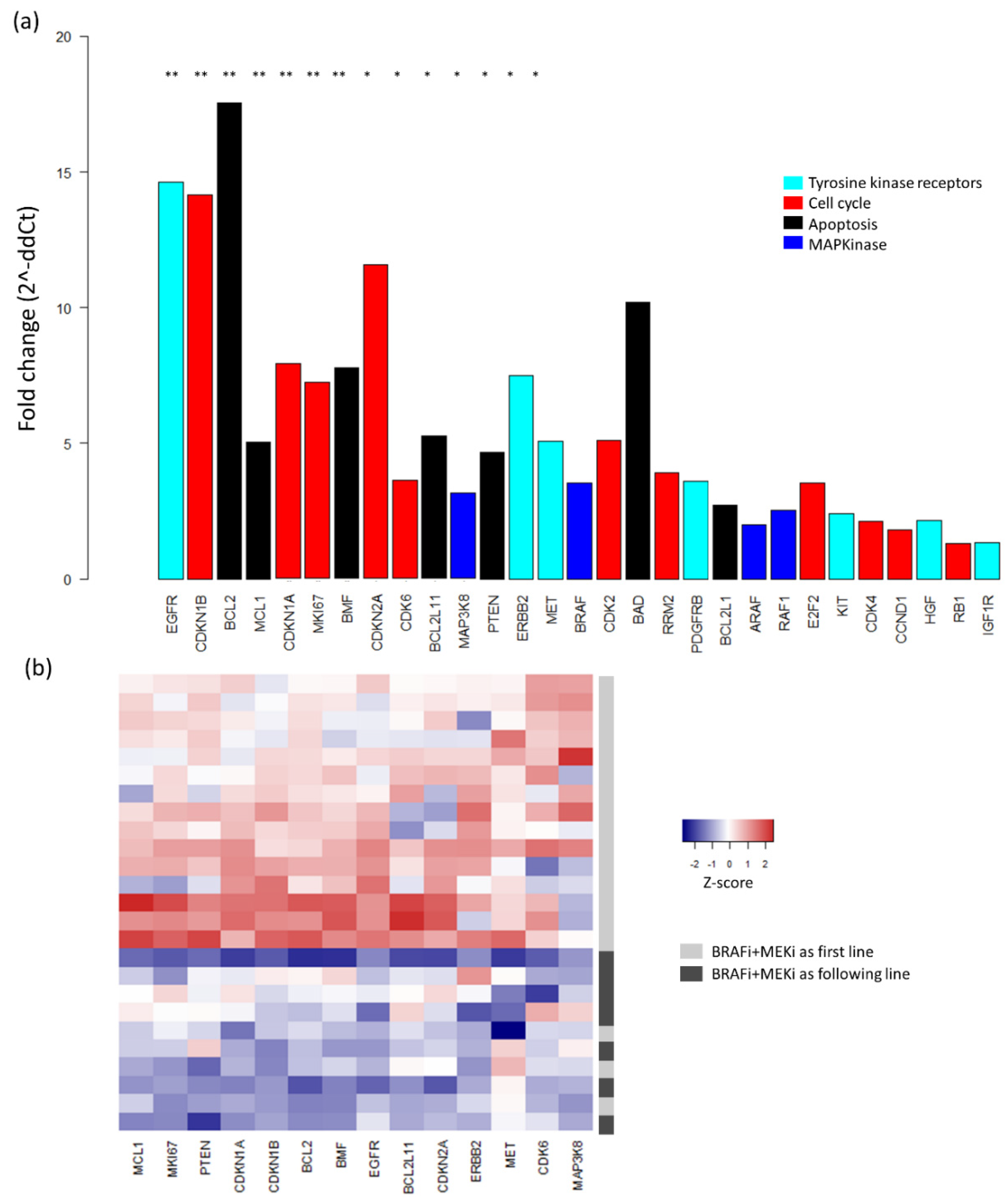
2.3. Baseline mRNA Expression Analysis

mRNA expression analysis was performed on 29 genes involved in the RAS–RAF–MAPK pathway, cell cycle, or apoptosis and implicated in MAPK inhibitor resistance mechanisms (Supplementary Table S1). Data were generated for baseline tumor samples from 25 patients (two patients with noninformative data). Principal component analysis (PCA) of the ∆Ct values was performed, revealing two clusters with different mRNA expression profiles (Figure 4).
In addition, differential transcript expression analysis was performed to identify expression profiles associated with clinical characteristics and to detect differentially expressed genes. Briefly, mRNA expression fold changes (2^-∆∆Ct) between predefined subgroups were calculated to assess relative quantification. First, patients were compared according to their clinical course during the BRAFi + MEKi treatment: occurrence of a PFS event vs. no PFS event and clinical response (complete or partial response) vs. no clinical response (stable or progressive disease). For most tested genes, higher mRNA expression levels were observed in patients with no PFS event vs. PFS event, with two genes coding for RTKs, KIT, and PDGFR, being significantly overexpressed in those with no PFS event (p < 0.01; Figure 5a). This association remained significant for KIT after false discovery rate (FDR) correction. However, no significant differential gene expression was found when comparing patients with or without a clinical response. These observations were confirmed when considering only patients with no brain metastases (n = 20). In this subgroup, KIT expression was significantly higher in patients with no PFS event vs. PFS event (p < 0.01; Figure 5b), though with no significant difference according to the occurrence of a clinical response. Due to the small number of patients with available data (n = 5), differential analysis was not performed specifically for the subgroup of “brain metastases”.
Second, analysis was performed to identify differential gene expression according to baseline clinical characteristics. Interestingly, comparison of the two clusters described in PCA showed that patients from Cluster 1 were more likely to have been treated with BRAFi + MEKi as the first-line than the following-line (91% vs. 57%, p = 0.06; Figure 4). According to differential expression analysis, 14 genes showed significantly higher expression in patients receiving BRAFi + MEKi as the first-line treatment vs. patients with BRAFi + MEKi as the following-line treatment, with seven remaining significant after FDR correction (Figure 6a). Using these 14 genes, an unsupervised heatmap was then plotted, confirming these different mRNA expression profiles between patients treated with BRAFi + MEKi as the first- and following-line (Figure 6b). Nonetheless, statistical significance for differential expression in patients with brain metastases vs. no brain metastases, or in patients with high LDH levels vs. low LDH levels, was not observed for any gene.

3. Discussion

Studies on the predictive potential of baseline genomic features in metastatic melanoma patients treated with targeted therapies have thus far primarily been performed using phase III clinical trial data. As a complement, data from patients followed as part of routine care are needed to enhance our understanding of these genomic features and their association with clinical characteristics.
Here, we present an exploratory analysis of a cohort of BRAFV600mut metastatic melanoma patients initiating BRAFi + MEKi treatment followed in a real-world setting. A median PFS of 10.1 months and a high rate of clinical response under BRAFi + MEKi (21/27 patients, 78%) were observed in our cohort, which is consistent with previous phase III trials [7,8,27,28] and recently published real-world studies [29,30,31]. Regarding clinical baseline characteristics, 16 patients (59%) were at stage IVM1c upon combined therapy initiation, which is similar to the rate reported by Long et al. [8] in the COMBI-D trial or by Luke et al. [31] in a real-world setting. The number of patients with brain metastases or with high LDH levels was also consistent with previous studies. Moreover, unlike clinical trials, seven patients (26%) who underwent previous systemic cancer therapy were included in our study, allowing a better representation of a real-life population. Indeed, in routine care, patients are likely to have undergone multiple treatments prior to BRAFi + MEKi [30].
Combining DNA mutations, copy number, and mRNA expression analyses, we conducted a targeted genomic approach to examine tumor samples prior to BRAFi + MEKi initiation. The studied genes were chosen based on the literature and were selected according to their potential implication in resistance to the MAPK inhibitor [32,33,34,35]. NRAS and MAP2K1 alterations concomitant with BRAF mutations were found in four patients (15%), which is consistent with the 24% rate reported by Johnson et al. in relapsed tumors [12]. However, we did not observe any associations between the type or total number of DNA alterations among our screened genes and clinical course under treatment. Although these observations must be considered carefully due to the small size of our cohort, they are consistent with recent analyses of vemurafenib + cobimetinib-treated patients from the BRIM-2, BRIM-3, BRIM-7, and coBRIM studies. Indeed, tumor mutational load, in addition to BRAF and CDKN2A alterations are reportedly similar between patients with complete response and those with rapid progressive disease [25]. Regarding mRNA expression, studies have evaluated levels in relapsed tumors, but few have characterized levels prior to MAPKi treatment. Overall, such predictive signatures are crucial for the identification of patients that are more likely to benefit from targeted therapy. Using data from the BRIM and coBRIM studies, Wongchenko et al. [24] and Wagle et al. [26] recently proposed an immune expression signature and a baseline MAPK pathway activity score, respectively, as predictors of clinical course under vemurafenib treatment. Our targeted approach has previously associated mRNA expression levels of a subset of genes (including RTKs, PGFRB, EGFR, and ERBB2) with survival in a real-life cohort of BRAFi-treated metastatic melanoma patients, highlighting a tumor proliferation/metabolic rate that might render it more sensitive to BRAFi [23]. In the present study, using the same targeted approach and focusing on patients treated with BRAFi + MEKi, we highlight the same trend with genes presenting higher expression in patients achieving clinical benefit from BRAFi + MEKi (no PFS event). Significance was observed for only a few genes, which may be related to the small size of our cohort and the occurrence of treatment discontinuation (adverse events or medical decision). Nonetheless, an interesting finding from our study is that one RTK-related gene, KIT, showed a high level of statistical significance. Low levels of KIT expression have been widely described in most cases of cutaneous melanoma, especially in BRAFV600mut melanoma, due to the endogenous activation of MAPK activation by BRAF mutations [35,36,37]. Furthermore, the loss of KIT has recently been described as a mechanism leading to increased BRAFV600E signaling. In BRAFV600E cells, KIT suppresses the RAS/MAPK pathway activity mediated by BRAF activation, and acts as a tumor suppressor [38]. As the majority of resistance mechanisms to BRAFi + MEKi are related to the MAPK pathway [11,22], we may assume that this baseline tumor-suppressor effect of KIT is emphasized in our cohort, improving the clinical course under BRAFi + MEKi. This alternative activation of the RAS/MAPK pathway via activated RTKs within the context of BRAF mutants has also been described by Grimm et al. [39].
We further analyzed the associations between genomic alterations and clinical baseline characteristics. Interestingly, CCND1 amplifications, RB1 deletions, and MET alterations were only observed for patients receiving BRAFi + MEKi as a first-line therapy, and none of the patients initiating BRAFi + MEKi as the following-line harbored these alterations. All these latter patients received and discontinued their immunotherapy treatment prior to BRAFi + MEKi initiation, suggesting that resistance mechanisms to immunotherapies may select tumor cells devoid of such genetic alterations.
Moreover, a difference in the baseline mRNA expression profile between patients treated with BRAFi + MEKi as the first-line and the following-line was also highlighted. Genomic biomarker studies performed to date in the field of melanoma have focused on first-line-treated patients, whereas little is known about tumor genomics in pretreated patients. In our study, we show that several genes involved in the cell cycle (CDKN1B, CDKN1A, MKI67) and apoptosis (BCL2, MCL1, BMF) pathways were more highly expressed in patients treated with BRAFi + MEKi as a first-line. Consistent with previous results [40], and because patients undergoing BRAFi + MEKi as the following-line do not present a satisfying response to previous treatment, our finding reveals the importance of presenting a high mRNA expression profile to obtain a benefit from immunotherapy. More broadly, these data show the importance of considering the treatment history when studying predictive genomic biomarkers and resistance mechanisms to targeted therapy.
Our targeted approach, combining DNA and mRNA alterations analyses, revealed the potential role of KIT as a predictive biomarker of response to BRAFi + MEKi. Within the context of metastatic melanoma, with frequent treatment discontinuation, our data also highlight the importance of performing tumor genomic analysis when switching therapy to account for molecular changes during successive targeted therapies/immunotherapies. However, considering the small size of our cohort, further studies are needed to support our findings.

4. Materials and Methods

4.1. Patients and Samples

Twenty-seven stage III or IV BRAFV600mut metastatic melanoma patients initiating combined treatment of BRAFi + MEKi as first- or following-line therapy and followed at Saint Louis Hospital (Paris, France) were included in this retrospective study. All patients were enrolled in MelBase, a multicenter French clinical database with a biobank dedicated to the prospective follow-up of advanced melanoma patients since March 2013. MelBase was approved by the French ethics committee (CPP Ile-de-france XI, n°12027, 2012) and registered in the NIH clinical trials database (NCT02828202). Written informed consent was obtained from all patients for their participation, including collection and analysis of their data. Patients were followed by a dermato-oncologist as part of the routine care at the Oncodermatology unit, and clinical response was evaluated using response evaluation criteria in solid tumor (RECIST) criteria [41]. For each patient, baseline (prior to BRAFi + MEKi initiation) tumor tissues were collected, stored as formalin-fixed, paraffin-embedded (FFPE), or frozen samples, and processed as previously described [23]. Samples harboring below 50% of tumor cells were macrodissected.

4.2. DNA and mRNA Extractions

DNA and mRNA were extracted from tumor samples as previously described [23]. Frozen tumor samples were processed with Maxwell RSC Tissue DNA and Maxwell RSC simplyRNA Tissue kits according to the manufacturer’s protocol for DNA and mRNA extraction, respectively (Promega, San Luis Obispo, CA, USA). A NanoDrop ND-1000 spectrophotometer (NanoDrop Technologies, Wilmington, NC, USA) was used for DNA and mRNA quantification and qualification. A Qubit 2.0 fluorometer (Thermo Fisher Scientific, Waltham, MA, USA) was also used for DNA quantification.

4.3. Copy Number Variation and Transcript Analyses

An off-the-shelf commercial personalized Human quantitative PCR (qPCR) SignArrays® 96 system (qPCR SignArrays®96 VPR1H1 kit, Anygenes, Paris, France) was used for gene copy number variation (CNV) and mRNA expression analyses. The PCR mixture, including 10 μL of Perfect MasterMix SYBR Green®, 8 μL of PCR grade water, and 2 μL of DNA (or cDNA after reverse transcription), was dropped into each well of the qPCR array. Amplification was performed using a LightCycler 480 (Roche, France) in duplicate according to the following sequence: (1) 10 min at 95 °C; (2) 40 cycles of 10 s at 95 °C; and (3) 30 s at 60 °C. The studied genes, involved in the RAS–RAF–MAPK pathway, cell cycle, or apoptosis as previously described [23], were selected for their validated or suggested role in BRAF inhibitor resistance. mRNA expression and copy number analyses were performed on 29 and 12 genes, respectively (Supplementary Table S1).
First-strand complementary DNA (cDNA) was synthesized from 1 µg of total RNA using a High-Capacity cDNA Archive Kit (Applied-Biosystems, Life Technologies, Carlsbad, CA, USA) according to the manufacturer’s protocol. The obtained cDNA samples were diluted 6 times for the qPCR assay. The raw mRNA expression data were normalized according to the ΔCt method (Ct: threshold cycle of amplification), as implemented in the ‘HTqPCR’ Bioconductor package (v1.36.0, https://www.bioconductor.org/packages/release/bioc/html/HTqPCR.html) [42]. For each targeted gene for each sample, mRNA expression values were generated in duplicate, and beta-2-microglobulin (B2M) was used as a housekeeping gene for normalization.
Gene copy number was quantified by comparison to glyceraldehyde 3-phosphate dehydrogenase (GAPDH) as an internal control. Two sets of primers were used for each gene, and relative copy numbers were calculated using the ΔΔCt method. Differences in the Ct of the targeted gene and GAPDH were assessed for each sample and compared to those in a reference pool of normal genomic DNA obtained from 10 samples of benign tissue. Relative copy number was calculated following the 2^-ΔΔCt formula, and conversion to absolute copy numbers was performed by assigning a value of 2 (diploid) to the reference pool and multiplying the relative copy number of samples by a factor of 2. Thresholds of 5 and 0.5 were set to define DNA amplification and DNA deletion, respectively [43,44].

4.4. Next-Generation Sequencing (NGS)

Targeted sequencing was performed with a customized AmpliSeq™ NGS panel (Thermo Fisher Scientific) designed with the Ion AmpliSeq™ designer software (Life Technologies). Seventy-four genes involved in the MAPK, PI3K, RTK, and cell cycle pathways were targeted (Supplementary Table S1). This panel of 35 kb generates 328 amplicons with an average size of 120 bp and a mean coverage of 92.79%. Sequencing amplicon libraries were synthesized from 50 ng of genomic DNA isolated from tissue samples using an Ion AmpliSeq™ Library Kit 2.0 (Thermo Fisher Scientific) and indexed with an Ion Xpress Barcode Adapters Kit (Life Technologies) following the manufacturer’s instructions. Amplification quality was defined with an Agilent 2100 BioAnalyzer. The library amplicon pool was sequenced with the Ion PGM™ Sequencer (Thermo Fisher Scientific) using Ion PGM Hi-Q Chef chemistry (single-end reads of 120 bp) and a 318-V2-BC sequencing flow cell. Base calling, alignment to the hg19 human reference sequence and variant calling were performed using Torrent Suite Software v5.8.0 (Thermo Fisher Scientific). ANNOVAR was employed for annotation through the Galaxy-APHP interface. Integrative Genomics Viewer (IGV) was used to visualize the read alignment and confirm the variant calls. Variant selection was performed using Alamut (Interactive Bio-Software) with the following criteria: (1) location in exonic or splice region; (2) nonsynonymous coding variant; (3) no previous annotation in the 1000 genomes project, ESP and ExAC databases or with an allelic frequency < 1%; (4) coverage ≥ 500×; (5) variant allele fraction (VAF) ≥ 5%; and (6) strand bias < 95%.

4.5. Statistical Analysis

Clinical characteristics are described in terms of median [IQR] for quantitative variables and in terms of number (%) for qualitative variables. PFS was defined as the time between BRAFi + MEKi initiation and disease progression or death of any cause under treatment, whichever occurred first. Patients were censored at the time of BRAFi + MEKi interruption. PFS was estimated using the Kaplan–Meier method, and comparisons were performed with the log rank test. Copy number variations were dichotomized as amplifications (>5) and as deletions (<0.5). The number of DNA alterations between groups was compared using a t-test. Differential expression analysis between groups of interest was performed on normalized data using ‘HTqPCR’ Biopackage [42]. ΔΔCt, defined as ΔCtgroup1−ΔCtgroup2, was calculated for each targeted gene, and the fold change between groups was set as 2^-ΔΔCt. A t-test was performed to assess any difference between groups, and FDR was used for multiple test corrections. Statistical significance was set at p ≤ 0.05, and all analyses were performed with R 3.5.1 (R foundation for statistical computing, Vienna, Austria).

5. Conclusions

Despite the small number of patients included in our study, our targeted genomic approach in a real-life cohort of patients initiating the BRAFi + MEKi treatment revealed a differential baseline transcriptomic profile with higher mRNA expression in BRAFV600mut metastatic melanoma patients receiving this combined therapy as a first-line treatment. KIT was found to be overexpressed in patients with an improved clinical course, suggesting its potential predictive value. Further studies on larger cohorts are needed to support our findings.

Supplementary Materials

The following are available online at https://www.mdpi.com/2072-6694/11/8/1203/s1, Table S1: List of studied genes, Table S2: Mutations detected by next-generation sequencing (panel of 74 genes) on the 24 baseline samples.

Author Contributions

Conceptualization, B.L., F.J., S.M., and C.L.; methodology, B.L. and F.J.; formal analysis, B.L., F.J., C.R.d.M., and A.S.; investigation, B.L., F.J., C.R.d.M., and A.S.; validation: C.R.d.M. and A.S.; resources, B.B., J.D., F.H., A.D.M., L.D.M. and M.B.; writing—original draft preparation, B.L. and F.J.; writing—review and editing, N.D., C.L. and S.M.; visualization, B.L.; supervision, C.L. and S.M.; project administration, C.L. and S.M.

Funding

This work was supported by the Assistance Publique-Hôpitaux de Paris, by the Universite de Paris, by La Societe Francaise de Dermatologie (SFD) and by La Fondation de l’Avenir (Bourse Legs Cancérologie).

Acknowledgments

The authors wish to thank patients who agreed to participate in this study and the MelBase team for help regarding clinical data acquisition.

Conflicts of Interest

B.L., F.J., C.R.d.M., A.S., A.D.M, L.D.M., and N.D. declare no conflict of interest. B.B. declares honoraria from BMS, MSD and Pierre Fabre, a consulting role for BMS and Pierre Fabre and travel accommodations from BMS. J.D. declares travel accommodations from Pierre Fabre. F.H. declares a consulting role for Sanofi-Aventis and travel accommodations from Sun Pharma. M.B. declares honoraria from Innate Pharma, Takeda, BMS and Leo Pharma. C.L. declares a consulting role for Amgen, BMS, MSD, Novartis, and Roche, research funding from BMS and Roche, honoraria from Amgen, BMS, Incyte, MSD, Novartis, Pfizer, Pierre Fabre and Roche and travel accommodations from BMS. S.M. declares a consulting role for Novartis, Biocartis, and Roche and research funding from Biocartis.

References

  1. Davies, H.; Bignell, G.R.; Cox, C.; Stephens, P.; Edkins, S.; Clegg, S.; Teague, J.; Woffendin, H.; Garnett, M.J.; Bottomley, W.; et al. Mutations of the BRAF gene in human cancer. Nature 2002, 417, 949–954. [Google Scholar] [CrossRef] [PubMed]
  2. Flaherty, K.T.; Puzanov, I.; Kim, K.B.; Ribas, A.; McArthur, G.A.; Sosman, J.A.; O’Dwyer, P.J.; Lee, R.J.; Grippo, J.F.; Nolop, K.; et al. Inhibition of mutated, activated BRAF in metastatic melanoma. N. Engl. J. Med. 2010, 363, 809–819. [Google Scholar] [CrossRef] [PubMed]
  3. Sosman, J.A.; Kim, K.B.; Schuchter, L.; Gonzalez, R.; Pavlick, A.C.; Weber, J.S.; McArthur, G.A.; Hutson, T.E.; Moschos, S.J.; Flaherty, K.T.; et al. Survival in BRAF V600-mutant advanced melanoma treated with vemurafenib. N. Engl. J. Med. 2012, 366, 707–714. [Google Scholar] [CrossRef] [PubMed]
  4. McArthur, G.A.; Chapman, P.B.; Robert, C.; Larkin, J.; Haanen, J.B.; Dummer, R.; Ribas, A.; Hogg, D.; Hamid, O.; Ascierto, P.A.; et al. Safety and efficacy of vemurafenib in BRAF(V600E) and BRAF(V600K) mutation-positive melanoma (BRIM-3): Extended follow-up of a phase 3, randomised, open-label study. Lancet Oncol. 2014, 15, 323–332. [Google Scholar] [CrossRef]
  5. Ascierto, P.A.; McArthur, G.A.; Dréno, B.; Atkinson, V.; Liszkay, G.; Di Giacomo, A.M.; Mandalà, M.; Demidov, L.; Stroyakovskiy, D.; Thomas, L.; et al. Cobimetinib combined with vemurafenib in advanced BRAF(V600)-mutant melanoma (coBRIM): Updated efficacy results from a randomised, double-blind, phase 3 trial. Lancet Oncol. 2016, 17, 1248–1260. [Google Scholar] [CrossRef]
  6. Hauschild, A.; Grob, J.-J.; Demidov, L.V.; Jouary, T.; Gutzmer, R.; Millward, M.; Rutkowski, P.; Blank, C.U.; Miller, W.H.; Kaempgen, E.; et al. Dabrafenib in BRAF-mutated metastatic melanoma: A multicentre, open-label, phase 3 randomised controlled trial. Lancet 2012, 380, 358–365. [Google Scholar] [CrossRef]
  7. Robert, C.; Karaszewska, B.; Schachter, J.; Rutkowski, P.; Mackiewicz, A.; Stroiakovski, D.; Lichinitser, M.; Dummer, R.; Grange, F.; Mortier, L.; et al. Improved overall survival in melanoma with combined dabrafenib and trametinib. N. Engl. J. Med. 2015, 372, 30–39. [Google Scholar] [CrossRef]
  8. Long, G.V.; Stroyakovskiy, D.; Gogas, H.; Levchenko, E.; de Braud, F.; Larkin, J.; Garbe, C.; Jouary, T.; Hauschild, A.; Grob, J.-J.; et al. Dabrafenib and trametinib versus dabrafenib and placebo for Val600 BRAF-mutant melanoma: A multicentre, double-blind, phase 3 randomised controlled trial. Lancet 2015, 386, 444–451. [Google Scholar] [CrossRef]
  9. Long, G.V.; Stroyakovskiy, D.; Gogas, H.; Levchenko, E.; de Braud, F.; Larkin, J.; Garbe, C.; Jouary, T.; Hauschild, A.; Grob, J.J.; et al. Combined BRAF and MEK inhibition versus BRAF inhibition alone in melanoma. N. Engl. J. Med. 2014, 371, 1877–1888. [Google Scholar] [CrossRef]
  10. Manzano, J.L.; Layos, L.; Bugés, C.; de los Llanos Gil, M.; Vila, L.; Martínez-Balibrea, E.; Martínez-Cardús, A. Resistant mechanisms to BRAF inhibitors in melanoma. Ann. Transl. Med. 2016, 4. [Google Scholar] [CrossRef]
  11. Rizos, H.; Menzies, A.M.; Pupo, G.M.; Carlino, M.S.; Fung, C.; Hyman, J.; Haydu, L.E.; Mijatov, B.; Becker, T.M.; Boyd, S.C.; et al. BRAF inhibitor resistance mechanisms in metastatic melanoma: Spectrum and clinical impact. Clin. Cancer Res. 2014, 20, 1965–1977. [Google Scholar] [CrossRef] [PubMed]
  12. Johnson, D.B.; Menzies, A.M.; Zimmer, L.; Eroglu, Z.; Ye, F.; Zhao, S.; Rizos, H.; Sucker, A.; Scolyer, R.A.; Gutzmer, R.; et al. Acquired BRAF inhibitor resistance: A multicenter meta-analysis of the spectrum and frequencies, clinical behaviour, and phenotypic associations of resistance mechanisms. Eur. J. Cancer 2015, 51, 2792–2799. [Google Scholar] [CrossRef] [PubMed] [Green Version]
  13. Rebecca, V.W.; Alicea, G.M.; Paraiso, K.H.T.; Lawrence, H.; Gibney, G.T.; Smalley, K.S.M. Vertical inhibition of the MAPK pathway enhances therapeutic responses in NRAS-mutant melanoma. Pigment Cell Melanoma Res. 2014, 27, 1154–1158. [Google Scholar] [CrossRef] [PubMed]
  14. Shi, H.; Hugo, W.; Kong, X.; Hong, A.; Koya, R.C.; Moriceau, G.; Chodon, T.; Guo, R.; Johnson, D.B.; Dahlman, K.B.; et al. Acquired resistance and clonal evolution in melanoma during BRAF inhibitor therapy. Cancer Discov. 2014, 4, 80–93. [Google Scholar] [CrossRef] [PubMed]
  15. Shi, H.; Kong, X.; Ribas, A.; Lo, R.S. Combinatorial treatments that overcome PDGFRβ-driven resistance of melanoma cells to V600EB-RAF inhibition. Cancer Res. 2011, 71, 5067–5074. [Google Scholar] [CrossRef] [PubMed]
  16. Deng, W.; Gopal, Y.N.V.; Scott, A.; Chen, G.; Woodman, S.E.; Davies, M.A. Role and therapeutic potential of PI3K-mTOR signaling in de novo resistance to BRAF inhibition. Pigment Cell Melanoma Res. 2012, 25, 248–258. [Google Scholar] [CrossRef] [PubMed]
  17. Straussman, R.; Morikawa, T.; Shee, K.; Barzily-Rokni, M.; Qian, Z.R.; Du, J.; Davis, A.; Mongare, M.M.; Gould, J.; Frederick, D.T.; et al. Tumour micro-environment elicits innate resistance to RAF inhibitors through HGF secretion. Nature 2012, 487, 500–504. [Google Scholar] [CrossRef] [PubMed] [Green Version]
  18. Shi, H.; Moriceau, G.; Kong, X.; Lee, M.-K.; Lee, H.; Koya, R.C.; Ng, C.; Chodon, T.; Scolyer, R.A.; Dahlman, K.B.; et al. Melanoma whole-exome sequencing identifies (V600E)B-RAF amplification-mediated acquired B-RAF inhibitor resistance. Nat. Commun. 2012, 3, 724. [Google Scholar] [CrossRef]
  19. Nazarian, R.; Shi, H.; Wang, Q.; Kong, X.; Koya, R.C.; Lee, H.; Chen, Z.; Lee, M.-K.; Attar, N.; Sazegar, H.; et al. Melanomas acquire resistance to B-RAF(V600E) inhibition by RTK or N-RAS upregulation. Nature 2010, 468, 973–977. [Google Scholar] [CrossRef]
  20. Poulikakos, P.I.; Persaud, Y.; Janakiraman, M.; Kong, X.; Ng, C.; Moriceau, G.; Shi, H.; Atefi, M.; Titz, B.; Gabay, M.T.; et al. RAF inhibitor resistance is mediated by dimerization of aberrantly spliced BRAF(V600E). Nature 2011, 480, 387–390. [Google Scholar] [CrossRef] [Green Version]
  21. Villanueva, J.; Vultur, A.; Lee, J.T.; Somasundaram, R.; Fukunaga-Kalabis, M.; Cipolla, A.K.; Wubbenhorst, B.; Xu, X.; Gimotty, P.A.; Kee, D.; et al. Acquired resistance to BRAF inhibitors mediated by a RAF kinase switch in melanoma can be overcome by cotargeting MEK and IGF-1R/PI3K. Cancer Cell 2010, 18, 683–695. [Google Scholar] [CrossRef]
  22. Van Allen, E.M.; Wagle, N.; Sucker, A.; Treacy, D.J.; Johannessen, C.M.; Goetz, E.M.; Place, C.S.; Taylor-Weiner, A.; Whittaker, S.; Kryukov, G.V.; et al. The genetic landscape of clinical resistance to RAF inhibition in metastatic melanoma. Cancer Discov. 2014, 4, 94–109. [Google Scholar] [CrossRef]
  23. Louveau, B.; Delyon, J.; De Moura, C.R.; Battistella, M.; Jouenne, F.; Golmard, L.; Sadoux, A.; Podgorniak, M.-P.; Chami, I.; Marco, O.; et al. A targeted genomic alteration analysis predicts survival of melanoma patients under BRAF inhibitors. Oncotarget 2019, 10, 1669–1687. [Google Scholar] [CrossRef] [Green Version]
  24. Wongchenko, M.J.; McArthur, G.A.; Dréno, B.; Larkin, J.; Ascierto, P.A.; Sosman, J.; Andries, L.; Kockx, M.; Hurst, S.D.; Caro, I.; et al. Gene Expression Profiling in BRAF-Mutated Melanoma Reveals Patient Subgroups with Poor Outcomes to Vemurafenib That May Be Overcome by Cobimetinib Plus Vemurafenib. Clin. Cancer Res. 2017, 23, 5238–5245. [Google Scholar] [CrossRef]
  25. Yan, Y.; Wongchenko, M.J.; Robert, C.; Larkin, J.; Ascierto, P.A.; Dréno, B.; Maio, M.; Garbe, C.; Chapman, P.B.; Sosman, J.A.; et al. Genomic Features of Exceptional Response in Vemurafenib ± Cobimetinib-treated Patients with BRAFV600-mutated Metastatic Melanoma. Clin. Cancer Res. 2019, 25, 3239–3246. [Google Scholar] [CrossRef]
  26. Wagle, M.-C.; Kirouac, D.; Klijn, C.; Liu, B.; Mahajan, S.; Junttila, M.; Moffat, J.; Merchant, M.; Huw, L.; Wongchenko, M.; et al. A transcriptional MAPK Pathway Activity Score (MPAS) is a clinically relevant biomarker in multiple cancer types. NPJ Precis. Oncol. 2018, 2, 7. [Google Scholar] [CrossRef]
  27. Flaherty, K.T.; Robert, C.; Hersey, P.; Nathan, P.; Garbe, C.; Milhem, M.; Demidov, L.V.; Hassel, J.C.; Rutkowski, P.; Mohr, P.; et al. Improved survival with MEK inhibition in BRAF-mutated melanoma. N. Engl. J. Med. 2012, 367, 107–114. [Google Scholar] [CrossRef]
  28. Larkin, J.M.G.; Yan, Y.; McArthur, G.A.; Ascierto, P.A.; Liszkay, G.; Maio, M.; Mandalà, M.; Demidov, L.V.; Stroyakovskiy, D.; Thomas, L.; et al. Update of progression-free survival (PFS) and correlative biomarker analysis from coBRIM: Phase III study of cobimetinib (cobi) plus vemurafenib (vem) in advanced BRAF-mutated melanoma. JCO 2015, 33, 9006. [Google Scholar] [CrossRef]
  29. Cavalieri, S.; Di Guardo, L.; Cimminiello, C.; Bono, A.; Tolomio, E.; Colombetti, A.; Valeri, B.; Di Tolla, G.; de Braud, F.; Del Vecchio, M. Combined therapy with dabrafenib and trametinib in BRAF-mutated metastatic melanoma in a real-life setting: The INT Milan experience. Tumori 2016, 102, 501–507. [Google Scholar] [CrossRef]
  30. Depenni, R.; De Rosa, F.; Greco, S.; Ridolfi, L.; Pellacani, G.; Ponti, G.; Cascinu, S.; Guidoboni, M. Dabrafenib-trametinib combination in “field-practice”: An Italian experience. Future Oncol. 2018, 14, 2045–2052. [Google Scholar] [CrossRef]
  31. Luke, J.J.; Ghate, S.R.; Kish, J.; Lee, C.H.; McAllister, L.; Mehta, S.; Ndife, B.; Feinberg, B.A. Targeted agents or immuno-oncology therapies as first-line therapy for BRAF-mutated metastatic melanoma: A real-world study. Future Oncol. 2019. [Google Scholar] [CrossRef]
  32. Carlino, M.S.; Gowrishankar, K.; Saunders, C.A.B.; Pupo, G.M.; Snoyman, S.; Zhang, X.D.; Saw, R.; Becker, T.M.; Kefford, R.F.; Long, G.V.; et al. Antiproliferative effects of continued mitogen-activated protein kinase pathway inhibition following acquired resistance to BRAF and/or MEK inhibition in melanoma. Mol. Cancer Ther. 2013, 12, 1332–1342. [Google Scholar] [CrossRef]
  33. Flaherty, K.; Davies, M.A.; Grob, J.J.; Long, G.V.; Nathan, P.; Ribas, A.; Robert, C. Genomic analysis and 3-year efficacy and safety update of COMBI-d. J. Clin. Oncol. 2019, 34, 9502. [Google Scholar] [CrossRef]
  34. Luke, J.J.; Flaherty, K.T.; Ribas, A.; Long, G.V. Targeted agents and immunotherapies: Optimizing outcomes in melanoma. Nat. Rev. Clin. Oncol. 2017, 14, 463–482. [Google Scholar] [CrossRef]
  35. Kunz, M.; Hölzel, M. The impact of melanoma genetics on treatment response and resistance in clinical and experimental studies. Cancer Metastasis Rev. 2017, 36, 53–75. [Google Scholar] [CrossRef]
  36. Akbani, R.; Akdemir, K.C.; Aksoy, B.A.; Albert, M.; Ally, A.; Amin, S.B.; Arachchi, H.; Arora, A.; Auman, J.T.; Ayala, B.; et al. Genomic Classification of Cutaneous Melanoma. Cell 2015, 161, 1681–1696. [Google Scholar] [CrossRef]
  37. Sun, C.; Wang, L.; Huang, S.; Heynen, G.J.J.E.; Prahallad, A.; Robert, C.; Haanen, J.; Blank, C.; Wesseling, J.; Willems, S.M.; et al. Reversible and adaptive resistance to BRAF(V600E) inhibition in melanoma. Nature 2014, 508, 118–122. [Google Scholar] [CrossRef]
  38. Neiswender, J.V.; Kortum, R.L.; Bourque, C.; Kasheta, M.; Zon, L.I.; Morrison, D.K.; Ceol, C.J. KIT Suppresses BRAFV600E-Mutant Melanoma by Attenuating Oncogenic RAS/MAPK Signaling. Cancer Res. 2017, 77, 5820–5830. [Google Scholar] [CrossRef]
  39. Grimm, J.; Hufnagel, A.; Wobser, M.; Borst, A.; Haferkamp, S.; Houben, R.; Meierjohann, S. BRAF inhibition causes resilience of melanoma cell lines by inducing the secretion of FGF1. Oncogenesis 2018, 7, 71. [Google Scholar] [CrossRef]
  40. Hugo, W.; Zaretsky, J.M.; Sun, L.; Song, C.; Moreno, B.H.; Hu-Lieskovan, S.; Berent-Maoz, B.; Pang, J.; Chmielowski, B.; Cherry, G.; et al. Genomic and Transcriptomic Features of Response to Anti-PD-1 Therapy in Metastatic Melanoma. Cell 2016, 165, 35–44. [Google Scholar] [CrossRef] [Green Version]
  41. Eisenhauer, E.A.; Therasse, P.; Bogaerts, J.; Schwartz, L.H.; Sargent, D.; Ford, R.; Dancey, J.; Arbuck, S.; Gwyther, S.; Mooney, M.; et al. New response evaluation criteria in solid tumours: Revised RECIST guideline (version 1.1). Eur. J. Cancer 2009, 45, 228–247. [Google Scholar] [CrossRef]
  42. Dvinge, H.; Bertone, P. HTqPCR: High-throughput analysis and visualization of quantitative real-time PCR data in R. Bioinformatics 2009, 25, 3325–3326. [Google Scholar] [CrossRef]
  43. Lebbé, C.; How-Kit, A.; Battistella, M.; Sadoux, A.; Podgorniak, M.-P.; Sidina, I.; Pages, C.; Roux, J.; Porcher, R.; Tost, J.; et al. BRAF(V600) mutation levels predict response to vemurafenib in metastatic melanoma. Melanoma Res. 2014, 24, 415–418. [Google Scholar] [CrossRef]
  44. Chraybi, M.; Abd Alsamad, I.; Copie-Bergman, C.; Baia, M.; André, J.; Dumaz, N.; Ortonne, N. Oncogene abnormalities in a series of primary melanomas of the sinonasal tract: NRAS mutations and cyclin D1 amplification are more frequent than KIT or BRAF mutations. Hum. Pathol. 2013, 44, 1902–1911. [Google Scholar] [CrossRef] [Green Version]
Figure 1. Clinical course of the 27 included patients. Events occurring during the BRAFi + MEKi treatment are shown. White bars indicate the absence of BRAFi + MEKi therapy. Patients were censored at the last available date of follow-up if death did not occur.
Cancers 11 01203 g001
Figure 2. Kaplan–Meier survival plot for progression-free survival of the 27 included patients.
Cancers 11 01203 g002
Figure 3. Landscape of baseline DNA alterations (mutations and copy number variations) for the 24 patients with available data. Amplifications and deletions were defined as CNV > 5 and CNV < 0.5, respectively. Patients are ranked according to the total number of alterations detected.
Cancers 11 01203 g003
Figure 4. Principal component analysis of mRNA expression for the 25 patients with available data (∆Ct, normalization on B2M). Patients from Cluster 1 were more likely to have been treated with BRAFi + MEKi as a first-line treatment.
Cancers 11 01203 g004
Figure 5. Differential gene expression analysis according to clinical course. (a) Differential gene expression in patients with no progression free survival (PFS) event vs. patients with PFS event (n = 25). (b) Differential gene expression in patients with no PFS event vs. PFS event (only patients with no brain metastases, n = 20). Differences in gene expression are expressed as fold change (2^-∆∆Ct), and genes are ranked by their level of significance. ** p < 0.01, * p < 0.05.
Cancers 11 01203 g005
Figure 6. Differential gene expression analysis according to line of treatment (a) Differential gene expression in patients with BRAFi + MEKi as the first-line vs. patients treated with BRAFi + MEKi as the following-line of treatment. Differences in gene expression are expressed as fold change (2^-∆∆Ct), and genes are ranked by their level of significance. ** p < 0.01, * p < 0.05. (b) Heatmap of mRNA expression. Colors represent the relative expression of a gene in each sample centered on the mean and scaled to the standard deviation (red: high and blue: low).
Cancers 11 01203 g006
Table 1. Patient characteristics at BRAFi + MEKi initiation.
All Included Patients
(n = 27)
Male sex13 (48%)
Age at melanoma diagnosis, years49.3 (35.5; 58.8)
Melanoma subtype
Superficial spreading melanoma15 (56%)
Nodular melanoma5 (19%)
Others2 (7%)
Undetermined5 (19%)
Breslow thickness, mm3 (1.4; 5.2)
Ulceration11/24 (46%)
Age at initiation, years49.9 (40.8; 62.7)
Disease stage
Unresectable stage III3 (11%)
Unresectable stage IV24 (89%)
M1c16 (67%)
Brain metastases6 (22%)
Elevated lactate dehydrogenase level7/24 (29%)
Number of disease sites
≤213/24 (54%)
≥311/24 (46%)
BRAFi + MEKi combination initiated
Dabrafenib + trametinib24 (89%)
Vemurafenib + cobimetinib3 (11%)
BRAFi + MEKi as first-line treatment
First-line treatment20 (74%)
Following-line treatment7 (26%)
Previous line of treatment (if any)
BRAFi monotherapy3 (11%)
Anti-CTLA42 (7%)
Anti-PD1/PDL16 (22%)
Anti-CTLA4 + anti-PD12 (7%)
Others1 (4%)
Data are median (interquartile range—IQR) and number (%). Elevated lactate dehydrogenase levels are defined as >480 UI. BRAFi: BRAF inhibitor, MEKi: MEK inhibitor, and mm: millimeters.

