Next Issue
Volume 10, July
Previous Issue
Volume 10, May
 
 
cancers-logo

Journal Browser

Journal Browser

Cancers, Volume 10, Issue 6 (June 2018) – 56 articles

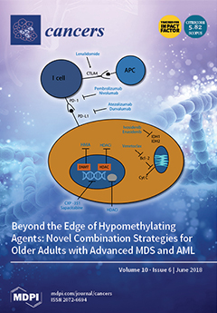
Cover Story (view full-size image): Until today, older patients with high-risk myelodysplastic syndrome (MDS) and acute myeloid leukemia (AML) have poor outcomes when treated with conventional treatment strategies. Response after a first-line treatment with hypomethylating agents (HMA) occurs in less than 50% of these patients. In our review, we focus on upcoming new treatment strategies in the field of HMA-based combination strategies. One of the current most promising approaches is preventing HMA resistance by using checkpoint inhibitors. We are sure that these new treatment options will guide the future of clinical research. Recent data also suggest a potential renaissance of intensive treatment strategies, such as therapies based on CPX-351. View this paper.
  • Issues are regarded as officially published after their release is announced to the table of contents alert mailing list.
  • You may sign up for e-mail alerts to receive table of contents of newly released issues.
  • PDF is the official format for papers published in both, html and pdf forms. To view the papers in pdf format, click on the "PDF Full-text" link, and use the free Adobe Reader to open them.
Order results
Result details
Section
Select all
Export citation of selected articles as:

Editorial

Jump to: Research, Review, Other

3 pages, 159 KiB  
Editorial
Mechanistic Target of Rapamycin (mTOR) in the Cancer Setting
by James T. Murray 1 and Andrew R. Tee 2,*
1 School of Biochemistry & Immunology, Trinity Biomedical Sciences Institute, Trinity College Dublin, Dublin 2, Ireland
2 Division of Cancer and Genetics, Cardiff University, Heath Park, Cardiff CF14 4XN, UK
Cancers 2018, 10(6), 168; https://doi.org/10.3390/cancers10060168 - 30 May 2018
Cited by 5 | Viewed by 4563
Abstract
This special issue on mammalian target of rapamycin (mTOR) explores the importance of mTOR in cell growth control and cancer. Cancer cells often exploit mTOR as a mechanism to enhance their capacity to grow. While protein synthesis is by far the best-characterized mTOR-driven [...] Read more.
This special issue on mammalian target of rapamycin (mTOR) explores the importance of mTOR in cell growth control and cancer. Cancer cells often exploit mTOR as a mechanism to enhance their capacity to grow. While protein synthesis is by far the best-characterized mTOR-driven process, this special issue also describes a wider array of mTOR-driven biological processes that cancer cells benefit from, including autophagy, cell cycle control, metabolic transformation, angiogenic signaling, and anabolic processes such as nucleotide biosynthesis and ribosomal biogenesis. Other areas of mTOR signaling covered in these reviews delve into cell migration, inflammation, and regulation of transcription factors linked to cancer progression. Full article
(This article belongs to the Special Issue mTOR Pathway in Cancer)

Research

Jump to: Editorial, Review, Other

12 pages, 1183 KiB  
Article
A Phase II Study of Pelareorep (REOLYSIN®) in Combination with Gemcitabine for Patients with Advanced Pancreatic Adenocarcinoma
by Devalingam Mahalingam 1,2,*, Sanjay Goel 3, Santiago Aparo 3, Sukeshi Patel Arora 2, Nicole Noronha 4, Hue Tran 4, Romit Chakrabarty 4, Giovanni Selvaggi 4, Andres Gutierrez 4, Matthew Coffey 4, Steffan T. Nawrocki 5, Gerard Nuovo 6 and Monica M. Mita 7
1 Division of Hematology/Oncology, Robert H. Lurie Comprehensive Cancer Center, Northwestern University, Chicago, IL 60611, USA
2 Cancer Therapy and Research Center, University of Texas Health Science Center, San Antonio, TX 78229, USA
3 Montefiore Medical Center, New York, NY 10467, USA
4 Oncolytics Biotech Inc., Calgary, AB T2N 1X7, Canada
5 Department of Medicine, Division of Translational and Regenerative Medicine, University of Arizona Cancer Center, Tucson, AZ 85724, USA
6 Comprehensive Cancer Center, Ohio State University, Columbus, OH and Phylogeny, Inc., Powell, OH 43065, USA
7 Samuel Oschin Comprehensive Cancer Institute, Los Angeles, CA 90048, USA
Cancers 2018, 10(6), 160; https://doi.org/10.3390/cancers10060160 - 25 May 2018
Cited by 101 | Viewed by 8906
Abstract
Pancreatic ductal adenocarcinoma (PDAC) has a poor prognosis, with 1 and 5-year survival rates of ~18% and 7% respectively. FOLFIRINOX or gemcitabine in combination with nab-paclitaxel are standard treatment options for metastatic disease. However, both regimens are more toxic than gemcitabine alone. Pelareorep [...] Read more.
Pancreatic ductal adenocarcinoma (PDAC) has a poor prognosis, with 1 and 5-year survival rates of ~18% and 7% respectively. FOLFIRINOX or gemcitabine in combination with nab-paclitaxel are standard treatment options for metastatic disease. However, both regimens are more toxic than gemcitabine alone. Pelareorep (REOLYSIN®), a proprietary isolate of reovirus Type 3 Dearing, has shown antitumor activity in clinical and preclinical models. In addition to direct cytotoxic effects, pelareorep can trigger antitumor immune responses. Due to the high frequency of RAS mutations in PDAC, we hypothesized that pelareorep would promote selective reovirus replication in pancreatic tumors and enhance the anticancer activity of gemcitabine. Chemotherapy-naïve patients with advanced PDAC were eligible for the study. The primary objective was Clinical Benefit Rate (complete response (CR) + partial response (PR) + stable disease (SD) ≥ 12 weeks) and secondary objectives include overall survival (OS), toxicity, and pharmacodynamics (PD) analysis. The study enrolled 34 patients; results included one partial response, 23 stable disease, and 5 progressive disease. The median OS was 10.2 months, with a 1- and 2-year survival rate of 45% and 24%, respectively. The treatment was well tolerated with manageable nonhematological toxicities. PD analysis revealed reovirus replication within pancreatic tumor and associated apoptosis. Upregulation of immune checkpoint marker PD-L1 suggests future consideration of combining oncolytic virus therapy with anti-PD-L1 inhibitors. We conclude that pelareorep complements single agent gemcitabine in PDAC. Full article
(This article belongs to the Special Issue Oncolytic Virotherapy)
Show Figures

Figure 1

19 pages, 4858 KiB  
Article
CRISPR-Mediated Reactivation of DKK3 Expression Attenuates TGF-β Signaling in Prostate Cancer
by Hoda Kardooni 1, Estela Gonzalez-Gualda 1, Emmanouil Stylianakis 1, Sina Saffaran 2, Jonathan Waxman 1 and Robert M. Kypta 1,3,*
1 Department of Surgery and Cancer, Imperial College London, London W12 0NN, UK
2 School of Engineering, University of Warwick, Coventry CV4 7AL, UK
3 Centre for Cooperative Research in Biosciences, CIC bioGUNE, 48160 Derio, Spain
Cancers 2018, 10(6), 165; https://doi.org/10.3390/cancers10060165 - 28 May 2018
Cited by 41 | Viewed by 7479
Abstract
The DKK3 gene encodes a secreted protein, Dkk-3, that inhibits prostate tumor growth and metastasis. DKK3 is downregulated by promoter methylation in many types of cancer, including prostate cancer. Gene silencing studies have shown that Dkk-3 maintains normal prostate epithelial cell homeostasis by [...] Read more.
The DKK3 gene encodes a secreted protein, Dkk-3, that inhibits prostate tumor growth and metastasis. DKK3 is downregulated by promoter methylation in many types of cancer, including prostate cancer. Gene silencing studies have shown that Dkk-3 maintains normal prostate epithelial cell homeostasis by limiting TGF-β/Smad signaling. While ectopic expression of Dkk-3 leads to prostate cancer cell apoptosis, it is unclear if Dkk-3 has a physiological role in cancer cells. Here, we show that treatment of PC3 prostate cancer cells with the DNA methyltransferase (DNMT) inhibitor decitabine demethylates the DKK3 promoter, induces DKK3 expression, and inhibits TGF-β/Smad-dependent transcriptional activity. Direct induction of DKK3 expression using CRISPR-dCas9-VPR also inhibited TGF-β/Smad-dependent transcription and attenuated PC3 cell migration and proliferation. These effects were not observed in C4-2B cells, which do not respond to TGF-β. TGF-β signals can regulate gene expression directly via SMAD proteins and indirectly by increasing DNMT expression, leading to promoter methylation. Analysis of genes downregulated by promoter methylation and predicted to be regulated by TGF-β found that DKK3 induction increased expression of PTGS2, which encodes cyclooxygenase-2. Together, these observations provide support for using CRISPR-mediated induction of DKK3 as a potential therapeutic approach for prostate cancer and highlight complexities in Dkk-3 regulation of TGF-β signaling. Full article
(This article belongs to the Special Issue TGF-Beta Signaling in Cancer)
Show Figures

Figure 1

17 pages, 5986 KiB  
Article
The Transition between Telomerase and ALT Mechanisms in Hodgkin Lymphoma and Its Predictive Value in Clinical Outcomes
by Radhia M’kacher 1,2,*, Corina Cuceu 1, Mustafa Al Jawhari 1, Luc Morat 1, Monika Frenzel 1, Grace Shim 1, Aude Lenain 1, William M. Hempel 1, Steffen Junker 3, Theodore Girinsky 4, Bruno Colicchio 5, Alain Dieterlen 5, Leonhard Heidingsfelder 6, Claire Borie 7, Noufissa Oudrhiri 7, Annelise Bennaceur-Griscelli 7, Olivier Moralès 8, Sarah Renaud 8, Zoé Van de Wyngaert 9, Eric Jeandidier 10, Nadira Delhem 8 and Patrice Carde 11add Show full author list remove Hide full author list
1 Laboratoire de Radiobiologie et d’Oncologie, IRCM/DSV/CEA, 92265 Fontenay aux Roses, France
2 Cell Environment, DNA Damages R&D, Oncology Section, 75020 Paris, France
3 Institute of Biomedicine, University of Aarhus, DK-8000 Aarhus C, Denmark
4 Department of Radiation Therapy, Gustave Roussy Cancer Campus, 94808 Villejuif, France
5 IRIMAS, Institut de Recherche en Informatique, Mathématiques, Automatique et Signal, Université de Haute-Alsace, 68093 Mulhouse, France
6 MetaSystems GmbH, Robert-Bosch-Str. 6, D-68804 Altlussheim, Germany
7 Université Paris Sud, Service d’hématologie moléculaire et cytogénétique Paul brousse CHU paris Sud, Inserm UMRS935, 94800 Villejuif, France
8 CNRS, Institut Pasteur de Lille, UMR 8161—Immunoregulation of Virus-induced Cancers Team, F-59000 Lille, France
9 CHRU Lille Service des Maladies du Sang, Hopital Huriez, 59000 Lille, France
10 Service de génétique, Groupe hospitalier de la région de Mulhouse Sud-Alsace, 68093 Mulhouse, France
11 Department of Medicine, Gustave Roussy Cancer Campus, 94808 Villejuif, France
add Show full affiliation list remove Hide full affiliation list
Cancers 2018, 10(6), 169; https://doi.org/10.3390/cancers10060169 - 30 May 2018
Cited by 18 | Viewed by 5811
Abstract
Background: We analyzed telomere maintenance mechanisms (TMMs) in lymph node samples from HL patients treated with standard therapy. The TMMs correlated with clinical outcomes of patients. Materials and Methods: Lymph node biopsies obtained from 38 HL patients and 24 patients with [...] Read more.
Background: We analyzed telomere maintenance mechanisms (TMMs) in lymph node samples from HL patients treated with standard therapy. The TMMs correlated with clinical outcomes of patients. Materials and Methods: Lymph node biopsies obtained from 38 HL patients and 24 patients with lymphadenitis were included in this study. Seven HL cell lines were used as in vitro models. Telomerase activity (TA) was assessed by TRAP assay and verified through hTERT immunofluorescence expression; alternative telomere lengthening (ALT) was also assessed, along with EBV status. Results: Both TA and ALT mechanisms were present in HL lymph nodes. Our findings were reproduced in HL cell lines. The highest levels of TA were expressed in CD30−/CD15− cells. Small cells were identified with ALT and TA. Hodgkin and Reed Sternberg cells contained high levels of PML bodies, but had very low hTERT expression. There was a significant correlation between overall survival (p < 10−3), event-free survival (p < 10−4), and freedom from progression (p < 10−3) and the presence of an ALT profile in lymph nodes of EBV+ patients. Conclusion: The presence of both types of TMMs in HL lymph nodes and in HL cell lines has not previously been reported. TMMs correlate with the treatment outcome of EBV+ HL patients. Full article
(This article belongs to the Special Issue Hodgkin's Lymphoma)
Show Figures

Figure 1

15 pages, 1931 KiB  
Article
The Cellular p53 Inhibitor MDM2 and the Growth Factor Receptor FLT3 as Biomarkers for Treatment Responses to the MDM2-Inhibitor Idasanutlin and the MEK1 Inhibitor Cobimetinib in Acute Myeloid Leukemia
by Katja Seipel 1,2,*, Miguel A. T. Marques 1, Corinne Sidler 1, Beatrice U. Mueller 2 and Thomas Pabst 2,*
1 Department for Biomedical Research (DBMR), University of Bern, 3008 Bern, Switzerland
2 Department of Medical Oncology, University Hospital Bern, 3010 Bern, Switzerland
Cancers 2018, 10(6), 170; https://doi.org/10.3390/cancers10060170 - 31 May 2018
Cited by 25 | Viewed by 6016
Abstract
The tumor suppressor protein p53 is inactivated in a large variety of cancer cells. Cellular p53 inhibitors like the mouse double minute 2 homolog (MDM2) commonly suppress the p53 function in acute myeloid leukemia (AML). Moreover, fms like tyrosine kinase 3 (FLT3) growth [...] Read more.
The tumor suppressor protein p53 is inactivated in a large variety of cancer cells. Cellular p53 inhibitors like the mouse double minute 2 homolog (MDM2) commonly suppress the p53 function in acute myeloid leukemia (AML). Moreover, fms like tyrosine kinase 3 (FLT3) growth factor signaling pathways including the mitogen-activated kinase (MAPK) cascade (RAS-RAF-MEK-ERK) are highly active in AML cells. Consequently, the combined administration of MDM2 and MEK inhibitors may present a promising anti-leukemic treatment strategy. Here we assessed the MDM2 antagonist idasanutlin and the MEK1 inhibitor cobimetinib as single agents and in combination in a variety of AML cell lines and primary AML blast cells for their ability to induce apoptosis and cell death. AML cell lines and blast cells comprised all major AML subtypes based on the mutational status of TP53, FLT3 and NPM1 genes. We observed a considerably varying anti-leukemic efficacy of idasanutlin and cobimetinib. AML cells with high sensitivity to the single compounds as well as to the combined treatment emerged with normal karyotype, wild-type TP53 and elevated FLT3 and MDM2 protein levels. Our data indicate that AML cells with normal karyotype (NK) and wild-type status of TP53 with elevated FLT3 and MDM2 expression emerge to be most sensitive to the combined treatment with cobimetinib and idasanutlin. FLT3 and MDM2 are biomarkers for treatment response to idasanutlin and cobimetinib in AML. Full article
(This article belongs to the Special Issue p53 Signaling in Cancers)
Show Figures

Figure 1

14 pages, 493 KiB  
Article
Factors Influencing the Clinical Presentation of Breakthrough Pain in Cancer Patients
by Sebastiano Mercadante 1,*, Paolo Marchetti 2, Arturo Cuomo 3, Augusto Caraceni 4, Rocco Domenico Mediati 5, Renato Vellucci 5, Massimo Mammucari 6, Silvia Natoli 7, Marzia Lazzari 7, Mario Dauri 7, Claudio Adile 1, Mario Airoldi 8, Giuseppe Azzarello 9, Mauro Bandera 10, Livio Blasi 11, Giacomo Cartenì 12, Bruno Chiurazzi 12, Benedetta Veruska Pierpaola Costanzo 13, Daniela Degiovanni 14, Flavio Fusco 15, Vittorio Guardamagna 16, Vincenzo Iaffaioli 17, Simeone Liguori 18, Loredana Palermo 19, Sergio Mameli 20, Francesco Masedu 21, Rodolfo Mattioli 22, Teresita Mazzei 23, Rita Maria Melotti 24, Valentino Menardo 25, Danilo Miotti 26, Stefano Moroso 27, Gaetano Pascoletti 28, Stefano De Santis 29, Remo Orsetti 30, Alfonso Papa 31, Sergio Ricci 32, Elvira Scelzi 33, Michele Sofia 34, Giuseppe Tonini 35, Alessandro Valle 36, Federica Aielli 37 and On behalf of the IOPS-MS Study Groupadd Show full author list remove Hide full author list
1 Anesthesia and Intensive Care & Pain Relief and Supportive Care, La Maddalena, 90146 Palermo, Italy
2 Molecular and Clinical Medicine Medical Oncology, La Sapienza University of Rome, 00155 Rome, Italy
3 Anesthesiology, Resuscitation, and Pain Therapy Department, National Cancer Institute, IRCCS Foundation Pascale, 80131 Naples, Italy
4 Palliative Care, Pain Therapy and Rehabilitation, National Cancer Institute, IRCCS Foundation, 20133 Milan, Italy
5 Palliative Care and Pain Therapy Unit, Careggi Hospital, 50139 Florence, Italy
6 Primary Care Unit, ASL RM1, 00165 Rome, Italy
7 Department of Clinical Science and Translational Medicine, University of Rome Tor Vergata, 00133 Rome, Italy
8 Second Medical Oncology Division, Città della Salute e della Scienza Hospital of Turin, 10126 Turin, Italy
9 Medical Specialties Department, Oncology and Oncologic Hematology, ASL 13 Mirano, 30035 Venice, Italy
10 Medical Oncology Unit, Ospedale di Circolo e Fondazione Macchi Hospital, 21100 Varese, Italy
11 Medical Oncology Unit, ARNAS Ospedale Civico Di Cristina Benfratelli, 90127 Palermo, Italy
12 Medical Oncology, A.O.R.N. Cardarelli, 80131 Naples, Italy
13 Palliative Care Unit, SAMO ONLUS, 95131 Catania, Italy
14 Palliative Care Unit, ASL AL, 15033 Casale Monferrato, Italy
15 Palliative Care Unit, Department of Primary and Community Care, ASL 3 Genovese, 16125 Genoa, Italy
16 Palliative Care and Pain Therapy Unit, European Oncology Institute IRCCS, 20141 Milan, Italy
17 Abdominal Medical Oncology, National Cancer Institute, IRCCS Foundation Pascale, 80131 Naples, Italy
18 Palliative Care and Pain Therapy Unit, Papa Giovanni XXIII Hospital, 24121 Bergamo, Italy
19 Medical Oncology Unit, National Cancer Research Center “Giovanni Paolo II”, 70124 Bari, Italy
20 Pain Therapy Unit, “A. Businco” Hospital, ASL 8, 09134 Cagliari, Italy
21 Department of Biotechnological and Applied Clinical Sciences, Section of Clinical Epidemiology and Environmental Medicine, University of L’Aquila, 67100 L’Aquila, Italy
22 Medical Oncology Unit, S. Croce Hospital, 61032 Fano-Pesaro, Italy
23 Section of Clinical Pharmacology and Oncology, Department of Health Sciences, University of Florence, 50139 Florence, Italy
24 Department of Medicine and Surgery Sciences, University of Bologna, 40126 Bologna, Italy
25 PainTherapy, S. Croce e Carle Hospital, 12100 Cuneo, Italy, vmenardo@gmail.com
26 Pain Therapy ICS Maugeri, IRCCS Foundation 27100 Pavia, Italy
27 Medical Oncology, Azienda Sanitaria Universitaria Integrata di Trieste, 34128 Trieste, Italy
28 Medical Oncology, Azienda Sanitaria Universitaria Integrata di Udine, 33100 Udine, Italy
29 Palliative Care and Oncologic Pain Service, S. Camillo-Forlanini Hospital, 00152 Rome, Italy
30 Pain Medicine Unit, S. Camillo-Forlanini Hospital, 00152 Rome, Italy
31 Pain Relief, A.O. Dei Colli, Monaldi Hospital, 80131 Naples, Italy
32 Division of Medical Oncology, Department of Oncology, S. Chiara University Hospital, 56126 Pisa, Italy
33 Medical Oncology, Castelfranco Veneto Hospital, 31033Treviso, Italy
34 Department of Palliative Care with Hospice and Pain therapy Unit, “G. Salvini” Hospital, Garbagnate Milanese, 20024 Milan, Italy
35 Department of Medical Oncology, Campus Bio-Medico University of Rome, 00128 Rome, Italy
36 Palliative Care, FARO Foundation, 10133 Turin, Italy
37 Department of Biotechnological and Applied Clinical Sciences, University of L’Aquila, 67100 L’Aquila, Italy
Membership of the Italian Oncologic Pain Multisetting Multicentric Survey (IOPS-MS) Study Group is provided in the Acknowledgments.
add Show full affiliation list remove Hide full affiliation list
Cancers 2018, 10(6), 175; https://doi.org/10.3390/cancers10060175 - 1 Jun 2018
Cited by 52 | Viewed by 7376
Abstract
Background: The aim of this study was to identify potential variables influencing the clinical presentation of breakthrough cancer pain (BTP). Methods: Cancer patients with a diagnosis of BTP were enrolled. Demographic and clinical characteristics, as well as background pain and BTP characteristics were [...] Read more.
Background: The aim of this study was to identify potential variables influencing the clinical presentation of breakthrough cancer pain (BTP). Methods: Cancer patients with a diagnosis of BTP were enrolled. Demographic and clinical characteristics, as well as background pain and BTP characteristics were collected. Multivariate analyses were conducted to assess the correlation between BTP characteristics and the variables examined. Results: Data of 4016 patients were analysed. Average daily number of BTP episodes was 2.4, mean intensity was 7.5, and a mean duration was 43.3 min. A short onset BTP was observed in 68.9% of patients. In 30.5% of patients BTP was predictable. There were 86.0% of participants who reported a marked interference of BTP with their daily activities. Furthermore, 86.8% of patients were receiving opioids for the management of BTP. The average time to meaningful pain relief was 16.5 min and 70.9% of patients were satisfied with their BTP medications. Age, head and neck cancer, Karnofsky, background pain intensity, predictable and fast onset BTP were independently associated with the number of BTP episodes. BTP pain intensity was independently associated with background pain intensity, fast onset BTP, and Karnofsky. Neuropathic pain mechanism was independently associated with unpredictable BTP. Variables independently associated with a longer duration of BTP were age, place of visit, cancer diagnosis, disease-oriented therapy, background pain intensity and mechanism, and unpredictable BTP. Age, Karnofsky, background pain intensity, fast onset, and long duration of BTP were independently associated with interference with daily activity. Conclusions: BTP has a variable presentation depending on interdependent relationships among its different characteristics. Full article
Show Figures

Figure 1

22 pages, 2074 KiB  
Article
Frequency of EBV LMP-1 Promoter and Coding Variations in Burkitt Lymphoma Samples in Africa and South America and Peripheral Blood in Uganda
by Hsiao-Mei Liao 1, Hebing Liu 1, Heiyan Lei 1, Bingjie Li 1, Pei-Ju Chin 1, Shien Tsai 1, Kishor Bhatia 2, Marina Gutierrez 3, Sidnei Epelman 4, Robert J. Biggar 5, Francis Nkrumah 6, Janet Neequaye 7, Martin D. Ogwang 8, Steven J. Reynolds 9, Shyh-Ching Lo 1,* and Sam M. Mbulaiteye 5,*
1 Center for Biologics Evaluation and Research, Food and Drug Administration, White Oak, MD 20993, USA
2 Cancer Genetics, Inc., Rutherford, NJ 07070, USA
3 Laboratorio Stamboulian, Laboratorio Stamboulian, Buenos Aires 1414, Argentina
4 Department of Pediatric Oncology, St Marcelina Hospital, Sao Paolo 08270-070, Brazil
5 Infections and Immunoepidemiology Branch, National Cancer Institute, Bethesda, MD 20892, USA
6 Noguchi Memorial Institute, Kor Le Bu University, P.O. Box LG 581 Legon, Accra, Ghana
7 Department of Child Health, University of Ghana, P.O. Box LG 25 Legon, Accra, Ghana
8 EMBLEM Study, St. Mary’s Hospital, Lacor, P.O. Box 180, Gulu, Uganda
9 Division of Intramural Research, National Institute of Allergy and Infectious Diseases, Bethesda, MD 20892, USA
Cancers 2018, 10(6), 177; https://doi.org/10.3390/cancers10060177 - 2 Jun 2018
Cited by 9 | Viewed by 5201
Abstract
Epstein-Barr virus (EBV) is linked to several cancers, including endemic Burkitt lymphoma (eBL), but causal variants are unknown. We recently reported novel sequence variants in the LMP-1 gene and promoter in EBV genomes sequenced from 13 of 14 BL biopsies. Alignments of the [...] Read more.
Epstein-Barr virus (EBV) is linked to several cancers, including endemic Burkitt lymphoma (eBL), but causal variants are unknown. We recently reported novel sequence variants in the LMP-1 gene and promoter in EBV genomes sequenced from 13 of 14 BL biopsies. Alignments of the novel sequence variants for 114 published EBV genomes, including 27 from BL cases, revealed four LMP-1 variant patterns, designated A to D. Pattern A variant was found in 48% of BL EBV genomes. Here, we used PCR-Sanger sequencing to evaluate 50 additional BL biopsies from Ghana, Brazil, and Argentina, and peripheral blood samples from 113 eBL cases and 115 controls in Uganda. Pattern A was found in 60.9% of 64 BL biopsies evaluated. Compared to PCR-negative subjects in Uganda, detection of Pattern A in peripheral blood was associated with eBL case status (odds ratio [OR] 31.7, 95% confidence interval: 6.8–149), controlling for relevant confounders. Variant Pattern A and Pattern D were associated with eBL case status, but with lower ORs (9.7 and 13.6, respectively). Our results support the hypothesis that EBV LMP-1 Pattern A may be associated with eBL, but it is not the sole associated variant. Further research is needed to replicate and elucidate our findings. Full article
(This article belongs to the Special Issue Epstein–Barr Virus Associated Cancers)
Show Figures

Figure 1

28 pages, 5341 KiB  
Article
KIAA0100 Modulates Cancer Cell Aggression Behavior of MDA-MB-231 through Microtubule and Heat Shock Proteins
by Zhenyu Zhong 1,2, Vaishali Pannu 1, Matthew Rosenow 1, Adam Stark 1 and David Spetzler 1,2,*
1 Caris Life Sciences, 4610 S. 44th Pl, Phoenix, AZ 85248, USA
2 Molecular and Cellular Biology, School of Life Sciences, Arizona State University, Tempe, AZ 85287, USA
Cancers 2018, 10(6), 180; https://doi.org/10.3390/cancers10060180 - 4 Jun 2018
Cited by 14 | Viewed by 4947
Abstract
The KIAA0100 gene was identified in the human immature myeloid cell line cDNA library. Recent studies have shown that its expression is elevated in breast cancer and associated with more aggressive cancer types as well as poor outcomes. However, its cellular and molecular [...] Read more.
The KIAA0100 gene was identified in the human immature myeloid cell line cDNA library. Recent studies have shown that its expression is elevated in breast cancer and associated with more aggressive cancer types as well as poor outcomes. However, its cellular and molecular function is yet to be understood. Here we show that silencing KIAA0100 by siRNA in the breast cancer cell line MDA-MB-231 significantly reduced the cancer cells’ aggressive behavior, including cell aggregation, reattachment, cell metastasis and invasion. Most importantly, silencing the expression of KIAA0100 particularly sensitized the quiescent cancer cells in suspension culture to anoikis. Immunoprecipitation, mass spectrometry and immunofluorescence analysis revealed that KIAA0100 may play multiple roles in the cancer cells, including stabilizing microtubule structure as a microtubule binding protein, and contributing to MDA-MB-231 cells Anoikis resistance by the interaction with stress protein HSPA1A. Our study also implies that the interaction between KIAA0100 and HSPA1A may be targeted for new drug development to specifically induce anoikis cell death in the cancer cell. Full article
Show Figures

Figure 1

17 pages, 4041 KiB  
Article
RIPK2: New Elements in Modulating Inflammatory Breast Cancer Pathogenesis
by Alaa Zare 1, Alexandra Petrova 1, Mehdi Agoumi 2, Heather Armstrong 1, Gilbert Bigras 3, Katia Tonkin 4, Eytan Wine 1 and Shairaz Baksh 1,5,6,7,8,*
1 Department of Pediatrics, Faculty of Medicine and Dentistry, University of Alberta, 113 Street 87 Avenue, Edmonton, AB T6G 2E1, Canada
2 Anatomic Pathologist at DynalifeDx, Diagnostic Laboratory Services, Department of Laboratory Medicine and Pathology, University of Alberta, 113 Street 87 Avenue, Edmonton, AB T6G 2R3, Canada
3 Cross Cancer Institute, Department of Laboratory Medicine and Pathology, University of Alberta, 11560 University Ave, Edmonton, AB T6G 1Z2, Canada
4 Division of Medical Oncology, Department of Oncology, Faculty of Medicine and Dentistry, University of Alberta, Edmonton, AB T6G 2R7, Canada
5 Division of Experimental Oncology, Department of Oncology, Faculty of Medicine and Dentistry, University of Alberta, 113 Street 87 Avenue, Edmonton, AB T6G 2E1, Canada
6 Department of Biochemistry, Faculty of Medicine and Dentistry, University of Alberta, 113 Street 87 Avenue, Edmonton, AB T6G 2E1, Canada
7 Cancer Research Institute of Northern Alberta, University of Alberta, Edmonton, AB T6G 2R7, Canada
8 Women and Children’s Health Research Institute, Edmonton Clinic Health Academy (ECHA), University of Alberta, 4-081 11405 87 Avenue NW Edmonton, AB T6G 1C9, Canada
Cancers 2018, 10(6), 184; https://doi.org/10.3390/cancers10060184 - 5 Jun 2018
Cited by 31 | Viewed by 6990
Abstract
Inflammatory breast cancer (IBC) is a rare and aggressive form of breast cancer that is associated with significantly high mortality. In spite of advances in IBC diagnoses, the prognosis is still poor compared to non-IBC. Due to the aggressive nature of the disease, [...] Read more.
Inflammatory breast cancer (IBC) is a rare and aggressive form of breast cancer that is associated with significantly high mortality. In spite of advances in IBC diagnoses, the prognosis is still poor compared to non-IBC. Due to the aggressive nature of the disease, we hypothesize that elevated levels of inflammatory mediators may drive tumorigenesis and metastasis in IBC patients. Utilizing IBC cell models and patient tumor samples, we can detect elevated NF-κB activity and hyperactivation of non-canonical drivers of NF-κB (nuclear factor kappaB)-directed inflammation such as tyrosine phosphorylated receptor-interacting protein kinase 2 (pY RIPK2), when compared to non-IBC cells or patients. Interestingly, elevated RIPK2 activity levels were present in a majority of pre-chemotherapy samples from IBC patients at the time of diagnosis to suggest that patients at diagnosis had molecular activation of NF-κB via RIPK2, a phenomenon we define as “molecular inflammation”. Surprisingly, chemotherapy did cause a significant increase in RIPK2 activity and thus molecular inflammation suggesting that chemotherapy does not resolve the molecular activation of NF-κB via RIPK2. This would impact on the metastatic potential of IBC cells. Indeed, we can demonstrate that RIPK2 activity correlated with advanced tumor, metastasis, and group stage as well as body mass index (BMI) to indicate that RIPK2 might be a useful prognostic marker for IBC and advanced stage breast cancer. Full article
(This article belongs to the Special Issue Inflammation and Cancer)
Show Figures

Figure 1

13 pages, 2494 KiB  
Article
Regulation of Constitutive Interferon-Stimulated Genes (Isgs) in Tumor Cells Contributes to Enhanced Antitumor Response of Newcastle Disease Virus-Infected Tumor Vaccines
by Mai Takamura-Ishii 1, Takaaki Nakaya 2 and Katsuro Hagiwara 1,*
1 School of Veterinary Medicine, Rakuno Gakuen University, 582 Bunkyodai, Ebetsu, Hokkaido 069-8501, Japan
2 Department of Infectious Disease, Kyoto Prefectural University of Medicine, Kajii-cho, Kawaramachi-Hirokoji, Kamigyo-ku, Kyoto 602-8566, Japan
Cancers 2018, 10(6), 186; https://doi.org/10.3390/cancers10060186 - 6 Jun 2018
Cited by 8 | Viewed by 5195
Abstract
Newcastle disease virus (NDV) is an oncolytic virus. As immunogenicity of tumor cells is enhanced by NDV infection, recombinant NDV-infected tumor vaccines (rNDV-TV) are effective methods for inducing specific immunity. However, several tumor cells resist NDV infection, and tumor specific immunity is not [...] Read more.
Newcastle disease virus (NDV) is an oncolytic virus. As immunogenicity of tumor cells is enhanced by NDV infection, recombinant NDV-infected tumor vaccines (rNDV-TV) are effective methods for inducing specific immunity. However, several tumor cells resist NDV infection, and tumor specific immunity is not sufficiently induced by rNDV-TV. Therefore, we clarified the factor contributing to the suppression of NDV infection and attempted to improve rNDV-TV. Initially we investigated the correlation between the NDV infection rate and interferon-related gene expression in six murine tumor cell lines. A significant negative correlation was observed between the constitutive gene expression of Interferon-stimulated genes (ISGs) and NDV infectivity. The NDV infection rate was examined in each tumor cell treated with the Janus kinase (JAK) inhibitor ruxolitinib (Rux). Furthermore, we evaluated the Th1 response induction by Rux-treated rNDV-TV (rNDV-TV-Rux). In Rux-treated tumor cells, Oasl2 gene expression was significantly decreased and viral infectivity was increased. In immunized mice, the number of CD8+ cells, and those expressing the IFN-γ gene, were significantly increased as compared with Rux-untreated rNDV-TV. The infectivity of the virus was dependent on the degree of ISGs expression in tumor cells. To remedy for this problem, rNDV-TV-Rux was expected to have a Th1 immune response. Full article
(This article belongs to the Special Issue Oncolytic Virotherapy)
Show Figures

Graphical abstract

14 pages, 39779 KiB  
Article
Dysregulated HAI-2 Plays an Important Role in Renal Cell Carcinoma Bone Metastasis through Ligand-Dependent MET Phosphorylation
by Koji Yamasaki 1, Shoichiro Mukai 1,*, Satoru Sugie 1, Takahiro Nagai 1, Kozue Nakahara 1, Toyoharu Kamibeppu 1, Hiromasa Sakamoto 2, Noboru Shibasaki 2, Naoki Terada 1, Yoshinobu Toda 3, Hiroaki Kataoka 4 and Toshiyuki Kamoto 1
1 Department of Urology, Faculty of Medicine, University of Miyazaki, Miyazaki 889-1692, Japan
2 Department of Urology, Faculty of Medicine, University of Kyoto, Kyoto 606-8507, Japan
3 Department of Clinical Laboratory Science, Tenri Health Care University, Nara 632-0018, Japan
4 Oncopathology and Regenerative Biology Section, Faculty of Medicine, University of Miyazaki, Miyazaki 889-1692, Japan
Cancers 2018, 10(6), 190; https://doi.org/10.3390/cancers10060190 - 8 Jun 2018
Cited by 5 | Viewed by 4560
Abstract
MET, a c-met proto-oncogene product and hepatocyte growth factor (HGF) receptor, is known to play an important role in cancer progression, including bone metastasis. In a previous study, we reported increased expression of MET and matriptase, a novel activator of HGF, in bone [...] Read more.
MET, a c-met proto-oncogene product and hepatocyte growth factor (HGF) receptor, is known to play an important role in cancer progression, including bone metastasis. In a previous study, we reported increased expression of MET and matriptase, a novel activator of HGF, in bone metastasis. In this study, we employed a mouse model of renal cell carcinoma (RCC) bone metastasis to clarify the significance of the HGF/MET signaling axis and the regulator of HGF activator inhibitor type-2 (HAI-2). Luciferase-transfected 786-O cells were injected into the left cardiac ventricle of mice to prepare the mouse model of bone metastasis. The formation of bone metastasis was confirmed by whole-body bioluminescent imaging, and specimens were extracted. Expression of HGF/MET-related molecules was analyzed. Based on the results, we produced HAI-2 stable knockdown 786-O cells, and analyzed invasiveness and motility. Expression of HGF and matriptase was increased in bone metastasis compared with the control, while that of HAI-2 was decreased. Furthermore, we confirmed increased phosphorylation of MET in bone metastasis. The expression of matriptase was upregulated, and both invasiveness and motility were increased significantly by knockdown of HAI-2. The significance of ligand-dependent MET activation in RCC bone metastasis is considered, and HAI-2 may be an important regulator in this system. Full article
(This article belongs to the Special Issue Targeting Bone Metastasis in Cancer)
Show Figures

Figure 1

10 pages, 2353 KiB  
Article
Recombinant TSR1 of ADAMTS5 Suppresses Melanoma Growth in Mice via an Anti-angiogenic Mechanism
by Bhuvanasundar Renganathan, Vinoth Durairaj, Dogan Can Kirman, Paa Kow A. Esubonteng, Swee Kim Ang and Ruowen Ge *
Department of Biological Sciences, Faculty of Science, National University of Singapore, Singapore 117543, Singapore
Cancers 2018, 10(6), 192; https://doi.org/10.3390/cancers10060192 - 11 Jun 2018
Cited by 10 | Viewed by 5076
Abstract
Inhibiting tumor angiogenesis is a well-established approach for anticancer therapeutic development. A Disintegrin-like and Metalloproteinase with ThromboSpondin Motifs 5 (ADAMTS5) is a secreted matrix metalloproteinase in the ADAMTS family that also functions as an anti-angiogenic/anti-tumorigenic molecule. Its anti-angiogenic/anti-tumorigenic function is independent from its [...] Read more.
Inhibiting tumor angiogenesis is a well-established approach for anticancer therapeutic development. A Disintegrin-like and Metalloproteinase with ThromboSpondin Motifs 5 (ADAMTS5) is a secreted matrix metalloproteinase in the ADAMTS family that also functions as an anti-angiogenic/anti-tumorigenic molecule. Its anti-angiogenic/anti-tumorigenic function is independent from its proteinase activity, but requires its first thrombospondin type 1 repeat (TSR1). However, it is not known if recombinant TSR1 (rTSR1) can function as an anticancer therapeutic. In this report, we expressed and purified a 75-residue recombinant TSR1 polypeptide from E. coli and investigated its ability to function as an anticancer therapeutic in mice. We demonstrate that rTSR1 is present in the blood circulation as well as in the tumor tissue at 15 min post intraperitoneal injection. Intraperitoneal delivery of rTSR1 potently suppressed subcutaneous B16F10 melanoma growth as a single agent, accompanied by diminished tumor angiogenesis, increased apoptosis, and reduced cell proliferation in the tumor tissue. Consistently, rTSR1 dose-dependently induced the apoptosis of cultured human umbilical vein endothelial cells (HUVECs) in a caspase-dependent manner. This work indicates that rTSR1 of ADAMTS5 can function as a potent anticancer therapy in mice. It thus has the potential to be further developed into an anticancer drug. Full article
(This article belongs to the Special Issue Tumor Angiogenesis: An Update)
Show Figures

Figure 1

15 pages, 1147 KiB  
Article
Ensuring the Safety and Security of Frozen Lung Cancer Tissue Collections through the Encapsulation of Dried DNA
by Kevin Washetine 1,†, Mehdi Kara-Borni 1,†, Simon Heeke 2,†, Christelle Bonnetaud 1, Jean-Marc Félix 1, Lydia Ribeyre 1, Coraline Bence 2,3, Marius Ilié 1,2,3,4, Olivier Bordone 1, Marine Pedro 1, Priscilla Maitre 1, Virginie Tanga 1, Emmanuelle Gormally 5, Pascal Mossuz 6, Philippe Lorimier 6, Charles Hugo Marquette 2,4,7, Jérôme Mouroux 8, Charlotte Cohen 8, Sandra Lassalle 1,2,3,4, Elodie Long-Mira 1,2,3,4, Bruno Clément 9, Georges Dagher 10, Véronique Hofman 1,2,3,4 and Paul Hofman 1,2,3,4,*add Show full author list remove Hide full author list
1 Hospital-Integrated Biobank (BB-0033-00025), Université Côte d’Azur, CHU de Nice, 06001 Nice CEDEX 1, France
2 Team 4, Institute of Research on Cancer and Aging of Nice (IRCAN), Inserm U1081, CNRS UMR7284, Université Côte d’Azur, CHU de Nice, 06107 Nice CEDEX 2, France
3 Laboratory of Clinical and Experimental Pathology, Université Côte d’Azur, CHU de Nice, University Hospital Federation OncoAge, 06001 Nice CEDEX 1, France
4 FHU OncoAge, University of Nice Sophia Antipolis, 06001 Nice CEDEX 1, France
5 Université de Lyon, UMRS 449; Laboratoire de Biologie générale, Université Catholique de Lyon; Reproduction et développement comparé, EPHE, 69002 Lyon, France
6 Biobank of Grenoble (BB-0033-00069), 38043 Grenoble, France
7 Department of Pulmonary Medicine and Oncology, Université Côte d’Azur, CHU de Nice, University Hospital Federation OncoAge, 06001 Nice CEDEX 1, France
8 Department of Thoracic Surgery, Université Côte d’Azur, CHU de Nice, University Hospital Federation OncoAge, 06001 Nice CEDEX 1, France
9 INSERM INRA, NuMeCan, Rennes University, CRB Santé, CHU Rennes, 35000 Rennes, France
10 INSERM, 75654 Paris, France
These authors contributed equally to this work.
add Show full affiliation list remove Hide full affiliation list
Cancers 2018, 10(6), 195; https://doi.org/10.3390/cancers10060195 - 11 Jun 2018
Cited by 6 | Viewed by 4469
Abstract
Collected specimens for research purposes may or may not be made available depending on their scarcity and/or on the project needs. Their protection against degradation or in the event of an incident is pivotal. Duplication and storage on a different site is the [...] Read more.
Collected specimens for research purposes may or may not be made available depending on their scarcity and/or on the project needs. Their protection against degradation or in the event of an incident is pivotal. Duplication and storage on a different site is the best way to assure their sustainability. The conservation of samples at room temperature (RT) by duplication can facilitate their protection. We describe a security system for the collection of non-small cell lung cancers (NSCLC) stored in the biobank of the Nice Hospital Center, France, by duplication and conservation of lyophilized (dried), encapsulated DNA kept at RT. Therefore, three frozen tissue collections from non-smoking, early stage and sarcomatoid carcinoma NSCLC patients were selected for this study. DNA was extracted, lyophilized and encapsulated at RT under anoxic conditions using the DNAshell technology. In total, 1974 samples from 987 patients were encapsulated. Six and two capsules from each sample were stored in the biobanks of the Nice and Grenoble (France) Hospitals, respectively. In conclusion, DNA maintained at RT allows for the conservation, duplication and durability of collections of interest stored in biobanks. This is a low-cost and safe technology that requires a limited amount of space and has a low environmental impact. Full article
Show Figures

Figure 1

15 pages, 1331 KiB  
Article
Overcoming Resistance of Human Non-Hodgkin’s Lymphoma to CD19-CAR CTL Therapy by Celecoxib and Histone Deacetylase Inhibitors
by Antoni Xavier Torres-Collado and Ali R. Jazirehi *
Department of Surgery, Division of Surgical Oncology, Jonsson Comprehensive Cancer Center, David Geffen School of Medicine, University of California, Los Angeles (UCLA), Los Angeles, CA 90095, USA
Cancers 2018, 10(6), 200; https://doi.org/10.3390/cancers10060200 - 14 Jun 2018
Cited by 40 | Viewed by 6554
Abstract
Patients with B-cell non-Hodgkin’s lymphoma (B-NHL) who fail to respond to first-line treatment regimens or develop resistance, exhibit poor prognosis. This signifies the need to develop alternative treatment strategies. CD19-chimeric antigen receptor (CAR) T cell-redirected immunotherapy is an attractive and novel option, which [...] Read more.
Patients with B-cell non-Hodgkin’s lymphoma (B-NHL) who fail to respond to first-line treatment regimens or develop resistance, exhibit poor prognosis. This signifies the need to develop alternative treatment strategies. CD19-chimeric antigen receptor (CAR) T cell-redirected immunotherapy is an attractive and novel option, which has shown encouraging outcomes in phase I clinical trials of relapsed/refractory NHL. However, the underlying mechanisms of, and approaches to overcome, acquired anti-CD19CAR CD8+ T cells (CTL)-resistance in NHL remain elusive. CD19CAR transduced primary human CTLs kill CD19+ human NHLs in a CD19- and caspase-dependent manner, mainly via the tumor necrosis factor-related apoptosis-inducing ligand (TRAIL) apoptotic pathway. To understand the dynamics of the development of resistance, we analyzed several anti-CD19CAR CTL-resistant NHL sublines (R-NHL) derived by serial exposure of sensitive parental lines to excessive numbers of anti-CD19CAR CTLs followed by a limiting dilution analysis. The R-NHLs retained surface CD19 expression and were efficiently recognized by CD19CAR CTLs. However, R-NHLs developed cross-resistance to CD19CAR transduced human primary CTLs and the Jurkat human T cell line, activated Jurkat, and lymphokine activated killer (LAK) cells, suggesting the acquisition of resistance is independent of CD19-loss and might be due to aberrant apoptotic machinery. We hypothesize that the R-NHL refractoriness to CD19CAR CTL killing could be partially rescued by small molecule sensitizers with apoptotic-gene regulatory effects. Chromatin modifiers and Celecoxib partially reversed the resistance of R-NHL cells to the cytotoxic effects of anti-CD19CAR CTLs and rhTRAIL. These in vitro results, though they require further examination, may provide a rational biological basis for combination treatment in the management of CD19CAR CTL-based therapy of NHL. Full article
(This article belongs to the Special Issue CAR-T Cell Therapy-Novel Approaches and Challenges)
Show Figures

Figure 1

11 pages, 1576 KiB  
Article
Loss of Cyclin-Dependent Kinase Inhibitor Alters Oncolytic Adenovirus Replication and Promotes More Efficient Virus Production
by Naseruddin Höti 1,†, Tamara Jane Johnson 2,†, Wasim H. Chowdhury 3 and Ronald Rodriguez 3,*
1 Department of Pathology, Johns Hopkins School of Medicine, Baltimore, MD 21205, USA
2 James Brady Urological Institute, The Johns Hopkins School of Medicine, Baltimore, MD 21205, USA
3 Department of Urology, University of Texas Health Science Center San Antonio, San Antonio, TX 78229, USA
These authors contributed equally to the work.
Cancers 2018, 10(6), 202; https://doi.org/10.3390/cancers10060202 - 15 Jun 2018
Cited by 2 | Viewed by 4146
Abstract
We elucidate the role of p21/Waf-1, a cyclin-dependent kinase inhibitor, on the oncolytic infection and replication cycle of adenovirus by studying both mRNA and adenoviral proteins expression. We found that infection in the absence of p21 causes a significant increase in adenoviral genomes [...] Read more.
We elucidate the role of p21/Waf-1, a cyclin-dependent kinase inhibitor, on the oncolytic infection and replication cycle of adenovirus by studying both mRNA and adenoviral proteins expression. We found that infection in the absence of p21 causes a significant increase in adenoviral genomes and late gene expression. Similarly, the oncolytic adenoviral infected p21−/− cells have earlier formation of replication foci and robust replication kinetics that were not observed in the wild type p21/Waf-1 intact cells. These findings suggest a culmination that the presence of intact p21 in host cells causes defects in the oncolytic viral life cycle which results in the production of immature and noninfectious particles. Full article
(This article belongs to the Special Issue Oncolytic Virotherapy)
Show Figures

Figure 1

18 pages, 2686 KiB  
Article
Oncolytic Reovirus and Immune Checkpoint Inhibition as a Novel Immunotherapeutic Strategy for Breast Cancer
by Ahmed A. Mostafa 1,2,†, Daniel E. Meyers 3,4,†, Chandini M. Thirukkumaran 3,4, Peter J. Liu 5, Kathy Gratton 3, Jason Spurrell 3,4, Qiao Shi 4, Satbir Thakur 3,4 and Don G. Morris 3,4,*
1 Department of Pathology and Laboratory Medicine, University of Calgary, 2500 University Drive NW, Calgary, AB T2N 1N4, Canada
2 Histocompatibility and Immunogenetics, Calgary Lab Services, 3535 Research Road NW, Calgary, AB T2L 2K8, Canada
3 Department of Oncology, University of Calgary, 1331 29 Street NW, Calgary, AB T2N 4N2, Canada
4 Tom Baker Cancer Centre, 1331 29 Street NW, Calgary, AB T2N 4N2, Canada
5 Faculty of Medicine, University of Toronto, King’s College Circle, Toronto, ON M5S 1A8, Canada
These authors contributed equally to this work.
Cancers 2018, 10(6), 205; https://doi.org/10.3390/cancers10060205 - 15 Jun 2018
Cited by 52 | Viewed by 9860
Abstract
As the current efficacy of oncolytic viruses (OVs) as monotherapy is limited, exploration of OVs as part of a broader immunotherapeutic treatment strategy for cancer is necessary. Here, we investigated the ability for immune checkpoint blockade to enhance the efficacy of oncolytic reovirus [...] Read more.
As the current efficacy of oncolytic viruses (OVs) as monotherapy is limited, exploration of OVs as part of a broader immunotherapeutic treatment strategy for cancer is necessary. Here, we investigated the ability for immune checkpoint blockade to enhance the efficacy of oncolytic reovirus (RV) for the treatment of breast cancer (BrCa). In vitro, oncolysis and cytokine production were assessed in human and murine BrCa cell lines following RV exposure. Furthermore, RV-induced upregulation of tumor cell PD-L1 was evaluated. In vivo, the immunocompetent, syngeneic EMT6 murine model of BrCa was employed to determine therapeutic and tumor-specific immune responses following treatment with RV, anti-PD-1 antibodies or in combination. RV-mediated oncolysis and cytokine production were observed following BrCa cell infection and RV upregulated tumor cell expression of PD-L1. In vivo, RV monotherapy significantly reduced disease burden and enhanced survival in treated mice, and was further enhanced by PD-1 blockade. RV therapy increased the number of intratumoral regulatory T cells, which was reversed by the addition of PD-1 blockade. Finally, dual treatment led to the generation of a systemic adaptive anti-tumor immune response evidenced by an increase in tumor-specific IFN-γ producing CD8+ T cells, and immunity from tumor re-challenge. The combination of PD-1 blockade and RV appears to be an efficacious immunotherapeutic strategy for the treatment of BrCa, and warrants further investigation in early-phase clinical trials. Full article
(This article belongs to the Special Issue Oncolytic Virotherapy)
Show Figures

Figure 1

18 pages, 2693 KiB  
Article
Constitutive Activation of STAT3 in Myeloma Cells Cultured in a Three-Dimensional, Reconstructed Bone Marrow Model
by Yung-Hsing Huang 1, Ommoleila Molavi 1,2, Abdulraheem Alshareef 1,†, Moinul Haque 1, Qian Wang 1, Michael P. Chu 3,4, Christopher P. Venner 3,4, Irwindeep Sandhu 3,4, Anthea C. Peters 3,4, Afsaneh Lavasanifar 5 and Raymond Lai 1,3,*
1 Department of Laboratory Medicine and Pathology, University of Alberta, Edmonton, AB T6G 2R3, Canada
2 Faculty of Pharmacy, Tabriz University of Medical Science, Tabriz P.O.Box 51664-14766, East Azerbaijan Province, Iran
3 Department of Oncology, University of Alberta, Edmonton, AB T6G2R7, Canada
4 Department of Medicine, University of Alberta, Edmonton, AB T6G2G3, Canada
5 Faculty of Pharmacy and Pharmaceutical Sciences, University of Alberta, Edmonton, AB T6G2H7, Canada
A.A. is now with the Department of Clinical Laboratory Sciences, College of Applied Medical Sciences, Taibah University, Medina P.O.Box 334, Saudi Arabia.
Cancers 2018, 10(6), 206; https://doi.org/10.3390/cancers10060206 - 16 Jun 2018
Cited by 23 | Viewed by 6207
Abstract
Malignant cells cultured in three-dimensional (3D) models have been found to be phenotypically and biochemically different from their counterparts cultured conventionally. Since most of these studies employed solid tumor types, how 3D culture affects multiple myeloma (MM) cells is not well understood. Here, [...] Read more.
Malignant cells cultured in three-dimensional (3D) models have been found to be phenotypically and biochemically different from their counterparts cultured conventionally. Since most of these studies employed solid tumor types, how 3D culture affects multiple myeloma (MM) cells is not well understood. Here, we compared MM cells (U266 and RPMI8226) in a 3D culture model with those in conventional culture. While the conventionally cultured cells were present in single cells or small clusters, MM-3D cells grew in large spheroids. We discovered that STAT3 was the pathway that was more activated in 3D in both cell lines. The active form of STAT3 (phospho-STAT3 or pSTAT3), which was absent in MM cells cultured conventionally, became detectable after 1–2 days in 3D culture. This elevated pSTAT3 level was dependent on the 3D environment, since it disappeared after transferring to conventional culture. STAT3 inhibition using a pharmacological agent, Stattic, significantly decreased the cell viability of MM cells and sensitized them to bortezomib in 3D culture. Using an oligonucleotide array, we found that 3D culture significantly increased the expression of several known STAT3 downstream genes implicated in oncogenesis. Since most primary MM tumors are naturally STAT3-active, studies of MM in 3D culture can generate results that are more representative of the disease. Full article
Show Figures

Figure 1

14 pages, 11192 KiB  
Article
Crosstalk between ERα and Receptor Tyrosine Kinase Signalling and Implications for the Development of Anti-Endocrine Resistance
by Rugaia Z. Montaser and Helen M. Coley *
Faculty of Health and Medical Sciences, University of Surrey, Guildford, Surrey GU2 7XH, UK
Cancers 2018, 10(6), 209; https://doi.org/10.3390/cancers10060209 - 20 Jun 2018
Cited by 13 | Viewed by 4424
Abstract
Although anti-endocrine therapies have significantly advanced the treatment of breast cancer, they pose the problem of acquired drug resistance. The oestrogen receptor (ER)-expressing breast cancer cell lines MCF-7 and T47D alongside their in vitro derived resistant counterparts MCF-7-TR (tamoxifen-resistant) and T47D-FR (fulvestrant-resistant) showed [...] Read more.
Although anti-endocrine therapies have significantly advanced the treatment of breast cancer, they pose the problem of acquired drug resistance. The oestrogen receptor (ER)-expressing breast cancer cell lines MCF-7 and T47D alongside their in vitro derived resistant counterparts MCF-7-TR (tamoxifen-resistant) and T47D-FR (fulvestrant-resistant) showed dual resistance to fulvestrant and tamoxifen in the presence of upregulated HER1 and HER2 growth factor receptors. Our study demonstrated that tamoxifen resistance and fulvestrant resistance are associated with collateral sensitivity to the tyrosine kinase inhibitors (TKIs) lapatinib (p < 0.0001) and afatinib (p < 0.0001). Further, we found that over time, the TKIs reactivated ERα protein and/or mRNA in tamoxifen- and fulvestrant-resistant cells. Combinations of anti-endocrine agents with afatinib gave rise to significantly enhanced levels of apoptosis in both T47D-FR and MCF-7-TR in a synergistic manner versus additive effects of agents used singly. This was associated with p27kip1 induction for anti-endocrine-resistant cells versus parental cells. Our data supports the use of combination treatment utilising dual HER1/2 inhibitors in breast cancer patients showing resistance to multiple anti-endocrine agents. Full article
Show Figures

Graphical abstract

Review

Jump to: Editorial, Research, Other

7 pages, 793 KiB  
Review
Treating p53 Mutant Aggregation-Associated Cancer
by Mathumai Kanapathipillai
Department of Mechanical Engineering, University of Michigan-Dearborn, Dearborn, MI 48128, USA
Cancers 2018, 10(6), 154; https://doi.org/10.3390/cancers10060154 - 23 May 2018
Cited by 180 | Viewed by 12831
Abstract
p53 is a tumor suppressor protein. Under stressful conditions, p53 tightly regulates cell growth by promoting apoptosis and DNA repair. When p53 becomes mutated, it loses its function, resulting in abnormal cell proliferation and tumor progression. Depending on the p53 mutation, it has [...] Read more.
p53 is a tumor suppressor protein. Under stressful conditions, p53 tightly regulates cell growth by promoting apoptosis and DNA repair. When p53 becomes mutated, it loses its function, resulting in abnormal cell proliferation and tumor progression. Depending on the p53 mutation, it has been shown to form aggregates leading to negative gain of function of the protein. p53 mutant associated aggregation has been observed in several cancer tissues and has been shown to promote tumor growth. Recent studies show correlation between p53 mutant aggregation, functional loss, and tumor growth. Moreover, p53 aggregation has been observed in biopsies, patient tissues, and in vivo studies. Given the fact that over fifty percent of cancers have p53 mutation and several of them are prone to aggregation, therapeutic strategies are needed for treating p53 mutant aggregation associated cancers. Recent studies using polyarginine analogues and designer peptides for inhibiting p53 aggregation and tumor growth gives further encouragement in treating cancer as a protein aggregation disease. In this review, we highlight the recent efforts in targeting p53 aggregation in cancer and propose the use of small stress molecules as potential p53-antiaggregation drugs. Full article
(This article belongs to the Special Issue p53 Signaling in Cancers)
Show Figures

Figure 1

20 pages, 1661 KiB  
Review
Targeted Tumor Therapy Remixed—An Update on the Use of Small-Molecule Drugs in Combination Therapies
by Martina V. Gatzka
Department of Dermatology and Allergic Diseases, University of Ulm, 89081 Ulm, Germany
Cancers 2018, 10(6), 155; https://doi.org/10.3390/cancers10060155 - 24 May 2018
Cited by 45 | Viewed by 11258
Abstract
Over the last decade, the treatment of tumor patients has been revolutionized by the highly successful introduction of novel targeted therapies, in particular small-molecule kinase inhibitors and monoclonal antibodies, as well as by immunotherapies. Depending on the mutational status, BRAF and MEK inhibitor [...] Read more.
Over the last decade, the treatment of tumor patients has been revolutionized by the highly successful introduction of novel targeted therapies, in particular small-molecule kinase inhibitors and monoclonal antibodies, as well as by immunotherapies. Depending on the mutational status, BRAF and MEK inhibitor combinations or immune checkpoint inhibitors are current first-line treatments for metastatic melanoma. However, despite great improvements of survival rates limitations due to tumor heterogeneity, primary and acquired therapy resistance, immune evasion, and economical considerations will need to be overcome. Accordingly, ongoing clinical trials explore the individualized use of small-molecule drugs in new targeted therapy combinations based on patient parameters and tumor biopsies. With focus on melanoma therapy this review aims at providing a comprehensive overview of such novel alternative and combinational therapy strategies currently emerging from basic research. The molecular principles and drug classes that may hold promise for improved tumor therapy combination regimens including kinase inhibition, induction of apoptosis, DNA-damage response inhibition, epigenetic reprogramming, telomerase inhibition, redox modulation, metabolic reprogramming, proteasome inhibition, cancer stem cell transdifferentiation, immune cell signaling modulation, and others, are explained in brief. In addition, relevant targeted therapy combinations in current clinical trials and individualized treatment strategies are highlighted. Full article
Show Figures

Graphical abstract

10 pages, 557 KiB  
Review
Multidisciplinary Approach for Bone Metastasis: A Review
by Takahiro Kimura
Department of Urology, Jikei University School of Medicine, 3-25-8, Nishi-Shimbashi, Minato-ku, Tokyo 105-8461, Japan
Cancers 2018, 10(6), 156; https://doi.org/10.3390/cancers10060156 - 24 May 2018
Cited by 104 | Viewed by 7826
Abstract
Progress in cancer treatment has improved the survival of patients with advanced-stage cancers. Consequently, the clinical courses of patients are prolonged and often accompanied by morbidity due to bone metastases. Skeletal-related events (SREs), such as pathological fractures and spinal paralysis, cause impairment in [...] Read more.
Progress in cancer treatment has improved the survival of patients with advanced-stage cancers. Consequently, the clinical courses of patients are prolonged and often accompanied by morbidity due to bone metastases. Skeletal-related events (SREs), such as pathological fractures and spinal paralysis, cause impairment in activities of daily life and quality of life (QOL). To avoid serious SREs causing impairment in QOL and survival, early diagnosis and a prophylactic approach are required. It is necessary to initiate a bone management program concurrently with the initiation of cancer treatment to prevent complications of bone metastasis. In addition, the requirement of a multidisciplinary approach through a cancer board focusing on the management of bone metastases and involving a team of specialists in oncology, palliative care, radiotherapy, orthopedics, nuclear medicine, radiology, and physiatrists has been emphasized. In the cancer board, a strong focus is placed on the prevention of complications due to bone metastases and on reductions in the high morbidity, hospitalization rate, and overall costs associated with advanced-stage cancers. Recent reports suggest the usefulness of such approaches. The multidisciplinary approach through a cancer board would improve QOL and prognosis of patients, leading to new or continued systemic therapy for primary cancers. Full article
(This article belongs to the Special Issue Targeting Bone Metastasis in Cancer)
Show Figures

Figure 1

19 pages, 305 KiB  
Review
Reversal of Resistance in Targeted Therapy of Metastatic Melanoma: Lessons Learned from Vemurafenib (BRAFV600E-Specific Inhibitor)
by Antoni Xavier Torres-Collado, Jeffrey Knott and Ali R. Jazirehi *
Department of Surgery, Division of Surgical Oncology, and the Jonsson Comprehensive Cancer Center, David Geffen School of Medicine at UCLA, University of California at Los Angeles, Los Angeles, CA 90095, USA
Cancers 2018, 10(6), 157; https://doi.org/10.3390/cancers10060157 - 24 May 2018
Cited by 43 | Viewed by 6609
Abstract
Malignant melanoma is the most aggressive form of skin cancer and has a very low survival rate. Over 50% of melanomas harbor various BRAF mutations with the most common being the V600E. BRAFV600E mutation that causes constitutive activation of the MAPK pathway [...] Read more.
Malignant melanoma is the most aggressive form of skin cancer and has a very low survival rate. Over 50% of melanomas harbor various BRAF mutations with the most common being the V600E. BRAFV600E mutation that causes constitutive activation of the MAPK pathway leading to drug-, immune-resistance, apoptosis evasion, proliferation, survival, and metastasis of melanomas. The ATP competitive BRAFV600E selective inhibitor, vemurafenib, has shown dramatic success in clinical trials; promoting tumor regression and an increase in overall survival of patients with metastatic melanoma. Regrettably, vemurafenib-resistance develops over an average of six months, which renders melanomas resistant to other therapeutic strategies. Elucidation of the underlying mechanism(s) of acquisition of vemurafenib-resistance and design of novel approaches to override resistance is the subject of intense clinical and basic research. In this review, we summarize recent developments in therapeutic approaches and clinical investigations on melanomas with BRAFV600E mutation to establish a new platform for the treatment of melanoma. Full article
(This article belongs to the Special Issue Sensitization Strategies in Cancer Treatment)
16 pages, 240 KiB  
Review
Beyond the Edge of Hypomethylating Agents: Novel Combination Strategies for Older Adults with Advanced MDS and AML
by Anne Sophie Kubasch 1 and Uwe Platzbecker 1,2,3,4,*
1 Medical Clinic and Policlinic I, University Hospital Carl Gustav Carus, TU Dresden, 01307 Dresden, Germany
2 National Center for Tumor Diseases (NCT), University Hospital Carl Gustav Carus, TU Dresden, 01307 Dresden, Germany
3 German Cancer Consortium (DKTK), 01307 Dresden, Germany
4 German Cancer Research Center (DKFZ), 69120 Heidelberg, Germany
Cancers 2018, 10(6), 158; https://doi.org/10.3390/cancers10060158 - 24 May 2018
Cited by 17 | Viewed by 5185
Abstract
Higher-risk myelodysplastic syndrome (MDS) and acute myeloid leukemia (AML) of the elderly exhibit several commonalities, including first line treatment with hypomethylating agents (HMA) like azacitidine (AZA) or decitabine (DAC). Until today, response to treatment occurs in less than 50 percent of patients, and [...] Read more.
Higher-risk myelodysplastic syndrome (MDS) and acute myeloid leukemia (AML) of the elderly exhibit several commonalities, including first line treatment with hypomethylating agents (HMA) like azacitidine (AZA) or decitabine (DAC). Until today, response to treatment occurs in less than 50 percent of patients, and is often short-lived. Moreover, patients failing HMA have a dismal prognosis. Current developments include combinations of HMA with novel drugs targeting epigenetic or immunomodulatory pathways. Other efforts focus on the prevention of resistance to HMA using checkpoint inhibitors to enhance immune attack. This review focuses on recent advances in the field of HMA-based front-line therapies in elderly patients with myeloid diseases. Full article
(This article belongs to the Special Issue Treatment of Older Adults with Acute Myeloid Leukemia)
19 pages, 979 KiB  
Review
Deregulation of Negative Controls on TGF-β1 Signaling in Tumor Progression
by Jiaqi Tang, Cody C. Gifford, Rohan Samarakoon * and Paul J. Higgins *
1 Department of Regenerative and Cancer Cell Biology, Albany Medical Center, 47 New Scotland Avenue, Albany, NY 12208, USA
These authors contributed equally to this work.
Cancers 2018, 10(6), 159; https://doi.org/10.3390/cancers10060159 - 25 May 2018
Cited by 61 | Viewed by 7002
Abstract
The multi-functional cytokine transforming growth factor-β1 (TGF-β1) has growth inhibitory and anti-inflammatory roles during homeostasis and the early stages of cancer. Aberrant TGF-β activation in the late-stages of tumorigenesis, however, promotes development of aggressive growth characteristics and metastatic spread. Given the critical importance [...] Read more.
The multi-functional cytokine transforming growth factor-β1 (TGF-β1) has growth inhibitory and anti-inflammatory roles during homeostasis and the early stages of cancer. Aberrant TGF-β activation in the late-stages of tumorigenesis, however, promotes development of aggressive growth characteristics and metastatic spread. Given the critical importance of this growth factor in fibrotic and neoplastic disorders, the TGF-β1 network is subject to extensive, multi-level negative controls that impact receptor function, mothers against decapentaplegic homolog 2/3 (SMAD2/3) activation, intracellular signal bifurcation into canonical and non-canonical pathways and target gene promotor engagement. Such negative regulators include phosphatase and tensin homologue (PTEN), protein phosphatase magnesium 1A (PPM1A), Klotho, bone morphogenic protein 7 (BMP7), SMAD7, Sloan-Kettering Institute proto-oncogene/ Ski related novel gene (Ski/SnoN), and bone morphogenetic protein and activin membrane-bound Inhibitor (BAMBI). The progression of certain cancers is accompanied by loss of expression, overexpression, mislocalization, mutation or deletion of several endogenous repressors of the TGF-β1 cascade, further modulating signal duration/intensity and phenotypic reprogramming. This review addresses how their aberrant regulation contributes to cellular plasticity, tumor progression/metastasis and reversal of cell cycle arrest and discusses the unexplored therapeutic value of restoring the expression and/or function of these factors as a novel approach to cancer treatment. Full article
(This article belongs to the Special Issue TGF-Beta Signaling in Cancer)
Show Figures

Figure 1

18 pages, 1307 KiB  
Review
KLF10 as a Tumor Suppressor Gene and Its TGF-β Signaling
by Azra Memon and Woon Kyu Lee *
Laboratory of Developmental Genetics, Department of Biomedical Sciences, School of Medicine, Inha University, Incheon 22212, Korea
Cancers 2018, 10(6), 161; https://doi.org/10.3390/cancers10060161 - 25 May 2018
Cited by 65 | Viewed by 7075
Abstract
Krüppel-like factor 10 (KLF10), originally named TGF-β (Transforming growth factor beta) inducible early gene 1 (TIEG1), is a DNA-binding transcriptional regulator containing a triple C2H2 zinc finger domain. By binding to Sp1 (specificity protein 1) sites on the DNA and interactions with other [...] Read more.
Krüppel-like factor 10 (KLF10), originally named TGF-β (Transforming growth factor beta) inducible early gene 1 (TIEG1), is a DNA-binding transcriptional regulator containing a triple C2H2 zinc finger domain. By binding to Sp1 (specificity protein 1) sites on the DNA and interactions with other regulatory transcription factors, KLF10 encourages and suppresses the expression of multiple genes in many cell types. Many studies have investigated its signaling cascade, but other than the TGF-β/Smad signaling pathway, these are still not clear. KLF10 plays a role in proliferation, differentiation as well as apoptosis, just like other members of the SP (specificity proteins)/KLF (Krüppel-like Factors). Recently, several studies reported that KLF10 KO (Knock out) is associated with defects in cell and organs such as osteopenia, abnormal tendon or cardiac hypertrophy. Since KLF10 was first discovered, several studies have defined its role in cancer as a tumor suppressor. KLF10 demonstrate anti-proliferative effects and induce apoptosis in various carcinoma cells including pancreatic cancer, leukemia, and osteoporosis. Collectively, these data indicate that KLF10 plays a significant role in various biological processes and diseases, but its role in cancer is still unclear. Therefore, this review was conducted to describe and discuss the role and function of KLF10 in diseases, including cancer, with a special emphasis on its signaling with TGF-β. Full article
(This article belongs to the Special Issue TGF-Beta Signaling in Cancer)
Show Figures

Figure 1

9 pages, 587 KiB  
Review
p53-Dependent and -Independent Epithelial Integrity: Beyond miRNAs and Metabolic Fluctuations
by Tsukasa Oikawa, Yutaro Otsuka and Hisataka Sabe *
Department of Molecular Biology, Graduate School of Medicine, Hokkaido University, North 15, West 7, Kita-ku, Sapporo, Hokkaido 060-8638, Japan
Cancers 2018, 10(6), 162; https://doi.org/10.3390/cancers10060162 - 25 May 2018
Cited by 16 | Viewed by 4160
Abstract
In addition to its classical roles as a tumor suppressor, p53 has also been shown to act as a guardian of epithelial integrity by inducing the microRNAs that target transcriptional factors driving epithelial–mesenchymal transition. On the other hand, the ENCODE project demonstrated an [...] Read more.
In addition to its classical roles as a tumor suppressor, p53 has also been shown to act as a guardian of epithelial integrity by inducing the microRNAs that target transcriptional factors driving epithelial–mesenchymal transition. On the other hand, the ENCODE project demonstrated an enrichment of putative motifs for the binding of p53 in epithelial-specific enhancers, such as CDH1 (encoding E-cadherin) enhancers although its biological significance remained unknown. Recently, we identified two novel modes of epithelial integrity (i.e., maintenance of CDH1 expression): one involves the binding of p53 to a CDH1 enhancer region and the other does not. In the former, the binding of p53 is necessary to maintain permissive histone modifications around the CDH1 transcription start site, whereas in the latter, p53 does not bind to this region nor affect histone modifications. Furthermore, these mechanisms likely coexisted within the same tissue. Thus, the mechanisms involved in epithelial integrity appear to be much more complex than previously thought. In this review, we describe our findings, which may instigate further experimental scrutiny towards understanding the whole picture of epithelial integrity as well as the related complex asymmetrical functions of p53. Such understanding will be important not only for cancer biology but also for the safety of regenerative medicine. Full article
(This article belongs to the Special Issue p53 Signaling in Cancers)
Show Figures

Figure 1

7 pages, 1063 KiB  
Review
Development of an Experimental Model for Analyzing Drug Resistance in Colorectal Cancer
by Mohamed Elbadawy 1,2,†, Tatsuya Usui 1,*,†, Hideyuki Yamawaki 3 and Kazuaki Sasaki 1
1 Laboratory of Veterinary Pharmacology, Department of Veterinary Medicine, Faculty of Agriculture, Tokyo University of Agriculture and Technology, 3-5-8 Saiwai-cho, Fuchu, Tokyo 183-8509, Japan
2 Department of Pharmacology, Faculty of Veterinary Medicine, Benha University, Moshtohor, Toukh, Elqaliobiya 13736, Egypt
3 Laboratory of Veterinary Pharmacology, School of Veterinary Medicine, Kitasato University, Towada, Aomori 034-8628, Japan
These authors contributed equally to this work.
Cancers 2018, 10(6), 164; https://doi.org/10.3390/cancers10060164 - 28 May 2018
Cited by 29 | Viewed by 5901
Abstract
Colorectal cancer (CRC) is one of the most common cancers, for which combination treatment of chemotherapy is employed. However, most patients develop drug resistance during the course of treatment. To clarify the mechanisms of drug resistance, various research models have been developed. Recently, [...] Read more.
Colorectal cancer (CRC) is one of the most common cancers, for which combination treatment of chemotherapy is employed. However, most patients develop drug resistance during the course of treatment. To clarify the mechanisms of drug resistance, various research models have been developed. Recently, we established a human CRC patients-derived three-dimensional (3D) culture system using an air-liquid interface organoid method. It contained numerous cancer stem cells and showed resistance to 5-fluorouracil and Irinotecan. In this review, we introduce conventional and our established models for studying drug resistance in CRC. Full article
(This article belongs to the Special Issue Drug Resistance in Cancers)
Show Figures

Figure 1

13 pages, 1062 KiB  
Review
Zinc Metallochaperones as Mutant p53 Reactivators: A New Paradigm in Cancer Therapeutics
by Samuel Kogan 1,2,3 and Darren R. Carpizo 1,2,3,4,*
1 Department of Surgery, Rutgers Robert Wood Johnson Medical School, New Brunswick, NJ 08901, USA
2 Rutgers Cancer Institute of New Jersey, 195 Little Albany Street, New Brunswick, NJ 08901, USA
3 Department of Pharmacology, Rutgers University, Piscataway, NJ 08854, USA
4 Z53 Therapeutics, Inc., Holmdel, NJ 07733, USA
Cancers 2018, 10(6), 166; https://doi.org/10.3390/cancers10060166 - 29 May 2018
Cited by 37 | Viewed by 6978
Abstract
Restoration of wild-type structure and function to mutant p53 with a small molecule (hereafter referred to as “reactivating” mutant p53) is one of the holy grails in cancer therapeutics. The majority of TP53 mutations are missense which generate a defective protein that is [...] Read more.
Restoration of wild-type structure and function to mutant p53 with a small molecule (hereafter referred to as “reactivating” mutant p53) is one of the holy grails in cancer therapeutics. The majority of TP53 mutations are missense which generate a defective protein that is targetable. We are currently developing a new class of mutant p53 reactivators called zinc metallochaperones (ZMCs) and, here, we review our current understanding of them. The p53 protein requires the binding of a single zinc ion, coordinated by four amino acids in the DNA binding domain, for proper structure and function. Loss of the wild-type structure by impairing zinc binding is a common mechanism of inactivating p53. ZMCs reactivate mutant p53 using a novel two-part mechanism that involves restoring the wild-type structure by reestablishing zinc binding and activating p53 through post-translational modifications induced by cellular reactive oxygen species (ROS). The former causes a wild-type conformation change, the later induces a p53-mediated apoptotic program to kill the cancer cell. ZMCs are small molecule metal ion chelators that bind zinc and other divalent metal ions strong enough to remove zinc from serum albumin, but weak enough to donate it to mutant p53. Recently we have extended our understanding of the mechanism of ZMCs to the role of cells’ response to this zinc surge. We found that cellular zinc homeostatic mechanisms, which normally function to maintain free intracellular zinc levels in the picomolar range, are induced by ZMCs. By normalizing zinc levels, they function as an OFF switch to ZMCs because zinc levels are no longer sufficiently high to maintain a wild-type structure. This on/off switch leads to a transient nature to the mechanism of ZMCs in which mutant p53 activity comes on in a few hours and then is turned off. This finding has important implications for the translation of ZMCs to the clinic because it indicates that ZMC concentrations need not be maintained at high levels for their activity. Indeed, we found that short exposures (as little as 15 min) were adequate to observe the mutant p53 reactivating activity. This switch mechanism imparts an advantage over other targeted therapeutics in that efficacy can be accomplished with minimal exposure which minimizes toxicity and maximizes the therapeutic window. This on/off switch mechanism is unique in targeted cancer therapeutics and will impact the design of human clinical trials. Full article
(This article belongs to the Special Issue p53 Signaling in Cancers)
Show Figures

Figure 1

13 pages, 1680 KiB  
Review
Clinical Importance of Epstein–Barr Virus-Associated Gastric Cancer
by Jun Nishikawa 1,*, Hisashi Iizasa 2, Hironori Yoshiyama 2, Kanami Shimokuri 1, Yuki Kobayashi 1, Sho Sasaki 3, Munetaka Nakamura 3, Hideo Yanai 4, Kohei Sakai 5, Yutaka Suehiro 5, Takahiro Yamasaki 5 and Isao Sakaida 3
1 Department of Laboratory Science, Yamaguchi University Graduate School of Medicine, 1-1-1 Minami-Kogushi, Ube, Yamaguchi 755-8505, Japan
2 Department of Microbiology, Shimane University Faculty of Medicine, 89-1 Enyacho, Izumo, Shimane 693-8501, Japan
3 Department of Gastroenterology and Hepatology, Yamaguchi University Graduate School of Medicine, 1-1-1 Minami-Kogushi, Ube, Yamaguchi 755-8505, Japan
4 Department of Clinical Research, National Hospital Organization Kanmon Medical Center, 1-1 Sotoura, Chofu, Shimonoseki, Yamaguchi 752-8510, Japan
5 Department of Oncology and Laboratory Medicine, Yamaguchi University Graduate School of Medicine, 1-1-1 Minami-Kogushi, Ube, Yamaguchi 755-8505, Japan
Cancers 2018, 10(6), 167; https://doi.org/10.3390/cancers10060167 - 29 May 2018
Cited by 72 | Viewed by 9088
Abstract
Epstein–Barr virus-associated gastric carcinoma (EBVaGC) is the most common malignancy caused by EBV infection. EBVaGC has definite histological characteristics similar to gastric carcinoma with lymphoid stroma. Clinically, EBVaGC has a significantly low frequency of lymph node metastasis compared with EBV-negative gastric cancer, resulting [...] Read more.
Epstein–Barr virus-associated gastric carcinoma (EBVaGC) is the most common malignancy caused by EBV infection. EBVaGC has definite histological characteristics similar to gastric carcinoma with lymphoid stroma. Clinically, EBVaGC has a significantly low frequency of lymph node metastasis compared with EBV-negative gastric cancer, resulting in a better prognosis. The Cancer Genome Atlas of gastric adenocarcinomas proposed a molecular classification divided into four molecular subtypes: (1) EBVaGC; (2) microsatellite instability; (3) chromosomal instability; and (4) genomically stable tumors. EBVaGC harbors a DNA methylation phenotype, PD-L1 and PD-L2 overexpression, and frequent alterations in the PIK3CA gene. We review clinical importance of EBVaGC and discuss novel therapeutic applications for EBVaGC. Full article
(This article belongs to the Special Issue Epstein–Barr Virus Associated Cancers)
Show Figures

Figure 1

19 pages, 626 KiB  
Review
Tune Up In Situ Autovaccination against Solid Tumors with Oncolytic Viruses
by Teresa Nguyen 1, Naze G. Avci 2, Dong Ho Shin 1, Naiara Martinez-Velez 3 and Hong Jiang 1,*
1 Department of Neuro-Oncology, The University of Texas MD Anderson Cancer Center, 6767 Bertner St., Houston, TX 77030, USA
2 Neurosurgery Research, Houston Methodist Research Institute, Houston, TX 77030, USA
3 Pediatric Department, Clínica Universidad de Navarra, 31008 Pamplona, Spain
Cancers 2018, 10(6), 171; https://doi.org/10.3390/cancers10060171 - 31 May 2018
Cited by 16 | Viewed by 7436
Abstract
With the progress of immunotherapy in cancer, oncolytic viruses (OVs) have attracted more and more attention during the past decade. Due to their cancer-selective and immunogenic properties, OVs are considered ideal candidates to be combined with immunotherapy to increase both specificity and efficacy [...] Read more.
With the progress of immunotherapy in cancer, oncolytic viruses (OVs) have attracted more and more attention during the past decade. Due to their cancer-selective and immunogenic properties, OVs are considered ideal candidates to be combined with immunotherapy to increase both specificity and efficacy in cancer treatment. OVs preferentially replicate in and lyse cancer cells, resulting in in situ autovaccination leading to adaptive anti-virus and anti-tumor immunity. The main challenge in OV approaches is how to redirect the host immunity from anti-virus to anti-tumor and optimize the clinical outcome of cancer patients. Here, we summarize the conceptual updates on oncolytic virotherapy and immunotherapy in cancer, and the development of strategies to enhance the virus-mediated anti-tumor immune response, including: (1) arm OVs with cytokines to modulate innate and adaptive immunity; (2) combining OVs with immune checkpoint inhibitors to release T cell inhibition; (3) combining OVs with immune co-stimulators to enhance T cell activation. Future studies need to be enforced on developing strategies to augment the systemic effect on metastasized tumors. Full article
(This article belongs to the Special Issue Oncolytic Virotherapy)
Show Figures

Figure 1

23 pages, 605 KiB  
Review
Good Guy or Bad Guy? The Duality of Wild-Type p53 in Hormone-Dependent Breast Cancer Origin, Treatment, and Recurrence
by Eileen M. McGowan 1,2,*, Yiguang Lin 2 and Diana Hatoum 2
1 Central Laboratory, The First Affiliated Hospital of Guangdong Pharmaceutical University, Guangzhou 510080, China
2 School of Life Sciences, University of Technology Sydney, Sydney 2007, Australia
Cancers 2018, 10(6), 172; https://doi.org/10.3390/cancers10060172 - 31 May 2018
Cited by 14 | Viewed by 5972
Abstract
Lactation is at one point perilously near becoming a cancerous process if it is at all arrested”, Beatson, 1896. Most breast cancers arise from the milk-producing cells that are characterized by aberrant cellular, molecular, and epigenetic translation. By understanding the [...] Read more.
Lactation is at one point perilously near becoming a cancerous process if it is at all arrested”, Beatson, 1896. Most breast cancers arise from the milk-producing cells that are characterized by aberrant cellular, molecular, and epigenetic translation. By understanding the underlying molecular disruptions leading to the origin of cancer, we might be able to design novel strategies for more efficacious treatments or, ambitiously, divert the cancerous process. It is an established reality that full-term pregnancy in a young woman provides a lifetime reduction in breast cancer risk, whereas delay in full-term pregnancy increases short-term breast cancer risk and the probability of latent breast cancer development. Hormonal activation of the p53 protein (encode by the TP53 gene) in the mammary gland at a critical time in pregnancy has been identified as one of the most important determinants of whether the mammary gland develops latent breast cancer. This review discusses what is known about the protective influence of female hormones in young parous women, with a specific focus on the opportune role of wild-type p53 reprogramming in mammary cell differentiation. The importance of p53 as a protector or perpetrator in hormone-dependent breast cancer, resistance to treatment, and recurrence is also explored. Full article
(This article belongs to the Special Issue p53 Signaling in Cancers)
Show Figures

Graphical abstract

10 pages, 1973 KiB  
Review
The Role of JMY in p53 Regulation
by Omanma Adighibe * and Francesco Pezzella *
Nuffield Division of Clinical Laboratory Science—Radcliffe Department of Medicine, University of Oxford, John Radcliffe Hospital, Oxford OX3 DU, UK
Cancers 2018, 10(6), 173; https://doi.org/10.3390/cancers10060173 - 31 May 2018
Cited by 18 | Viewed by 4969
Abstract
Following the event of DNA damage, the level of tumour suppressor protein p53 increases inducing either cell cycle arrest or apoptosis. Junctional Mediating and Regulating Y protein (JMY) is a transcription co-factor involved in p53 regulation. In event of DNA damage, JMY levels [...] Read more.
Following the event of DNA damage, the level of tumour suppressor protein p53 increases inducing either cell cycle arrest or apoptosis. Junctional Mediating and Regulating Y protein (JMY) is a transcription co-factor involved in p53 regulation. In event of DNA damage, JMY levels also upregulate in the nucleus where JMY forms a co-activator complex with p300/CREB-binding protein (p300/CBP), Apoptosis-stimulating protein of p53 (ASPP) and Stress responsive activator of p53 (Strap). This co-activator complex then binds to and increases the ability of p53 to induce transcription of proteins triggering apoptosis but not cell cycle arrest. This then suggests that the increase of JMY levels due to DNA damage putatively “directs” p53 activity toward triggering apoptosis. JMY expression is also linked to increased cell motility as it: (1) downregulates the expression of adhesion molecules of the Cadherin family and (2) induces actin nucleation, making cells less adhesive and more mobile, favouring metastasis. All these characteristics taken together imply that JMY possesses both tumour suppressive and tumour metastasis promoting capabilities. Full article
(This article belongs to the Special Issue p53 Signaling in Cancers)
Show Figures

Figure 1

25 pages, 1214 KiB  
Review
Signal-Targeted Therapies and Resistance Mechanisms in Pancreatic Cancer: Future Developments Reside in Proteomics
by Célia Cintas 1,2, Thibaut Douché 1,2, Nicole Therville 1,2, Silvia Arcucci 1,2, Fernanda Ramos-Delgado 1,2, Céline Basset 1,2,3, Benoît Thibault 1,2 and Julie Guillermet-Guibert 1,2,*
1 INSERM U1037, CRCT, Université Paul Sabatier, 31037 Toulouse, France
2 Laboratoire d’Excellence TouCAN, 31037 Toulouse, France
3 Pathology Department, IUCT-O, Hôpitaux de Toulouse, 31037 Toulouse, France
Cancers 2018, 10(6), 174; https://doi.org/10.3390/cancers10060174 - 1 Jun 2018
Cited by 13 | Viewed by 4535
Abstract
For patients with metastatic pancreatic cancer that are not eligible for surgery, signal-targeted therapies have so far failed to significantly improve survival. These therapeutic options have been tested in phase II/III clinical trials mostly in combination with the reference treatment gemcitabine. Innovative therapies [...] Read more.
For patients with metastatic pancreatic cancer that are not eligible for surgery, signal-targeted therapies have so far failed to significantly improve survival. These therapeutic options have been tested in phase II/III clinical trials mostly in combination with the reference treatment gemcitabine. Innovative therapies aim to annihilate oncogenic dependency, or to normalize the tumoural stroma to allow immune cells to function and/or re-vascularisation to occur. Large scale transcriptomic and genomic analysis revealed that pancreatic cancers display great heterogeneity but failed to clearly delineate specific oncogene dependency, besides oncogenic Kras. Beyond these approaches, proteomics appears to be an appropriate approach to classify signal dependency and to identify specific alterations at the targetable level. However, due to difficulties in sampling, proteomic data for this pathology are scarce. In this review, we will discuss the current state of clinical trials for targeted therapies against pancreatic cancer. We will then highlight the most recent proteomic data for pancreatic tumours and their metastasis, which could help to identify major oncogenic signalling dependencies, as well as provide future leads to explain why pancreatic tumours are intrinsically resistant to signal-targeted therapies. We will finally discuss how studies on phosphatidylinositol-3-kinase (PI3K) signalling, as the paradigmatic pro-tumoural signal downstream of oncogenic Kras in pancreatic cancer, would benefit from exploratory proteomics to increase the efficiency of targeted therapies. Full article
Show Figures

Graphical abstract

15 pages, 536 KiB  
Review
Emerging and Established Models of Bone Metastasis
by Alexander H. Jinnah 1, Benjamin C. Zacks 2, Chukwuweike U. Gwam 1 and Bethany A. Kerr 1,2,3,4,*
1 Department of Orthopaedic Surgery, Wake Forest School of Medicine, Winston Salem, NC 27157, USA
2 Department of Cancer Biology, Wake Forest School of Medicine, Winston Salem, NC 27157, USA
3 Wake Forest Baptist Comprehensive Cancer Center, Winston Salem, NC 27157, USA
4 Virginia Tech-Wake Forest University School of Biomedical Engineering and Sciences, Winston Salem, NC 27157, USA
Cancers 2018, 10(6), 176; https://doi.org/10.3390/cancers10060176 - 1 Jun 2018
Cited by 34 | Viewed by 7938
Abstract
Metastasis is the leading cause of cancer-related death and drives patient morbidity as well as healthcare costs. Bone is the primary site of metastasis for several cancers—breast and prostate cancers in particular. Efforts to treat bone metastases have been stymied by a lack [...] Read more.
Metastasis is the leading cause of cancer-related death and drives patient morbidity as well as healthcare costs. Bone is the primary site of metastasis for several cancers—breast and prostate cancers in particular. Efforts to treat bone metastases have been stymied by a lack of models to study the progression, cellular players, and signaling pathways driving bone metastasis. In this review, we examine newly described and classic models of bone metastasis. Through the use of current in vivo, microfluidic, and in silico computational bone metastasis models we may eventually understand how cells escape the primary tumor and how these circulating tumor cells then home to and colonize the bone marrow. Further, future models may uncover how cells enter and then escape dormancy to develop into overt metastases. Recreating the metastatic process will lead to the discovery of therapeutic targets for disrupting and treating bone metastasis. Full article
(This article belongs to the Special Issue Targeting Bone Metastasis in Cancer)
Show Figures

Figure 1

11 pages, 862 KiB  
Review
p53 and the Viral Connection: Back into the Future
by Ronit Aloni-Grinstein 1,2,†, Meital Charni-Natan 1,†, Hilla Solomon 1 and Varda Rotter 1,*
1 Department of Molecular Cell Biology, Weizmann Institute of Science, 76100 Rehovot, Israel
2 Department of Biochemistry and Molecular Genetics, Israel Institute for Biological Research, Box 19, 74100 Ness-Ziona, Israel
These authors contributed equally to this work.
This review is dedicated to the 25th memorial year of Prof. Yosef Aloni of the Weizmann Institute of Science, for his seminal and important pioneering research in the field of molecular biology of the SV40 virus.
Cancers 2018, 10(6), 178; https://doi.org/10.3390/cancers10060178 - 4 Jun 2018
Cited by 66 | Viewed by 6672
Abstract
The discovery of the tumor suppressor p53, through its interactions with proteins of tumor-promoting viruses, paved the way to the understanding of p53 roles in tumor virology. Over the years, accumulating data suggest that WTp53 is involved in the viral life cycle of [...] Read more.
The discovery of the tumor suppressor p53, through its interactions with proteins of tumor-promoting viruses, paved the way to the understanding of p53 roles in tumor virology. Over the years, accumulating data suggest that WTp53 is involved in the viral life cycle of non-tumor-promoting viruses as well. These include the influenza virus, smallpox and vaccinia viruses, the Zika virus, West Nile virus, Japanese encephalitis virus, Human Immunodeficiency Virus Type 1, Human herpes simplex virus-1, and more. Viruses have learned to manipulate WTp53 through different strategies to improve their replication and spreading in a stage-specific, bidirectional way. While some viruses require active WTp53 for efficient viral replication, others require reduction/inhibition of WTp53 activity. A better understanding of WTp53 functionality in viral life may offer new future clinical approaches, based on WTp53 manipulation, for viral infections. Full article
(This article belongs to the Special Issue p53 Signaling in Cancers)
Show Figures

Figure 1

18 pages, 526 KiB  
Review
Allogeneic Hematopoietic Cell Transplantation for Older Adults with Acute Myeloid Leukemia
by Jodi J. Lipof, Kah Poh Loh, Kristen O’Dwyer and Jane L. Liesveld *
1 James P.Wilmot Cancer Institute, University of Rochester Medical Center, 601 Elmwood Avenue, P.O. Box 704, Rochester, NY 14642, USA
Co-first author.
Cancers 2018, 10(6), 179; https://doi.org/10.3390/cancers10060179 - 4 Jun 2018
Cited by 23 | Viewed by 6811
Abstract
Acute myeloid leukemia (AML) is a disease that affects adults aged 65 years and above, and survival in this population is poor. Allogeneic hematopoietic cell transplantation (allo-HCT) is a potentially curative therapy for these patients but is underutilized due to frequent comorbidities and [...] Read more.
Acute myeloid leukemia (AML) is a disease that affects adults aged 65 years and above, and survival in this population is poor. Allogeneic hematopoietic cell transplantation (allo-HCT) is a potentially curative therapy for these patients but is underutilized due to frequent comorbidities and perceived higher risk of treatment-related mortality and non-relapse mortality. Increasing data supports the utility of allo-HCT in fit older patients after intensive chemotherapy resulting in improvement of outcomes. With the development of reduced intensity and non-myeloablative conditioning regimens that are associated with lower rates of treatment-related toxicity and mortality, this has allowed more older patients with AML to receive allo-HCT. In this review, we provide some guidance on appropriate selection of older patients as transplant candidates, benefits and risks associated with allo-HCT, conditioning regimen choice, and stem cell transplant sources as they relate to the conduct of stem cell transplantation in older patients. Full article
(This article belongs to the Special Issue Treatment of Older Adults with Acute Myeloid Leukemia)
Show Figures

Figure 1

16 pages, 916 KiB  
Review
The Glucose-Regulated MiR-483-3p Influences Key Signaling Pathways in Cancer
by Felice Pepe 1, Rosa Visone 2,3 and Angelo Veronese 2,3,*
1 Comprehensive Cancer Center, The Ohio State University, Columbus, OH 43210, USA
2 Ageing Research Center and Translational Medicine-CeSI-MeT, 66100 Chieti, Italy
3 Department of Medical, Oral and Biotechnological Sciences, G. d’Annunzio University Chieti-Pescara, 66100 Chieti, Italy
Cancers 2018, 10(6), 181; https://doi.org/10.3390/cancers10060181 - 4 Jun 2018
Cited by 37 | Viewed by 6947
Abstract
The hsa-mir-483 gene, located within the IGF2 locus, transcribes for two mature microRNAs, miR-483-5p and miR-483-3p. This gene, whose regulation is mediated by the the CTNNB1/USF1 complex, shows an independent expression from its host gene IGF2. The miR-483-3p affects the Wnt/β-catenin, [...] Read more.
The hsa-mir-483 gene, located within the IGF2 locus, transcribes for two mature microRNAs, miR-483-5p and miR-483-3p. This gene, whose regulation is mediated by the the CTNNB1/USF1 complex, shows an independent expression from its host gene IGF2. The miR-483-3p affects the Wnt/β-catenin, the TGF-β, and the TP53 signaling pathways by targeting several genes as CTNNB1, SMAD4, IGF1, and BBC3. Accordingly, miR-483-3p is associated with various tissues specific physiological properties as insulin and melanin production, as well as with cellular physiological functions such as wounding, differentiation, proliferation, and survival. Deregulation of miR-483-3p is observed in different types of cancer, and its overexpression can inhibit the pro-apoptotic pathway induced by the TP53 target effectors. As a result, the oncogenic characteristics of miR-483-3p are linked to the effect of some of the most relevant cancer-related genes, TP53 and CTNNB1, as well as to one of the most important cancer hallmark: the aberrant glucose metabolism of tumor cells. In this review, we summarize the recent findings regarding the miR-483-3p, to elucidate its functional role in physiological and pathological contexts, focusing overall on its involvement in cancer and in the TP53 pathway. Full article
(This article belongs to the Special Issue p53 Signaling in Cancers)
Show Figures

Graphical abstract

37 pages, 3829 KiB  
Review
Cancer Metastases to Bone: Concepts, Mechanisms, and Interactions with Bone Osteoblasts
by Alison B. Shupp, Alexus D. Kolb, Dimpi Mukhopadhyay and Karen M. Bussard *
1 Department of Cancer Biology, Thomas Jefferson University, Philadelphia, PA 19107, USA
These authors contributed equally to this work.
Cancers 2018, 10(6), 182; https://doi.org/10.3390/cancers10060182 - 4 Jun 2018
Cited by 109 | Viewed by 11173
Abstract
The skeleton is a unique structure capable of providing support for the body. Bone resorption and deposition are controlled in a tightly regulated balance between osteoblasts and osteoclasts with no net bone gain or loss. However, under conditions of disease, the balance between [...] Read more.
The skeleton is a unique structure capable of providing support for the body. Bone resorption and deposition are controlled in a tightly regulated balance between osteoblasts and osteoclasts with no net bone gain or loss. However, under conditions of disease, the balance between bone resorption and deposition is upset. Osteoblasts play an important role in bone homeostasis by depositing new bone osteoid into resorption pits. It is becoming increasingly evident that osteoblasts additionally play key roles in cancer cell dissemination to bone and subsequent metastasis. Our laboratory has evidence that when osteoblasts come into contact with disseminated breast cancer cells, the osteoblasts produce factors that initially reduce breast cancer cell proliferation, yet promote cancer cell survival in bone. Other laboratories have demonstrated that osteoblasts both directly and indirectly contribute to dormant cancer cell reactivation in bone. Moreover, we have demonstrated that osteoblasts undergo an inflammatory stress response in late stages of breast cancer, and produce inflammatory cytokines that are maintenance and survival factors for breast cancer cells and osteoclasts. Advances in understanding interactions between osteoblasts, osteoclasts, and bone metastatic cancer cells will aid in controlling and ultimately preventing cancer cell metastasis to bone. Full article
(This article belongs to the Special Issue Targeting Bone Metastasis in Cancer)
Show Figures

Figure 1

17 pages, 1227 KiB  
Review
Clinico-Pathological Importance of TGF-β/Phospho-Smad Signaling during Human Hepatic Fibrocarcinogenesis
by Katsunori Yoshida *, Koichi Matsuzaki, Miki Murata, Takashi Yamaguchi, Kanehiko Suwa and Kazuichi Okazaki
Department of Gastroenterology and Hepatology, Kansai Medical University 2-5-1, Shin-Machi, Hirakata, Osaka 573-1010, Japan
Cancers 2018, 10(6), 183; https://doi.org/10.3390/cancers10060183 - 5 Jun 2018
Cited by 68 | Viewed by 9348
Abstract
Chronic viral hepatitis is a global public health problem, with approximately 570 million persons chronically infected. Hepatitis B and C viruses increase the risk of morbidity and mortality from liver cirrhosis, hepatocellular carcinoma (HCC), and extrahepatic complications that develop. Hepatitis virus infection induces [...] Read more.
Chronic viral hepatitis is a global public health problem, with approximately 570 million persons chronically infected. Hepatitis B and C viruses increase the risk of morbidity and mortality from liver cirrhosis, hepatocellular carcinoma (HCC), and extrahepatic complications that develop. Hepatitis virus infection induces transforming growth factor (TGF)-β, which influences microenvironments within the infected liver. TGF-β promotes liver fibrosis by up-regulating extracellular matrix production by hepatic stellate cells. TGF-β is also up-regulated in patients with HCC, in whom it contributes importantly to bringing about a favorable microenvironment for tumor growth. Thus, TGF-β is thought to be a major factor regulating liver fibrosis and carcinogenesis. Since TGF-β carries out regulatory signaling by influencing the phosphorylation of Smads, we have generated several kinds of phospho-specific antibodies to Smad2/3. Using these, we have identified three types of phospohorylated forms: COOH-terminally phosphorylated Smad2/3 (pSmad2C and pSmad3C), linker phosphorylated Smad2/3 (pSmad2L and pSmad3L), and dually phosphorylated Smad3 (pSmad2L/C and pSmad3L/C). TGF-β-mediated pSmad2/3C signaling terminates cell proliferation; on the other hand, cytokine-induced pSmad3L signaling accelerates cell proliferation and promotes fibrogenesis. This review addresses TGF-β/Smad signal transduction in chronic liver injuries and carcinogenic processes. We also discuss the reversibility of Smad signaling after antiviral therapy. Full article
(This article belongs to the Special Issue TGF-Beta Signaling in Cancer)
Show Figures

Figure 1

14 pages, 1308 KiB  
Review
Cutting to the Chase: How Matrix Metalloproteinase-2 Activity Controls Breast-Cancer-to-Bone Metastasis
by Marilena Tauro and Conor C. Lynch *
Department of Tumor Biology, H. Lee Moffitt Cancer Research Center and Institute, 12902 Magnolia Dr., Tampa, FL 33612, USA
Cancers 2018, 10(6), 185; https://doi.org/10.3390/cancers10060185 - 5 Jun 2018
Cited by 62 | Viewed by 8767
Abstract
Bone metastatic breast cancer is currently incurable and will be evident in more than 70% of patients that succumb to the disease. Understanding the factors that contribute to the progression and metastasis of breast cancer can reveal therapeutic opportunities. Matrix metalloproteinases (MMPs) are [...] Read more.
Bone metastatic breast cancer is currently incurable and will be evident in more than 70% of patients that succumb to the disease. Understanding the factors that contribute to the progression and metastasis of breast cancer can reveal therapeutic opportunities. Matrix metalloproteinases (MMPs) are a family of proteolytic enzymes whose role in cancer has been widely documented. They are capable of contributing to every step of the metastatic cascade, but enthusiasm for the use of MMP inhibition as a therapeutic approach has been dampened by the disappointing results of clinical trials conducted more than 20 years ago. Since the trials, our knowledge of MMP biology has expanded greatly. Combined with advances in the selective targeting of individual MMPs and the specific delivery of therapeutics to the tumor microenvironment, we may be on the verge of finally realizing the promise of MMP inhibition as a treatment strategy. Here, as a case in point, we focus specifically on MMP-2 as an example to show how it can contribute to each stage of breast-cancer-to-bone metastasis and also discuss novel approaches for the selective targeting of MMP-2 in the setting of the bone-cancer microenvironment. Full article
(This article belongs to the Special Issue Targeting Bone Metastasis in Cancer)
Show Figures

Figure 1

20 pages, 640 KiB  
Review
Evolving Treatment Strategies for Elderly Leukemia Patients with IDH Mutations
by Michael J. Buege 1, Adam J. DiPippo 2 and Courtney D. DiNardo 2,*
1 Pharmacy Clinical Programs, The University of Texas MD Anderson Cancer Center, Houston, TX 77030, USA
2 Department of Leukemia, The University of Texas MD Anderson Cancer Center, Houston, TX 77030, USA
Cancers 2018, 10(6), 187; https://doi.org/10.3390/cancers10060187 - 6 Jun 2018
Cited by 29 | Viewed by 5435
Abstract
Acute myeloid leukemia (AML) is a debilitating and life-threatening condition, especially for elderly patients who account for over 50% of diagnoses. For over four decades, standard induction therapy with intensive cytotoxic chemotherapy for AML had remained unchanged. However, for most patients, standard therapy [...] Read more.
Acute myeloid leukemia (AML) is a debilitating and life-threatening condition, especially for elderly patients who account for over 50% of diagnoses. For over four decades, standard induction therapy with intensive cytotoxic chemotherapy for AML had remained unchanged. However, for most patients, standard therapy continues to have its shortcomings, especially for elderly patients who may not be able to tolerate the complications from intensive cytotoxic chemotherapy. New research into the development of targeted and alternative therapies has led to a new era in AML therapy. For the nearly 20% of diagnoses harboring a mutation in isocitrate dehydrogenase 1 or 2 (IDH1/2), potential treatment options have undergone a paradigm shift away from intensive cytotoxic chemotherapy and towards targeted therapy alone or in combination with lower intensity chemotherapy. The first FDA approved IDH2 inhibitor was enasidenib in 2017. In addition, IDH1 inhibitors are in ongoing clinical studies, and the oral BCL-2 inhibitor venetoclax shows preliminary efficacy in this subset of patients. These new tools aim to improve outcomes and change the treatment paradigm for elderly patients with IDH mutant AML. However, the challenge of how to best incorporate these agents into standard practice remains. Full article
(This article belongs to the Special Issue Treatment of Older Adults with Acute Myeloid Leukemia)
Show Figures

Figure 1

16 pages, 424 KiB  
Review
Gain-of-Function (GOF) Mutant p53 as Actionable Therapeutic Target
by Ramona Schulz-Heddergott 1,* and Ute M. Moll 1,2
1 Institute of Molecular Oncology, University of Göttingen, 37077 Göttingen, Germany
2 Department of Pathology, Stony Brook University, Stony Brook, NY 11794, USA
Cancers 2018, 10(6), 188; https://doi.org/10.3390/cancers10060188 - 7 Jun 2018
Cited by 94 | Viewed by 9804
Abstract
p53 missense mutant alleles are present in nearly 40% of all human tumors. Such mutated alleles generate aberrant proteins that not only lose their tumor-suppressive functions but also frequently act as driver oncogenes, which promote malignant progression, invasion, metastasis, and chemoresistance, leading to [...] Read more.
p53 missense mutant alleles are present in nearly 40% of all human tumors. Such mutated alleles generate aberrant proteins that not only lose their tumor-suppressive functions but also frequently act as driver oncogenes, which promote malignant progression, invasion, metastasis, and chemoresistance, leading to reduced survival in patients and mice. Notably, these oncogenic gain-of-function (GOF) missense mutant p53 proteins (mutp53) are constitutively and tumor-specific stabilised. This stabilisation is one key pre-requisite for their GOF and is largely due to mutp53 protection from the E3 ubiquitin ligases Mdm2 and CHIP by the HSP90/HDAC6 chaperone machinery. Recent mouse models provide convincing evidence that tumors with highly stabilized GOF mutp53 proteins depend on them for growth, maintenance, and metastasis, thus creating exploitable tumor-specific vulnerabilities that markedly increase lifespan if intercepted. This identifies mutp53 as a promising cancer-specific drug target. This review discusses direct mutp53 protein-targeting drug strategies that are currently being developed at various preclinical levels. Full article
(This article belongs to the Special Issue p53 Signaling in Cancers)
Show Figures

Figure 1

22 pages, 1504 KiB  
Review
The Roles of p53 in Mitochondrial Dynamics and Cancer Metabolism: The Pendulum between Survival and Death in Breast Cancer?
by David E. Moulder 1, Diana Hatoum 1, Enoch Tay 2, Yiguang Lin 1,* and Eileen M. McGowan 3,*
1 School of Life Sciences, University of Technology Sydney, 15 Broadway, Ultimo NSW 2007, Australia
2 Viral Hepatitis Pathogenesis Group, The Westmead Institute for Medical Research, University of Sydney, 176 Hawkesbury Road, Westmead NSW 2145, Australia
3 Central Laboratory, The First Affiliated Hospital of Guangdong Pharmaceutical University, Guangzhou 510080, China
Cancers 2018, 10(6), 189; https://doi.org/10.3390/cancers10060189 - 8 Jun 2018
Cited by 59 | Viewed by 18454
Abstract
Cancer research has been heavily geared towards genomic events in the development and progression of cancer. In contrast, metabolic regulation, such as aberrant metabolism in cancer, is poorly understood. Alteration in cellular metabolism was once regarded simply as a consequence of cancer rather [...] Read more.
Cancer research has been heavily geared towards genomic events in the development and progression of cancer. In contrast, metabolic regulation, such as aberrant metabolism in cancer, is poorly understood. Alteration in cellular metabolism was once regarded simply as a consequence of cancer rather than as playing a primary role in cancer promotion and maintenance. Resurgence of cancer metabolism research has identified critical metabolic reprogramming events within biosynthetic and bioenergetic pathways needed to fulfill the requirements of cancer cell growth and maintenance. The tumor suppressor protein p53 is emerging as a key regulator of metabolic processes and metabolic reprogramming in cancer cells—balancing the pendulum between cell death and survival. This review provides an overview of the classical and emerging non-classical tumor suppressor roles of p53 in regulating mitochondrial dynamics: mitochondrial engagement in cell death processes in the prevention of cancer. On the other hand, we discuss p53 as a key metabolic switch in cellular function and survival. The focus is then on the conceivable roles of p53 in breast cancer metabolism. Understanding the metabolic functions of p53 within breast cancer metabolism will, in due course, reveal critical metabolic hotspots that cancers advantageously re-engineer for sustenance. Illustration of these events will pave the way for finding novel therapeutics that target cancer metabolism and serve to overcome the breast cancer burden. Full article
(This article belongs to the Special Issue p53 Signaling in Cancers)
Show Figures

Figure 1

14 pages, 402 KiB  
Review
Emerging Therapies and Future Directions in Targeting the Tumor Stroma and Immune System in the Treatment of Pancreatic Adenocarcinoma
by Daniel H. Ahn, Ramesh K. Ramanathan and Tanios Bekaii-Saab *
Department of Hematology/Medical Oncology, Mayo Clinic Cancer Center, 5777 E. Mayo Blvd, Phoenix, AZ 85054, USA
Cancers 2018, 10(6), 193; https://doi.org/10.3390/cancers10060193 - 11 Jun 2018
Cited by 19 | Viewed by 5245
Abstract
Pancreatic adenocarcinoma is typically refractory to conventional treatments and associated with poor prognosis. While therapeutic advances over the past several years have improved patient outcomes, the observed benefits have been modest at best, highlighting the need for continued development of alternate treatment strategies. [...] Read more.
Pancreatic adenocarcinoma is typically refractory to conventional treatments and associated with poor prognosis. While therapeutic advances over the past several years have improved patient outcomes, the observed benefits have been modest at best, highlighting the need for continued development of alternate treatment strategies. The tumor microenvironment has been identified as being integral to oncogenesis through its direct effect on cellular pathway communication, immune inhibition, and promoting chemo-resistance. A more in depth understanding of the biology of the disease, in addition with our ability to develop more effective novel therapies have led to ongoing studies that are investigating several promising treatment options in this disease. Herein, we highlight and review the therapeutic landscape in pancreatic adenocarcinoma. Full article
(This article belongs to the Special Issue Latest Development in Pancreatic Cancer)
Show Figures

Figure 1

21 pages, 950 KiB  
Review
TGF-β in T Cell Biology: Implications for Cancer Immunotherapy
by Amina Dahmani 1 and Jean-Sébastien Delisle 1,2,*
1 Centre de Recherche de L’hôpital Maisonneuve-Rosemont, 5415 Boul. de L’Assomption, Montréal, QC H1T 2M4, Canada
2 Hematology-Oncology service, Hôpital Maisonneuve-Rosemont, Department of Medicine, Université de Montréal, Montréal, QC H1T 2M4, Canada
Cancers 2018, 10(6), 194; https://doi.org/10.3390/cancers10060194 - 11 Jun 2018
Cited by 158 | Viewed by 14890
Abstract
Transforming Growth Factor beta (TGF-β) is a pleiotropic cytokine produced in large amounts within cancer microenvironments that will ultimately promote neoplastic progression, notably by suppressing the host’s T-cell immunosurveillance. This effect is mostly due to the well-known inhibitory effect of TGF-β on T [...] Read more.
Transforming Growth Factor beta (TGF-β) is a pleiotropic cytokine produced in large amounts within cancer microenvironments that will ultimately promote neoplastic progression, notably by suppressing the host’s T-cell immunosurveillance. This effect is mostly due to the well-known inhibitory effect of TGF-β on T cell proliferation, activation, and effector functions. Moreover, TGF-β subverts T cell immunity by favoring regulatory T-cell differentiation, further reinforcing immunosuppression within tumor microenvironments. These findings stimulated the development of many strategies to block TGF-β or its signaling pathways, either as monotherapy or in combination with other therapies, to restore anti-cancer immunity. Paradoxically, recent studies provided evidence that TGF-β can also promote differentiation of certain inflammatory populations of T cells, such as Th17, Th9, and resident-memory T cells (Trm), which have been associated with improved tumor control in several models. Here, we review current advances in our understanding of the many roles of TGF-β in T cell biology in the context of tumor immunity and discuss the possibility to manipulate TGF-β signaling to improve cancer immunotherapy. Full article
(This article belongs to the Special Issue TGF-Beta Signaling in Cancer)
Show Figures

Figure 1

28 pages, 2302 KiB  
Review
Endogenous Control Mechanisms of FAK and PYK2 and Their Relevance to Cancer Development
by Rayan Naser, Abdullah Aldehaiman, Escarlet Díaz-Galicia and Stefan T. Arold *
1 King Abdullah University of Science and Technology (KAUST), Computational Bioscience Research Center (CBRC), Division of Biological and Environmental Sciences and Engineering (BESE), Thuwal 23955-6900, Saudi Arabia
Contributed equally.
Cancers 2018, 10(6), 196; https://doi.org/10.3390/cancers10060196 - 11 Jun 2018
Cited by 58 | Viewed by 9320
Abstract
Focal adhesion kinase (FAK) and its close paralogue, proline-rich tyrosine kinase 2 (PYK2), are key regulators of aggressive spreading and metastasis of cancer cells. While targeted small-molecule inhibitors of FAK and PYK2 have been found to have promising antitumor activity, their clinical long-term [...] Read more.
Focal adhesion kinase (FAK) and its close paralogue, proline-rich tyrosine kinase 2 (PYK2), are key regulators of aggressive spreading and metastasis of cancer cells. While targeted small-molecule inhibitors of FAK and PYK2 have been found to have promising antitumor activity, their clinical long-term efficacy may be undermined by the strong capacity of cancer cells to evade anti-kinase drugs. In healthy cells, the expression and/or function of FAK and PYK2 is tightly controlled via modulation of gene expression, competing alternatively spliced forms, non-coding RNAs, and proteins that directly or indirectly affect kinase activation or protein stability. The molecular factors involved in this control are frequently deregulated in cancer cells. Here, we review the endogenous mechanisms controlling FAK and PYK2, and with particular focus on how these mechanisms could inspire or improve anticancer therapies. Full article
(This article belongs to the Special Issue FAK Signaling Pathway in Cancers)
Show Figures

Figure 1

16 pages, 961 KiB  
Review
Oncolytic Viruses for Multiple Myeloma Therapy
by Christine M. Calton 1, Kevin R. Kelly 2, Faiz Anwer 3, Jennifer S. Carew 1 and Steffan T. Nawrocki 1,*
1 Division of Translational and Regenerative Medicine, Department of Medicine and The University of Arizona Cancer Center, Tucson, AZ 85724, USA
2 Jane Anne Nohl Division of Hematology and Center for the Study of Blood Diseases, University of Southern California Norris Comprehensive Cancer Center, Los Angeles, CA 90033, USA
3 Division of Hematology and Oncology, University of Arizona Cancer Center, Tucson, AZ 85724, USA
Cancers 2018, 10(6), 198; https://doi.org/10.3390/cancers10060198 - 14 Jun 2018
Cited by 20 | Viewed by 7415
Abstract
Although recent treatment advances have improved outcomes for patients with multiple myeloma (MM), the disease frequently becomes refractory to current therapies. MM thus remains incurable for most patients and new therapies are urgently needed. Oncolytic viruses are a promising new class of therapeutics [...] Read more.
Although recent treatment advances have improved outcomes for patients with multiple myeloma (MM), the disease frequently becomes refractory to current therapies. MM thus remains incurable for most patients and new therapies are urgently needed. Oncolytic viruses are a promising new class of therapeutics that provide tumor-targeted therapy by specifically infecting and replicating within cancerous cells. Oncolytic therapy yields results from both direct killing of malignant cells and induction of an anti-tumor immune response. In this review, we will describe oncolytic viruses that are being tested for MM therapy with a focus on those agents that have advanced into clinical trials. Full article
(This article belongs to the Special Issue Oncolytic Virotherapy)
Show Figures

Figure 1

18 pages, 10279 KiB  
Review
TGF-β Sustains Tumor Progression through Biochemical and Mechanical Signal Transduction
by Robert L. Furler 1,*, Douglas F. Nixon 1, Christine A. Brantner 2, Anastas Popratiloff 2 and Christel H. Uittenbogaart 3
1 Department of Medicine, Division of Infectious Diseases, Weill Cornell Medicine, 413 E 69th St., Belfer Research Building, New York, NY 10021, USA
2 GW Nanofabrication and Imaging Center, Office of the Vice President for Research, George Washington University, Washington, DC 20052, USA
3 Departments of Microbiology, Immunology and Molecular Genetics, Medicine, Pediatrics, UCLA AIDS Institute and the Jonsson Comprehensive Cancer Center, University of California, 615 Charles E. Young Drive South, BSRB2, Los Angeles, CA 90095, USA
Cancers 2018, 10(6), 199; https://doi.org/10.3390/cancers10060199 - 14 Jun 2018
Cited by 41 | Viewed by 7572
Abstract
Transforming growth factor β (TGF-β) signaling transduces immunosuppressive biochemical and mechanical signals in the tumor microenvironment. In addition to canonical SMAD transcription factor signaling, TGF-β can promote tumor growth and survival by inhibiting proinflammatory signaling and extracellular matrix (ECM) remodeling. In this article, [...] Read more.
Transforming growth factor β (TGF-β) signaling transduces immunosuppressive biochemical and mechanical signals in the tumor microenvironment. In addition to canonical SMAD transcription factor signaling, TGF-β can promote tumor growth and survival by inhibiting proinflammatory signaling and extracellular matrix (ECM) remodeling. In this article, we review how TGF-β activated kinase 1 (TAK1) activation lies at the intersection of proinflammatory signaling by immune receptors and anti-inflammatory signaling by TGF-β receptors. Additionally, we discuss the role of TGF-β in the mechanobiology of cancer. Understanding how TGF-β dampens proinflammatory responses and induces pro-survival mechanical signals throughout cancer development is critical for designing therapeutics that inhibit tumor progression while bolstering the immune response. Full article
(This article belongs to the Special Issue TGF-Beta Signaling in Cancer)
Show Figures

Figure 1

39 pages, 4806 KiB  
Review
Designer Oncolytic Adenovirus: Coming of Age
by Alexander T. Baker 1, Carmen Aguirre-Hernández 2, Gunnel Halldén 2 and Alan L. Parker 1,*
1 Division of Cancer and Genetics, Cardiff University School of Medicine, Cardiff CF14 4XN, UK
2 Centre for Molecular Oncology, Barts Cancer Institute, Queen Mary University of London, London EC1M 6BQ, UK
Cancers 2018, 10(6), 201; https://doi.org/10.3390/cancers10060201 - 14 Jun 2018
Cited by 69 | Viewed by 14567
Abstract
The licensing of talimogene laherparepvec (T-Vec) represented a landmark moment for oncolytic virotherapy, since it provided unequivocal evidence for the long-touted potential of genetically modified replicating viruses as anti-cancer agents. Whilst T-Vec is promising as a locally delivered virotherapy, especially in combination with [...] Read more.
The licensing of talimogene laherparepvec (T-Vec) represented a landmark moment for oncolytic virotherapy, since it provided unequivocal evidence for the long-touted potential of genetically modified replicating viruses as anti-cancer agents. Whilst T-Vec is promising as a locally delivered virotherapy, especially in combination with immune-checkpoint inhibitors, the quest continues for a virus capable of specific tumour cell killing via systemic administration. One candidate is oncolytic adenovirus (Ad); it’s double stranded DNA genome is easily manipulated and a wide range of strategies and technologies have been employed to empower the vector with improved pharmacokinetics and tumour targeting ability. As well characterised clinical and experimental agents, we have detailed knowledge of adenoviruses’ mechanisms of pathogenicity, supported by detailed virological studies and in vivo interactions. In this review we highlight the strides made in the engineering of bespoke adenoviral vectors to specifically infect, replicate within, and destroy tumour cells. We discuss how mutations in genes regulating adenoviral replication after cell entry can be used to restrict replication to the tumour, and summarise how detailed knowledge of viral capsid interactions enable rational modification to eliminate native tropisms, and simultaneously promote active uptake by cancerous tissues. We argue that these designer-viruses, exploiting the viruses natural mechanisms and regulated at every level of replication, represent the ideal platforms for local overexpression of therapeutic transgenes such as immunomodulatory agents. Where T-Vec has paved the way, Ad-based vectors now follow. The era of designer oncolytic virotherapies looks decidedly as though it will soon become a reality. Full article
(This article belongs to the Special Issue Oncolytic Virotherapy)
Show Figures

Figure 1

15 pages, 980 KiB  
Review
The Role of Immune Checkpoint Inhibitors in Classical Hodgkin Lymphoma
by Nicholas Meti, Khashayar Esfahani and Nathalie A. Johnson *
Department of Medicine, Jewish General Hospital, McGill University, Montreal, QC H3T 1E2, Canada
Cancers 2018, 10(6), 204; https://doi.org/10.3390/cancers10060204 - 15 Jun 2018
Cited by 36 | Viewed by 8433
Abstract
Hodgkin Lymphoma (HL) is a unique disease entity both in its pathology and the young patient population that it primarily affects. Although cure rates are high, survivorship can be linked with significant long-term morbidity associated with both chemotherapy and radiotherapy. The most significant [...] Read more.
Hodgkin Lymphoma (HL) is a unique disease entity both in its pathology and the young patient population that it primarily affects. Although cure rates are high, survivorship can be linked with significant long-term morbidity associated with both chemotherapy and radiotherapy. The most significant recent advances have been with the use of the anti-CD30-drug conjugated antibody brentuximab vedotin (BV) and inhibitors of program death 1 (PD-1). HL is genetically wired to up-regulate program death ligand 1 (PD-L1) in >95% of cases, creating a state of so-called “T cell exhaustion”, which can be reversed with immune checkpoint-inhibitor blockade. The overall and complete response rates to PD-1 inhibitors in patients with relapsed or refractory HL are 70% and 20%, respectively, with a long median duration of response of ~16 months. In fact, PD-1 inhibitors can benefit a wide spectrum of relapsed HL patients, including some who have “progressive disease” by strict response criteria. We review the biology of HL, with a focus on the immune micro-environment and mechanisms of immune evasion. We also provide the rationale supporting the use of PD-1 inhibitors in HL and highlight some of the challenges of monitoring disease response in patients treated with this immunotherapy. Full article
(This article belongs to the Special Issue Hodgkin's Lymphoma)
Show Figures

Figure 1

19 pages, 1433 KiB  
Review
Hypersialylation in Cancer: Modulation of Inflammation and Therapeutic Opportunities
by Emily Rodrigues 1 and Matthew S. Macauley 1,2,*
1 Department of Chemistry, University of Alberta, Edmonton, AB T6G 2G2, Canada
2 Department of Medical Microbiology and Immunology, University of Alberta, Edmonton, AB T6G 2G2, Canada
Cancers 2018, 10(6), 207; https://doi.org/10.3390/cancers10060207 - 18 Jun 2018
Cited by 177 | Viewed by 11666
Abstract
Cell surface glycosylation is dynamic and often changes in response to cellular differentiation under physiological or pathophysiological conditions. Altered glycosylation on cancers cells is gaining attention due its wide-spread occurrence across a variety of cancer types and recent studies that have documented functional [...] Read more.
Cell surface glycosylation is dynamic and often changes in response to cellular differentiation under physiological or pathophysiological conditions. Altered glycosylation on cancers cells is gaining attention due its wide-spread occurrence across a variety of cancer types and recent studies that have documented functional roles for aberrant glycosylation in driving cancer progression at various stages. One change in glycosylation that can correlate with cancer stage and disease prognosis is hypersialylation. Increased levels of sialic acid are pervasive in cancer and a growing body of evidence demonstrates how hypersialylation is advantageous to cancer cells, particularly from the perspective of modulating immune cell responses. Sialic acid-binding receptors, such as Siglecs and Selectins, are well-positioned to be exploited by cancer hypersialylation. Evidence is also mounting that Siglecs modulate key immune cell types in the tumor microenvironment, particularly those responsible for maintaining the appropriate inflammatory environment. From these studies have come new and innovative ways to block the effects of hypersialylation by directly reducing sialic acid on cancer cells or blocking interactions between sialic acid and Siglecs or Selectins. Here we review recent works examining how cancer cells become hypersialylated, how hypersialylation benefits cancer cells and tumors, and proposed therapies to abrogate hypersialylation of cancer. Full article
(This article belongs to the Special Issue Inflammation and Cancer)
Show Figures

Figure 1

Other

7 pages, 850 KiB  
Commentary
Epstein Barr Virus-Associated Hodgkin Lymphoma
by Antonino Carbone 1,* and Annunziata Gloghini 2
1 Department of Pathology, Centro di Riferimento Oncologico Aviano, Istituto Nazionale Tumori, IRCCS, Via F. Gallini 2, 33081 Aviano, Italy
2 Department of Diagnostic Pathology and Laboratory Medicine, Fondazione IRCCS, Istituto Nazionale Tumori, Via G. Venezian 1, I-20133 Milano, Italy
Cancers 2018, 10(6), 163; https://doi.org/10.3390/cancers10060163 - 25 May 2018
Cited by 28 | Viewed by 6243
Abstract
Classical Hodgkin lymphoma (cHL) is a distinct clinical and pathological entity with heterogeneous genetic and virological features, with regards to Epstein–Barr virus (EBV) infection. The variable association of cHL with EBV infection is probably related to the different levels of patient immunosuppression, both [...] Read more.
Classical Hodgkin lymphoma (cHL) is a distinct clinical and pathological entity with heterogeneous genetic and virological features, with regards to Epstein–Barr virus (EBV) infection. The variable association of cHL with EBV infection is probably related to the different levels of patient immunosuppression, both locally in the tumour tissue and at the systemic level. This review paper focuses on EBV-related cHL highlighting pathogenetic and pathological features that may impact pathobiology-driven treatment for the affected patients. Full article
(This article belongs to the Special Issue Epstein–Barr Virus Associated Cancers)
Show Figures

Figure 1

10 pages, 664 KiB  
Commentary
WIP-YAP/TAZ as A New Pro-Oncogenic Pathway in Glioma
by Sergio Rivas 1,2,3,4, Inés M. Antón 1,2,4,* and Francisco Wandosell 3,4,*
1 Centro Nacional de Biotecnología (CNB-CSIC), Darwin 3, 28049 Madrid, Spain
2 Centro Nacional de Biotecnología (CNB-CSIC), 28031 Madrid, Spain
3 Centro de Biología Molecular Severo Ochoa (CSIC-UAM), Nicolás Cabrera 1, 28049 Madrid, Spain
4 Centro de Investigación Biomédica en Red de Enfermedades Neurodegenerativas (CIBERNED), Valderrebollo 5, 28031 Madrid, Spain
Cancers 2018, 10(6), 191; https://doi.org/10.3390/cancers10060191 - 9 Jun 2018
Cited by 19 | Viewed by 6183
Abstract
Wild-type p53 (wtp53) is described as a tumour suppressor gene, and mutations in p53 occur in many human cancers. Indeed, in high-grade malignant glioma, numerous molecular genetics studies have established central roles of RTK-PI3K-PTEN and ARF-MDM2-p53 INK4a-RB pathways in promoting oncogenic capacity. Deregulation [...] Read more.
Wild-type p53 (wtp53) is described as a tumour suppressor gene, and mutations in p53 occur in many human cancers. Indeed, in high-grade malignant glioma, numerous molecular genetics studies have established central roles of RTK-PI3K-PTEN and ARF-MDM2-p53 INK4a-RB pathways in promoting oncogenic capacity. Deregulation of these signalling pathways, among others, drives changes in the glial/stem cell state and environment that permit autonomous growth. The initially transformed cell may undergo subsequent modifications, acquiring a more complete tumour-initiating phenotype responsible for disease advancement to stages that are more aggressive. We recently established that the oncogenic activity of mutant p53 (mtp53) is driven by the actin cytoskeleton-associated protein WIP (WASP-interacting protein), correlated with tumour growth, and more importantly that both proteins are responsible for the tumour-initiating cell phenotype. We reported that WIP knockdown in mtp53-expressing glioblastoma greatly reduced proliferation and growth capacity of cancer stem cell (CSC)-like cells and decreased CSC-like markers, such as hyaluronic acid receptor (CD44), prominin-1 (CD133), yes-associated protein (YAP) and transcriptional co-activator with PDZ-binding motif (TAZ). We thus propose a new CSC signalling pathway downstream of mtp53 in which Akt regulates WIP and controls YAP/TAZ stability. WIP drives a mechanism that stimulates growth signals, promoting YAP/TAZ and β-catenin stability in a Hippo-independent fashion, which allows cells to coordinate processes such as proliferation, stemness and invasiveness, which are key factors in cancer progression. Based on this multistep tumourigenic model, it is tantalizing to propose that WIP inhibitors may be applied as an effective anti-cancer therapy. Full article
Show Figures

Graphical abstract

6 pages, 211 KiB  
Commentary
Antiviral Drugs for EBV
by Joseph S. Pagano 1,*, Christopher B. Whitehurst 2 and Graciela Andrei 3
1 Department of Medicine, Lineberger Comprehensive Cancer Center, University of North Carolina at Chapel Hill, Chapel Hill, NC 27599, USA
2 Department of Microbiology and Immunology, University of North Carolina at Chapel Hill, Chapel Hill, NC 27599, USA
3 Department of Microbiology and Immunology, University of Leuven, BE-3000 Leuven, Belgium
Cancers 2018, 10(6), 197; https://doi.org/10.3390/cancers10060197 - 13 Jun 2018
Cited by 69 | Viewed by 10204
Abstract
Epstein–Barr virus (EBV) infects up to 95% of the adult human population, with primary infection typically occurring during childhood and usually asymptomatic. However, EBV can cause infectious mononucleosis in approximately 35–50% cases when infection occurs during adolescence and early adulthood. Epstein–Barr virus is [...] Read more.
Epstein–Barr virus (EBV) infects up to 95% of the adult human population, with primary infection typically occurring during childhood and usually asymptomatic. However, EBV can cause infectious mononucleosis in approximately 35–50% cases when infection occurs during adolescence and early adulthood. Epstein–Barr virus is also associated with several B-cell malignancies including Burkitt lymphoma, Hodgkin lymphoma, and post-transplant lymphoproliferative disease. A number of antiviral drugs have proven to be effective inhibitors of EBV replication, yet have resulted in limited success clinically, and none of them has been approved for treatment of EBV infections. Full article
(This article belongs to the Special Issue Epstein–Barr Virus Associated Cancers)
13 pages, 260 KiB  
Perspective
Biomarkers for Early Diagnosis and Prognosis of Malignant Pleural Mesothelioma: The Quest Goes on
by Caterina Ledda *, Paola Senia and Venerando Rapisarda
Occupational Medicine, Department of Clinical and Experimental Medicine, University of Catania, Catania 95123, Italy
Cancers 2018, 10(6), 203; https://doi.org/10.3390/cancers10060203 - 15 Jun 2018
Cited by 42 | Viewed by 12324
Abstract
Malignant pleural mesothelioma (MM) is a highly aggressive tumor characterized by a poor prognosis. Although its carcinogenesis mechanism has not been strictly understood, about 80% of MM can be attributed to occupational and/or environmental exposure to asbestos fibers. The identification of non-invasive molecular [...] Read more.
Malignant pleural mesothelioma (MM) is a highly aggressive tumor characterized by a poor prognosis. Although its carcinogenesis mechanism has not been strictly understood, about 80% of MM can be attributed to occupational and/or environmental exposure to asbestos fibers. The identification of non-invasive molecular markers for an early diagnosis of MM has been the subject of several studies aimed at diagnosing the disease at an early stage. The most studied biomarker is mesothelin, characterized by a good specificity, but it has low sensitivity, especially for non-epithelioid MM. Other protein markers are Fibulin-3 and osteopontin which have not, however, showed a superior diagnostic performance. Recently, interesting results have been reported for the HMGB1 protein in a small but limited series. An increase in channel proteins involved in water transport, aquaporins, have been identified as positive prognostic factors in MM, high levels of expression of aquaporins in tumor cells predict an increase in survival. MicroRNAs and protein panels are among the new indicators of interest. None of the markers available today are sufficiently reliable to be used in the surveillance of subjects exposed to asbestos or in the early detection of MM. Our aim is to give a detailed account of biomarkers available for MM. Full article
(This article belongs to the Special Issue Cancer Biomarkers)
9 pages, 219 KiB  
Conference Report
Innovative Technologies Changing Cancer Treatment
by Sara Charmsaz 1,†, Maria Prencipe 2,†, Maeve Kiely 3,†, Graham P. Pidgeon 4 and Denis M. Collins 5,*
1 RCSI Surgery, Royal College of Surgeons in Ireland, 31A York Street, Dublin 2, Ireland
2 School of Biomolecular and Biomedical Research, UCD Conway Institute of Biomolecular and Biomedical Research, University College Dublin, Belfield, Dublin 4, Ireland
3 Graduate Entry Medical School, University of Limerick, Limerick, Ireland
4 Trinity Translational Medicine Institute (TTMI), St. James’s Hospital and Trinity College Dublin, Dublin 2, Ireland
5 Cancer Biotherapeutics, National Institute for Cellular Biotechnology, Dublin City University, Glasnevin, Dublin 9, Ireland
These authors contributed equally to this work.
Cancers 2018, 10(6), 208; https://doi.org/10.3390/cancers10060208 - 19 Jun 2018
Cited by 25 | Viewed by 5654
Abstract
Conventional therapies for cancer such as chemotherapy and radiotherapy remain a mainstay in treatment, but in many cases a targeted approach is lacking, and patients can be vulnerable to drug resistance. In recent years, novel concepts have been emerging to improve the traditional [...] Read more.
Conventional therapies for cancer such as chemotherapy and radiotherapy remain a mainstay in treatment, but in many cases a targeted approach is lacking, and patients can be vulnerable to drug resistance. In recent years, novel concepts have been emerging to improve the traditional therapeutic options in cancers with poor survival outcomes. New therapeutic strategies involving areas like energy metabolism and extracellular vesicles along with advances in immunotherapy and nanotechnology are driving the next generation of cancer treatments. The development of fields such as theranostics in nanomedicine is also opening new doors for targeted drug delivery and nano-imaging. Here we discuss the use of innovative technologies presented at the Irish Association for Cancer Research (IACR) Annual Meeting, highlighting examples of where new approaches may lead to promising new treatment options for a range of cancer types. Full article
Previous Issue
Next Issue
Back to TopTop