Next Issue
Volume 13, January
Previous Issue
Volume 12, November


Brain Sci., Volume 12, Issue 12 (December 2022) – 145 articles

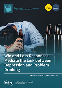
Cover Story (view full-size image): Depression and alcohol misuse, frequently comorbid, are associated with altered reward processing. However, no study has examined whether and how the neural markers of reward processing are shared between depression and alcohol misuse. We studied 43 otherwise healthy drinking adults in a monetary incentive delay task (MIDT) during fMRI. All participants were evaluated with the Alcohol Use Disorders Identification Test (AUDIT) and Beck’s Depression Inventory (BDI-II) to assess the severity of drinking and depression. The findings suggest shared neural correlates interlinking depression and problem drinking during both win and loss processing and provide evidence for co-morbid etiological processes of depressive and alcohol use disorders. View this paper
  • Issues are regarded as officially published after their release is announced to the table of contents alert mailing list.
  • You may sign up for e-mail alerts to receive table of contents of newly released issues.
  • PDF is the official format for papers published in both, html and pdf forms. To view the papers in pdf format, click on the "PDF Full-text" link, and use the free Adobe Reader to open them.
Order results
Result details
Section
Select all
Export citation of selected articles as:
16 pages, 2492 KiB  
Article
Neural Aspects of Prospective Control through Resonating Taus in an Interceptive Timing Task
by F. R. (Ruud) van der Weel, Ingemārs Sokolovskis, Vicente Raja and Audrey L. H. van der Meer
Brain Sci. 2022, 12(12), 1737; https://doi.org/10.3390/brainsci12121737 - 19 Dec 2022
Cited by 5 | Viewed by 2885
Abstract
High-density electroencephalography from visual and motor cortices in addition to kinematic hand and target movement recordings were used to investigate τ-coupling between brain activity patterns and physical movements in an interceptive timing task. Twelve adult participants were presented with a target car moving [...] Read more.
High-density electroencephalography from visual and motor cortices in addition to kinematic hand and target movement recordings were used to investigate τ-coupling between brain activity patterns and physical movements in an interceptive timing task. Twelve adult participants were presented with a target car moving towards a destination at three constant accelerations, and an effector dot was available to intercept the car at the destination with a swift movement of the finger. A τ-coupling analysis was used to investigate involvement of perception and action variables at both the ecological scale of behavior and neural scale. By introducing the concept of resonance, the underlying dynamics of interceptive actions were investigated. A variety of one- and two-scale τ-coupling analyses showed significant differences in distinguishing between slow, medium, and fast target speed when car motion and finger movement, VEP and MRP brain activity, VEP and car motion, and MRP and finger movement were involved. These results suggested that the temporal structure present at the ecological scale is reflected at the neural scale. The results further showed a strong effect of target speed, indicating that τ-coupling constants k and kres increased with higher speeds of the moving target. It was concluded that τ-coupling can be considered a valuable tool when combining different types of variables at both the ecological and neural levels of analysis. Full article
(This article belongs to the Section Sensory and Motor Neuroscience)
Show Figures

Figure 1

39 pages, 1336 KiB  
Review
Cognition and Neuropsychological Changes at Altitude—A Systematic Review of Literature
by Kathrin Bliemsrieder, Elisabeth Margarete Weiss, Rainald Fischer, Hermann Brugger, Barbara Sperner-Unterweger and Katharina Hüfner
Brain Sci. 2022, 12(12), 1736; https://doi.org/10.3390/brainsci12121736 - 19 Dec 2022
Cited by 11 | Viewed by 3455
Abstract
High-altitude (HA) exposure affects cognitive functions, but studies have found inconsistent results. The aim of this systematic review was to evaluate the effects of HA exposure on cognitive functions in healthy subjects. A structural overview of the applied neuropsychological tests was provided with [...] Read more.
High-altitude (HA) exposure affects cognitive functions, but studies have found inconsistent results. The aim of this systematic review was to evaluate the effects of HA exposure on cognitive functions in healthy subjects. A structural overview of the applied neuropsychological tests was provided with a classification of superordinate cognitive domains. A literature search was performed using PubMed up to October 2021 according to PRISMA guidelines. Eligibility criteria included a healthy human cohort exposed to altitude in the field (at minimum 2440 m [8000 ft]) or in a hypoxic environment in a laboratory, and an assessment of cognitive domains. The literature search identified 52 studies (29 of these were field studies; altitude range: 2440 m–8848 m [8000–29,029 ft]). Researchers applied 112 different neuropsychological tests. Attentional capacity, concentration, and executive functions were the most frequently studied. In the laboratory, the ratio of altitude-induced impairments (64.7%) was twice as high compared to results showing no change or improved results (35.3%), but altitudes studied were similar in the chamber compared to field studies. In the field, the opposite results were found (66.4 % no change or improvements, 33.6% impairments). Since better acclimatization can be assumed in the field studies, the findings support the hypothesis that sufficient acclimatization has beneficial effects on cognitive functions at HA. However, it also becomes apparent that research in this area would benefit most if a consensus could be reached on a standardized framework of freely available neurocognitive tests. Full article
(This article belongs to the Section Neuropsychology)
Show Figures

Figure 1

10 pages, 1008 KiB  
Perspective
Telemedicine in the Management of Parkinson’s Disease: Achievements, Challenges, and Future Perspectives
by Esther Cubo and Pedro David Delgado-López
Brain Sci. 2022, 12(12), 1735; https://doi.org/10.3390/brainsci12121735 - 19 Dec 2022
Cited by 9 | Viewed by 2626
Abstract
Background: As the global population grows, there is an increasing demand for neurologic consultation that prompts new ways to reach more patients. Telemedicine can provide an accessible, cost-effective, and high-quality healthcare services. Objectives: In this article, we highlight recent developments, achievements, and challenges [...] Read more.
Background: As the global population grows, there is an increasing demand for neurologic consultation that prompts new ways to reach more patients. Telemedicine can provide an accessible, cost-effective, and high-quality healthcare services. Objectives: In this article, we highlight recent developments, achievements, and challenges regarding outcomes, clinical care, tele-education, teletreatment, teleresearch, and cybersecurity for telemedicine applied to Parkinson´s disease (PD) and other neurological conditions. Results: A growing body of evidence supports the feasibility and effectiveness of telemedicine tools for PD and other movement disorders. Outcome variables regarding satisfaction and efficacy in clinical care and specific issues about education, research, and treatment are reviewed. Additionally, a specific legal framework for teleconsultation has been developed in some centers worldwide. Yet, the implementation of telemedicine is conditioned by the limitations inherent to remote neurological examination, the variable computer usage literacy among patients, and the availability of a reliable internet connection. At present, telemedicine can be considered an additional tool in the clinical management of PD patients. Conclusions: There is an increasing use of remote clinical practice regarding the management of PD and other neurological conditions. Telemedicine is a new and promising tool aimed at special settings and subpopulations. Full article
22 pages, 635 KiB  
Review
Effect of Physical Exercise in Real-World Settings on Executive Function of Typical Children and Adolescents: A Systematic Review
by Peng Shi, Yan Tang, Ziyun Zhang, Xiaosu Feng and Chenyang Li
Brain Sci. 2022, 12(12), 1734; https://doi.org/10.3390/brainsci12121734 - 18 Dec 2022
Cited by 12 | Viewed by 3083
Abstract
Objective: The aim of this paper is to provide a systematic review of research on physical exercise in real-world settings on executive function of typical children and adolescents. Methods: The CNKI, WOS, PubMed, ScienceDirect, and SPORTDiscus databases were searched by computer. Two researchers [...] Read more.
Objective: The aim of this paper is to provide a systematic review of research on physical exercise in real-world settings on executive function of typical children and adolescents. Methods: The CNKI, WOS, PubMed, ScienceDirect, and SPORTDiscus databases were searched by computer. Two researchers independently screened the literature, extracted data, and evaluated the risk of bias in the included literature. Statistical analysis was performed using frequency and percentage and the χ2 test. Results: A total of 49 articles was included. Acute (moderate intensity lasting 30–50 min) and long-term (interventions of moderate intensity of 30–50 min at least 3 times a week for 17 weeks or more) physical exercises in real-world settings have positive intervention effects on executive function. Furthermore, for acute interventions, closed skills are more efficient for inhibitory control, open skills are more efficient for working memory and cognitive flexibility, and open-continuous and closed-sequential skills are the most efficient; long-term interventions with open skills, sequential skills, and open-sequential skills are more effective. Conclusion: Physical exercise in real-world settings has a good promotion effect on typical children and adolescents, and motor skills with open and/or sequential attributes are more helpful in improving executive function. Full article
(This article belongs to the Special Issue Physical Exercise for Brain Health and Longevity)
Show Figures

Figure 1

8 pages, 709 KiB  
Case Report
COVID-19 Still Surprising Us—A Rare Movement Disorder Induced by Infection
by Carmen Adella Sirbu, Diana Popescu, Ion Stefan, Constantin Stefani, Marian Mitrica and Daniela Anghel
Brain Sci. 2022, 12(12), 1733; https://doi.org/10.3390/brainsci12121733 - 17 Dec 2022
Viewed by 2750
Abstract
Background: Many neurological manifestations are part of COVID-19 infections, including movement disorders, but a clinical picture closely resembling stiff-person syndrome has not yet been described. Case presentation: We report a case of a stiff-person-like syndrome in the context of COVID-19 infection. A 79-year-old [...] Read more.
Background: Many neurological manifestations are part of COVID-19 infections, including movement disorders, but a clinical picture closely resembling stiff-person syndrome has not yet been described. Case presentation: We report a case of a stiff-person-like syndrome in the context of COVID-19 infection. A 79-year-old woman, with no prior history of diseases, presented global reversible stiffness associated with SARS-CoV-2 infection. We aim to shed light on several particularities regarding this clinical picture and its evolution in close relationship with the infectious disease progression, with full regression of symptoms and signs once the infectious process ceased. The impairment of speech and motility caused the wrong diagnosis of stroke in the Emergency Room. In addition, we would also like to emphasize the concomitant rhabdomyolysis, closely linked to the grade of muscle rigidity. Conclusions: We would like to raise awareness regarding this clinical setting and its association with SARS-COV-2 infection, to aid in its future recognition and management. To our knowledge, this is the first case of a stiff-person-like syndrome to be described in association with COVID-19 infection. Full article
(This article belongs to the Topic Pathophysiology of Aging and Age-related Diseases)
Show Figures

Figure 1

17 pages, 4025 KiB  
Article
Electroacupuncture Alleviates Neuroinflammation by Inhibiting the HMGB1 Signaling Pathway in Rats with Sepsis-Associated Encephalopathy
by Yueyang Xin, Jinxu Wang, Tiantian Chu, Yaqun Zhou, Cheng Liu and Aijun Xu
Brain Sci. 2022, 12(12), 1732; https://doi.org/10.3390/brainsci12121732 - 17 Dec 2022
Cited by 12 | Viewed by 2557
Abstract
Sepsis-Associated Encephalopathy (SAE) is common in sepsis patients, with high mortality rates. It is believed that neuroinflammation is an important mechanism involved in SAE. High mobility group box 1 protein (HMGB1), as a late pro-inflammatory factor, is significantly increased during sepsis in different [...] Read more.
Sepsis-Associated Encephalopathy (SAE) is common in sepsis patients, with high mortality rates. It is believed that neuroinflammation is an important mechanism involved in SAE. High mobility group box 1 protein (HMGB1), as a late pro-inflammatory factor, is significantly increased during sepsis in different brain regions, including the hippocampus. HMGB1 causes neuroinflammation and cognitive impairment through direct binding to advanced glycation end products (RAGE) and Toll-like receptor 4 (TLR4). Electroacupuncture (EA) at Baihui (GV20) and Zusanli (ST36) is beneficial for neurological diseases and experimental sepsis. Our study used EA to treat SAE induced by lipopolysaccharide (LPS) in male Sprague–Dawley rats. The Y maze test was performed to assess working memory. Immunofluorescence (IF) and Western blotting (WB) were used to determine neuroinflammation and the HMGB1 signaling pathway. Results showed that EA could improve working memory impairment in rats with SAE. EA alleviated neuroinflammation by downregulating the hippocampus’s HMGB1/TLR4 and HMGB1/RAGE signaling, reducing the levels of pro-inflammatory factors, and relieving microglial and astrocyte activation. However, EA did not affect the tight junctions’ expression of the blood–brain barrier (BBB) in the hippocampus. Full article
(This article belongs to the Section Neuropharmacology and Neuropathology)
Show Figures

Figure 1

16 pages, 1727 KiB  
Article
EEG Microstate Features as an Automatic Recognition Model of High-Density Epileptic EEG Using Support Vector Machine
by Li Yang, Jiaxiu He, Ding Liu, Wen Zheng and Zhi Song
Brain Sci. 2022, 12(12), 1731; https://doi.org/10.3390/brainsci12121731 - 17 Dec 2022
Cited by 7 | Viewed by 2271
Abstract
Epilepsy is one of the most serious nervous system diseases; it can be diagnosed accurately by video electroencephalogram. In this study, we analyzed microstate epileptic electroencephalogram (EEG) to aid in the diagnosis and identification of epilepsy. We recruited patients with focal epilepsy and [...] Read more.
Epilepsy is one of the most serious nervous system diseases; it can be diagnosed accurately by video electroencephalogram. In this study, we analyzed microstate epileptic electroencephalogram (EEG) to aid in the diagnosis and identification of epilepsy. We recruited patients with focal epilepsy and healthy participants from the Third Xiangya Hospital and recorded their resting EEG data. In this study, the EEG data were analyzed by microstate analysis, and the support vector machine (SVM) classifier was used for automatic epileptic EEG classification based on features of the EEG microstate series, including microstate parameters (duration, occurrence, and coverage), linear features (median, second quartile, mean, kurtosis, and skewness) and non-linear features (Petrosian fractal dimension, approximate entropy, sample entropy, fuzzy entropy, and Lempel–Ziv complexity). In the gamma sub-band, the microstate parameters as a model were the best for interictal epilepsy recognition, with an accuracy of 87.18%, recall of 70.59%, and an area under the curve of 94.52%. There was a recognition effect of interictal epilepsy through the features extracted from the EEG microstate, which varied within the 4~45 Hz band with an accuracy of 79.55%. Based on the SVM classifier, microstate parameters and EEG features can be effectively used to classify epileptic EEG, and microstate parameters can better classify epileptic EEG compared with EEG features. Full article
(This article belongs to the Section Computational Neuroscience and Neuroinformatics)
Show Figures

Figure 1

14 pages, 1058 KiB  
Article
Prolonged Longitudinal Transcutaneous Auricular Vagus Nerve Stimulation Effect on Striatal Functional Connectivity in Patients with Major Depressive Disorder
by Shuai Zhang, Jia-Kai He, Gang-Liang Zhong, Yu Wang, Ya-Nan Zhao, Lei Wang, Shao-Yuan Li, Xue Xiao, Zheng-Yi Yang, Bin Zhao, Jin-Ling Zhang, Tian-Zi Jiang, Ji-Liang Fang and Pei-Jing Rong
Brain Sci. 2022, 12(12), 1730; https://doi.org/10.3390/brainsci12121730 - 17 Dec 2022
Cited by 3 | Viewed by 2273
Abstract
Background: Transcutaneous auricular vagus nerve stimulation (taVNS) is effective for treating major depressive disorder (MDD). We aimed to explore the modulating effect of prolonged longitudinal taVNS on the striatal subregions’ functional connectivity (FC) in MDD patients. Methods: Sixteen MDD patients were enrolled and [...] Read more.
Background: Transcutaneous auricular vagus nerve stimulation (taVNS) is effective for treating major depressive disorder (MDD). We aimed to explore the modulating effect of prolonged longitudinal taVNS on the striatal subregions’ functional connectivity (FC) in MDD patients. Methods: Sixteen MDD patients were enrolled and treated with taVNS for 8 weeks. Sixteen healthy control subjects (HCs) were recruited without intervention. The resting-state FC (rsFC) based on striatal subregion seed points and the Hamilton Depression Scale (HAMD) were evaluated in the MDD patients and HCs at baseline and after 8 weeks. A two-way ANCOVA test was performed on each rsFC metric to obtain the (group-by-time) interactions. Results: The rsFC values between the left ventral caudate (vCa) and right ventral prefrontal cortex (vPFC), and between the right nucleus accumbens (NAc) and right dorsal medial prefrontal cortex (dmPFC) and ventrolateral prefrontal cortex (vlPFC) are lower in the MDD patients compared to the HCs at baseline, and increase following taVNS; the rsFC values between the left vCa and right, superior occipital gyrus (SOG), and between the left dorsal caudate (dCa) and right cuneus are higher in MDD patients and decrease following taVNS. Conclusions: Prolonged longitudinal taVNS can modulate the striatum rsFC with the prefrontal cortex, occipital cortex, temporal cortex, and intra-striatum, and these changes partly underlie any symptomatic improvements. The results indicate that prolonged longitudinal taVNS may produce beneficial treatment effects by modulating the cortical striatum circuitry in patients with MDD. Full article
(This article belongs to the Section Psychiatric Diseases)
Show Figures

Figure 1

9 pages, 1769 KiB  
Article
Aβ42 as a Biomarker of Alzheimer’s Disease: Is Saliva a Viable Alternative to Cerebrospinal Fluid?
by Silvia Boschi, Fausto Roveta, Alberto Grassini, Andrea Marcinnò, Aurora Cermelli, Fabio Ferrandes, Innocenzo Rainero and Elisa Rubino
Brain Sci. 2022, 12(12), 1729; https://doi.org/10.3390/brainsci12121729 - 17 Dec 2022
Cited by 4 | Viewed by 2415
Abstract
The identification of reliable biomarkers in biological fluids is paramount to optimizing the diagnosis of Alzheimer’s disease (AD). Measurement of Aβ42, t-tau, and p-tau in cerebrospinal fluid (CSF) is the most accepted method to support the diagnosis of AD. However, lumbar puncture represents [...] Read more.
The identification of reliable biomarkers in biological fluids is paramount to optimizing the diagnosis of Alzheimer’s disease (AD). Measurement of Aβ42, t-tau, and p-tau in cerebrospinal fluid (CSF) is the most accepted method to support the diagnosis of AD. However, lumbar puncture represents an invasive investigation, whereas saliva is one of the most accessible body fluids. The aim of our study was to investigate salivary concentrations in AD and evaluate the correlation between salivary and CSF Aβ42 concentrations in AD patients, patients with non-AD dementias, and controls. We recruited 100 subjects: 18 AD patients, 64 patients with non-AD dementias, and 18 controls. The mean saliva Aβ42 concentrations in AD patients were higher than in controls (p < 0.001), and to patients with non-AD dementias (p = 0.001). A significant negative correlation between salivary and CSF Aβ42 concentrations was found in the overall group (r = −0.562, p < 0.001) and in non-AD patients (r = −0.443, p < 0.001). Salivary Aβ42 concentrations positively correlated with CSF t-tau (r = 0.321, p = 0.001) and p-tau (r = 0.297, p = 0.001). Our study showed that in AD patients’ saliva, Aβ42 concentrations are specifically increased, and we found an interesting negative correlation between CSF and salivary Aβ42 concentrations that warrants further investigation. Full article
(This article belongs to the Section Molecular and Cellular Neuroscience)
Show Figures

Figure 1

9 pages, 245 KiB  
Article
Fatigue in Patients with Idiopathic/Isolated REM Sleep Behavior Disorder
by Yajie Zang, Hui Zhang, Yuan Li, Yanning Cai, Jagadish K. Chhetri, Piu Chan and Wei Mao
Brain Sci. 2022, 12(12), 1728; https://doi.org/10.3390/brainsci12121728 - 17 Dec 2022
Cited by 1 | Viewed by 1749
Abstract
Introduction: Fatigue is one of the most common and disabling symptoms of Parkinson’s Disease (PD). The occurrence and clinical features of fatigue in patients with prodromal PD remain largely elusive. This study aimed to investigate the prevalence and clinical characteristics of fatigue in [...] Read more.
Introduction: Fatigue is one of the most common and disabling symptoms of Parkinson’s Disease (PD). The occurrence and clinical features of fatigue in patients with prodromal PD remain largely elusive. This study aimed to investigate the prevalence and clinical characteristics of fatigue in patients with idiopathic/isolated REM sleep behavior disorders (iRBD). Methods: A total of 97 polysomnography-confirmed iRBD patients were enrolled in this study. A comprehensive neurological assessment (including motor and non-motor assessment) was performed. Fatigue was assessed using the Fatigue Severity Scale (FSS). Motor and non-motor characteristics were compared between iRBD patients with and without fatigue. Logistic regression was used to identify the factors associated with fatigue. Results: The prevalence of fatigue was 35.05%. Compared to the non-fatigue patients, patients with fatigue had higher non-motor symptom scale (NMSS) score (p = 0.009), higher Hamilton Depression Rating Scale (HAMD) score (p = 0.002), and a higher prevalence of orthostatic hypotension (p = 0.021). Multivariate regression analysis showed that depression (OR 4.17, 95% CI 1.13–15.49, p = 0.033) and orthostatic hypotension (OR 2.80, 95% CI 1.09–7.18, p = 0.032) were significantly associated with fatigue in iRBD patients. Additionally, both NMSS (rs = 0.310, p = 0.002) and HAMD (rs = 0.385, p < 0.001) scores were mildly correlated with fatigue severity. Conclusion: Our study showed that fatigue is common in patients with iRBD. In addition, depression and orthostatic hypotension were independently associated with fatigue in iRBD patients. Full article
(This article belongs to the Section Neurodegenerative Diseases)
13 pages, 679 KiB  
Review
Review of the Treatments for Central Neuropathic Pain
by Breanna L. Sheldon, Zachary T. Olmsted, Shelby Sabourin, Ehsaun Heydari, Tessa A. Harland and Julie G. Pilitsis
Brain Sci. 2022, 12(12), 1727; https://doi.org/10.3390/brainsci12121727 - 16 Dec 2022
Cited by 11 | Viewed by 2850
Abstract
Central neuropathic pain (CNP) affects millions worldwide, with an estimated prevalence of around 10% globally. Although there are a wide variety of treatment options available, due to the complex and multidimensional nature in which CNP arises and presents symptomatically, many patients still experience [...] Read more.
Central neuropathic pain (CNP) affects millions worldwide, with an estimated prevalence of around 10% globally. Although there are a wide variety of treatment options available, due to the complex and multidimensional nature in which CNP arises and presents symptomatically, many patients still experience painful symptoms. Pharmaceutical, surgical, non-invasive, cognitive and combination treatment options offer a generalized starting point for alleviating symptoms; however, a more customized approach may provide greater benefit. Here, we comment on the current treatment options that exist for CNP and further suggest the need for additional research regarding the use of biomarkers to help individualize treatment options for patients. Full article
Show Figures

Figure 1

15 pages, 2568 KiB  
Article
Local Field Potential-Guided Contact Selection Using Chronically Implanted Sensing Devices for Deep Brain Stimulation in Parkinson’s Disease
by Joshua N. Strelow, Till A. Dembek, Juan C. Baldermann, Pablo Andrade, Hannah Jergas, Veerle Visser-Vandewalle and Michael T. Barbe
Brain Sci. 2022, 12(12), 1726; https://doi.org/10.3390/brainsci12121726 - 16 Dec 2022
Cited by 14 | Viewed by 2673
Abstract
Intra- and perioperatively recorded local field potential (LFP) activity of the nucleus subthalamicus (STN) has been suggested to guide contact selection in patients undergoing deep brain stimulation (DBS) for Parkinson’s disease (PD). Despite the invention of sensing capacities in chronically implanted devices, a [...] Read more.
Intra- and perioperatively recorded local field potential (LFP) activity of the nucleus subthalamicus (STN) has been suggested to guide contact selection in patients undergoing deep brain stimulation (DBS) for Parkinson’s disease (PD). Despite the invention of sensing capacities in chronically implanted devices, a comprehensible algorithm that enables contact selection using such recordings is still lacking. We evaluated a fully automated algorithm that uses the weighted average of bipolar recordings to determine effective monopolar contacts based on elevated activity in the beta band. LFPs from 14 hemispheres in seven PD patients with newly implanted directional DBS leads of the STN were recorded. First, the algorithm determined the stimulation level with the highest beta activity. Based on the prior determined level, the directional contact with the highest beta activity was chosen in the second step. The mean clinical efficacy of the contacts chosen using the algorithm did not statistically differ from the mean clinical efficacy of standard contact selection as performed in clinical routine. All recording sites were projected into MNI standard space to investigate the feasibility of the algorithm with respect to the anatomical boundaries of the STN. We conclude that the proposed algorithm is a first step towards LFP-based contact selection in STN-DBS for PD using chronically implanted devices. Full article
(This article belongs to the Special Issue Advances in Deep Brain Stimulation for Movement Disorders)
Show Figures

Figure 1

13 pages, 1631 KiB  
Review
Sleep and Neuroimmunomodulation for Maintenance of Optimum Brain Function: Role of Noradrenaline
by Rachna Mehta, Rohosen Bhattacharya and Birendra Nath Mallick
Brain Sci. 2022, 12(12), 1725; https://doi.org/10.3390/brainsci12121725 - 16 Dec 2022
Cited by 7 | Viewed by 5024
Abstract
Immune function and sleep are two normal physiological processes to protect the living organism from falling sick. There is hardly any disease in which they remain unaffected, though the quantum of effect may differ. Therefore, we propose the existence of a strong correlation [...] Read more.
Immune function and sleep are two normal physiological processes to protect the living organism from falling sick. There is hardly any disease in which they remain unaffected, though the quantum of effect may differ. Therefore, we propose the existence of a strong correlation between sleep (quality or quantity) and immune response. This may be supported by the fact that sleep loss modulates many of the immunological molecules, which includes interferons; however, not much is known about their mechanism of action. Sleep is divided into rapid eye movement sleep (REMS) and non-REMS. For practical reasons, experimental studies have been conducted mostly by inducing loss of REMS. It has been shown that withdrawal of noradrenaline (NA) is a necessity for generation of REMS. Moreover, NA level increases in the brain upon REMS loss and the elevated NA is responsible for many of the sleep loss-associated symptoms. In this review, we describe how sleep (and its disturbance/loss) modulates the immune system by modulating the NA level in the brain or vice versa to maintain immune functions, physiological homeostasis, and normal healthy living. The increased levels of NA during REMS loss may cause neuroinflammation possibly by glial activation (as NA is a key modulator of microglia). Therefore, maintaining sleep hygiene plays a crucial role for a normal healthy living. Full article
(This article belongs to the Section Behavioral Neuroscience)
Show Figures

Figure 1

14 pages, 2165 KiB  
Article
Brain Correlates of Chinese Handwriting and Their Relation to Reading Development in Children: An fMRI Study
by Jun Zhang, Liying Kang, Junjun Li, Yizhen Li, Hongyan Bi and Yang Yang
Brain Sci. 2022, 12(12), 1724; https://doi.org/10.3390/brainsci12121724 - 16 Dec 2022
Cited by 1 | Viewed by 3565
Abstract
Handwriting plays an important role in written communication, reading, and academic success. However, little is known about the neural correlates of handwriting in children. Using functional magnetic resonance imaging (fMRI) and a copying task, we investigated regional brain activation and functional lateralization associated [...] Read more.
Handwriting plays an important role in written communication, reading, and academic success. However, little is known about the neural correlates of handwriting in children. Using functional magnetic resonance imaging (fMRI) and a copying task, we investigated regional brain activation and functional lateralization associated with Chinese handwriting in children (N = 36, 9–11 years old), as well as their relations to reading skills. We found significant activation of the bilateral frontal motor cortices, somatosensory cortex, intraparietal sulcus (IPS), fusiform gyrus (FuG), and cerebellum during handwriting, suggesting that an adult-like brain activation pattern emerges by middle childhood. Moreover, children showed left-lateralized and bilateral activation of motor regions and right-lateralized activation of the FuG and cerebellum during handwriting, suggesting that functional lateralization of handwriting is not fully established by this age. Finally, the activation of Exner’s area and the lateralization of the IPS and cerebellum during handwriting were correlated with reading skills, possibly representing a neural link between handwriting and reading in children. Collectively, this study reveals the brain correlates of handwriting and their relation to reading development in Chinese children, offering new insight into the development of handwriting and reading skills. Full article
(This article belongs to the Section Neuropsychology)
Show Figures

Figure 1

16 pages, 885 KiB  
Article
Occupational Burnout Is Linked with Inefficient Executive Functioning, Elevated Average Heart Rate, and Decreased Physical Activity in Daily Life - Initial Evidence from Teaching Professionals
by Mia Pihlaja, Pipsa P. A. Tuominen, Jari Peräkylä and Kaisa M. Hartikainen
Brain Sci. 2022, 12(12), 1723; https://doi.org/10.3390/brainsci12121723 - 16 Dec 2022
Cited by 10 | Viewed by 5863
Abstract
Burnout is becoming a global pandemic jeopardizing brain health, with a huge impact on quality of life, available workforce, and the economy. Knowledge of the impact of burnout on cognition, physiology, and physical activity (PA) in daily life allows for an improved understanding [...] Read more.
Burnout is becoming a global pandemic jeopardizing brain health, with a huge impact on quality of life, available workforce, and the economy. Knowledge of the impact of burnout on cognition, physiology, and physical activity (PA) in daily life allows for an improved understanding of the health consequences and everyday ramifications of burnout. Twenty-eight volunteers participated in a three-day recording of daily physiology and PA, including heart rate (HR) and daily steps, with a wearable device. They filled in questionnaires screening for burnout (BBI-15), depression (BDI), and executive functions (EFs) in daily life (BRIEF-A). The subjects with burnout had more challenges in EFs, higher average HRs and lower numbers of steps in daily life than those without it. The BBI-15 scores correlated positively with the BDI scores and BRIEF-A indices and negatively with the awake HR variability (HRV) and daily steps. The metacognition index correlated negatively with the HRV. In conclusion, burnout is linked with compromised EFs along with alterations in cardiac physiology and PA in daily life. Such alterations may be easily detected with wearable devices, opening possibilities for novel biomarkers of burnout and other neuropsychiatric disorders. We suggest that physical activity and heart and brain health are intimately intertwined and that burnout interacts with each of them bidirectionally. Full article
(This article belongs to the Special Issue The Biomarkers in Neuropsychiatric Disorders)
Show Figures

Figure 1

14 pages, 1484 KiB  
Article
An fNIRS Study of Applicability of the Unity–Diversity Model of Executive Functions in Preschoolers
by Sha Xie, Chaohui Gong, Jiahao Lu, Hao Zhang, Dandan Wu, Xinli Chi, Hui Li and Chunqi Chang
Brain Sci. 2022, 12(12), 1722; https://doi.org/10.3390/brainsci12121722 - 16 Dec 2022
Cited by 1 | Viewed by 2225
Abstract
Executive function (EF) includes a set of higher-order abilities that control one’s actions and thoughts consciously and has a protracted developmental trajectory that parallels the maturation of the frontal lobes, which develop speedily over the preschool period. To fully understand the development of [...] Read more.
Executive function (EF) includes a set of higher-order abilities that control one’s actions and thoughts consciously and has a protracted developmental trajectory that parallels the maturation of the frontal lobes, which develop speedily over the preschool period. To fully understand the development of EF in preschoolers, this study examined the relationship among the three domains of executive function (cognitive shifting, inhibitory control, and working memory) to test the applicability of the unity–diversity model in preschoolers using both behavioral and fNIRS approaches. Altogether, 58 Chinese preschoolers (34 boys, 24 girls, Mage = 5.86 years, SD = 0.53, age range = 4.83–6.67 years) were administered the Dimensional Card Change Sort (DCCS), go/no-go, and missing scan task. Their brain activations in the prefrontal cortex during the tasks were examined using fNIRS. First, the behavioral results indicated that the missing scan task scores (working memory) correlated with the DCCS (cognitive shifting) and go/no-go tasks (inhibitory control). However, the latter two did not correlate with each other. Second, the fNIRS results demonstrated that the prefrontal activations during the working memory task correlated with those in the same regions during the cognitive shifting and inhibitory control tasks. However, the latter two still did not correlate. The behavioral and neuroimaging evidence jointly indicates that the unity–diversity model of EF does apply to Chinese preschoolers. Full article
(This article belongs to the Section Developmental Neuroscience)
Show Figures

Figure 1

10 pages, 279 KiB  
Case Report
Neuropsychiatric Symptoms after Liver Transplantation in a 65-Year-Old Male Patient
by Cesar Bugallo-Carrera, David Facal, Cristina Domínguez-Lenogue, Vanessa Álvarez-Vidal, Manuel Gandoy-Crego and José Caamaño-Ponte
Brain Sci. 2022, 12(12), 1721; https://doi.org/10.3390/brainsci12121721 - 16 Dec 2022
Cited by 3 | Viewed by 2518
Abstract
The development of immunosuppressants has been key for the advancement of solid organ transplant surgery. Specifically, cyclosporine, tacrolimus, or everolimus have significantly increased the survival rate of patients by reducing the risk of a rejection of the transplanted organ and limiting graft-versus-host disease. [...] Read more.
The development of immunosuppressants has been key for the advancement of solid organ transplant surgery. Specifically, cyclosporine, tacrolimus, or everolimus have significantly increased the survival rate of patients by reducing the risk of a rejection of the transplanted organ and limiting graft-versus-host disease. We report the case of a 65-year-old man who, after undergoing a liver transplantation and receiving an immunosuppressive treatment with cyclosporine and everolimus, presented severe obsessive, psychotic, and behavioral symptoms over the past three years, and describe the pharmacological and non-pharmacological interventions implemented against these symptoms. In this case, the immunosuppressants used have been cyclosporine and, preferably, everolimus. On the other hand, potential adverse reactions to the treatment have been observed, including neuropsychiatric symptoms such as tremor, anxiety, dysthymia, psychosis, and behavioral disorders, which make it necessary to use corrective psychoactive drugs such as benzodiazepines, antidepressants, and antipsychotics, combined with non-pharmacological interventions. A transversal approach, from the medical and psychosocial disciplines, facilitates success in managing neuropsychiatric symptoms after soft organ transplants. Full article
13 pages, 2403 KiB  
Article
Dexmedetomidine Preserves Activity of Neurons in Primary Somatosensory Cortex Compared to Propofol and Ketamine
by Mu-Chao Xia, Juan Guo, Yan Ding, Zi-Qi Shi, Fang Du, Kai Wang, Chang-Hong Miao and Chao Liang
Brain Sci. 2022, 12(12), 1720; https://doi.org/10.3390/brainsci12121720 - 15 Dec 2022
Viewed by 1947
Abstract
General anesthesia has been shown to induce significant changes in the functional connectivity of the cerebral cortex. However, traditional methods such as electroencephalography (EEG) or functional magnetic resonance imaging (fMRI) lack the spatial resolution to study the effects of general anesthesia on individual [...] Read more.
General anesthesia has been shown to induce significant changes in the functional connectivity of the cerebral cortex. However, traditional methods such as electroencephalography (EEG) or functional magnetic resonance imaging (fMRI) lack the spatial resolution to study the effects of general anesthesia on individual cortical neurons. This study aimed to use high-resolution two-photon imaging, which can provide single-neuron resolution, to investigate the characteristics of consciousness under general anesthesia. We used C57BL/6J and Thy1-GCamp6s mice and found that at similar levels of sedation, as measured by EEG, dexmedetomidine did not significantly inhibit the spontaneous activity of neuronal somata in the S1 cortex, but preserved the frequency of calcium events in neuronal spines. In contrast, propofol and ketamine dramatically inhibited the spontaneous activity of both neuronal somata and spines. The S1 cortex still responded to whisker stimulation under dexmedetomidine anesthesia, but not under propofol or ketamine anesthesia. Our results suggest that dexmedetomidine anesthesia has unique neuronal properties associated with its ability to facilitate easy awakening in the clinic. These findings provide insights into the development of more effective strategies for monitoring consciousness during general anesthesia. Full article
(This article belongs to the Section Neuropharmacology and Neuropathology)
Show Figures

Figure 1

16 pages, 5757 KiB  
Article
Age Effects in Emotional Memory and Associated Eye Movements
by Daphne Stam, Laura Colman, Kristof Vansteelandt, Mathieu Vandenbulcke and Jan Van den Stock
Brain Sci. 2022, 12(12), 1719; https://doi.org/10.3390/brainsci12121719 - 15 Dec 2022
Cited by 2 | Viewed by 1911
Abstract
Mnemonic enhanced memory has been observed for negative events. Here, we investigate its association with spatiotemporal attention, consolidation, and age. An ingenious method to study visual attention for emotional stimuli is eye tracking. Twenty young adults and twenty-one older adults encoded stimuli depicting [...] Read more.
Mnemonic enhanced memory has been observed for negative events. Here, we investigate its association with spatiotemporal attention, consolidation, and age. An ingenious method to study visual attention for emotional stimuli is eye tracking. Twenty young adults and twenty-one older adults encoded stimuli depicting neutral faces, angry faces, and houses while eye movements were recorded. The encoding phase was followed by an immediate and delayed (48 h) recognition assessment. Linear mixed model analyses of recognition performance with group, emotion, and their interaction as fixed effects revealed increased performance for angry compared to neutral faces in the young adults group only. Furthermore, young adults showed enhanced memory for angry faces compared to older adults. This effect was associated with a shorter fixation duration for angry faces compared to neutral faces in the older adults group. Furthermore, the results revealed that total fixation duration was a strong predictor for face memory performance. Full article
(This article belongs to the Special Issue Social Cognition across Healthy and Neuropsychiatric Conditions)
Show Figures

Figure 1

14 pages, 7945 KiB  
Article
Role for Astrocytes in mGluR-Dependent LTD in the Neocortex and Hippocampus
by Ulyana Lalo and Yuriy Pankratov
Brain Sci. 2022, 12(12), 1718; https://doi.org/10.3390/brainsci12121718 - 15 Dec 2022
Cited by 4 | Viewed by 2220
Abstract
Astroglia are an active element of brain plasticity, capable to release small molecule gliotransmitters by various mechanisms and regulate synaptic strength. While importance of glia-neuron communications for long-term potentiation has been rather widely reported, research into role for astrocytes in long-depression (LTD) is [...] Read more.
Astroglia are an active element of brain plasticity, capable to release small molecule gliotransmitters by various mechanisms and regulate synaptic strength. While importance of glia-neuron communications for long-term potentiation has been rather widely reported, research into role for astrocytes in long-depression (LTD) is just gaining momentum. Here, we explored the role for astrocytes in the prominent form of synaptic plasticity—mGluR-dependent LTD. We found out the substantial contribution of the Group I receptors, especially mGluR1 subtype, into Ca2+-signaling in hippocampal and neocortical astrocytes, which can be activated during synaptic stimulation used for LTD induction. Our data demonstrate that mGluR receptors can activate SNARE-dependent release of ATP from astrocytes which in turn can directly activate postsynaptic P2X receptors in the hippocampal and neocortical neurons. The latter mechanism has recently been shown to cause the synaptic depression via triggering the internalisation of AMPA receptors. Using mouse model of impaired glial exocytosis (dnSNARE mice), we demonstrated that mGluR-activated release of ATP from astrocytes is essential for regulation of mGluR-dependent LTD in CA3-CA1 and layer 2/3 synapses. Our data also suggest that astrocyte-related pathway relies mainly on mGluR1 receptors and act synergistically with neuronal mechanisms dependent mainly on mGluR5. Full article
(This article belongs to the Special Issue How Do Astrocytes Shape Synaptic Transmission and Plasticity?)
Show Figures

Figure 1

11 pages, 648 KiB  
Article
Validation of the Longitudinal Interval Follow-Up Evaluation for the Long-Term Measurement of Mood Symptoms in Bipolar Disorder
by Richard J. Porter, Will Moot, Maree L. Inder, Marie T. Crowe, Katie M. Douglas, Janet D. Carter and Christopher Frampton
Brain Sci. 2022, 12(12), 1717; https://doi.org/10.3390/brainsci12121717 - 15 Dec 2022
Cited by 5 | Viewed by 1851
Abstract
The long-term burden of symptoms is an important outcome in bipolar disorder (BD). A method which has minimal burden of assessment uses a retrospective interview, the Longitudinal Interval Follow-up Examination (LIFE), although this may be subject to problems with recall. This study examines [...] Read more.
The long-term burden of symptoms is an important outcome in bipolar disorder (BD). A method which has minimal burden of assessment uses a retrospective interview, the Longitudinal Interval Follow-up Examination (LIFE), although this may be subject to problems with recall. This study examines the relationship between the retrospective LIFE scale and concurrently-rated mood rating scales in two clinical trials of 18 months of psychotherapy for patients with BD. The Montgomery-Asberg Depression Rating Scale (MADRS) and Young Mania Rating Scale (YMRS) were administered every eight to nine weeks and the LIFE was carried out every 6 months. Correlations between scores on mood rating scales and at equivalent times on the LIFE were examined, as well as of potential clinical moderators. There were significant correlations between LIFE depression ratings and concurrent MADRS score (r = 0.57) and between LIFE mania ratings and YMRS score (r = 0.40). In determining “mild depression” on the MADRS, a receiver operating characteristics (ROC) analysis showed an AUC of 0.78 for LIFE scores. Correlations, particularly for depression scores, were high even when the LIFE rating was several months before the interview, suggesting that the LIFE has validity in examining the burden of mood symptoms over time, with relatively little burden of assessment. Future research should examine the relationship between symptom burden and quality of life measured in this way. Full article
(This article belongs to the Special Issue Bipolar Disorders: Progressing from Bench to Bedside)
Show Figures

Figure 1

13 pages, 920 KiB  
Article
Effects of Faces and Voices on the Encoding of Biographic Information
by Sarah Fransson, Sherryse Corrow, Shanna Yeung, Heidi Schaefer and Jason J. S. Barton
Brain Sci. 2022, 12(12), 1716; https://doi.org/10.3390/brainsci12121716 - 15 Dec 2022
Cited by 2 | Viewed by 1379
Abstract
There are multiple forms of knowledge about people. Whether diverse person-related data interact is of interest regarding the more general issue of integration of multi-source information about the world. Our goal was to examine whether perception of a person’s face or voice enhanced [...] Read more.
There are multiple forms of knowledge about people. Whether diverse person-related data interact is of interest regarding the more general issue of integration of multi-source information about the world. Our goal was to examine whether perception of a person’s face or voice enhanced the encoding of their biographic data. We performed three experiments. In the first experiment, subjects learned the biographic data of a character with or without a video clip of their face. In the second experiment, they learned the character’s data with an audio clip of either a generic narrator’s voice or the character’s voice relating the same biographic information. In the third experiment, an audiovisual clip of both the face and voice of either a generic narrator or the character accompanied the learning of biographic data. After learning, a test phase presented biographic data alone, and subjects were tested first for familiarity and second for matching of biographic data to the name. The results showed equivalent learning of biographic data across all three experiments, and none showed evidence that a character’s face or voice enhanced the learning of biographic information. We conclude that the simultaneous processing of perceptual representations of people may not modulate the encoding of biographic data. Full article
(This article belongs to the Special Issue People Recognition through Face, Voice, Name and Their Interactions)
Show Figures

Figure 1

7 pages, 3328 KiB  
Case Report
Hardware-Related Skin Erosion in Deep Brain Stimulation for Parkinson’s Disease: How Far Can We Go? An Illustrative Case Report
by Pietro Zeppa, Marco Fraccalvieri, Chiara Fronda, Enrico Lo Bue, Laura Rizzi, Virginia Caliendo and Michele Maria Lanotte
Brain Sci. 2022, 12(12), 1715; https://doi.org/10.3390/brainsci12121715 - 15 Dec 2022
Cited by 4 | Viewed by 2826
Abstract
Skin erosion is a hardware-related complication commonly described after deep brain stimulation (DBS). Hardware exposure is often associated with the development of infection that can lead to implant removal. However, in selected cases, it is possible to manage skin erosion without having to [...] Read more.
Skin erosion is a hardware-related complication commonly described after deep brain stimulation (DBS). Hardware exposure is often associated with the development of infection that can lead to implant removal. However, in selected cases, it is possible to manage skin erosion without having to remove the hardware. This article presents the case of a patient with recurrent skin erosions above the IPG, who underwent multiple surgeries. Given the failure of less invasive approaches, a more complex surgery with the employment of a pedunculated flap of pectoralis major in order to cover the IPG was attempted. Nevertheless, the IPG removal was finally unavoidable, resulting in a rapid decline in clinical performance. This illustrative case suggests how, in patients with sustained stimulation who benefit from a good degree of autonomy, it may be useful to use invasive surgical techniques to resolve skin erosions and save the DBS system. In spite of everything, sometimes complete or partial removal of the implant still becomes unavoidable, but this can lead to a severe worsening of PD symptoms. Definitive removal of the system should therefore be considered only in cases of frank infection or after failure of all other approaches. Full article
(This article belongs to the Section Neurosurgery and Neuroanatomy)
Show Figures

Figure 1

25 pages, 19360 KiB  
Article
Individual- and Connectivity-Based Real-Time fMRI Neurofeedback to Modulate Emotion-Related Brain Responses in Patients with Depression: A Pilot Study
by Maximilian Maywald, Marco Paolini, Boris Stephan Rauchmann, Christian Gerz, Jan Lars Heppe, Annika Wolf, Linda Lerchenberger, Igor Tominschek, Sophia Stöcklein, Paul Reidler, Nadja Tschentscher, Birgit Ertl-Wagner, Oliver Pogarell, Daniel Keeser and Susanne Karch
Brain Sci. 2022, 12(12), 1714; https://doi.org/10.3390/brainsci12121714 - 14 Dec 2022
Cited by 3 | Viewed by 2868
Abstract
Introduction: Individual real-time functional magnetic resonance imaging neurofeedback (rtfMRI NF) might be a promising adjuvant in treating depressive symptoms. Further studies showed functional variations and connectivity-related changes in the dorsolateral prefrontal cortex (dlPFC) and the insular cortex. Objectives: The aim of this pilot [...] Read more.
Introduction: Individual real-time functional magnetic resonance imaging neurofeedback (rtfMRI NF) might be a promising adjuvant in treating depressive symptoms. Further studies showed functional variations and connectivity-related changes in the dorsolateral prefrontal cortex (dlPFC) and the insular cortex. Objectives: The aim of this pilot study was to investigate whether individualized connectivity-based rtfMRI NF training can improve symptoms in depressed patients as an adjunct to a psychotherapeutic programme. The novel strategy chosen for this was to increase connectivity between individualized regions of interest, namely the insula and the dlPFC. Methods: Sixteen patients diagnosed with major depressive disorder (MDD, ICD-10) and 19 matched healthy controls (HC) participated in a rtfMRI NF training consisting of two sessions with three runs each, within an interval of one week. RtfMRI NF was applied during a sequence of negative emotional pictures to modulate the connectivity between the dlPFC and the insula. The MDD REAL group was divided into a Responder and a Non-Responder group. Patients with an increased connectivity during the second NF session or during both the first and the second NF session were identified as “MDD REAL Responder” (N = 6). Patients that did not show any increase in connectivity and/or a decreased connectivity were identified as “MDD REAL Non-Responder” (N = 7). Results: Before the rtfMRI sessions, patients with MDD showed higher neural activation levels in ventromedial PFC and the insula than HC; by contrast, HC revealed increased hemodynamic activity in visual processing areas (primary visual cortex and visual association cortex) compared to patients with MDD. The comparison of hemodynamic responses during the first compared to during the last NF session demonstrated significantly increased BOLD-activation in the medial orbitofrontal cortex (mOFC) in patients and HC, and additionally in the lateral OFC in patients with MDD. These findings were particularly due to the MDD Responder group, as the MDD Non-Responder group showed no increase in this region during the last NF run. There was a decrease of neural activation in emotional processing brain regions in both groups in the last NF run compared to the first: HC showed differences in the insula, parahippocampal gyrus, basal ganglia, and cingulate gyrus. Patients with MDD demonstrated deceased responses in the parahippocampal gyrus. There was no significant reduction of BDI scores after NF training in patients. Conclusions: Increased neural activation in the insula and vmPFC in MDD suggests an increased emotional reaction in patients with MDD. The activation of the mOFC could be associated with improved control-strategies and association-learning processes. The increased lOFC activation could indicate a stronger sensitivity to failed NF attempts in MDD. A stronger involvement of visual processing areas in HC may indicate better adaptation to negative emotional stimuli after repeated presentation. Overall, the rtfMRI NF had an impact on neurobiological mechanisms, but not on psychometric measures in patients with MDD. Full article
(This article belongs to the Section Behavioral Neuroscience)
Show Figures

Figure 1

23 pages, 2039 KiB  
Systematic Review
Cranial Ultrasound Abnormalities in Small for Gestational Age or Growth-Restricted Infants Born over 32 Weeks Gestation: A Systematic Review and Meta-Analysis
by Charlene Roufaeil, Abdul Razak and Atul Malhotra
Brain Sci. 2022, 12(12), 1713; https://doi.org/10.3390/brainsci12121713 - 14 Dec 2022
Cited by 5 | Viewed by 2405
Abstract
Aim: To perform a systematic review and meta-analysis of existing literature to evaluate the incidence of cranial ultrasound abnormalities (CUAs) amongst moderate to late preterm (MLPT) and term infants, affected by fetal growth restriction (FGR) or those classified as small for gestational age [...] Read more.
Aim: To perform a systematic review and meta-analysis of existing literature to evaluate the incidence of cranial ultrasound abnormalities (CUAs) amongst moderate to late preterm (MLPT) and term infants, affected by fetal growth restriction (FGR) or those classified as small for gestational age (SGA). Methods: A systematic review methodology was performed, and Preferred Reporting Items for Systematic Review and Meta-Analyses (PRISMA) statement was utilised. Descriptive and observational studies reporting cranial ultrasound outcomes on FGR/SGA MLPT and term infants were included. Primary outcomes reported was incidence of CUAs in MLPT and term infants affected by FGR or SGA, with secondary outcomes including brain structure development and growth, and cerebral artery Dopplers. A random-effects model meta-analysis was performed. Risk of Bias was assessed using the Newcastle-Ottawa scale for case–control and cohort studies, and Joanna Briggs Institute Critical Appraisal Checklist for studies reporting prevalence data. GRADE was used to assess for certainty of evidence. Results: Out of a total of 2085 studies identified through the search, seventeen were deemed to be relevant and included. Nine studies assessed CUAs in MLPT FGR/SGA infants, seven studies assessed CUAs in late preterm and term FGR/SGA infants, and one study assessed CUAs in both MLPT and term FGR/SGA infants. The incidence of CUAs in MLPT, and late preterm to term FGR/SGA infants ranged from 0.4 to 33% and 0 to 70%, respectively. A meta-analysis of 7 studies involving 168,136 infants showed an increased risk of any CUA in FGR infants compared to appropriate for gestational age (AGA) infants (RR 1.96, [95% CI 1.26–3.04], I2 = 68%). The certainty of evidence was very low due to non-randomised studies, methodological limitations, and heterogeneity. Another meta-analysis looking at 4 studies with 167,060 infants showed an increased risk of intraventricular haemorrhage in FGR/SGA infants compared to AGA infants (RR 2.40, [95% CI 2.03–2.84], I2 = 0%). This was also of low certainty. Conclusions: The incidence of CUAs in MLPT and term growth-restricted infants varied widely between studies. Findings from the meta-analyses suggest the risk of CUAs and IVH may indeed be increased in these FGR/SGA infants when compared with infants not affected by FGR, however the evidence is of low to very low certainty. Further specific cohort studies are needed to fully evaluate the benefits and prognostic value of cranial ultrasonography to ascertain the need for, and timing of a cranial ultrasound screening protocol in this infant population, along with follow-up studies to ascertain the significance of CUAs identified. Full article
(This article belongs to the Section Developmental Neuroscience)
Show Figures

Figure 1

18 pages, 3362 KiB  
Review
P3b Amplitude and Latency in Tic Disorders: A Meta-Analysis
by Yue Yang, Hua Yang, Yao Deng, Tao Yu and Rong Luo
Brain Sci. 2022, 12(12), 1712; https://doi.org/10.3390/brainsci12121712 - 14 Dec 2022
Cited by 1 | Viewed by 1853
Abstract
P3b is an event-related potential (ERP) that may be abnormal in patients with tic disorders (TD), but evidence has been inconsistent. Given the possible association between P3b and TD and the need for biomarkers for TD, the primary objective of this meta-analysis was [...] Read more.
P3b is an event-related potential (ERP) that may be abnormal in patients with tic disorders (TD), but evidence has been inconsistent. Given the possible association between P3b and TD and the need for biomarkers for TD, the primary objective of this meta-analysis was to characterize P3b in patients with TD in comparison to healthy controls (HCs). Methods: By searching PubMed, Embase, Web of Science, SCOPUS, Medline, and Google Scholar, we identified studies that compared P3b between TD patients and HCs. The amplitude and latency of P3b were then analyzed. Subgroup analyses were conducted to investigate the influence of different experimental factors on P3b indices. Result: Overall, 19 articles involving 388 cases and 414 controls were evaluated. There were no significant abnormalities in P3b amplitude and latency in TD patients. The P3b amplitude of the TD patients was significantly decreased during the oddball task, and the P3b amplitude of the adult TD patients was also significantly decreased. Conclusion: TD patients may have an abnormal P3b compared to HCs under specified conditions. Full article
(This article belongs to the Special Issue New Insights in Neurobiology and Genetics of Tourette Syndrome)
Show Figures

Figure 1

7 pages, 1269 KiB  
Brief Report
Stress Reduction Effects during Block-Tapping Task of Jaw in Healthy Participants: Functional Near-Infrared Spectroscopy (fNIRS) Measurements of Prefrontal Cortex Activity
by Takahiro Kishimoto, Takaharu Goto and Tetsuo Ichikawa
Brain Sci. 2022, 12(12), 1711; https://doi.org/10.3390/brainsci12121711 - 14 Dec 2022
Viewed by 1450
Abstract
The purpose of this study was to explore the influence of simple jaw opening and closing task of chewing movements on the activities of the prefrontal cortex (PFC) from the viewpoint of stress reduction. We measured cerebral blood flow (CBF) in the PFC [...] Read more.
The purpose of this study was to explore the influence of simple jaw opening and closing task of chewing movements on the activities of the prefrontal cortex (PFC) from the viewpoint of stress reduction. We measured cerebral blood flow (CBF) in the PFC during a block-tapping task of the jaw in healthy participants. Eleven young healthy individuals with normal dentition (7 males and 4 females, mean age 28.0 ± 3.7 years) volunteered for this study. CBF was measured using a wearable, functional near-infrared spectroscopy device. Measurements were taken using the central incisors and first molars at interocclusal distances of 5 and 10 mm. The participants were asked to bite a hard/soft block. CBF in all conditions showed limited variability or decreasing trend compared to resting state before the task. The main effect was observed for interocclusal distance (p = 0.008), and there were no significant differences for measurement area of the PFC, tooth type, and material type. An interaction was found between tooth type and material type. In conclusion, these results suggest that simple and rhythmical chewing motion has an effect of reducing CBF in the PFC and resting the PFC, which is an especially notable aspect of periodontal sensory information in the molar. Full article
(This article belongs to the Section Systems Neuroscience)
Show Figures

Figure 1

6 pages, 1224 KiB  
Case Report
Ictal fMRI: Mapping Seizure Topography with Rhythmic BOLD Oscillations
by David Fischer, Otto Rapalino, Matteo Fecchio and Brian L. Edlow
Brain Sci. 2022, 12(12), 1710; https://doi.org/10.3390/brainsci12121710 - 13 Dec 2022
Cited by 2 | Viewed by 2757
Abstract
Functional magnetic resonance imaging (fMRI) has shown elevations in the blood-oxygen-level-dependent (BOLD) signal associated with, but insensitive for, seizure. Rather than evaluating absolute BOLD signal elevations, assessing rhythmic oscillations in the BOLD signal with fMRI may improve the accuracy of seizure mapping. We [...] Read more.
Functional magnetic resonance imaging (fMRI) has shown elevations in the blood-oxygen-level-dependent (BOLD) signal associated with, but insensitive for, seizure. Rather than evaluating absolute BOLD signal elevations, assessing rhythmic oscillations in the BOLD signal with fMRI may improve the accuracy of seizure mapping. We report a case of a patient with non-convulsive, right hemispheric seizures who underwent fMRI. Unbiased processing methods revealed a map of rhythmically oscillating BOLD signal over the cortical region affected by seizure, and synchronous BOLD signal in the contralateral cerebellum. High-resolution fMRI may help identify the spatial topography of seizure and provide insights into seizure physiology. Full article
(This article belongs to the Special Issue Advancements in Neuroimaging Approaches for Brain Disorders)
Show Figures

Graphical abstract

3 pages, 491 KiB  
Correction
Correction: La Torre et al. Role of Vitamin E and the Orexin System in Neuroprotection. Brain Sci. 2021, 11, 1098
by Maria Ester La Torre, Ines Villano, Marcellino Monda, Antonietta Messina, Giuseppe Cibelli, Anna Valenzano, Daniela Pisanelli, Maria Antonietta Panaro, Nicola Tartaglia, Antonio Ambrosi, Marco Carotenuto, Vincenzo Monda, Giovanni Messina and Chiara Porro
Brain Sci. 2022, 12(12), 1709; https://doi.org/10.3390/brainsci12121709 - 13 Dec 2022
Viewed by 1066
Abstract
In the original article [...] Full article
(This article belongs to the Special Issue Neuroprotection and Oxidative Distress)
Show Figures

Figure 1

8 pages, 259 KiB  
Article
Serum Inflammatory Profile in Hereditary Transthyretin Amyloidosis: Mechanisms and Possible Therapeutic Implications
by Marco Luigetti, Angela Romano, Valeria Guglielmino, Maria Ausilia Sciarrone, Francesca Vitali, Carmine Carbone, Geny Piro, Andrea Sabino, Nicola De Stefano, Domenico Plantone and Guido Primiano
Brain Sci. 2022, 12(12), 1708; https://doi.org/10.3390/brainsci12121708 - 12 Dec 2022
Cited by 3 | Viewed by 1690
Abstract
Hereditary transthyretin (ATTRv) amyloidosis is a severe, progressive, and heterogeneous multisystemic condition due to mutations in the TTR gene. Although multiple aspects of its molecular pathophysiological mechanisms have been elucidated over the years, it is possible to hypothesize different pathogenetic pathways. Indeed, we [...] Read more.
Hereditary transthyretin (ATTRv) amyloidosis is a severe, progressive, and heterogeneous multisystemic condition due to mutations in the TTR gene. Although multiple aspects of its molecular pathophysiological mechanisms have been elucidated over the years, it is possible to hypothesize different pathogenetic pathways. Indeed, we extensively investigated the serum levels of several molecules involved in the immune response, in a cohort of ATTRv patients and healthy controls (HCs). Sixteen ATTRv patients and twenty-five HCs were included in the study. IFN-alpha levels were higher in ATTRv patients than in HCs, as well as IFN-gamma levels. By contrast, IL-7 levels were lower in ATTRv patients than in HCs. No significant difference between groups was found regarding IL-1Ra, IL-6, IL-2, IL-4, and IL-33 levels. Correlation analysis did not reveal any significant correlation between IFN-α, IFN-γ, IL-7, and demographic and clinical data. Larger and longitudinal studies using ultrasensitive methods to perform a full cytokine profiling are needed to better elucidate the role of inflammation in ATTRv pathogenesis and to test the reliability of these molecules as possible biomarkers in monitoring patients’ progression. Full article
(This article belongs to the Special Issue Immunological Implications in Neuromuscular Disorders)
Previous Issue
Next Issue
Back to TopTop