Next Issue
Volume 15, July
Previous Issue
Volume 15, May


Brain Sci., Volume 15, Issue 6 (June 2025) – 127 articles

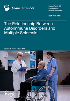
Cover Story (view full-size image): Multiple sclerosis (MS) and autoimmune diseases (AIDs) share immunological underpinnings, leading to frequent co-occurrence. This study investigated the prevalence of AIDs among Polish patients with MS (PwMSs) and its potential effects on disease characteristics. The aims were to compare clinical and demographic characteristics between PwMSs with and without coexisting AIDs.  Methods: A retrospective analysis was conducted on data from 580 PwMSs who were treated at the Department of Neurology, University Clinical Center in Katowice, Poland, between February 2018 and August 2023. Variables analyzed included age, sex, MS type, disease duration, treatment, Expanded Disability Status Scale (EDSS) scores, thyroid-stimulating hormone (TSH), and vitamin D3 serum concentrations. View this paper
  • Issues are regarded as officially published after their release is announced to the table of contents alert mailing list.
  • You may sign up for e-mail alerts to receive table of contents of newly released issues.
  • PDF is the official format for papers published in both, html and pdf forms. To view the papers in pdf format, click on the "PDF Full-text" link, and use the free Adobe Reader to open them.
Order results
Result details
Section
Select all
Export citation of selected articles as:
12 pages, 923 KiB  
Article
Cortical and Striatal Functional Connectivity in Juvenile-Onset Huntington’s Disease
by Amy Barry and Peg C. Nopoulos
Brain Sci. 2025, 15(6), 663; https://doi.org/10.3390/brainsci15060663 - 19 Jun 2025
Viewed by 411
Abstract
Background: Huntington’s disease (HD) is a neurodegenerative disorder caused by a CAG repeat expansion in the HTT gene, with a rare juvenile-onset form (JoHD) marked by early, rigid motor symptoms. This study examined cortical and subcortical resting-state connectivity in JoHD, hypothesizing preserved cortical [...] Read more.
Background: Huntington’s disease (HD) is a neurodegenerative disorder caused by a CAG repeat expansion in the HTT gene, with a rare juvenile-onset form (JoHD) marked by early, rigid motor symptoms. This study examined cortical and subcortical resting-state connectivity in JoHD, hypothesizing preserved cortical networks but altered striatal connectivity, in line with early subcortical atrophy despite relatively spared cortical volume. Methods: Participants included children and young adults with clinician-confirmed Juvenile-Onset Huntington’s Disease (JoHD; n = 19) and gene-non-expanded (GNE) controls (n = 64), both drawn from longitudinal studies at the University of Iowa. Resting-state functional MRI scans were analyzed to assess canonical cortical network and striatal connectivity, and linear mixed-effects models tested group differences and associations with motor, cognitive, and clinical outcomes. Results: JoHD participants showed reduced connectivity within the left somatomotor network and striatal circuits, despite largely typical cortical network connectivity. Striatal connectivity was associated with disease burden and cognitive ability, while left somatomotor connectivity was unrelated to clinical outcomes. Conclusions: These findings support the hypothesis of antagonistic pleiotropy in JoHD, where early neural advantages—such as relatively preserved or possibly enhanced cortical function—may contribute to later striatal vulnerability and degeneration. The observed left-lateralized somatomotor hypoconnectivity aligns with prior volumetric and gene expression research, highlighting the role of excitotoxic glutamatergic input and the selective vulnerability of high-functioning circuits in disease progression. Full article
(This article belongs to the Section Neurodegenerative Diseases)
Show Figures

Figure 1

21 pages, 1327 KiB  
Article
Give or Take: Semantic Priming from Sentences to Two-Digit Operations
by Miguel Ayala-Cuesta, Sofía Castro and Pedro Macizo
Brain Sci. 2025, 15(6), 662; https://doi.org/10.3390/brainsci15060662 - 19 Jun 2025
Viewed by 820
Abstract
Objectives: The objective of this study was to assess the potential existence of shared semantics between linguistic (e.g., reading a sentence) and numerical information (e.g., performing an arithmetic operation). Methods: To evaluate this proposal, we devised a paradigm with blocks of two trials. [...] Read more.
Objectives: The objective of this study was to assess the potential existence of shared semantics between linguistic (e.g., reading a sentence) and numerical information (e.g., performing an arithmetic operation). Methods: To evaluate this proposal, we devised a paradigm with blocks of two trials. In the first trial, participants were presented with sentences containing verbs that conveyed either an increase (e.g., “to give”) or a decrease (e.g., “to take away”). In the subsequent trial, participants were required to perform additions (e.g., 61 + 1) and subtractions (e.g., 52 − 4). We hypothesized that addition and subtraction would exhibit shared semantic processing with sentences denoting increase and decrease, respectively, resulting in cross-domain effects. Results: Participants exhibited enhanced speed and accuracy in addition problem-solving when preceded by increase sentences, whereas subtractions were solved with higher accuracy when preceded by decrease sentences. Moreover, these effects were found to be subject to modulation by the complexity of the numerical operation. Conclusions: The findings of this study support the hypothesis that there is a shared semantic processing between language and mathematics. Full article
(This article belongs to the Section Behavioral Neuroscience)
Show Figures

Figure 1

10 pages, 1434 KiB  
Article
Geographic Distribution and Future Projections of Mild Cognitive Impairment and Dementia in Greece: Analysis from 1991 to 2050
by Themis P. Exarchos, Konstantina Skolariki, Vasiliki Mahairaki, Constantine G. Lyketsos, Panagiotis Vlamos, Nikolaos Scarmeas, Efthimios Dardiotis and on behalf of the Hellenic Initiative Against Alzheimer’s Disease (HIAAD)
Brain Sci. 2025, 15(6), 661; https://doi.org/10.3390/brainsci15060661 - 19 Jun 2025
Viewed by 387
Abstract
Background: Greece is among the fastest-aging countries globally, with one of the highest proportions of elderly individuals. As a result, the prevalence of mild cognitive impairment (MCI) and dementia is among the highest in Europe. The distribution of affected individuals varies considerably across [...] Read more.
Background: Greece is among the fastest-aging countries globally, with one of the highest proportions of elderly individuals. As a result, the prevalence of mild cognitive impairment (MCI) and dementia is among the highest in Europe. The distribution of affected individuals varies considerably across different regions of the country. Method: We estimated the number of people living with MCI or dementia in Greece and visualized these estimates using heatmaps by regions for four census years: 1991, 2001, 2011, and 2023 (the 2023 census was delayed due to the COVID-19 pandemic). Age- and sex-specific prevalence rates of MCI and dementia were obtained from the Hellenic Longitudinal Investigation of Aging and Diet. These prevalence rates were then applied to population data from each census to estimate the number of affected individuals per region. Results: There was a consistent increase in the number of people living with MCI, rising from 177,898 in 1991 to 311,189 in 2023. Dementia cases increased from 103,535 in 1991 to 206,939 in 2023. Projections based on future census data for 2035 and 2050 suggest that the number of people with MCI will reach 375,000 and 440,000, respectively, while dementia cases will increase to 250,000 in 2035 and 310,000 in 2050. Conclusion: Given that each person with dementia typically requires care from at least two caregivers over time, these projections highlight the profound impact the dementia epidemic will have on Greece. The heatmaps developed in this study can serve as valuable tools for policymakers in designing and implementing clinical care programs tailored to the needs of each region based on the projected burden of disease. Full article
Show Figures

Figure 1

2 pages, 136 KiB  
Retraction
RETRACTED: Erira et al. Differential Regulation of the EGFR/PI3K/AKT/PTEN Pathway between Low- and High-Grade Gliomas. Brain Sci. 2021, 11, 1655
by Alveiro Erira, Fernando Velandia, José Penagos, Camilo Zubieta and Gonzalo Arboleda
Brain Sci. 2025, 15(6), 660; https://doi.org/10.3390/brainsci15060660 - 19 Jun 2025
Viewed by 207
Abstract
The Journal retracts the article “Differential Regulation of the EGFR/PI3K/AKT/PTEN Pathway between Low- and High-Grade Gliomas” [...] Full article
11 pages, 3146 KiB  
Article
Kyphoplasty as a Treatment Option for Traumatic Burst Fractures: A Case Series Evaluating Patient Outcomes and Functional Benefits
by Anoop S. Chinthala, Barnabas Obeng-Gyasi, Trenton A. Line, Matthew K. Tobin, Gordon Mao and Bradley N. Bohnstedt
Brain Sci. 2025, 15(6), 659; https://doi.org/10.3390/brainsci15060659 - 19 Jun 2025
Viewed by 310
Abstract
Background/Objectives: Kyphoplasty and vertebroplasty are minimally invasive approaches for spinal fractures aiming to reduce pain, increase mobilization, and prevent further vertebral height loss. Their efficacy in treating burst fractures has been questioned due to fragment mobility and concerns for cement leakage. We aim [...] Read more.
Background/Objectives: Kyphoplasty and vertebroplasty are minimally invasive approaches for spinal fractures aiming to reduce pain, increase mobilization, and prevent further vertebral height loss. Their efficacy in treating burst fractures has been questioned due to fragment mobility and concerns for cement leakage. We aim to report outcomes in patients who underwent kyphoplasty for spinal burst fractures. Methods: We conducted a retrospective review of patients with burst fractures treated from 2018 to 2023. Those who underwent kyphoplasty or vertebroplasty and had follow-up imaging were included. Clinical characteristics and follow-up outcomes were obtained through chart review. The primary outcome was the need for surgical intervention after kyphoplasty. Results: We identified ten patients (mean age 67.9 years, range 36–93 years) with burst fractures who underwent kyphoplasty/vertebroplasty. Six received kyphoplasty/vertebroplasty within 1 week of injury and four between 1 and 4 months post-injury. Nine patients had a TLICS score of 2, and one had a TLICS score of 5. Kyphoplasty/vertebroplasty was performed for pain management in seven patients and significant/worsening vertebral height loss in three patients. At follow-up, 70% of patients reported an improvement in pain and 75% of patients reported improved mobility. One patient experienced progression of an L2 burst fracture but improved with conservative management. No patient required additional surgical fixation. Conclusions: In this series of ten patients with spinal burst fractures, standalone kyphoplasty was a safe and effective treatment. Our findings suggest kyphoplasty may be a viable treatment option for select spinal traumatic burst fractures, offering potential pain relief and mobility improvement in the short term. Full article
Show Figures

Figure 1

19 pages, 1222 KiB  
Systematic Review
Effectiveness of Pain Neuroscience Education in Physical Therapy: A Systematic Review and Meta-Analysis
by Andrea Sánchez-Robalino, Hugo Sinchi-Sinchi and Andrés Ramírez
Brain Sci. 2025, 15(6), 658; https://doi.org/10.3390/brainsci15060658 - 18 Jun 2025
Viewed by 801
Abstract
Background: Pain neuroscience education (PNE), when combined with physical therapy interventions, has been recognized as an effective strategy for improving pain management and reducing disability in individuals with chronic pain. Objective: This systematic review and meta-analysis aimed to evaluate the effectiveness [...] Read more.
Background: Pain neuroscience education (PNE), when combined with physical therapy interventions, has been recognized as an effective strategy for improving pain management and reducing disability in individuals with chronic pain. Objective: This systematic review and meta-analysis aimed to evaluate the effectiveness of PNE in combination with rehabilitation modalities, with a focus on pain reduction and functional improvement. Methods: A comprehensive systematic search was conducted in Cochrane, PsycInfo, PubMed, ScienceDirect, Scopus, and Web of Science databases to identify randomized clinical trials examining the effects of combining PNE with physical therapy. Nineteen studies met the inclusion criteria. Data extraction focused on demographic and methodological characteristics, as well as outcomes related to pain and disability. Results: The findings indicate that PNE combined with physical therapy significantly reduces pain intensity and enhances functionality. The mean pain score decreased from 5.89 (pre-intervention) to 3.03 (post-intervention), with similar improvements observed in disability outcomes. However, heterogeneity among studies—attributable to sociocultural and methodological differences—suggests the need for a cautious interpretation of the results. Conclusions: The integration of PNE with physical therapy appears to be an effective approach for reducing pain and improving functional outcomes in patients with chronic pain. Nevertheless, further research is recommended to address existing heterogeneity and to refine standardized intervention protocols. Full article
(This article belongs to the Section Sensory and Motor Neuroscience)
Show Figures

Graphical abstract

62 pages, 1422 KiB  
Review
The Neural Correlates of Chewing Gum—A Neuroimaging Review of Its Effects on Brain Activity
by James Chmiel and Agnieszka Malinowska
Brain Sci. 2025, 15(6), 657; https://doi.org/10.3390/brainsci15060657 - 18 Jun 2025
Viewed by 777
Abstract
Introduction: Chewing gum is a widespread, seemingly mundane behaviour that has been linked to diverse benefits such as improved cognitive performance, reduced stress, and enhanced alertness. While animal and human research indicate that mastication engages extensive sensorimotor networks and may also modulate higher-order [...] Read more.
Introduction: Chewing gum is a widespread, seemingly mundane behaviour that has been linked to diverse benefits such as improved cognitive performance, reduced stress, and enhanced alertness. While animal and human research indicate that mastication engages extensive sensorimotor networks and may also modulate higher-order cognitive and emotional processes, questions remain about the specific neural mechanisms involved. This review combines findings from neuroimaging studies—including fMRI, fNIRS, and EEG—that investigate how chewing gum alters brain activity in humans. Methods: Using a targeted search strategy, we screened the major databases (PubMed/Medline, Scopus, ResearchGate, Google Scholar, and Cochrane) from January 1980 to March 2025 for clinical studies published in English. Eligible studies explicitly measured brain activity during gum chewing using EEG, fNIRS, or fMRI. Results: After a title/abstract screening and a full-text review, thirty-two studies met the inclusion criteria for this review: 15 utilising fMRI, 10 using fNIRS, 2 using both fNIRS and EEG, and 5 employing EEG. Overall, the fMRI investigations consistently reported strong activation in bilateral motor and somatosensory cortices, the supplementary motor area, the insula, the cerebellum, and the thalamus, during gum chewing, with several studies also noting involvement of higher-order prefrontal and cingulate regions, particularly under stress conditions or when participants chewed flavoured gum. The fNIRS findings indicated that chewing gum increased oxygenated haemoglobin in the prefrontal cortex, reflecting heightened cortical blood flow; these effects were often amplified when the gum was flavoured or when participants were exposed to stressful stimuli, suggesting that both sensory and emotional variables can influence chewing-related cortical responses. Finally, the EEG studies documented transient increases in alpha and beta wave power during gum chewing, particularly when flavoured gum was used, and reported short-lived enhancements in vigilance or alertness, which tended to subside soon after participants ceased chewing. Conclusions: Neuroimaging data indicate that chewing gum reliably engages broad sensorimotor circuits while also influencing regions tied to attention, stress regulation, and possibly memory. Although these effects are often short-lived, the range of outcomes—from changes in cortical oxygenation to shifts in EEG power—underscores chewing gum’s capacity to modulate brain function beyond simple oral motor control. However, at this time, the neural changes associated with gum chewing cannot be directly linked to the positive behavioural and functional outcomes observed in studies that measure these effects without the use of neuroimaging techniques. Future research should address longer-term impacts, refine methods to isolate flavour or stress variables, and explore potential therapeutic applications for mastication-based interventions. Full article
(This article belongs to the Special Issue Brain Network Connectivity Analysis in Neuroscience)
Show Figures

Figure 1

14 pages, 439 KiB  
Article
Trajectories of Emotional Exhaustion and Their Contribution to Depression: Optimism as a Buffer in Young People
by Martha Cruz-Soto, Emmanuel Said Baeza-Torres, Luis Castañeda Pelaez, Jesús Rojas Jaimes and Jorge Palacios-Delgado
Brain Sci. 2025, 15(6), 656; https://doi.org/10.3390/brainsci15060656 - 18 Jun 2025
Viewed by 808
Abstract
Background: In Mexico, depression is one of the main mental health problems, with university students being particularly susceptible. Recent studies have explored the relationship between emotional factors and depression in young people. Our study investigates whether optimism buffers the indirect relationship between burnout, [...] Read more.
Background: In Mexico, depression is one of the main mental health problems, with university students being particularly susceptible. Recent studies have explored the relationship between emotional factors and depression in young people. Our study investigates whether optimism buffers the indirect relationship between burnout, stress and coping in mitigating the negative effects on depressive symptoms in young university students. We hypothesized that optimism would moderate the negative impacts of stress and emotional exhaustion on depression. Methods: In total, 497 students of a university in Mexico participated (63% female and 36.6% male), ranging in age from 18 to 29. Students completed screenings for depression, emotional scales and optimism measures. Results: Emotional exhaustion and stress are direct predictors of depression. Although coping strategies did not have a direct effect, optimism mediated the relationship between stress and depression. Conclusions: These findings suggest that promoting optimism in university students could be an effective strategy to reduce depressive symptoms, especially in the context of socioemotional vulnerability. Full article
(This article belongs to the Topic Psychopathology and Developmental Trajectories)
Show Figures

Figure 1

11 pages, 241 KiB  
Article
Comparison of Glutathione, Retinol and α- and γ-Tocopherols Concentrations Between Children with and Without Epilepsy: A Single-Center Case–Control Study
by Izabela Szołtysek-Bołdys, Wioleta Zielińska-Danch, Łucja Gajowska, Ilona Kopyta and Beata Sarecka-Hujar
Brain Sci. 2025, 15(6), 655; https://doi.org/10.3390/brainsci15060655 - 18 Jun 2025
Viewed by 286
Abstract
Background: Oxidative stress is associated with the pathogenesis of epilepsy. Long-term treatment with anti-seizure medications (ASMs) may reduce antioxidant levels, which consequently impairs the brain’s ability to counteract oxidative damage. This study aimed to assess the concentrations of selected antioxidants (i.e., glutathione, retinol, [...] Read more.
Background: Oxidative stress is associated with the pathogenesis of epilepsy. Long-term treatment with anti-seizure medications (ASMs) may reduce antioxidant levels, which consequently impairs the brain’s ability to counteract oxidative damage. This study aimed to assess the concentrations of selected antioxidants (i.e., glutathione, retinol, and α- and γ-tocopherols) in children with epilepsy treated with polytherapy. Methods: The study included 21 children with epilepsy treated with ≥2 ASMs for at least 6 months (mean age 7.1 ± 4.4 years) and 23 control children without epilepsy (mean age 7.4 ± 3.9 years). Both groups were recruited at the Department of Pediatric Neurology, the Medical University of Silesia in Katowice (Poland). The concentrations of glutathione, retinol, and α- and γ-tocopherols were determined in blood serum by HPLC. The antioxidant levels were compared between sex and age subgroups of individuals with epilepsy. Results: In the group of individuals with epilepsy, the percentage of females was 38% and in the control group it was 30%. There were no differences in antioxidant levels between female and male individuals with epilepsy, nor between younger epileptic children (0–6 years) and older children (>6 years). Individuals with epilepsy had significantly lower glutathione levels than the control group (1.5 ± 0.3 µmol/L vs. 2.4 ± 1.2 µmol/L, respectively, p < 0.001). In turn, the ratios of both α-tocopherol/glutathione and γ-tocopherol/glutathione were higher in individuals with epilepsy than in the control group (p = 0.042 and p = 0.004, respectively). Individuals with epilepsy taking ASM combinations other than valproic acid (VPA) and levetiracetam (LEV) had a lower level of both retinol and glutathione than individuals on VPA and LEV treatment (for retinol 0.44 ± 0.13 µmol/L vs. 0.6 ± 0.1 µmol/L, respectively, p = 0.047, and for glutathione 1.3 ± 0.3 µmol/L vs. 1.8 ± 0.3 µmol/L, respectively, p = 0.003). In the individuals with epilepsy, the level of α-tocopherol decreased with age (r = −0.505, p = 0.019). In turn, in the control group, the levels of retinol and γ-tocopherol increased with age (r = 0.573, p = 0.004 and r = 0.461, p = 0.027, respectively). Conclusions: Glutathione levels significantly differed between children with and without epilepsy. The concentration of α-tocopherol decreased with age in pediatric individuals with epilepsy. The levels of both retinol and glutathione were higher in individuals with epilepsy taking VPA and LEV treatment compared to individuals on ASMs combination other than VPA and LEV. Full article
38 pages, 1158 KiB  
Review
An Updated and Comprehensive Review Exploring the Gut–Brain Axis in Neurodegenerative Disorders and Neurotraumas: Implications for Therapeutic Strategies
by Ahmed Hasan, Sarah Adriana Scuderi, Anna Paola Capra, Domenico Giosa, Andrea Bonomo, Alessio Ardizzone and Emanuela Esposito
Brain Sci. 2025, 15(6), 654; https://doi.org/10.3390/brainsci15060654 - 18 Jun 2025
Viewed by 644
Abstract
The gut–brain axis (GBA) refers to the biochemical bidirectional communication between the central nervous system (CNS) and the gastrointestinal tract, linking brain and gut functions. It comprises a complex network of interactions involving the endocrine, immune, autonomic, and enteric nervous systems. The balance [...] Read more.
The gut–brain axis (GBA) refers to the biochemical bidirectional communication between the central nervous system (CNS) and the gastrointestinal tract, linking brain and gut functions. It comprises a complex network of interactions involving the endocrine, immune, autonomic, and enteric nervous systems. The balance of this bidirectional pathway depends on the composition of the gut microbiome and its metabolites. While the causes of neurodegenerative diseases (NDDs) vary, the gut microbiome plays a crucial role in their development and prognosis. NDDs are often associated with an inflammation-related gut microbiome. However, restoring balance to the gut microbiome and reducing inflammation may have therapeutic benefits. In particular, introducing short-chain fatty acid-producing bacteria, key metabolites that support gut homeostasis, can help counteract the inflammatory microbiome. This strong pathological link between the gut and NDDs underscores the gut–brain axis (GBA) as a promising target for therapeutic intervention. This review, by scrutinizing the more recent original research articles published in PubMed (MEDLINE) database, emphasizes the emerging notion that GBA is an equally important pathological marker for neurological movement disorders, particularly in Alzheimer’s disease, Parkinson’s disease, multiple sclerosis, amyotrophic lateral sclerosis, Huntington’s disease and neurotraumatic disorders such as traumatic brain injury and spinal cord injury. Additionally, the GBA presents a promising therapeutic target for managing these diseases. Full article
Show Figures

Figure 1

15 pages, 1434 KiB  
Article
Measuring Multisensory Integration in Clinical Settings: Comparing an Established Laboratory Method with a Novel Digital Health App
by Valerie Nunez, James Gordon, Mooyeon Oh-Park, Jessica Silvers, Tanya Verghese, Vance Zemon and Jeannette R. Mahoney
Brain Sci. 2025, 15(6), 653; https://doi.org/10.3390/brainsci15060653 - 17 Jun 2025
Viewed by 541
Abstract
Background/Objectives: Recent research has correlated an inability to integrate sensory information with several adverse clinical outcomes, including slow gait, poor balance, and falls. For this reason, a digital health iPhone app (CatchU® v3.1.2) has been strategically designed to bring the measurement of [...] Read more.
Background/Objectives: Recent research has correlated an inability to integrate sensory information with several adverse clinical outcomes, including slow gait, poor balance, and falls. For this reason, a digital health iPhone app (CatchU® v3.1.2) has been strategically designed to bring the measurement of visual–somatosensory integration into clinical settings. The purpose of this study was to determine whether CatchU could reliably capture the phenomenon of multisensory integration compared to a validated piece of laboratory apparatus (“tristimulator”). Methods: Using both the established tristimulator and CatchU, 50 participants (76.5 ± 6.2 years of age, 60% female) completed a simple reaction time test in response to visual, somatosensory, and combined visual–somatosensory stimulation. A reaction time cumulative distribution frequency (CDF) curve was calculated for each stimulus condition, and together these were used to calculate the CDF difference function (the multisensory visual–somatosensory CDF minus a magnitude-limited sum of the unisensory visual and somatosensory CDFs). From this, the magnitude of visual–somatosensory integration (VSI) was obtained. Results: CatchU captured multisensory integration in both average reaction times and the CDF difference function. It also produced a similar magnitude of VSI and showed no systematic bias compared to the laboratory stimulator. Additionally, CatchU responses were significantly less variable than responses recorded using the tristimulator. Conclusions: Despite using different forms of stimulation and different methods to record responses, these results reveal that CatchU can be used to produce the same inferences as laboratory apparatus. This confirms the ability of CatchU to reliably capture VSI. Full article
Show Figures

Figure 1

15 pages, 306 KiB  
Article
How Cognitive Reserve Could Protect from Dementia? An Analysis of Everyday Activities and Social Behaviors During Lifespan
by Francesca Morganti and Ilia Negri
Brain Sci. 2025, 15(6), 652; https://doi.org/10.3390/brainsci15060652 - 17 Jun 2025
Viewed by 430
Abstract
Background/Objectives: In the last decade, there has been a notable increase in the prevalence of cognitive decline among the elderly population. This phenomenon is further compounded by the concurrent rise in life expectancy, indicating a growing concern for the health and well-being of [...] Read more.
Background/Objectives: In the last decade, there has been a notable increase in the prevalence of cognitive decline among the elderly population. This phenomenon is further compounded by the concurrent rise in life expectancy, indicating a growing concern for the health and well-being of individuals in this demographic. Dementia has become a disease with a strong social impact, not exclusively limited to its health dimension. It is generally accepted that lifestyle factors and psychological attitudes toward life challenges may serve as protective mechanisms against pathological cognitive decline. The objective of this contribution is to evaluate the impact of lifestyle factors (e.g., physical activity, employment history, nutrition, technology use, etc.), stressors (e.g., illness, rare events, abandonments, home moving, etc.), and sociability (e.g., marriage, active friend network, children proximity, work relationships, etc.) at the onset of pathological cognitive frailty. Methods: In this study, a semi-structured interview was administered to 32 individuals over the age of 65 during their initial neuropsychological evaluation for suspected dementia. Results: Linear regressions with Mini Mental State Examination scores indicated that lifestyle and sociability factors offer a degree of protection against cognitive decline, while stressors were found to be unrelated to this phenomenon. Conclusions: The utilization of contemporary technologies, the possession of a driver’s license, and the maintenance of an active social network have been demonstrated to possess a high degree of predictive value with respect to cognitive reserve in the context of aging. Full article
(This article belongs to the Section Neurodegenerative Diseases)
Show Figures

Figure 1

16 pages, 2453 KiB  
Systematic Review
The Neuroanatomical Correlates of Visceral Pain: An Activation Likelihood Estimation Meta-Analysis
by Christoph Müller and Hagen Maxeiner
Brain Sci. 2025, 15(6), 651; https://doi.org/10.3390/brainsci15060651 - 17 Jun 2025
Viewed by 297
Abstract
Background: Acute visceral pain is among the most common symptoms of patients seeking in-hospital treatment and is related to various thoracic, abdominal, and pelvic diseases. It is characterized by distinguishable sensory qualities and can be described on a sensory-discriminative and affective-motivational level. These [...] Read more.
Background: Acute visceral pain is among the most common symptoms of patients seeking in-hospital treatment and is related to various thoracic, abdominal, and pelvic diseases. It is characterized by distinguishable sensory qualities and can be described on a sensory-discriminative and affective-motivational level. These sensory qualities correlate with the activation of cerebral areas involved in the neuronal processing of visceral pain and can be visualized using functional neuroimaging. Methods: An ALE (activation likelihood estimation) meta-analysis of a total of 21 studies investigating different balloon distention paradigms during either PET or fMRI was performed to demonstrate the neuroanatomical correlates of visceral pain. The ALE meta-analysis was performed using the GingerAle software version 3.0.2 and was displayed with the Mango software 4.1 on an anatomical MNI template. Results: Summarizing studies investigating the functional neuroanatomy of visceral pain, bihemispheric activation of the insula, the thalamus, and clusters involving the right inferior parietal lobe/postcentral gyrus as well as the left postcentral gyrus/parietal inferior lobe were observed. Conclusions: This ALE meta-analysis substantiates the concept of two distinguishable neuroanatomical pathways of visceral pain which are related to either the sensory-discriminative or the affective-motivational dimension of pain processing. Full article
(This article belongs to the Section Neurosurgery and Neuroanatomy)
Show Figures

Figure 1

14 pages, 874 KiB  
Case Report
Robotic-Assisted Gait Training Combined with Multimodal Rehabilitation for Functional Recovery in Acute Dermatomyositis: A Case Report
by Wilmer Esparza, Rebeca Benalcazar-Aguilar, Gabriela Moreno-Andrade and Israel Vinueza-Fernández
Brain Sci. 2025, 15(6), 650; https://doi.org/10.3390/brainsci15060650 - 17 Jun 2025
Viewed by 281
Abstract
This case report examines the impact of robotic-assisted therapy (Lokomat) on functional recovery in a 28-year-old male patient with acute dermatomyositis (DM), an autoimmune inflammatory myopathy causing progressive muscle weakness and disability. The patient underwent 21 sessions of robotic therapy combined with physical [...] Read more.
This case report examines the impact of robotic-assisted therapy (Lokomat) on functional recovery in a 28-year-old male patient with acute dermatomyositis (DM), an autoimmune inflammatory myopathy causing progressive muscle weakness and disability. The patient underwent 21 sessions of robotic therapy combined with physical therapy, and occupational therapy over seven weeks. Assessments were conducted at baseline, week 10, and week 21 using standardized measures for balance, muscle strength, and functionality. Results demonstrated significant improvements across all domains: balance scores progressed from severe impairment (4/56 Berg, 0/28 Tinetti) to near-normal function (55/56, 24/28, respectively); muscle strength increased from grade 1/5 to 4/5 (MMT-8) in all tested muscle groups; and functionality improved from moderate dependence (59/126 FIM) to complete independence (126/126). The trunk functionality scores showed remarkable recovery from 12/100 to 100/100 (TCT), indicating restored trunk control. Lokomat-assisted therapy combined with conventional rehabilitation effectively improves proximal weakness and postural instability in DM. Robotic therapy enhances motor learning via repetitive movements and reduces therapist workload. Though limited by a single-case design, this study offers preliminary evidence for robotic rehabilitation in DM, previously unexplored. Controlled studies are needed to standardize protocols and validate results in larger cohorts. Advanced technologies show promise for functional recovery in inflammatory myopathies. Full article
Show Figures

Figure 1

35 pages, 994 KiB  
Review
Understanding the Radiobiology of Central Nervous System Diseases in the Golden Age of Radiosurgery—Does It Matter?
by Fred C. Lam, John Byun, Santosh Guru, Deyaldeen AbuReesh, Yusuke S. Hori, Elham Rahimy, Erqi Liu Pollom, Scott Soltys, David J. Park and Steven D. Chang
Brain Sci. 2025, 15(6), 649; https://doi.org/10.3390/brainsci15060649 - 17 Jun 2025
Viewed by 374
Abstract
Stereotactic radiosurgery (SRS) deploys image-guidance to deliver multiple beams of highly focused ionizing radiation to tightly conformed anatomical targets, leading to precise dosing of radiation-induced cellular injury and predictable biological responses that can be applied to treat a multitude of central nervous system [...] Read more.
Stereotactic radiosurgery (SRS) deploys image-guidance to deliver multiple beams of highly focused ionizing radiation to tightly conformed anatomical targets, leading to precise dosing of radiation-induced cellular injury and predictable biological responses that can be applied to treat a multitude of central nervous system (CNS) disorders. Herein we review the principles of CNS radiobiology, comparing differences between SRS and conventional radiation therapy. We then review the radiobiology of SRS as it pertains to the treatment of CNS tumors and vascular malformations and the emerging application of SRS for the treatment of functional and psychiatric neurological disorders. Finally, we look toward the future in combining SRS with other novel technologies to improve treatment outcomes for patients with CNS disorders. Full article
Show Figures

Figure 1

12 pages, 261 KiB  
Article
Sleep in Juvenile Idiopathic Arthritis: An Exploratory Investigation of Heart Rate Variability
by M. C. Lopes, S. Roizenblatt, L. M. A. Soster and K. Spruyt
Brain Sci. 2025, 15(6), 648; https://doi.org/10.3390/brainsci15060648 - 17 Jun 2025
Viewed by 281
Abstract
Introduction: The monitoring of autonomic nervous balance during childhood remains underexplored. However, heart rate variability (HRV) is widely recognized as a biomarker of health risk across the lifespan. Juvenile idiopathic arthritis (JIA), a group of chronic inflammatory joint disorders, is associated with persistent [...] Read more.
Introduction: The monitoring of autonomic nervous balance during childhood remains underexplored. However, heart rate variability (HRV) is widely recognized as a biomarker of health risk across the lifespan. Juvenile idiopathic arthritis (JIA), a group of chronic inflammatory joint disorders, is associated with persistent inflammation and pain, both of which contribute to increased cardiovascular risk, commonly linked to reduced HRV. Among HRV parameters, very-low frequency (VLF) components have been associated with physiological recovery processes. This study aimed to assess HRV during sleep in patients with JIA. Methods: We studied 10 patients with JIA and 10 age-, gender-, and Tanner stage-matched healthy controls. All participants underwent polysomnographic monitoring following an adaptation night in the sleep laboratory. HRV was analyzed using standard time and frequency domain measures over 5 min epochs across all sleep stages. Frequency components were classified into low- and high-frequency bands, and time domain measures included the standard deviation of the beat-to-beat intervals. Group differences in HRV parameters were assessed using nonparametric tests for independent samples, with a significance level set at p < 0.05. Results: JIA exhibited greater sleep disruption than controls, including reduced NREM sleep, longer total sleep time, and increased wake time after sleep onset. HRV analyses in both time and frequency domains revealed significant differences between groups across all stages of sleep. In JIA patients, the standard deviation of the normal-to-normal interval during slow wave sleep (SWS) and total power across all sleep stages (p < 0.05) was reduced. In JIA patients, the standard deviation of the normal-to-normal interval during slow wave sleep and total power across all sleep stages were significantly reduced (p < 0.05). VLF power was also significantly lower in JIA patients across all sleep stages (p = 0.002), with pronounced reductions during N2 and SWS (p = 0.03 and p = 0.02, respectively). A group effect was observed for total power across all stages, mirroring the VLF findings. Additionally, group differences were detected in LF/HF ratio analyses, although values during N2, SWS, and REM sleep did not differ significantly between groups. Notably, the number of affected joints showed a moderate positive correlation with the parasympathetic HRV parameter. Conclusions: Patients with JIA exhibited sleep disruption and alterations in cardiovascular autonomic functioning during sleep. Reduced HRV across all sleep stages in these patients suggests underlying autonomic nervous dysfunction. Addressing sleep disturbances in patients with chronic pain may serve as an effective strategy for managing their cardiovascular risk. Full article
(This article belongs to the Special Issue Advances in Global Sleep and Circadian Health)
23 pages, 862 KiB  
Review
Shaping the Future of Psychiatric Neurosurgery: From Connectomic Precision to Technological Integration
by Cristina V. Torres Díaz, Marta Navas García, Paloma Pulido Rivas, Mónica Lara Almunia and José Antonio Fernández Alén
Brain Sci. 2025, 15(6), 647; https://doi.org/10.3390/brainsci15060647 - 16 Jun 2025
Viewed by 351
Abstract
Psychiatric neurosurgery is undergoing a profound transformation, propelled by advances in neurotechnology, connectomics, and personalized medicine. Once controversial, surgical interventions are now guided by detailed functional brain mapping and precise neuromodulation techniques, such as deep brain stimulation (DBS), which offer therapeutic options for [...] Read more.
Psychiatric neurosurgery is undergoing a profound transformation, propelled by advances in neurotechnology, connectomics, and personalized medicine. Once controversial, surgical interventions are now guided by detailed functional brain mapping and precise neuromodulation techniques, such as deep brain stimulation (DBS), which offer therapeutic options for patients with severe, treatment-resistant psychiatric disorders. This manuscript reviews the current techniques, including lesion-based procedures and DBS, and explores their mechanisms of action, from synaptic plasticity to large-scale network modulation. It highlights recent progress in neuroimaging, connectomic targeting, and artificial intelligence applications for surgical planning and the prediction of treatment responses. Ethical considerations—including informed consent, identity, and long-term follow-up—are critically examined in light of these advances. Furthermore, the growing role of minimally invasive procedures and wearable integrated neurotechnologies is discussed as part of a shift toward dynamic and adaptive interventions. Although still investigational, psychiatric neurosurgery is emerging as a technologically sophisticated field that demands rigorous clinical evaluation, ethical accountability, and an individualized approach to restoring function and autonomy in some of the most disabling mental illnesses. Full article
Show Figures

Figure 1

19 pages, 1732 KiB  
Article
Pain Hypersensitivity and Increased Urinary Tetrahydrobiopterin Levels in Mice Submitted to High-Fat Diet
by Tuany Eichwald, Débora da Luz Scheffer, Leonardo Barros, Alexandre Solano, Vivian De Souza Menegassi, Ananda Christina Staats Pires, Camila Sartor Spivakoski, Rodrigo A. Da Silva, Joana M. Gaspar, Marcelo Fernando Ronsoni and Alexandra Latini
Brain Sci. 2025, 15(6), 646; https://doi.org/10.3390/brainsci15060646 - 16 Jun 2025
Viewed by 320
Abstract
Objective: This study aimed to examine the impact of high-fat-diet(HFD)-induced obesity on pain sensitivity and tetrahydrobiopterin (BH4) levels. The effect of moderate-intensity physical exercise, an anti-inflammatory non-pharmacological intervention, on pain scores was also investigated. Methods: Adult male C57BL/J6 mice were fed standard [...] Read more.
Objective: This study aimed to examine the impact of high-fat-diet(HFD)-induced obesity on pain sensitivity and tetrahydrobiopterin (BH4) levels. The effect of moderate-intensity physical exercise, an anti-inflammatory non-pharmacological intervention, on pain scores was also investigated. Methods: Adult male C57BL/J6 mice were fed standard or an HFD for eight weeks. Their total body weight, food intake, locomotor and motivational behavior, and pain reflexes were measured. A subgroup of animals underwent physical exercise for five days per week over six weeks. Blood was collected for glucose tolerance testing and levels of lactate. Urine samples were collected to measure BH4 levels. Results: We showed that the HFD increased weight gain, epididymal white adipose tissue, and the percentage of body weight as epididymal fat. These anthropometric alterations were characterized by impaired glucose tolerance at four and eight weeks of the dietary intervention. It was also observed that reduced locomotor activity and higher pain scores in the HFD-fed mice were prevented by a physical exercise intervention. The HFD also induced an increase in urinary BH4 levels at four and eight weeks of intervention. Conclusions: The HFD increased scores of chemical and mechanical hyperalgesias, as well as urinary BH4 levels. Urinary BH4 can be proposed as a potential easy-to-access, sensitive, and reliable biomarker of pain development, and a promising target for the control of pain hypersensitivity in obesity. Full article
(This article belongs to the Special Issue Novel Insights into Neuroinflammation and Brain Disease)
Show Figures

Figure 1

19 pages, 966 KiB  
Article
Sensitivity to Instruction Strategies in Motor Learning Is Predicted by Anterior–Posterior TMS Motor Thresholds
by Michael L. Perrier, Kylee R. Graham, Jessica E. Vander Vaart, W. Richard Staines and Sean K. Meehan
Brain Sci. 2025, 15(6), 645; https://doi.org/10.3390/brainsci15060645 - 16 Jun 2025
Viewed by 357
Abstract
Background: The impact of exogenous explicit knowledge on early motor learning is highly variable and may be influenced by excitability within the procedural sensorimotor network. Recent transcranial magnetic stimulation (TMS) studies suggest that variability in interneuron recruitment by anterior–posterior (AP) currents is linked [...] Read more.
Background: The impact of exogenous explicit knowledge on early motor learning is highly variable and may be influenced by excitability within the procedural sensorimotor network. Recent transcranial magnetic stimulation (TMS) studies suggest that variability in interneuron recruitment by anterior–posterior (AP) currents is linked to differences in functional connectivity between premotor and motor regions. Objectives: This study used controllable pulse parameter TMS (cTMS) to assess how AP-sensitive interneuron excitability interacts with explicit knowledge to influence motor learning. Methods: Seventy-two participants were grouped as AP-positive (n = 36) and AP-negative groups (n = 36) based on whether an AP threshold could be obtained before reaching maximal stimulator output. A narrow (30 µs) stimulus was employed to target the longest latency corticospinal inputs selectively. Participants then practiced a continuous visuomotor tracking task and completed a delayed retention test. Half of each group received explicit knowledge of a repeated sequence embedded between random sequences. Random sequence tracking performance assessed general sensorimotor efficiency; repeated sequence performance assessed sequence-specific learning. Results: Both AP30-positive participants, with and without explicit knowledge, and the AP30-negative without explicit knowledge demonstrated similar improvements in sensorimotor efficiency driven by offline consolidation. However, AP30-negative participants given explicit instruction exhibited significantly reduced improvement in sensorimotor efficiency, primarily due to impaired offline consolidation. Conclusions: These findings suggest that individuals with low excitability in long-latency AP-sensitive inputs may be more vulnerable to interference from explicit instruction. The current results highlight the importance of accounting for individual differences in interneuron excitability when developing instructional strategies for motor learning. Full article
Show Figures

Figure 1

17 pages, 1891 KiB  
Article
Exploring the Impact of Robotic Hand Rehabilitation on Functional Recovery in Parkinson’s Disease: A Randomized Controlled Trial
by Loredana Raciti, Desiree Latella, Gianfranco Raciti, Chiara Sorbera, Mirjam Bonanno, Laura Ciatto, Giuseppe Andronaco, Angelo Quartarone, Giuseppe Di Lorenzo and Rocco Salvatore Calabrò
Brain Sci. 2025, 15(6), 644; https://doi.org/10.3390/brainsci15060644 - 15 Jun 2025
Viewed by 485
Abstract
Background/Objective: Parkinson’s disease (PD) is characterized by motor and cognitive impairments that significantly affect quality of life. Robotic-assisted therapies, such as the AMADEO® system, have shown potential in rehabilitating upper limb function but are underexplored in PD. This study aimed to assess [...] Read more.
Background/Objective: Parkinson’s disease (PD) is characterized by motor and cognitive impairments that significantly affect quality of life. Robotic-assisted therapies, such as the AMADEO® system, have shown potential in rehabilitating upper limb function but are underexplored in PD. This study aimed to assess the effects of Robotic-Assisted Therapy (RAT) compared to Conventional Physical Therapy (CPT) on cognitive, motor, and functional outcomes in PD patients. Methods: A single-blind, randomized controlled trial was conducted with PD patients allocated to RAT or CPT. Participants were assessed at baseline (T0) and post-intervention (T1) using measures including MoCA, FAB, UPDRS-III, 9-Hole Peg Test, FMA-UE, FIM, and PDQ-39. Statistical analyses included ANCOVA and regression models. Results: RAT led to significant improvements in global cognition (MoCA, p < 0.001) and executive functioning (FAB, p = 0.0002) compared to CPT. Motor function improved, particularly in wrist and hand control (FMA-UE), whereas changes in fine motor dexterity (9-Hole Peg Test) were less consistent and did not reach robust significance. No significant improvements were observed in broader quality of life domains, depressive symptoms, or memory-related cognitive measures. However, quality of life improved significantly in the stigma subdomain of the PDQ-39 (p = 0.0075). Regression analyses showed that baseline motor impairment predicted cognitive outcomes. Conclusions: RAT demonstrated superior cognitive and motor benefits in PD patients compared to CPT. These results support the integration of robotic rehabilitation into PD management. Further studies with larger sample sizes and long-term follow-up are needed to validate these findings and assess their sustainability. Full article
Show Figures

Figure 1

20 pages, 1885 KiB  
Review
Hypoxia’s Impact on Hippocampal Functional Connectivity: Insights from Resting-State fMRI Studies
by Julia Micaux, Abir Troudi Habibi, Franck Mauconduit and Marion Noulhiane
Brain Sci. 2025, 15(6), 643; https://doi.org/10.3390/brainsci15060643 - 14 Jun 2025
Viewed by 722
Abstract
The hippocampus is one of the brain’s most vulnerable structures to hypoxia, playing a crucial role in memory and spatial navigation. This sensitivity makes it a key region for understanding the effects of hypoxia on brain connectivity. This review examines the effects of [...] Read more.
The hippocampus is one of the brain’s most vulnerable structures to hypoxia, playing a crucial role in memory and spatial navigation. This sensitivity makes it a key region for understanding the effects of hypoxia on brain connectivity. This review examines the effects of both acute and chronic hypoxia on resting-state networks (RSNs) that contribute to hippocampal functional connectivity (FC). Hypoxia, characterized by a reduced oxygen supply to the brain, can result from environmental factors (such as high-altitude exposure) or hypoxia-induced pathological conditions (including obstructive sleep apnea and hypoxic–ischemic encephalopathy). The hippocampus’s susceptibility to hypoxic damage significantly impairs brain connectivity. This review examines through rs-fMRI studies how hypoxia alters hippocampal FC, focusing on its effects on RSNs involved in hippocampal functions, and compares acute and chronic hypoxic states. We seek to determine whether distinct or shared patterns of FC changes exist between acute and chronic hypoxia, and how hypoxia indirectly changes hippocampal FC, given the challenges of studying it in isolation. By addressing these questions, this review aims to deepen our understanding of hypoxia-induced changes in hippocampal FC and provide insights into potential therapeutic strategies to mitigate its effects on cognitive functions. Full article
(This article belongs to the Special Issue Brain Network Connectivity Analysis in Neuroscience)
Show Figures

Figure 1

15 pages, 2337 KiB  
Article
Is It About Speech or About Prediction? Testing Between Two Accounts of the Rhythm–Reading Link
by Susana Silva, Ana Rita Batista, Nathércia Lima Torres, José Sousa, Aikaterini Liapi, Styliani Bairami and Vasiliki Folia
Brain Sci. 2025, 15(6), 642; https://doi.org/10.3390/brainsci15060642 - 14 Jun 2025
Viewed by 635
Abstract
Background/Objectives: The mechanisms underlying the positive association between reading and rhythmic skills remain unclear. Our goal was to systematically test between two major explanations: the Temporal Sampling Framework (TSF), which highlights the relation between rhythm and speech encoding, and a competing explanation based [...] Read more.
Background/Objectives: The mechanisms underlying the positive association between reading and rhythmic skills remain unclear. Our goal was to systematically test between two major explanations: the Temporal Sampling Framework (TSF), which highlights the relation between rhythm and speech encoding, and a competing explanation based on rhythm’s role in enhancing prediction within visual and auditory sequences. Methods: We compared beat versus duration perception for their associations with encoding and sequence learning (prediction-related) tasks, using both visual and auditory sequences. We also compared these associations for Portuguese vs. Greek participants, since Portuguese stress-timed rhythm is more compatible with music-like beats lasting around 500 ms, in contrast to the syllable-timed rhythm of Greek. If rhythm acts via speech encoding, its effects should be more salient in Portuguese. Results: Consistent with the TSF’s predictions, we found a significant association between beat perception and auditory encoding in Portuguese but not in Greek participants. Correlations between time perception and sequence learning in both modalities were either null or insufficiently supported in both groups. Conclusions: Altogether, the evidence supported the TSF-related predictions in detriment of the Rhythm-as-Predictor (RaP) hypothesis. Full article
(This article belongs to the Section Neurolinguistics)
Show Figures

Figure 1

31 pages, 3880 KiB  
Review
Sleep Deprivation and Alzheimer’s Disease: A Review of the Bidirectional Interactions and Therapeutic Potential of Omega-3
by Nasar Ullah Khan Niazi, Jiahui Jiang, Haiyan Ou, Ruiye Chen and Zhiyou Yang
Brain Sci. 2025, 15(6), 641; https://doi.org/10.3390/brainsci15060641 - 14 Jun 2025
Viewed by 834
Abstract
Sleep is essential for physical and mental health, playing a critical role in memory consolidation, behavioral stability, and the regulation of immune and metabolic functions. The incidence of sleep disorders, particularly sleep deprivation (SD), increases with age and is prevalent in neurodegenerative and [...] Read more.
Sleep is essential for physical and mental health, playing a critical role in memory consolidation, behavioral stability, and the regulation of immune and metabolic functions. The incidence of sleep disorders, particularly sleep deprivation (SD), increases with age and is prevalent in neurodegenerative and psychiatric disorders such as Alzheimer’s disease (AD). Nearly 40% of AD patients experience significant chronic sleep impairments. The clinical distinction between late-life sleep disorders and AD is often challenging due to overlapping symptoms, including cognitive decline and behavioral impairments. Although the exact causal relationship between SD and AD remains complex and multifaceted, strong evidence suggests a bidirectional link, with AD patients frequently exhibiting disrupted sleep architecture, reduced slow-wave activity, and shorter total sleep duration. On a pathophysiological level, SD contributes to neuroinflammation, amyloid-β plaque deposition, and tau tangles, which are key features of AD. Current treatments, such as sedatives and antidepressants, often have limitations, including inconsistent efficacy, dependency risks, and poor long-term outcomes/recurrence, highlighting the need for safer and more effective alternatives. This review examines the interplay between SD and AD and proposes omega (n)-3 fatty acids (FAs) as a potential therapeutic intervention. Preclinical and clinical studies suggest that n-3 supplementation may improve sleep onset/quality, reduce neuroinflammation, support synaptic function, and decrease amyloid-β aggregation, thereby alleviating early AD-related neurological changes. Given their safety profile and neuroprotective effects, n-3 FAs represent a promising strategy for managing the comorbidity of sleep disorders in AD. Full article
(This article belongs to the Special Issue What Impact Does Lack of Sleep Have on Mental Health?)
Show Figures

Figure 1

34 pages, 1488 KiB  
Review
Self, Me, or I? Unravelling the Triumvirate of Selfhood in Pathological Consciousness
by Alexander A. Fingelkurts and Andrew A. Fingelkurts
Brain Sci. 2025, 15(6), 640; https://doi.org/10.3390/brainsci15060640 - 13 Jun 2025
Viewed by 497
Abstract
In this conceptual review, we explore how alterations in the configuration and expression of the three core aspects of experiential Selfhood—‘Self,’ ‘Me’, and ‘I’—both reflect and shape an individual’s susceptibility to neuropsychopathology. Drawing on empirical neurophenomenological evidence and theoretical insights, we examine a [...] Read more.
In this conceptual review, we explore how alterations in the configuration and expression of the three core aspects of experiential Selfhood—‘Self,’ ‘Me’, and ‘I’—both reflect and shape an individual’s susceptibility to neuropsychopathology. Drawing on empirical neurophenomenological evidence and theoretical insights, we examine a range of psychiatric and neurological disorders through the lens of the Selfhood triumvirate. Our findings indicate that, despite variations in the expression of Selfhood aspects across different pathologies, their proportional configuration remains remarkably stable in most conditions, with the ‘Self’ aspect consistently dominant, followed by the ‘Me’ aspect, and finally the ‘I’ aspect. This stability suggests a fundamental neurophenomenological hierarchy in Selfhood organization, which seems to be disrupted only in extreme cases such as vegetative (unresponsive) states and also schizophrenia. Ultimately, we propose that all neuropsychopathologies are best understood as disorders of Selfhood, where disruptions in the dynamic balance and configuration of the ‘Self’, ‘Me’, and ‘I’ aspects accompany neurophenomenological manifestations in distinct dysfunctions and pathologies. Full article
(This article belongs to the Section Neuropsychiatry)
Show Figures

Figure 1

16 pages, 1868 KiB  
Article
Bridging the Gap: Missing Data Imputation Methods and Their Effect on Dementia Classification Performance
by Federica Aracri, Maria Giovanna Bianco, Andrea Quattrone and Alessia Sarica
Brain Sci. 2025, 15(6), 639; https://doi.org/10.3390/brainsci15060639 - 13 Jun 2025
Viewed by 333
Abstract
Background/Objectives: Missing data is a common challenge in neuroscience and neuroimaging studies, especially in the context of neurodegenerative disorders such as Mild Cognitive Impairment (MCI) and Alzheimer’s Disease (AD). Inadequate handling of missing values can compromise the performance and interpretability of machine learning [...] Read more.
Background/Objectives: Missing data is a common challenge in neuroscience and neuroimaging studies, especially in the context of neurodegenerative disorders such as Mild Cognitive Impairment (MCI) and Alzheimer’s Disease (AD). Inadequate handling of missing values can compromise the performance and interpretability of machine learning (ML) models. This study aimed to systematically compare the impacts of five imputation methods on classification performance using multimodal data from the Alzheimer’s Disease Neuroimaging Initiative (ADNI). Methods: We analyzed a dataset including clinical, cognitive, and neuroimaging features from ADNI participants diagnosed with MCI or AD. Five imputation techniques—mean, median, k-Nearest Neighbors (kNNs), Multiple Imputation by Chained Equations (MICE), and missForest (MF)—were applied. Classification tasks were performed using Random Forest (RF), Logistic Regression (LR), and Support Vector Machine (SVM). Models were trained on the imputed datasets and evaluated on a test set without missing values. The statistical significance of performance differences was assessed using McNemar’s test. Results: On the test set, MICE imputation yielded the highest accuracy for both RF (0.76) and LR (0.81), while SVM performed best with median imputation (0.81). McNemar’s test revealed significant differences between RF and both LR and SVM (p < 0.01), but not between LR and SVM. Simpler methods like mean and median performed adequately but were generally outperformed by MICE. The performance of kNNs and MF was less consistent. Conclusions: Overall, the choice of imputation method significantly affects classification accuracy. Selecting strategies tailored to both data structure and classifier is essential for robust predictive modeling in clinical neuroscience. Full article
Show Figures

Figure 1

18 pages, 1771 KiB  
Article
Analysis of Early EEG Changes After Tocilizumab Treatment in New-Onset Refractory Status Epilepticus
by Yong-Won Shin, Sang Bin Hong and Sang Kun Lee
Brain Sci. 2025, 15(6), 638; https://doi.org/10.3390/brainsci15060638 - 13 Jun 2025
Viewed by 432
Abstract
Background/Objectives: New-onset refractory status epilepticus (NORSE) is a rare neurologic emergency that often requires immunotherapy despite an unclear etiology and poor response to standard treatments. Tocilizumab, an anti-interleukin-6 monoclonal antibody, has shown promise in case reports; however, objective early biomarkers of treatment [...] Read more.
Background/Objectives: New-onset refractory status epilepticus (NORSE) is a rare neurologic emergency that often requires immunotherapy despite an unclear etiology and poor response to standard treatments. Tocilizumab, an anti-interleukin-6 monoclonal antibody, has shown promise in case reports; however, objective early biomarkers of treatment response remain lacking. We investigated early electroencephalography (EEG) changes following tocilizumab administration in NORSE patients using both quantitative and qualitative analyses. Methods: We retrospectively analyzed six NORSE patients who received tocilizumab and underwent continuous EEG monitoring during the period of its administration, following the failure of first- and second-line immunotherapies. Clinical characteristics, treatment history, and EEG recordings were collected. EEG features were analyzed from 2 h before to 1 day after tocilizumab treatment. Quantitative EEG metrics included relative band power, spectral ratios, permutation and spectral entropy, and connectivity metrics (coherence, weighted phase lag index [wPLI]). Temporal EEG trajectories were clustered to identify distinct response patterns. Results: Changes in spectral power and band ratios were heterogeneous and not statistically significant. Among entropy metrics, spectral entropy in the theta band showed a significant reduction at 1 day post-treatment. Connectivity metrics, particularly wPLI, demonstrated a consistent decline after treatment. Clustering of subject–channel trajectories revealed distinct patterns including monotonic changes, indicating individual variation in response. Visual EEG review corroborated qualitative improvements in all cases. Conclusions: Tocilizumab was associated with measurable early EEG changes in NORSE, supported by visually noticeable EEG changes. Quantitative EEG may serve as a useful early biomarker for treatment response in NORSE and assist in monitoring the critical phase. Further validation in larger cohorts and standardized protocols is warranted to confirm these findings and refine EEG-based biomarkers. Full article
(This article belongs to the Section Neurotechnology and Neuroimaging)
Show Figures

Figure 1

19 pages, 5014 KiB  
Article
Integrated Workflow for Drug Repurposing in Glioblastoma: Computational Prediction and Preclinical Validation of Therapeutic Candidates
by Nazareno Gonzalez, Melanie Pérez Küper, Matías Garcia Fallit, Jorge A. Peña Agudelo, Alejandro Nicola Candia, Maicol Suarez Velandia, Ana Clara Romero, Guillermo Videla Richardson and Marianela Candolfi
Brain Sci. 2025, 15(6), 637; https://doi.org/10.3390/brainsci15060637 - 13 Jun 2025
Cited by 1 | Viewed by 463
Abstract
Background: Glioblastoma (GBM) remains a significant challenge in oncology due to its resistance to standard treatments including temozolomide. This study aimed to develop and validate an integrated model for predicting GBM sensitivity to alternative chemotherapeutics and identifying new drugs and combinations with therapeutic [...] Read more.
Background: Glioblastoma (GBM) remains a significant challenge in oncology due to its resistance to standard treatments including temozolomide. This study aimed to develop and validate an integrated model for predicting GBM sensitivity to alternative chemotherapeutics and identifying new drugs and combinations with therapeutic potential. Research Design and Methods: We analyzed drug sensitivity data for 272 compounds from CancerRxTissue and employed in silico algorithms to assess blood-brain barrier permeability. The model was used to predict GBM sensitivity to various drugs, which was then validated using GBM cellular models. Alternative drugs targeting overexpressed and negative prognostic biomarkers in GBM were experimentally validated. Results: The model predicted that GBM is more sensitive to Etoposide and Cisplatin compared to Temozolomide, which was confirmed by experimental validation in GBM cells. We also identified novel drugs with high predicted sensitivity in GBM. Daporinad, a NAMPT inhibitor that permeates the blood-brain barrier was selected for further preclinical evaluation. This evaluation supported the in silico predictions of high potential efficacy and safety in GBM. Conclusions: Our findings using different cellular models suggest that this computational prediction model could constitute a valuable tool for drug repurposing in GBM and potentially in other tumors, which could accelerate the development of more effective cancer treatments. Full article
Show Figures

Figure 1

20 pages, 3404 KiB  
Article
Dynamic Synergy Network Analysis Reveals Stage-Specific Regional Dysfunction in Alzheimer’s Disease
by Xiaoyan Zhang, Chao Han, Jingbo Xia, Lingli Deng and Jiyang Dong
Brain Sci. 2025, 15(6), 636; https://doi.org/10.3390/brainsci15060636 - 12 Jun 2025
Viewed by 311
Abstract
Background: Alzheimer’s disease (AD) is a prevalent neurodegenerative disorder characterized by progressive neurodegeneration and connectivity deterioration. While resting-state functional magnetic resonance imaging (fMRI) provides critical insights into brain network abnormalities, traditional mutual information-based methods exhibit inherent limitations in characterizing the dynamic synergistic mechanisms [...] Read more.
Background: Alzheimer’s disease (AD) is a prevalent neurodegenerative disorder characterized by progressive neurodegeneration and connectivity deterioration. While resting-state functional magnetic resonance imaging (fMRI) provides critical insights into brain network abnormalities, traditional mutual information-based methods exhibit inherent limitations in characterizing the dynamic synergistic mechanisms between cerebral regions. Method: This study pioneered the application of an Integrated Information Decomposition (ΦID) framework in AD brain network analysis, constructing single-sample network models based on ΦID-derived synergy metrics to systematically compare their differences with mutual information-based methods in pathological sensitivity, computational robustness, and network representation capability, while detecting brain regions with declining dynamic synergy during AD progression through intergroup t-tests. Result: The key finding are as follows: (1) synergy metrics exhibited lower intra-group coefficient of variation than mutual information metrics, indicating higher computational stability; (2) single-sample reconstruction significantly enhanced the statistical power in intergroup difference detection; (3) synergy metrics captured brain network features that are undetectable by traditional mutual information methods, with more pronounced differences between networks; (4) key node analysis demonstrated spatiotemporal degradation patterns progressing from initial dysfunction in orbitofrontal–striatal–temporoparietal pathways accompanied by multi-regional impairments during prodromal stages, through moderate-phase decline located in the right middle frontal and postcentral gyri, to advanced-stage degeneration of the right supramarginal gyrus and left inferior parietal lobule. ΦID-driven dynamic synergy network analysis provides novel information integration theory-based biomarkers for AD progression diagnosis and potentially lays the foundation for pathological understanding and subsequent targeted therapy development. Full article
(This article belongs to the Special Issue Using Neuroimaging to Explore Neurodegenerative Diseases)
Show Figures

Figure 1

24 pages, 3394 KiB  
Article
Resilience of Neural Networks Underlying the Stroop Effect in the Aftermath of Severe COVID-19: fMRI Pilot Study
by Valérie Beaud, Nicolas Farron, Eleonora Fornari, Vincent Dunet, Sonia Crottaz-Herbette and Stephanie Clarke
Brain Sci. 2025, 15(6), 635; https://doi.org/10.3390/brainsci15060635 - 12 Jun 2025
Viewed by 564
Abstract
Background: Alterations in resting-state functional connectivity and in activation patterns elicited during cognitive tasks were reported in acute to chronic stages of mild, moderate and critical SARS-CoV-2 infection, suggesting the dysregulation of specialised neural networks. In this pilot study, we report on activation [...] Read more.
Background: Alterations in resting-state functional connectivity and in activation patterns elicited during cognitive tasks were reported in acute to chronic stages of mild, moderate and critical SARS-CoV-2 infection, suggesting the dysregulation of specialised neural networks. In this pilot study, we report on activation patterns elicited by the colour–word Stroop task in patients who suffered from severe COVID-19 requiring Intensive Care Unit hospitalisation but who had no prior or COVID-19-related brain damage. Methods: Neural activity elicited during a 16 min long colour–word Stroop task was investigated with 3T fMRI 9 months after severe SARS-CoV-2 infection in six patients and in twenty-four control subjects. Results: Patients’ performance in the Stroop task was within normal limits, with the exception of one (out of six) response time in one patient and one (out of six) accuracy measure in another patient. Activation elicited by the Stroop effect, i.e., the contrasting Incongruent vs. Congruent condition, differed between the first and second parts of the task. In controls, the Stroop effect yielded an increase in activity in prefrontal, cingulate and parieto-temporal clusters as well as in the nucleus accumbens during the first part, and the activity receded during the second part in most regions. Two distinct response profiles were found among patients: (i) a Stroop effect-linked increase during the first part followed by a partial decrease during the second part, as in healthy subjects; and (ii) a weak or absent Stroop effect increase during the first part followed by a partial increase during the second part. Conclusions: The normal performance presented by patients on the Stroop task was associated with two distinct activation patterns. They may represent different resilience profiles of the corresponding neural networks and be indicative of propensity for further recovery and/or susceptibility to therapeutic interventions. Full article
(This article belongs to the Section Neurorehabilitation)
Show Figures

Figure 1

16 pages, 4024 KiB  
Systematic Review
Epileptic Seizure Detection Using Machine Learning: A Systematic Review and Meta-Analysis
by Lin Bai, Gerhard Litscher and Xiaoning Li
Brain Sci. 2025, 15(6), 634; https://doi.org/10.3390/brainsci15060634 - 12 Jun 2025
Viewed by 590
Abstract
Background/Objectives: Epileptic seizures significantly impact patients’ lives due to their unpredictability, making early and accurate detection crucial for effective treatment. Machine learning (ML) models based on electroencephalogram (EEG) signals have been explored for automated seizure detection. This meta-analysis reviews the performance of ML [...] Read more.
Background/Objectives: Epileptic seizures significantly impact patients’ lives due to their unpredictability, making early and accurate detection crucial for effective treatment. Machine learning (ML) models based on electroencephalogram (EEG) signals have been explored for automated seizure detection. This meta-analysis reviews the performance of ML models in seizure detection and analyzes factors such as the model type (deep learning vs. traditional ML), data preprocessing methods, and dataset types. Aim: This study aims to provide an evidence-based foundation for the future development of intelligent tools by evaluating the performance of ML models in detecting epileptic seizures through a meta-analysis. Methods: A systematic search of multiple databases up to April 2025 identified 60 studies and 93 datasets. The pooled sensitivity, specificity, and area under the curve (AUC) were calculated using Stata 17.0. Subgroup analyses were performed to identify sources of heterogeneity. Publication bias was assessed using Deek’s test and funnel plots. Results: The pooled sensitivity, specificity, and AUC were 0.96 (95% CI 0.95–0.97), 0.97 (95% CI 0.96–0.98), and 0.99 (95% CI 0.98–1.00), respectively, indicating a good performance of ML in seizure detection. Subgroup analyses revealed that the model type, data preprocessing methods, and dataset type contributed to heterogeneity. Conclusions: ML shows a strong potential for EEG-based seizure detection. Imaging devices integrating ML may serve as effective tools for early epilepsy diagnosis. However, larger, multicenter clinical studies are needed to validate these algorithms and enhance their interpretability, safety, and applicability in real-world clinical settings. Full article
(This article belongs to the Section Neurotechnology and Neuroimaging)
Show Figures

Figure 1

Previous Issue
Next Issue
Back to TopTop