

Crystals, Volume 15, Issue 8 (August 2025) – 79 articles
Cover Story (view full-size image):
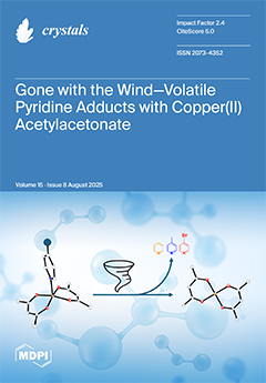
This work expands the library of copper acetylacetonate adducts to include both volatile and non-volatile ligands. Four pentacoordinate complexes, two coordination polymers and one oligomer were synthesized and are discussed. The use of bidentate ligands yielded one-dimensional coordination polymers, while the use of monodentate ligands produced weaker pentacoordinate adducts. Of particular note is the synthesis and characterization of the pyridine adduct, which, despite its simplicity, has been structurally elucidated for the first time. Furthermore, the new adducts enable correlations to be established between the electronic properties of the ligand substituent and the structural diversity of the complexes. Additionally, Hirshfeld surface analysis was employed to investigate intermolecular interactions in the pyridine derivatives. View this paper
- Issues are regarded as officially published after their release is announced to the table of contents alert mailing list.
- You may sign up for e-mail alerts to receive table of contents of newly released issues.
- PDF is the official format for papers published in both, html and pdf forms. To view the papers in pdf format, click on the "PDF Full-text" link, and use the free Adobe Reader to open them.
Previous Issue
Next Issue