Next Issue
Volume 14, September-2
Previous Issue
Volume 14, August-2


Cancers, Volume 14, Issue 17 (September-1 2022) – 280 articles

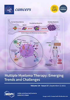
Cover Story (view full-size image): Multiple myeloma (MM) is a complex hematologic malignancy caused by the uncontrolled expansion of clonal plasma cells. In recent days, several mechanism-driven therapeutics have entered the routine clinical practice, offering a significant survival benefit and improved quality of life. The current treatment paradigm includes advanced plasma-directed therapies, such as new generation of proteasome inhibitors and immunomodulators, targeted therapies, immunotherapies, and most recently engineered T-cell therapies. Despite these advancements, mortality remains high, with most patients becoming resistant/refractory to available therapeutics, emphasizing the urgent need for new strategies. A deeper insight into the genetic and epigenetic basis of the molecular evolution from precursor states to MM will help us to develop newer therapeutic strategies to combat this incurable disease. View this paper
  • Issues are regarded as officially published after their release is announced to the table of contents alert mailing list.
  • You may sign up for e-mail alerts to receive table of contents of newly released issues.
  • PDF is the official format for papers published in both, html and pdf forms. To view the papers in pdf format, click on the "PDF Full-text" link, and use the free Adobe Reader to open them.
Order results
Result details
Section
Select all
Export citation of selected articles as:
13 pages, 1757 KiB  
Article
Endometrial Cancer Detection Using a Cervical DNA Methylation Assay (MPap) in Women with Abnormal Uterine Bleeding: A Multicenter Hospital-Based Validation Study
by Kuo-Chang Wen, Rui-Lan Huang, Lin-Yu Chen, Tzu-I Wu, Chien-Hsing Lu, Tang-Yuan Chu, Yu-Che Ou, Chen-Hsuan Wu, Shih-Tien Hsu, Dah-Ching Ding, Ling-Hui Chu, Chien-Wen Chen, Heng-Cheng Chang, Yu-Shu Liu, Hui-Chen Wang, Yu-Chun Weng, Po-Hsuan Su, Hao Lin and Hung-Cheng Lai
Cancers 2022, 14(17), 4343; https://doi.org/10.3390/cancers14174343 - 5 Sep 2022
Cited by 5 | Viewed by 3218
Abstract
Background: We describe a DNA methylation assay, named MPap test, using cervical scraping as an alternative technique for endometrial cancer detection. Methods: A multicenter hospital-based, two-stage validation study was conducted to validate the cancer detection performance of the MPap test. The MPap value [...] Read more.
Background: We describe a DNA methylation assay, named MPap test, using cervical scraping as an alternative technique for endometrial cancer detection. Methods: A multicenter hospital-based, two-stage validation study was conducted to validate the cancer detection performance of the MPap test. The MPap value was determined from the DNA methylation status of two genes (BHLHE22, CDO1) and combined with two other clinical variables (age, BMI). The cutoff threshold of the MPap value was established in stage 1 and validated in stage 2. A total of 592 women with abnormal uterine bleeding were enrolled from five medical centers throughout Taiwan. Results: In stage 1, the sensitivity, specificity, and positive and negative predictive values of the MPap test for detecting endometrial cancer were 92.9%, 71.5%, 39.8%, and 98.0%, respectively. These values were validated in stage 2, being 92.5%, 73.8%, 40.2%, and 98.1%. Moreover, MPap outperformed transvaginal ultrasound in sensitivity and negative predictive values for detecting endometrial cancer. When we applied the algorithm for triage of endometrial cancer detection by MPap in the Taiwan National Health Insurance dataset, we found that it may reduce invasive procedures by 69~73%. Conclusions: MPap may provide a feasible alternative for endometrial cancer detection and can be considered as a triage test to reduce unnecessary invasive procedures. Full article
Show Figures

Figure 1

10 pages, 1509 KiB  
Article
Multi-Class Cancer Subtyping in Salivary Gland Carcinomas with MALDI Imaging and Deep Learning
by David Pertzborn, Christoph Arolt, Günther Ernst, Oliver J. Lechtenfeld, Jan Kaesler, Daniela Pelzel, Orlando Guntinas-Lichius, Ferdinand von Eggeling and Franziska Hoffmann
Cancers 2022, 14(17), 4342; https://doi.org/10.3390/cancers14174342 - 5 Sep 2022
Cited by 12 | Viewed by 2438
Abstract
Salivary gland carcinomas (SGC) are a heterogeneous group of tumors. The prognosis varies strongly according to its type, and even the distinction between benign and malign tumor is challenging. Adenoid cystic carcinoma (AdCy) is one subgroup of SGCs that is prone to late [...] Read more.
Salivary gland carcinomas (SGC) are a heterogeneous group of tumors. The prognosis varies strongly according to its type, and even the distinction between benign and malign tumor is challenging. Adenoid cystic carcinoma (AdCy) is one subgroup of SGCs that is prone to late metastasis. This makes accurate tumor subtyping an important task. Matrix-assisted laser desorption/ionization (MALDI) imaging is a label-free technique capable of providing spatially resolved information about the abundance of biomolecules according to their mass-to-charge ratio. We analyzed tissue micro arrays (TMAs) of 25 patients (including six different SGC subtypes and a healthy control group of six patients) with high mass resolution MALDI imaging using a 12-Tesla magnetic resonance mass spectrometer. The high mass resolution allowed us to accurately detect single masses, with strong contributions to each class prediction. To address the added complexity created by the high mass resolution and multiple classes, we propose a deep-learning model. We showed that our deep-learning model provides a per-class classification accuracy of greater than 80% with little preprocessing. Based on this classification, we employed methods of explainable artificial intelligence (AI) to gain further insights into the spectrometric features of AdCys. Full article
(This article belongs to the Special Issue Salivary Gland Adenoid Cystic Carcinoma)
Show Figures

Graphical abstract

16 pages, 2215 KiB  
Review
Electrochemotherapy: An Alternative Strategy for Improving Therapy in Drug-Resistant SOLID Tumors
by Maria Condello, Gloria D’Avack, Enrico Pierluigi Spugnini and Stefania Meschini
Cancers 2022, 14(17), 4341; https://doi.org/10.3390/cancers14174341 - 5 Sep 2022
Cited by 11 | Viewed by 3682
Abstract
Electrochemotherapy (ECT) is one of the innovative strategies to overcome the multi drug resistance (MDR) that often occurs in cancer. Resistance to anticancer drugs results from a variety of factors, such as genetic or epigenetic changes, an up-regulated outflow of drugs, and various [...] Read more.
Electrochemotherapy (ECT) is one of the innovative strategies to overcome the multi drug resistance (MDR) that often occurs in cancer. Resistance to anticancer drugs results from a variety of factors, such as genetic or epigenetic changes, an up-regulated outflow of drugs, and various cellular and molecular mechanisms. This technology combines the administration of chemotherapy with the application of electrical pulses, with waveforms capable of increasing drug uptake in a non-toxic and well tolerated mechanical system. ECT is used as a first-line adjuvant therapy in veterinary oncology, where it improves the efficacy of many chemotherapeutic agents by increasing their uptake into cancer cells. The chemotherapeutic agents that have been enhanced by this technique are bleomycin, cisplatin, mitomycin C, and 5-fluorouracil. After their use, a better localized control of the neoplasm has been observed. In humans, the use of ECT was initially limited to local palliative therapy for cutaneous metastases of melanoma, but phase I/II studies are currently ongoing for several histotypes of cancer, with promising results. In this review, we described the preclinical and clinical use of ECT on drug-resistant solid tumors, such as head and neck squamous cell carcinoma, breast cancer, gynecological cancer and, finally, colorectal cancer. Full article
(This article belongs to the Collection Drug Resistance and Novel Therapies in Cancers)
Show Figures

Figure 1

15 pages, 1607 KiB  
Article
Impact of Preoperative Chemotherapy Features on Patient Outcomes after Hepatectomy for Initially Unresectable Colorectal Cancer Liver Metastases: A LiverMetSurvey Analysis
by Pasquale F. Innominato, Valérie Cailliez, Marc-Antoine Allard, Santiago Lopez-Ben, Alessandro Ferrero, Hugo Marques, Catherine Hubert, Felice Giuliante, Fernando Pereira, Esteban Cugat, Darius F. Mirza, Jose Costa-Maia, Alejandro Serrablo, Real Lapointe, Cristina Dopazo, Jose Tralhao, Gernot Kaiser, Jinn-Shiun Chen, Francisco Garcia-Borobia, Jean-Marc Regimbeau, Oleg Skipenko, Jen-Kou Lin, Christophe Laurent, Enrico Opocher, Yuichi Goto, Benoist Chibaudel, Aimery de Gramont and René Adamadd Show full author list remove Hide full author list
Cancers 2022, 14(17), 4340; https://doi.org/10.3390/cancers14174340 - 5 Sep 2022
Cited by 2 | Viewed by 2284
Abstract
Background: Prognostic factors have been extensively reported after resection of colorectal liver metastases (CLM); however, specific analyses of the impact of preoperative systemic anticancer therapy (PO-SACT) features on outcomes is lacking. Methods: For this real-world evidence study, we used prospectively collected data within [...] Read more.
Background: Prognostic factors have been extensively reported after resection of colorectal liver metastases (CLM); however, specific analyses of the impact of preoperative systemic anticancer therapy (PO-SACT) features on outcomes is lacking. Methods: For this real-world evidence study, we used prospectively collected data within the international surgical LiverMetSurvey database from all patients with initially-irresectable CLM. The main outcome was Overall Survival (OS) after surgery. Disease-free (DFS) and hepatic-specific relapse-free survival (HS-RFS) were secondary outcomes. PO-SACT features included duration (cumulative number of cycles), choice of the cytotoxic backbone (oxaliplatin- or irinotecan-based), fluoropyrimidine (infusional or oral) and addition or not of targeted monoclonal antibodies (anti-EGFR or anti-VEGF). Results: A total of 2793 patients in the database had received PO-SACT for initially irresectable diseases. Short (<7 or <13 cycles in 1st or 2nd line) PO-SACT duration was independently associated with longer OS (HR: 0.85 p = 0.046), DFS (HR: 0.81; p = 0.016) and HS-RFS (HR: 0.80; p = 0.05). All other PO-SACT features yielded basically comparable results. Conclusions: In this international cohort, provided that PO-SACT allowed conversion to resectability in initially irresectable CLM, surgery performed as soon as technically feasible resulted in the best outcomes. When resection was achieved, our findings indicate that the choice of PO-SACT regimen had a marginal if any, impact on outcomes. Full article
Show Figures

Figure 1

14 pages, 2095 KiB  
Article
Investigation of the Optimal Prime Boost Spacing Regimen for a Cancer Therapeutic Vaccine Targeting Human Papillomavirus
by Diane M. Da Silva, Emma A. Martinez, Lies Bogaert and W. Martin Kast
Cancers 2022, 14(17), 4339; https://doi.org/10.3390/cancers14174339 - 5 Sep 2022
Cited by 2 | Viewed by 2102
Abstract
Therapeutic vaccine studies should be designed to elicit durable, high magnitude, and efficacious T cell responses, all of which can be impacted by the choice of the vaccination schedule. Here, we compare different prime-boost intervals (PBI) in a human papillomavirus (HPV) model using [...] Read more.
Therapeutic vaccine studies should be designed to elicit durable, high magnitude, and efficacious T cell responses, all of which can be impacted by the choice of the vaccination schedule. Here, we compare different prime-boost intervals (PBI) in a human papillomavirus (HPV) model using a HPV16E7E6 Venezuelan equine encephalitis virus replicon particle (VRP) vaccination to address the optimal boosting schedule, quality of immune response, and overall in vivo efficacy. Six different vaccine regimens were tested with each group receiving booster vaccinations at different time intervals. Analysis of T-cell responses demonstrated a significant HPV16 E7 specific CD8+ T cell response with at minimum a one-week PBI between antigen re-exposure. Significant E7-specific in vivo cytotoxicity was also observed with longer PBIs. Additionally, longer PBIs led to an enhanced memory recall response to tumor challenge, which correlated with differential expansion of T cell memory subsets. Our findings imply that when using alphavirus vector platforms as a vaccination strategy, a one-week PBI is sufficient to induce high magnitude effector T cells with potent anti-tumor activity. However, longer PBIs lead to enhanced long-term protective anti-tumor immunity. These findings have implications for therapeutic vaccine clinical trials in which shorter intervals of prime-boost regimens may lead to suboptimal durable immune responses. Full article
(This article belongs to the Special Issue Advances in Immuno-Oncology Research)
Show Figures

Figure 1

11 pages, 685 KiB  
Article
Impact of COVID-19 Pandemic on Thyroid Surgery in a University Hospital in South Korea
by Seong Hoon Kim, Euna Min, Young Mi Hwang, Yun Suk Choi and Jin Wook Yi
Cancers 2022, 14(17), 4338; https://doi.org/10.3390/cancers14174338 - 5 Sep 2022
Cited by 8 | Viewed by 2302
Abstract
The COVID-19 pandemic has changed healthcare systems around the world. Medical personnel concentrated on infectious disease management and treatments for non-emergency diseases and scheduled surgeries were delayed. We aimed to investigate the change in the severity of thyroid cancer before and after the [...] Read more.
The COVID-19 pandemic has changed healthcare systems around the world. Medical personnel concentrated on infectious disease management and treatments for non-emergency diseases and scheduled surgeries were delayed. We aimed to investigate the change in the severity of thyroid cancer before and after the outbreak of COVID-19 in Korea. We collected three years of data (2019, 2020, and 2021) on patients who received thyroid surgery in a university hospital in South Korea and grouped them as “Before COVID-19”, “After COVID-19 1-year” and “After COVID-19 2-years”. The total number of annual outpatients declined significantly after the outbreak of COVID-19 in both new (1303, 939, and 1098 patients) and follow-up patients (5584, 4609, and 4739 patients). Clinical characteristics, including age, sex, BMI, preoperative cytology results, surgical extent, and final pathologic diagnosis, were not significantly changed after the outbreak of COVID-19. However, the number of days from the first visit to surgery was significantly increased (38.3 ± 32.2, 58.3 ± 105.2, 47.8 ± 124.7 days, p = 0.027). Papillary thyroid carcinoma (PTC) patients showed increased proportions of extrathyroidal extension, lymphatic invasion, vascular invasion, and cervical lymph node metastasis. Increased tumor size was observed in patients with follicular tumor (3.5 ± 2.2, 4.0 ± 1.9, 4.3 ± 2.3 cm, p = 0.019). After the COVID-19 outbreak, poor prognostic factors for thyroid cancer increased, and an increase in the size of follicular tumors was observed. Due to our study being confined to a single tertiary institution in Incheon city, Korea, nationwide studies that include primary clinics should be required to identify the actual impact of COVID-19 on thyroid disease treatment. Full article
(This article belongs to the Special Issue Coronavirus Disease (COVID-19) and Its Impact on Patients with Cancer)
Show Figures

Figure 1

12 pages, 2150 KiB  
Article
A Score to Predict the Malignancy of a Breast Lesion Based on Different Contrast Enhancement Patterns in Contrast-Enhanced Spectral Mammography
by Luca Nicosia, Anna Carla Bozzini, Simone Palma, Marta Montesano, Filippo Pesapane, Federica Ferrari, Valeria Dominelli, Anna Rotili, Lorenza Meneghetti, Samuele Frassoni, Vincenzo Bagnardi, Claudia Sangalli and Enrico Cassano
Cancers 2022, 14(17), 4337; https://doi.org/10.3390/cancers14174337 - 5 Sep 2022
Cited by 11 | Viewed by 1856
Abstract
Background: To create a predictive score of malignancy of a breast lesion based on the main contrast enhancement features ascertained by contrast-enhanced spectral mammography (CESM). Methods: In this single-centre prospective study, patients with suspicious breast lesions (BIRADS > 3) were enrolled between January [...] Read more.
Background: To create a predictive score of malignancy of a breast lesion based on the main contrast enhancement features ascertained by contrast-enhanced spectral mammography (CESM). Methods: In this single-centre prospective study, patients with suspicious breast lesions (BIRADS > 3) were enrolled between January 2013 and February 2022. All participants underwent CESM prior to breast biopsy, and eventually surgery. A radiologist with 20 years’ experience in breast imaging evaluated the presence or absence of enhancement and the following enhancement descriptors: intensity, pattern, margin, and ground glass. A score of 0 or 1 was given for each descriptor, depending on whether the enhancement characteristic was predictive of benignity or malignancy (both in situ and invasive). Then, an overall enhancement score ranging from 0 to 4 was obtained. The histological results were considered the gold standard in the evaluation of the relationship between enhancement patterns and malignancy. Results: A total of 321 women (median age: 51 years; range: 22–83) with 377 suspicious breast lesions were evaluated. Two hundred forty-nine lesions (66%) have malignant histological results (217 invasive and 32 in situ). Considering an overall enhancement score ≥ 2 as predictive of malignancy, we obtain an overall sensitivity of 92.4%; specificity of 89.8%; positive predictive value of 94.7%; and negative predictive value of 85.8%. Conclusions: Our proposed predictive score on the enhancement descriptors of CESM to predict the malignancy of a breast lesion shows excellent results and can help in early breast cancer diagnosis and in avoiding unnecessary biopsies. Full article
(This article belongs to the Special Issue New Challenges in Breast Cancer Diagnosis and Management)
Show Figures

Figure 1

21 pages, 7989 KiB  
Article
Interleukin 4 Controls the Pro-Tumoral Role of Macrophages in Mammary Cancer Pulmonary Metastasis in Mice
by Carolina Rodriguez-Tirado, David Entenberg, Jiufeng Li, Bin-Zhi Qian, John S. Condeelis and Jeffrey W. Pollard
Cancers 2022, 14(17), 4336; https://doi.org/10.3390/cancers14174336 - 5 Sep 2022
Cited by 12 | Viewed by 4337
Abstract
Metastasis is the systemic manifestation of cancer and the main cause of death from breast cancer. In mouse models of lung metastases, recruitment of classical monocytes from blood to the lung and their differentiation to metastasis-associated macrophages (MAMs) facilitate cancer cell extravasation, survival [...] Read more.
Metastasis is the systemic manifestation of cancer and the main cause of death from breast cancer. In mouse models of lung metastases, recruitment of classical monocytes from blood to the lung and their differentiation to metastasis-associated macrophages (MAMs) facilitate cancer cell extravasation, survival and growth. Ablation of MAMs or their monocytic progenitors inhibits metastasis. We hypothesized that factors controlling macrophage polarization modulate tumor cell extravasation in the lung. We evaluated whether signaling by Th1 or Th2 cytokines in macrophages affected transendothelial migration of tumor cells in vitro. Interferon gamma and LPS inhibited macrophage-dependent tumor cell extravasation while the Th2 cytokine interleukin-4 (IL4) enhanced this process. We demonstrated that IL4 receptor (IL4rα)-null mice developed fewer and smaller lung metastasis in E0771-LG mammary cancer models of this disease. Adoptive transfer of wild-type monocytes to IL4rα-deficient mice partially rescued this phenotype. IL4 signaling in macrophages controlled the expression of the chemokine receptor CXCR2, necessary for IL4-mediated tumor cell extravasation in vitro. Furthermore, IL4 signaling in macrophages regulated the transcript abundance of several other genes already causally associated with mammary cancer lung metastasis including Ccl2, Csf1, Ccr1, Hgf and Flt1. The central role of IL4 signaling in MAMs was confirmed by high-resolution intravital imaging of the lung in mice at the time of metastatic seeding, which showed reduced physical interaction between tumor cells and IL4rα-deficient macrophages. This interaction with wild-type MAMs enhanced tumor cell survival and seeding, which was lost in the IL4rα mice. These data indicate that IL4 signaling in monocytes and macrophages is key during seeding and growth of breast metastasis in the lung, as it regulates pro-tumoral paracrine signaling between cancer cells and macrophages. Full article
Show Figures

Figure 1

13 pages, 952 KiB  
Article
Demographics, Outcomes, and Risk Factors for Patients with Sarcoma and COVID-19: A CCC19-Registry Based Retrospective Cohort Study
by Michael J. Wagner, Cassandra Hennessy, Alicia Beeghly, Benjamin French, Dimpy P. Shah, Sarah Croessmann, Diana Vilar-Compte, Erika Ruiz-Garcia, Matthew Ingham, Gary K. Schwartz, Corrie A. Painter, Rashmi Chugh, Leslie Fecher, Cathleen Park, Olga Zamulko, Jonathan C. Trent, Vivek Subbiah, Ali Raza Khaki, Lisa Tachiki, Elizabeth S. Nakasone, Elizabeth T. Loggers, Chris Labaki, Renee Maria Saliby, Rana R. McKay, Archana Ajmera, Elizabeth A. Griffiths, Igor Puzanov, William D. Tap, Clara Hwang, Sheela Tejwani, Sachin R. Jhawar, Brandon Hayes-Lattin, Elizabeth Wulff-Burchfield, Anup Kasi, Daniel Y. Reuben, Gayathri Nagaraj, Monika Joshi, Hyma Polimera, Amit A. Kulkarni, Khashayar Esfahani, Daniel H. Kwon, Luca Paoluzzi, Mehmet A. Bilen, Eric B. Durbin, Petros Grivas, Jeremy L. Warner and Elizabeth J. Davisadd Show full author list remove Hide full author list
Cancers 2022, 14(17), 4334; https://doi.org/10.3390/cancers14174334 - 5 Sep 2022
Cited by 3 | Viewed by 3891
Abstract
Background: Patients with sarcoma often require individualized treatment strategies and are likely to receive aggressive immunosuppressive therapies, which may place them at higher risk for severe COVID-19. We aimed to describe demographics, risk factors, and outcomes for patients with sarcoma and COVID-19. Methods: [...] Read more.
Background: Patients with sarcoma often require individualized treatment strategies and are likely to receive aggressive immunosuppressive therapies, which may place them at higher risk for severe COVID-19. We aimed to describe demographics, risk factors, and outcomes for patients with sarcoma and COVID-19. Methods: We performed a retrospective cohort study of patients with sarcoma and COVID-19 reported to the COVID-19 and Cancer Consortium (CCC19) registry (NCT04354701) from 17 March 2020 to 30 September 2021. Demographics, sarcoma histologic type, treatments, and COVID-19 outcomes were analyzed. Results: of 281 patients, 49% (n = 139) were hospitalized, 33% (n = 93) received supplemental oxygen, 11% (n = 31) were admitted to the ICU, and 6% (n = 16) received mechanical ventilation. A total of 23 (8%) died within 30 days of COVID-19 diagnosis and 44 (16%) died overall at the time of analysis. When evaluated by sarcoma subtype, patients with bone sarcoma and COVID-19 had a higher mortality rate than patients from a matched SEER cohort (13.5% vs 4.4%). Older age, poor performance status, recent systemic anti-cancer therapy, and lung metastases all contributed to higher COVID-19 severity. Conclusions: Patients with sarcoma have high rates of severe COVID-19 and those with bone sarcoma may have the greatest risk of death. Full article
Show Figures

Figure 1

15 pages, 2874 KiB  
Article
The Efficacy of PD-1/PD-L1 Inhibitors in Patients with Liver Metastasis of Non-Small Cell Lung Cancer: A Real-World Study
by Mingying Xie, Na Li, Xiaoling Xu, Yanjun Xu, Hui Li, Liang Zhu, Jiamin Sheng, Zichao Zhou and Yun Fan
Cancers 2022, 14(17), 4333; https://doi.org/10.3390/cancers14174333 - 5 Sep 2022
Cited by 9 | Viewed by 2912
Abstract
Background: A controversy exists regarding the efficacy of programmed death-1 (PD-1)/ programmed death ligand-1 (PD-L1) inhibitors for patients with non-small cell lung cancer (NSCLC) and liver metastases. Our study retrospectively evaluated the efficacy of PD-1/PD-L1 inhibitors in NSCLC patients with liver metastases. Methods: [...] Read more.
Background: A controversy exists regarding the efficacy of programmed death-1 (PD-1)/ programmed death ligand-1 (PD-L1) inhibitors for patients with non-small cell lung cancer (NSCLC) and liver metastases. Our study retrospectively evaluated the efficacy of PD-1/PD-L1 inhibitors in NSCLC patients with liver metastases. Methods: This retrospective study included 1627 lung cancer patients who received immunotherapy. Among 648 patients who had advanced NSCLC and received PD-1/PD-L1 inhibitors, 61 had liver metastases and 587 did not have. We analyzed patient characteristics, progression-free survival (PFS) and overall survival (OS). An exploratory analysis of biomarkers including CD4, CD8 and CD68 for efficacy in patients with liver metastases was also performed. Results: In liver metastasis patients receiving PD-1/PD-L1 inhibitors, the objective response rate (ORR) was 29.5%, the disease control rate (DCR) was 72.1%, PFS was 6.4 months and OS was 15.2 months, which were all worse than those of patients without liver metastases (ORR: 35.8%; DCR: 81.8%; PFS: 7.9 months, p = 0.001; OS: 20.6 months, p = 0.008). When compared to non-liver lesions, the ORR (26.2 vs. 39.3%) and DCR (75.4 vs. 88.5%) of liver lesions were lower. During the analysis of PD-L1 expression, 27 PD-L1-positive patients had a longer PFS than 21 patients in the negative group (p = 0.012). Being PD-L1 positive was the independent prognostic indicators for PFS (p = 0.006). Additionally, the PD-L1 and CD8 dual-positive group responded favorably to PD-1/PD-L1 inhibitors. Conclusions: PD-1/PD-L1 inhibitors are effective in liver metastasis–NSCLC patients. However, the efficacy is inferior when compared to those of patients without liver metastases. In NSCLC patients with liver metastases, PD-L1 expression and CD8+ T cell infiltration can predict the response of PD-1/PD-L1-directed immunotherapy. Full article
(This article belongs to the Special Issue Advances in Lung Cancer Therapy)
Show Figures

Figure 1

18 pages, 767 KiB  
Review
PARP Inhibitors for Breast Cancer: Germline BRCA1/2 and Beyond
by Maria Clara Saad Menezes, Farah Raheem, Lida Mina, Brenda Ernst and Felipe Batalini
Cancers 2022, 14(17), 4332; https://doi.org/10.3390/cancers14174332 - 5 Sep 2022
Cited by 20 | Viewed by 4086
Abstract
Poly-adenosine diphosphate ribose polymerase (PARP) inhibitors (PARPi) are approved for BRCA1/2 carriers with HER2-negative breast cancer in the adjuvant setting with a high risk of recurrence as well as the metastatic setting. However, the indications for PARPi are broader for patients with other [...] Read more.
Poly-adenosine diphosphate ribose polymerase (PARP) inhibitors (PARPi) are approved for BRCA1/2 carriers with HER2-negative breast cancer in the adjuvant setting with a high risk of recurrence as well as the metastatic setting. However, the indications for PARPi are broader for patients with other cancer types (e.g., prostate and ovarian cancer), involving additional biomarkers (e.g., ATM, PALB2, and CHEK) and genomic instability scores. Herein, we summarize the data on PARPi and breast cancer and discuss their use beyond BRCA carriers. Full article
(This article belongs to the Special Issue Genomic Landscape of Breast Cancer: From Primary to Metastasis)
Show Figures

Figure 1

13 pages, 1393 KiB  
Article
Comparing Apparent Diffusion Coefficient and FNCLCC Grading to Improve Pretreatment Grading of Soft Tissue Sarcoma—A Translational Feasibility Study on Fusion Imaging
by Madelaine Hettler, Julia Kitz, Ali Seif Amir Hosseini, Manuel Guhlich, Babak Panahi, Jennifer Ernst, Lena-Christin Conradi, Michael Ghadimi, Philipp Ströbel and Jens Jakob
Cancers 2022, 14(17), 4331; https://doi.org/10.3390/cancers14174331 - 5 Sep 2022
Cited by 4 | Viewed by 3276
Abstract
Histological subtype and grading are cornerstones of treatment decisions in soft tissue sarcoma (STS). Due to intratumoral heterogeneity, pretreatment grading assessment is frequently unreliable and may be improved through functional imaging. In this pilot study, 12 patients with histologically confirmed STS were included. [...] Read more.
Histological subtype and grading are cornerstones of treatment decisions in soft tissue sarcoma (STS). Due to intratumoral heterogeneity, pretreatment grading assessment is frequently unreliable and may be improved through functional imaging. In this pilot study, 12 patients with histologically confirmed STS were included. Preoperative functional magnetic resonance imaging was fused with a computed tomography scan of the resected specimen after collecting core needle biopsies and placing radiopaque markers at distinct tumor sites. The Fédération Nationale des Centres de Lutte Contre le Cancer (FNCLCC) grading criteria of the biopsies and apparent diffusion coefficients (ADCs) of the biopsy sites were correlated. Concordance in grading between the specimen and at least one biopsy was achieved in 9 of 11 cases (81.8%). In 7 of 12 cases, fusion imaging was feasible without relevant contour deviation. Functional analysis revealed a tendency for high-grade regions (Grade 2/3 (G2/G3)) (median (range) ± standard deviation: 1.13 (0.78–1.70) ± 0.23 × 10⁻³ mm2/s) to have lower ADC values than low-grade regions (G1; 1.43 (0.64–2.03) ± 0.46 × 10⁻³ mm2/s). In addition, FNCLCC scoring of multiple tumor biopsies proved intratumoral heterogeneity as expected. The ADC appears to correlate with the FNCLCC grading criteria. Further studies are needed to determine whether functional imaging may supplement histopathological grading. Full article
(This article belongs to the Special Issue Soft Tissue Sarcoma: Imaging, Mechanisms and Therapy)
Show Figures

Graphical abstract

17 pages, 523 KiB  
Review
COVID-19 vs. Cancer Immunosurveillance: A Game of Thrones within an Inflamed Microenviroment
by Ioannis Liapis and Stavroula Baritaki
Cancers 2022, 14(17), 4330; https://doi.org/10.3390/cancers14174330 - 5 Sep 2022
Cited by 5 | Viewed by 3072
Abstract
The COVID-19 pandemic accounts for more than 500 million confirmed infections and over 6 million deaths worldwide in the last 2 years. SARS-CoV-2 causes a highly complex form of inflammation that affects the human organism both acutely and chronically. In the same line, [...] Read more.
The COVID-19 pandemic accounts for more than 500 million confirmed infections and over 6 million deaths worldwide in the last 2 years. SARS-CoV-2 causes a highly complex form of inflammation that affects the human organism both acutely and chronically. In the same line, cancer as an inflammation-induced and immune-editing disease appears to cross-react with immune system at different levels including early interactions during carcinogenesis and later cross-talks within the tumor microenvironment. With all that in mind, a reasonable question one might address is whether the SARS-CoV-2 infection and the derived “long lasting inflammatory status” that is frequently observed in patients, might affect the cancer immunosurveillance mechanisms and consequently their risk of developing cancer, as well as the tumor and immune cell behaviors within the inflamed microenvironment. On this context, this review intends to outline and discuss the existing knowledge on SARS-CoV-2-mediated immunomodulation under the prism of changes that might be able to interfere with cancer cell immunoescape and the overall tumor progression and response to conventional therapeutics. Our goal is to highlight a potential interplay between the COVID-19 immunopathology and cancer immune-microenvironment that may pave the way for thorough investigation in the future. Full article
Show Figures

Figure 1

15 pages, 1605 KiB  
Article
Epidemiologic Trends of Cutaneous T-Cell Lymphoma in Arkansas Reveals Demographic Disparities
by Delice Kayishunge, Sophia Ly, Joseph Su and Henry K. Wong
Cancers 2022, 14(17), 4329; https://doi.org/10.3390/cancers14174329 - 4 Sep 2022
Cited by 5 | Viewed by 1885
Abstract
Accurate demographic data are critical for comprehending and treating cutaneous T-cell lymphoma (CTCL). Our research aimed to determine the demographics and incidence trends of CTCL patients in Arkansas compared to those of the national CTCL population to recognize the underlying disparities. We collected [...] Read more.
Accurate demographic data are critical for comprehending and treating cutaneous T-cell lymphoma (CTCL). Our research aimed to determine the demographics and incidence trends of CTCL patients in Arkansas compared to those of the national CTCL population to recognize the underlying disparities. We collected data from 143 CTCL patients at the University of Arkansas for Medical Sciences (UAMS) and national CTCL patient data from the Surveillance, Epidemiology, and End Results (SEER) database. Our analysis revealed that males are affected more than females across all ages and races. CTCL incidence and mortality data show that CTCL has a steady increase at the national level and in Arkansas while disproportionately affecting the young black male population. In Arkansas, more than one-third of black patients presented at an advanced stage (IIB+) compared to one-fifth in the white population, and the mean age of death was more than a decade younger for black (60 years) than for white patients (74.6 years). Nationally, black male patients had the greatest mortality rate (0.5) compared to 0.32 for white males. CTCL is 2.23 and 2.38 times more prevalent in urban versus rural areas in Arkansas and nationally, respectively. Most Arkansas patients reside near major interstates and chemical-emitting sites. In conclusion, our demographic analysis of Arkansas and national CTCL patients verifies recent trends toward more aggressive presentations in young black male patients, and our geographic findings suggest possible environmental risk factors. Full article
(This article belongs to the Special Issue Population-Based Research on Modifiable Risk Factors for Cancer)
Show Figures

Figure 1

15 pages, 1003 KiB  
Systematic Review
Neoadjuvant Stereotactic Radiotherapy for Brain Metastases: Systematic Review and Meta-Analysis of the Literature and Ongoing Clinical Trials
by Paolo Palmisciano, Gianluca Ferini, Ramlah Khan, Othman Bin-Alamer, Giuseppe E. Umana, Kenny Yu, Aaron A. Cohen-Gadol, Tarek Y. El Ahmadieh and Ali S. Haider
Cancers 2022, 14(17), 4328; https://doi.org/10.3390/cancers14174328 - 4 Sep 2022
Cited by 9 | Viewed by 2833
Abstract
Background: Brain metastases (BMs) carry a high morbidity and mortality burden. Neoadjuvant stereotactic radiotherapy (NaSRT) has shown promising results. We systematically reviewed the literature on NaSRT for BMs. Methods: PubMed, EMBASE, Scopus, Web-of-Science, Cochrane, and ClinicalTrial.gov were searched following the PRISMA guidelines to [...] Read more.
Background: Brain metastases (BMs) carry a high morbidity and mortality burden. Neoadjuvant stereotactic radiotherapy (NaSRT) has shown promising results. We systematically reviewed the literature on NaSRT for BMs. Methods: PubMed, EMBASE, Scopus, Web-of-Science, Cochrane, and ClinicalTrial.gov were searched following the PRISMA guidelines to include studies and ongoing trials reporting NaSRT for BMs. Indications, protocols, and outcomes were analyzed using indirect random-effect meta-analyses. Results: We included 7 studies comprising 460 patients with 483 BMs, and 13 ongoing trials. Most BMs originated from non-small lung cell carcinoma (41.4%), breast cancer (18.7%) and melanoma (43.6%). Most patients had single-BM (69.8%) located supratentorial (77.8%). Patients were eligible if they had histologically-proven primary tumors and ≤4 synchronous BMs candidate for non-urgent surgery and radiation. Patients with primary tumors clinically responsive to radiotherapy, prior brain radiation, and leptomeningeal metastases were deemed non-eligible. Median planning target volume was 9.9 cm3 (range, 2.9–57.1), and NaSRT was delivered in 1-fraction (90.9%), 5-fraction (4.8%), or 3-fraction (4.3%), with a median biological effective dose of 39.6 Gy10 (range, 35.7–60). Most patients received piecemeal (76.3%) and gross-total (94%) resection after a median of 1-day (range, 1–10) post-NaSRT. Median follow-up was 19.2-months (range, 1–41.3). Actuarial post-treatment rates were 4% (95%CI: 2–6%) for symptomatic radiation necrosis, 15% (95%CI: 12–18%) and 47% (95%CI: 42–52%) for local and distant recurrences, 6% (95%CI: 3–8%) for leptomeningeal metastases, 81% (95%CI: 75–87%) and 59% (95%CI: 54–63%) for 1-year local tumor control and overall survival. Conclusion: NaSRT is effective and safe for BMs. Ongoing trials will provide high-level evidence on long-term post-treatment outcomes, further compared to adjuvant stereotactic radiotherapy. Full article
(This article belongs to the Special Issue Advances in Stereotactic Radiotherapy of Brain Metastases)
Show Figures

Figure 1

3 pages, 227 KiB  
Editorial
The Third Joint Meeting on Lung Cancer of the FHU OncoAge (University Côte d’Azur, Nice, France) and the University of Texas MD Anderson Cancer Center (Houston, TX, USA). Understanding New Therapeutic Options and Promising Predictive Biomarkers for Lung Cancer Patients
by Paul Hofman, George A. Calin, Sandurai A. Mani, Christophe Bontoux, Marius Ilié and Ignacio I. Wistuba
Cancers 2022, 14(17), 4327; https://doi.org/10.3390/cancers14174327 - 4 Sep 2022
Viewed by 1724
Abstract
We are proud and happy to present this Special Issue, a follow-up to the third joint meeting on lung cancer of the FHU OncoAge (University Côte d’Azur, Nice, France) and the University of Texas MD Anderson Cancer Center (Houston, TX, USA), which was [...] Read more.
We are proud and happy to present this Special Issue, a follow-up to the third joint meeting on lung cancer of the FHU OncoAge (University Côte d’Azur, Nice, France) and the University of Texas MD Anderson Cancer Center (Houston, TX, USA), which was held virtually on 4 October 2021 [...] Full article
13 pages, 2887 KiB  
Article
Clone Phylogenetics Reveals Metastatic Tumor Migrations, Maps, and Models
by Antonia Chroni, Sayaka Miura, Lauren Hamilton, Tracy Vu, Stephen G. Gaffney, Vivian Aly, Sajjad Karim, Maxwell Sanderford, Jeffrey P. Townsend and Sudhir Kumar
Cancers 2022, 14(17), 4326; https://doi.org/10.3390/cancers14174326 - 4 Sep 2022
Cited by 5 | Viewed by 2975
Abstract
Dispersal routes of metastatic cells are not medically detected or even visible. A molecular evolutionary analysis of tumor variation provides a way to retrospectively infer metastatic migration histories and answer questions such as whether the majority of metastases are seeded from clones within [...] Read more.
Dispersal routes of metastatic cells are not medically detected or even visible. A molecular evolutionary analysis of tumor variation provides a way to retrospectively infer metastatic migration histories and answer questions such as whether the majority of metastases are seeded from clones within primary tumors or seeded from clones within pre-existing metastases, as well as whether the evolution of metastases is generally consistent with any proposed models. We seek answers to these fundamental questions through a systematic patient-centric retrospective analysis that maps the dynamic evolutionary history of tumor cell migrations in many cancers. We analyzed tumor genetic heterogeneity in 51 cancer patients and found that most metastatic migration histories were best described by a hybrid of models of metastatic tumor evolution. Synthesizing across metastatic migration histories, we found new tumor seedings arising from clones of pre-existing metastases as often as they arose from clones from primary tumors. There were also many clone exchanges between the source and recipient tumors. Therefore, a molecular phylogenetic analysis of tumor variation provides a retrospective glimpse into general patterns of metastatic migration histories in cancer patients. Full article
(This article belongs to the Special Issue Advance in Computational Methods in Cancer Research)
Show Figures

Figure 1

21 pages, 530 KiB  
Systematic Review
Guidelines for Cancer Treatment during Pregnancy: Ethics-Related Content Evolution and Implications for Clinicians
by Alma Linkeviciute, Rita Canario, Fedro Alessandro Peccatori and Kris Dierickx
Cancers 2022, 14(17), 4325; https://doi.org/10.3390/cancers14174325 - 3 Sep 2022
Cited by 10 | Viewed by 3622
Abstract
(1) Background: Current scientific evidence suggests that most cancers, including breast cancer, can be treated during pregnancy without compromising maternal and fetal outcomes. This, however, raises questions regarding the ethical implications of clinical care. (2) Methods: Using a systematic literature search, 32 clinical [...] Read more.
(1) Background: Current scientific evidence suggests that most cancers, including breast cancer, can be treated during pregnancy without compromising maternal and fetal outcomes. This, however, raises questions regarding the ethical implications of clinical care. (2) Methods: Using a systematic literature search, 32 clinical practice guidelines for cancer treatment during pregnancy published between 2002 and 2021 were selected for analysis and 25 of them mentioned or made references to medical ethics when offering clinical management guidance for clinicians. (3) Results: Four bioethical themes were identified: respect for patient’s autonomy, balanced approach to maternal and fetal beneficence, protection of the vulnerable and justice in resource allocation. Most guidelines recommended informing the pregnant patient about available evidence-based treatment options, offering counselling and support in the process of decision making. The relational aspect of a pregnant patient’s autonomy was also recognized and endorsed in a significant number of available guidelines. (4) Conclusions: Recognition and support of a patient’s autonomy and its relational aspects should remain an integral part of future clinical practice guidelines. Nevertheless, a more structured approach is needed when addressing existing and potential ethical issues in clinical practice guidelines for cancer treatment during pregnancy. Full article
(This article belongs to the Special Issue Breast Cancer: Clinical Trial and Translational Research)
Show Figures

Figure 1

14 pages, 686 KiB  
Review
An Overview of Pituitary Incidentalomas: Diagnosis, Clinical Features, and Management
by Shigeyuki Tahara, Yujiro Hattori, Koji Suzuki, Eitaro Ishisaka, Shinichiro Teramoto and Akio Morita
Cancers 2022, 14(17), 4324; https://doi.org/10.3390/cancers14174324 - 3 Sep 2022
Cited by 5 | Viewed by 2691
Abstract
Pituitary incidentalomas are tumors or mass lesions of the pituitary gland. These are incidentally discovered during imaging studies for symptoms that are not causally related to pituitary diseases. The most common symptom that triggers an examination is headache, and the most common type [...] Read more.
Pituitary incidentalomas are tumors or mass lesions of the pituitary gland. These are incidentally discovered during imaging studies for symptoms that are not causally related to pituitary diseases. The most common symptom that triggers an examination is headache, and the most common type of pituitary incidentalomas are pituitary neuroendocrine tumors (PitNETs) and Rathke cleft cysts. The existing treatment strategy is controversial; however, surgical resection is recommended in cases of clinically non-functioning PitNETs with optic chiasm compression. In contrast, cystic lesions, such as Rathke cleft cysts, should be followed if the patients are asymptomatic. In this case, MRI and pituitary function tests are recommended every six months to one year; if there is no change, the follow-up period should be extended. The natural history of PitNET is partially known, and the management of pituitary incidentalomas is determined by this history. However, the pathogenesis of PitNET has significantly changed with the new World Health Organization classification, and follow-up is important based on this new classification. Therefore, a high level of evidence-based research is needed to consider treatment guidelines for pituitary incidentalomas in the future. Full article
(This article belongs to the Special Issue Pituitary Tumors: Molecular Insights, Diagnosis, and Targeted Therapy)
Show Figures

Figure 1

10 pages, 865 KiB  
Article
Prognostic Factors in Patients Treated with Pembrolizumab as a Second-Line Treatment for Advanced Biliary Tract Cancer
by Chan Su Park, Min Je Sung, So Jeong Kim, Jung Hyun Jo, Hee Seung Lee, Moon Jae Chung, Seungmin Bang, Seung Woo Park, Si Young Song and Jeong Youp Park
Cancers 2022, 14(17), 4323; https://doi.org/10.3390/cancers14174323 - 3 Sep 2022
Cited by 5 | Viewed by 1647
Abstract
Some BTC types respond to pembrolizumab, but there are no known prognostic factors to predict its treatment benefits. In this study, we attempted to identify the prognostic factors associated with pembrolizumab as a second-line treatment for gemcitabine-refractory BTC. This retrospective and single tertiary-center [...] Read more.
Some BTC types respond to pembrolizumab, but there are no known prognostic factors to predict its treatment benefits. In this study, we attempted to identify the prognostic factors associated with pembrolizumab as a second-line treatment for gemcitabine-refractory BTC. This retrospective and single tertiary-center study involved all the consecutive patients (n = 80) with refractory advanced BTC, who were diagnosed as programmed cell death ligand 1-positive and treated with pembrolizumab between August 2017 and February 2021. The overall survival (OS) was analyzed using Cox regression analysis. The median OS was 6.0 months [95% confidence interval (CI): 3.87–8.20]; median progression-free survival was 1.9 months (95% CI: 1.82–1.98); and the response rate was 15.9%. In the multivariate Cox regression analysis, the TB [adjusted hazard ratio (HR) = 2.286; 95% CI: 1.177–4.440; p = 0.015), albumin levels (adjusted HR = 0.392; 95% CI: 0.211–0.725; p = 0.003), ALP levels (adjusted HR = 1.938; 95% CI: 1.105–3.400; p = 0.021), and LMR (adjusted HR = 0.325; 95% CI: 0.173–0.609; p < 0.001) were identified as significant variables associated with the OS. High albumin levels and LMR and low ALP levels and TB were significantly associated with better OS in patients treated with pembrolizumab. Full article
Show Figures

Figure 1

15 pages, 2039 KiB  
Article
Deep View of HCC Gene Expression Signatures and Their Comparison with Other Cancers
by Yuquan Qian, Timo Itzel, Matthias Ebert and Andreas Teufel
Cancers 2022, 14(17), 4322; https://doi.org/10.3390/cancers14174322 - 3 Sep 2022
Cited by 2 | Viewed by 1897
Abstract
Background: Gene expression signatures correlate genetic alterations with specific clinical features, providing the potential for clinical usage. A plethora of HCC-dependent gene signatures have been developed in the last two decades. However, none of them has made its way into clinical practice. Thus, [...] Read more.
Background: Gene expression signatures correlate genetic alterations with specific clinical features, providing the potential for clinical usage. A plethora of HCC-dependent gene signatures have been developed in the last two decades. However, none of them has made its way into clinical practice. Thus, we investigated the specificity of public gene signatures to HCC by establishing a comparative transcriptomic analysis, as this may be essential for clinical applications. Methods: We collected 10 public HCC gene signatures and evaluated them by utilizing four different (commercial and non-commercial) gene expression profile comparison tools: Oncomine Premium, SigCom LINCS, ProfileChaser (modified version), and GENEVA, which can assign similar pre-analyzed profiles of patients with tumors or cancer cell lines to our gene signatures of interests. Among the query results of each tool, different cancer entities were screened. In addition, seven breast and colorectal cancer gene signatures were included in order to further challenge tumor specificity of gene expression signatures. Results: Although the specificity of the evaluated HCC gene signatures varied considerably, none of the gene signatures showed strict specificity to HCC. All gene signatures exhibited potential significant specificity to other cancers, particularly for colorectal and breast cancer. Since signature specificity proved challenging, we furthermore investigated common core genes and overlapping enriched pathways among all gene signatures, which, however, showed no or only very little overlap, respectively. Conclusion: Our study demonstrates that specificity, independent validation, and clinical use of HCC genetic signatures solely relying on gene expression remains challenging. Furthermore, our work made clear that standards in signature generation and statistical methods but potentially also in tissue preparation are urgently needed. Full article
Show Figures

Graphical abstract

20 pages, 2228 KiB  
Article
The Role of HPV in Determining Treatment, Survival, and Prognosis of Head and Neck Squamous Cell Carcinoma
by Imogen Sharkey Ochoa, Esther O’Regan, Mary Toner, Elaine Kay, Peter Faul, Connor O’Keane, Roisin O’Connor, Dorinda Mullen, Mataz Nur, Eamon O’Murchu, Jacqui Barry-O’Crowley, Niamh Kernan, Prerna Tewari, Helen Keegan, Sharon O’Toole, Robbie Woods, Susan Kennedy, Kenneth Feeley, Linda Sharp, Tarik Gheit, Massimo Tommasino, John J. O’Leary and Cara M. Martinadd Show full author list remove Hide full author list
Cancers 2022, 14(17), 4321; https://doi.org/10.3390/cancers14174321 - 3 Sep 2022
Cited by 10 | Viewed by 3364
Abstract
Human papillomavirus (HPV) infection has been identified as a significant etiological agent in the development of head and neck squamous cell carcinoma (HNSCC). HPV’s involvement has alluded to better survival and prognosis in patients and suggests that different treatment strategies may be appropriate [...] Read more.
Human papillomavirus (HPV) infection has been identified as a significant etiological agent in the development of head and neck squamous cell carcinoma (HNSCC). HPV’s involvement has alluded to better survival and prognosis in patients and suggests that different treatment strategies may be appropriate for them. Only some data on the epidemiology of HPV infection in the oropharyngeal, oral cavity, and laryngeal SCC exists in Europe. Thus, this study was carried out to investigate HPV’s impact on HNSCC patient outcomes in the Irish population, one of the largest studies of its kind using consistent HPV testing techniques. A total of 861 primary oropharyngeal, oral cavity, and laryngeal SCC (OPSCC, OSCC, LSCC) cases diagnosed between 1994 and 2013, identified through the National Cancer Registry of Ireland (NCRI), were obtained from hospitals across Ireland and tested for HPV DNA using Multiplex PCR Luminex technology based in and sanctioned by the International Agency for Research on Cancer (IARC). Both overall and cancer-specific survival were significantly improved amongst all HPV-positive patients together, though HPV status was only a significant predictor of survival in the oropharynx. Amongst HPV-positive patients in the oropharynx, surgery alone was associated with prolonged survival, alluding to the potential for de-escalation of treatment in HPV-related OPSCC in particular. Cumulatively, these findings highlight the need for continued investigation into treatment pathways for HPV-related OPSCC, the relevance of introducing boys into national HPV vaccination programs, and the relevance of the nona-valent Gardasil-9 vaccine to HNSCC prevention. Full article
Show Figures

Figure 1

15 pages, 1803 KiB  
Article
Systemic Therapy Is Associated with Improved Oncologic Outcomes in Resectable Stage II/III Intrahepatic Cholangiocarcinoma: An Examination of the National Cancer Database over the Past Decade
by Rebecca Marcus, Wade Christopher, Jennifer Keller, Sean Nassoiy, Shu-Ching Chang, Melanie Goldfarb, Ronald Wolf and Zeljka Jutric
Cancers 2022, 14(17), 4320; https://doi.org/10.3390/cancers14174320 - 3 Sep 2022
Cited by 3 | Viewed by 1941
Abstract
Limited evidence-based management guidelines for resectable intrahepatic cholangiocarcinoma (ICC) currently exist. Using a large population-based cancer registry; the utilization rates and outcomes for patients with clinical stages I-III ICC treated with neoadjuvant chemotherapy (NAT) in relation to other treatment strategies were investigated, as [...] Read more.
Limited evidence-based management guidelines for resectable intrahepatic cholangiocarcinoma (ICC) currently exist. Using a large population-based cancer registry; the utilization rates and outcomes for patients with clinical stages I-III ICC treated with neoadjuvant chemotherapy (NAT) in relation to other treatment strategies were investigated, as were the predictors of treatment regimen utilization. Oncologic outcomes were compared between treatment strategies. Amongst 2736 patients, chemotherapy utilization was low; however, NAT use increased from 4.3% to 7.2% (p = 0.011) over the study period. A higher clinical stage was predictive of the use of NAT, while higher pathologic stage and margin-positive resections were predictive of the use of adjuvant therapy (AT). For patients with more advanced disease, the receipt of NAT or AT was associated with significantly improved survival compared to surgery alone (cStage II, p = 0.040; cStage III, p = 0.003). Furthermore, patients receiving NAT were more likely to undergo margin-negative resections compared to those treated with AT (72.5% vs. 62.6%, p = 0.027), despite having higher-risk tumors. This analysis of treatment strategies for resectable ICC suggests a benefit for systemic therapy. Prospective and randomized studies evaluating the sequencing of treatments for patients with high-risk resectable ICC are needed. Full article
(This article belongs to the Special Issue Translational Research of Liver Cancer)
Show Figures

Figure 1

9 pages, 936 KiB  
Article
Probe-Based Confocal Laser Endomicroscopy versus White-Light Endoscopy with Narrow-Band Imaging for Predicting and Collecting Residual Cancer Tissue in Patients with Gastric Cancer Receiving Chemotherapy
by Yuna Kim, Hyunki Kim, Minkyu Jung, Sun Young Rha, Hyun Cheol Chung and Sang Kil Lee
Cancers 2022, 14(17), 4319; https://doi.org/10.3390/cancers14174319 - 3 Sep 2022
Cited by 1 | Viewed by 1646
Abstract
In cases of progression despite chemotherapy, collecting gastric cancer (GC) tissues might be helpful for molecular biology research or the development of new target drugs for treating cases that are refractory to chemotherapy. Chemotherapy, however, may reduce or alter the distribution of GC [...] Read more.
In cases of progression despite chemotherapy, collecting gastric cancer (GC) tissues might be helpful for molecular biology research or the development of new target drugs for treating cases that are refractory to chemotherapy. Chemotherapy, however, may reduce or alter the distribution of GC tissue on the surface, making the detection of GC tissue during upper endoscopy challenging. Probe-based confocal laser endomicroscopy (pCLE) is a new technology that enables histological diagnosis by magnifying the mucous membrane to a microscopic level. Here, we evaluated whether pCLE could increase the yield of endoscopic biopsy for GC compared to white-light endoscopy (WLE) with magnifying narrow-band imaging (M-NBI) in GC patients receiving chemotherapy with its powerful imaging technique. Patients underwent WLE/M-NBI and pCLE for the detection of residual GC for the purpose of response evaluation or clinical trial registration. After WLE/M-NBI and pCLE, each residual GC lesion was biopsied for histological analysis. A total of 23 patients were enrolled between January 2018 and June 2020. Overall, pCLE showed significantly higher sensitivity and negative predictive value than WLE/M-NBI. The accuracy of pCLE was superior to that of WLE/M-NBI. Moreover, pCLE showed better predictive ability for residual GC than WLE/M-NBI, while WLE/M-NBI and pCLE showed inconsistent results. pCLE diagnosed residual GC more accurately than WLE/M-NBI, which resulted in an increased number of GC tissues collected during the endoscopic biopsy. Full article
Show Figures

Figure 1

24 pages, 1725 KiB  
Review
Current Progress of CAR-NK Therapy in Cancer Treatment
by Zhaojun Pang, Zhongyi Wang, Fengqi Li, Chunjing Feng and Xin Mu
Cancers 2022, 14(17), 4318; https://doi.org/10.3390/cancers14174318 - 2 Sep 2022
Cited by 22 | Viewed by 5191
Abstract
CD8+ T cells and natural killer (NK) cells eliminate target cells through the release of lytic granules and Fas ligand (FasL)-induced target cell apoptosis. The introduction of chimeric antigen receptor (CAR) makes these two types of cells selective and effective in killing [...] Read more.
CD8+ T cells and natural killer (NK) cells eliminate target cells through the release of lytic granules and Fas ligand (FasL)-induced target cell apoptosis. The introduction of chimeric antigen receptor (CAR) makes these two types of cells selective and effective in killing cancer cells. The success of CAR-T therapy in the treatment of acute lymphoblastic leukemia (ALL) and other types of blood cancers proved that the immunotherapy is an effective approach in fighting against cancers, yet adverse effects, such as graft versus host disease (GvHD) and cytokine release syndrome (CRS), cannot be ignored for the CAR-T therapy. CAR-NK therapy, then, has its advantage in lacking these adverse effects and works as effective as CAR-T in terms of killing. Despite these, NK cells are known to be hard to transduce, expand in vitro, and sustain shorter in vivo comparing to infiltrated T cells. Moreover, CAR-NK therapy faces challenges as CAR-T therapy does, e.g., the time, the cost, and the potential biohazard due to the use of animal-derived products. Thus, enormous efforts are needed to develop safe, effective, and large-scalable protocols for obtaining CAR-NK cells. Here, we reviewed current progress of CAR-NK therapy, including its biological properties, CAR compositions, preparation of CAR-NK cells, and clinical progresses. We also discussed safety issues raised from genetic engineering. We hope this review is instructive to the research community and a broad range of readers. Full article
(This article belongs to the Special Issue Cancer Immunotherapy and Immune-Related Adverse Events)
Show Figures

Figure 1

11 pages, 1397 KiB  
Article
Differences in Stakeholders’ Perception of the Impact of COVID-19 on Clinical Care and Decision-Making
by Joerg Haier, Johannes Beller, Kristina Adorjan, Stefan Bleich, Moritz de Greck, Frank Griesinger, Markus V. Heppt, René Hurlemann, Soeren Torge Mees, Alexandra Philipsen, Gernot Rohde, Georgia Schilling, Karolin Trautmann, Stephanie E. Combs, Siegfried Geyer and Juergen Schaefers
Cancers 2022, 14(17), 4317; https://doi.org/10.3390/cancers14174317 - 2 Sep 2022
Cited by 3 | Viewed by 1616
Abstract
Background: Pandemics are related to changes in clinical management. Factors that are associated with individual perceptions of related risks and decision-making processes focused on prevention and vaccination, but perceptions of other healthcare consequences are less investigated. Different perceptions of patients, nurses, and physicians [...] Read more.
Background: Pandemics are related to changes in clinical management. Factors that are associated with individual perceptions of related risks and decision-making processes focused on prevention and vaccination, but perceptions of other healthcare consequences are less investigated. Different perceptions of patients, nurses, and physicians on consequences regarding clinical management, decisional criteria, and burden were compared. Study Design: Cross-sectional OnCoVID questionnaire studies. Methods: Data that involved 1231 patients, physicians, and nurses from 11 German institutions that were actively involved in clinical treatment or decision-making in oncology or psychiatry were collected. Multivariate statistical approaches were used to analyze the stakeholder comparisons. Results: A total of 29.2% of professionals reported extensive changes in workload. Professionals in psychiatry returned severe impact of pandemic on all major aspects of their clinical care, but less changes were reported in oncology (p < 0.001). Both patient groups reported much lower recognition of treatment modifications and consequences for their own care. Decisional and pandemic burden was intensively attributed from professionals towards patients, but less in the opposite direction. Conclusions: All of the groups share concerns about the impact of the COVID-19 pandemic on healthcare management and clinical processes, but to very different extent. The perception of changes is dissociated in projection towards other stakeholders. Specific awareness should avoid the dissociated impact perception between patients and professionals potentially resulting in impaired shared decision-making. Full article
(This article belongs to the Special Issue Cancer Care in the Era of COVID-19 Pandemic)
Show Figures

Figure 1

20 pages, 7006 KiB  
Article
Nrf2 Downregulation Contributes to Epithelial-to-Mesenchymal Transition in Helicobacter pylori-Infected Cells
by Sarah Bacon, Lornella Seeneevassen, Alison Fratacci, Faustine Rose, Camille Tiffon, Elodie Sifré, Maria M. Haykal, Maya M. Moubarak, Astrid Ducournau, Lucie Bruhl, Stéphane Claverol, Caroline Tokarski, Alina-Roxani Gouloumi, Ioannis S. Pateras, Thomas Daubon, Philippe Lehours, Christine Varon and Océane C. B. Martin
Cancers 2022, 14(17), 4316; https://doi.org/10.3390/cancers14174316 - 2 Sep 2022
Cited by 5 | Viewed by 2508
Abstract
Background: Gastric cancer, the fifth most common cancer worldwide, is mainly linked to Helicobacter pylori infection. H. pylori induces chronic inflammation of the gastric mucosa associated with high oxidative stress. Our study aimed at assessing the implication of Nrf2, a major regulator of [...] Read more.
Background: Gastric cancer, the fifth most common cancer worldwide, is mainly linked to Helicobacter pylori infection. H. pylori induces chronic inflammation of the gastric mucosa associated with high oxidative stress. Our study aimed at assessing the implication of Nrf2, a major regulator of cellular redox homeostasis, in H. pylori-induced gastric carcinogenesis. Methods: Using three different gastric epithelial cell lines, a non-cancerous (HFE-145) and two different subtypes of gastric cancer (AGS and MKN74), we analyzed the modulation of Nrf2 expression over time. After invalidation of Nrf2 by CRISPR-cas9, we assessed its role in H. pylori-induced epithelial-to-mesenchymal transition (EMT). Finally, we evaluated the expression of Nrf2 and ZEB1, a central EMT transcription factor, in human gastric tissues. Results: We first demonstrated that the Nrf2 signaling pathway is differentially regulated depending on the infection stage. Rapidly and transiently activated, Nrf2 was downregulated 24 h post-infection in a VacA-dependent manner. We then demonstrated that Nrf2 invalidation leads to increased EMT, which is even exacerbated after H. pylori infection. Finally, Nrf2 expression tended to decrease in human patients’ gastric mucosa infected with H. pylori. Conclusions: Our work supports the hypothesis that Nrf2 downregulation upon H. pylori infection participates in EMT, one of the most important events in gastric carcinogenesis. Full article
(This article belongs to the Special Issue New Molecular Insights for GC Characterization and Treatment)
Show Figures

Figure 1

27 pages, 2730 KiB  
Review
Overcoming Resistance: FLT3 Inhibitors Past, Present, Future and the Challenge of Cure
by Debora Capelli, Diego Menotti, Alessandro Fiorentini, Francesco Saraceni and Attilio Olivieri
Cancers 2022, 14(17), 4315; https://doi.org/10.3390/cancers14174315 - 2 Sep 2022
Cited by 15 | Viewed by 5064
Abstract
FLT3 ITD and TKD mutations occur in 20% and 10% of Acute Myeloid Leukemia (AML), respectively, and they represent the target of the first approved anti-leukemic therapies in the 2000s. Type I and type II FLT3 inhibitors (FLT3i) are active against FLT3 TKD/ITD [...] Read more.
FLT3 ITD and TKD mutations occur in 20% and 10% of Acute Myeloid Leukemia (AML), respectively, and they represent the target of the first approved anti-leukemic therapies in the 2000s. Type I and type II FLT3 inhibitors (FLT3i) are active against FLT3 TKD/ITD and FLT3 ITD mutations alone respectively, but they still fail remissions in 30–40% of patients due to primary and secondary mechanisms of resistance, with variable relapse rate of 30–50%, influenced by NPM status and FLT3 allelic ratio. Mechanisms of resistance to FLT3i have recently been analyzed through NGS and single cell assays that have identified and elucidated the polyclonal nature of relapse in clinical and preclinical studies, summarized here. Knowledge of tumor escape pathways has helped in the identification of new targeted drugs to overcome resistance. Immunotherapy and combination or sequential use of BCL2 inhibitors and experimental drugs including aurora kinases, menin and JAK2 inhibitors will be the goal of present and future clinical trials, especially in patients with FLT3-mutated (FLT3mut) AML who are not eligible for allogeneic transplantation. Full article
(This article belongs to the Collection Acute Myeloid Leukemia (AML))
Show Figures

Figure 1

13 pages, 702 KiB  
Review
IRF4 as an Oncogenic Master Transcription Factor
by Regina Wan Ju Wong, Jolynn Zu Lin Ong, Madelaine Skolastika Theardy and Takaomi Sanda
Cancers 2022, 14(17), 4314; https://doi.org/10.3390/cancers14174314 - 2 Sep 2022
Cited by 16 | Viewed by 5144
Abstract
IRF4 is a transcription factor in the interferon regulatory factor (IRF) family. Since the discovery of this gene, various research fields including immunology and oncology have highlighted the unique characteristics and the importance of IRF4 in several biological processes that distinguish it from [...] Read more.
IRF4 is a transcription factor in the interferon regulatory factor (IRF) family. Since the discovery of this gene, various research fields including immunology and oncology have highlighted the unique characteristics and the importance of IRF4 in several biological processes that distinguish it from other IRF family members. In normal lymphocyte development and immunity, IRF4 mediates critical immune responses via interactions with upstream signaling pathways, such as the T-cell receptor and B-cell receptor pathways, as well as their binding partners, which are uniquely expressed in each cell type. On the other hand, IRF4 acts as an oncogene in various mature lymphoid neoplasms when abnormally expressed. IRF4 induces several oncogenes, such as MYC, as well as genes that characterize each cell type by utilizing its ability as a master regulator of immunity. IRF4 and its upstream factor NF-κB form a transcriptional regulatory circuit, including feedback and feedforward loops, to maintain the oncogenic transcriptional program in malignant lymphoid cells. In this review article, we provide an overview of the molecular functions of IRF4 in mature lymphoid neoplasms and highlight its upstream and downstream pathways, as well as the regulatory circuits mediated by IRF4. Full article
Show Figures

Figure 1

14 pages, 1593 KiB  
Article
Exploratory Circular RNA Profiling in Adrenocortical Tumors
by Péter István Turai, Gábor Nyirő, Katalin Borka, Tamás Micsik, István Likó, Attila Patócs and Peter Igaz
Cancers 2022, 14(17), 4313; https://doi.org/10.3390/cancers14174313 - 2 Sep 2022
Cited by 1 | Viewed by 1827
Abstract
Differentiation of adrenocortical adenoma (ACA) and carcinoma (ACC) is often challenging even in the histological analysis. Circular RNAs (circRNAs) belonging to the group of non-coding RNAs have been implicated as relevant factors in tumorigenesis. Our aim was to explore circRNA expression profiles in [...] Read more.
Differentiation of adrenocortical adenoma (ACA) and carcinoma (ACC) is often challenging even in the histological analysis. Circular RNAs (circRNAs) belonging to the group of non-coding RNAs have been implicated as relevant factors in tumorigenesis. Our aim was to explore circRNA expression profiles in adrenocortical tumors by next-generation sequencing followed by RT-qPCR validation. Archived FFPE (formalin-fixed, paraffin embedded) including 8 ACC, 8 ACA and 8 normal adrenal cortices (NAC) were used in the discovery cohort. For de novo and known circRNA expression profiling, a next-generation sequencing platform was used. CIRI2, CircExplorer2, AutoCirc bioinformatics tools were used for the discovery of circRNAs. The top five most differentially circRNAs were measured by RT-qPCR in an independent validation cohort (10 ACC, 8 ACA, 8 NAC). In silico predicted, interacting microRNAs potentially sponged by differentially expressed circRNAs were studied by individual RT-qPCR assays. We focused on overexpressed circRNAs here. Significantly differentially expressed circRNAs have been revealed between the cohorts by NGS. Only circPHC3 could be confirmed to be significantly overexpressed in ACC, ACA vs. NAC samples by RT-qPCR. We could not observe microRNA expression changes fully corresponding to our sponging hypothesis. To the best of our knowledge, our study is the first to investigate circRNAs in adrenocortical tumors. Further studies are warranted to explore their biological and diagnostic relevance. Full article
Show Figures

Figure 1

Previous Issue
Back to TopTop