




Next Issue
Volume 18, January-2
Previous Issue
Volume 17, December-2
 
 
cancers-logo

Journal Browser

Journal Browser

Cancers, Volume 18, Issue 1 (January-1 2026) – 178 articles
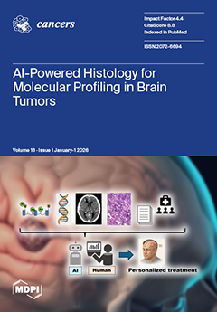
Cover Story (view full-size image): The integration of molecular features into histopathological diagnosis is central to the WHO classification of central nervous system tumors, improving prognostic stratification and precision medicine. However, limited access to molecular testing hampers the widespread use of integrated diagnoses. To address this gap, artificial intelligence (AI) approaches have been developed to predict molecular alterations directly from histological images. In gliomas, deep learning applied to whole-slide images achieves neuropathologist-level performance in predicting biomarkers such as IDH mutation and 1p/19q codeletion. Recent advances have extended these methods to intraoperative settings and other brain tumors. This review summarizes recent progress in AI-assisted molecular prediction and its potential for accessible neuro-oncologic diagnostics. View this paper
  • Issues are regarded as officially published after their release is announced to the table of contents alert mailing list.
  • You may sign up for e-mail alerts to receive table of contents of newly released issues.
  • PDF is the official format for papers published in both, html and pdf forms. To view the papers in pdf format, click on the "PDF Full-text" link, and use the free Adobe Reader to open them.
Order results
Result details
Section
Select all
Export citation of selected articles as:
11 pages, 1343 KB  
Article
Predictors of Survival in Patients Aged ≥70 with Glioblastoma: A Time-Dependent Multivariable Analysis
by Ahmad M. S. Ali, Viraj Parmar, Cathal J. Hannan and Jibril Osman Farah
Cancers 2026, 18(1), 178; https://doi.org/10.3390/cancers18010178 - 5 Jan 2026
Viewed by 2111
Abstract
Background: Glioblastoma (GB) carries a dismal prognosis, with survival outcomes particularly poor in older patients. With the fastest-growing global demographic being those aged over 65, the incidence of GB is expected to rise. Objective: To evaluate predictors of survival in patients aged ≥70 [...] Read more.
Background: Glioblastoma (GB) carries a dismal prognosis, with survival outcomes particularly poor in older patients. With the fastest-growing global demographic being those aged over 65, the incidence of GB is expected to rise. Objective: To evaluate predictors of survival in patients aged ≥70 years with histologically confirmed GB, focusing on surgical resection, adjuvant therapy, and comorbidities. Methods: A retrospective review was performed of all patients aged ≥70 undergoing index surgery for GB between January 2021 and March 2025 at a single tertiary neurosurgical centre. Demographics, pre-operative fitness scores (Karnofsky Performance Status [KPS]., Charlson Comorbidity Index [CCI].), tumour characteristics, extent of resection, adjuvant treatment, and survival were analysed. Tumour volume was estimated using the ABC/2 method. Survival outcomes were assessed using Kaplan–Meier curves and multivariable Cox proportional hazards regression. Results: A total of 124 patients aged ≥70 years (median 74 years, range 70–86) were included. Median overall survival was 8 months (IQR 4–15). On multivariable analysis, adjuvant chemoradiotherapy (HR = 0.30, 95% CI 0.17–0.52; p < 0.001) and gross total resection (GTR) (HR = 0.41, 95% CI 0.20–0.86; p = 0.019) were independently associated with improved survival. Smoking history was associated with increased hazard of death (HR = 2.02, 95% CI 1.07–3.81; p = 0.029), an effect robust to multiple sensitivity analyses. No significant associations were found for age, pre-operative KPS, comorbidity index, tumour volume, or methylation status (all p > 0.10). Tests for non-proportional hazards indicated that the survival benefit of adjuvant therapy diminished over time (interaction p = 0.0002), with early post-operative benefit (HR ≈ 0.35 at 1 month) that attenuated towards unity by 6–12 months. The effects of GTR and smoking were time-invariant. RMST analysis suggested a modest, non-significant absolute survival advantage of GTR over STR (mean difference = 2.0 months at 18 months; p = 0.11). After exclusion of early post-operative deaths (<6 weeks), adjuvant therapy (HR = 0.34; p < 0.001) and GTR (HR = 0.33; p = 0.005) remained independent predictors of improved survival. Conclusions: Among patients aged ≥70 years with glioblastoma, adjuvant therapy and extent of resection remain key independent predictors of survival, while smoking is associated with poorer outcomes. The survival benefit of adjuvant chemoradiotherapy is strongest in the early post-operative period and diminishes over time, underscoring the importance of early multidisciplinary intervention. These findings highlight that aggressive multimodal treatment may confer survival advantage even in older patients. Full article
(This article belongs to the Special Issue Combination Therapy in Geriatric Population with Cancer (2nd Edition))
Show Figures

Figure 1

13 pages, 874 KB  
Article
Outcomes of pPCL Diagnosed Using the IMWG 2021 Consensus Definition: A Retrospective Multicenter Analysis
by Priyanka Venkatesh, Razan Mansour, Yara Shatnawi, Akhil Jain, Christopher Strouse, Nausheen Ahmed, Muhammad Umair Mushtaq, Al-Ola Abdallah, Shebli Atrash and Barry Paul
Cancers 2026, 18(1), 177; https://doi.org/10.3390/cancers18010177 - 5 Jan 2026
Viewed by 1324
Abstract
Background: Primary plasma cell leukemia (pPCL) represents the most aggressive plasma cell dyscrasia with a poor prognosis and survival of <3 years. The International Myeloma Working Group (IMWG) adopted more inclusive diagnostic criteria for pPCL in 2021, including patients with 5% or more [...] Read more.
Background: Primary plasma cell leukemia (pPCL) represents the most aggressive plasma cell dyscrasia with a poor prognosis and survival of <3 years. The International Myeloma Working Group (IMWG) adopted more inclusive diagnostic criteria for pPCL in 2021, including patients with 5% or more circulating plasma cells (down from 20%). Most published studies of pPCL do not include patients who meet the criteria for pPCL based on the newer diagnostic guidelines, and the data on the optimal treatment of pPCL is scarce. In our multi-center retrospective analysis, we report data on treatment regimens used in 67 pPCL patients to characterize outcomes in this population. Methods: We included patients with newly diagnosed pPCL between 2010 and 2023 based on the 2021 IMWG definition at one of three academic centers. Results: Our results suggest significant improvement in overall response rate (ORR) and progression-free survival (PFS) with the use of autologous stem cell transplant, but without additional benefit for a tandem transplant. The presence of high-risk cytogenetics was an independent risk factor for progression in the cohort. Conclusions: Our dataset represents one of the largest cohorts to date using the expanded definition of pPCL adopted by the IMWG in 2021 and stresses the importance of taking pPCL patients to transplant. Unfortunately, our study was not powered to determine the efficacy of individual induction and maintenance regimens, and many patients diagnosed with pPCL are ineligible for transplant based on end-organ damage at diagnosis or from disease that is refractory to induction therapy, underscoring the need for early diagnosis and treatment in hopes of preserving transplant eligibility. Full article
(This article belongs to the Section Cancer Causes, Screening and Diagnosis)
Show Figures

Figure 1

15 pages, 672 KB  
Article
Effects of Tobacco Use on Oral Cancer Screening Algorithm Performance
by Elyse Kanagandram, Aksel Alp, Thair Takesh, Cherie Wink, Susan Yang, Amber Davis, Michelle Hurlbutt, Jerica Block and Petra Wilder-Smith
Cancers 2026, 18(1), 176; https://doi.org/10.3390/cancers18010176 - 5 Jan 2026
Cited by 1 | Viewed by 1584
Abstract
Background/Objectives: Effective screening for oral cancer (OC) remains challenging. Inaccuracies contribute to delayed diagnosis and poor outcomes. Tobacco-related changes in oral mucosa may compromise the accuracy of oral screening approaches, and, in emerging “smart” screening modalities, they may overshadow the influence of other [...] Read more.
Background/Objectives: Effective screening for oral cancer (OC) remains challenging. Inaccuracies contribute to delayed diagnosis and poor outcomes. Tobacco-related changes in oral mucosa may compromise the accuracy of oral screening approaches, and, in emerging “smart” screening modalities, they may overshadow the influence of other predictive variables. The objective of this study was to evaluate the screening accuracy of an imaging- and risk factor-based OC screening platform in individuals practicing different types of tobacco usage. Methods: 318 subjects who had previously screened positive for increased OC risk were recruited and sorted into “tobacco smoker”, “tobacco vaper”, “tobacco chewer”, “hookah user”, “multiple tobacco usage”, or “tobacco non-user” groups. Next, demographic information, risk factors, outcome of clinical examination, as well as AFI and pWLI were recorded using a prototype OC screening platform. The OC risk assessment outcome from the OC screening platform was compared to that from an oral medicine specialist. Results: The screening platform demonstrated high sensitivity in tobacco chewers and hookah users, and it also exceeded 90% in smokers, vapers, and multi-product users. In tobacco non-users, 80% screening sensitivity was recorded. Screening specificity was considerably better in tobacco non-users than in the tobacco-user groups, and low in tobacco chewers (33.3%), vapers (55.6%), and smokers (62.5%). Across all groups, agreement between the screening platform outcome and specialist evaluation exceeded 80%. Significant differences in probe accuracy were noted between tobacco non-users and users (p < 0.05), except for tobacco vapers. Conclusions: These findings highlight the need to consider the effects of type of tobacco use on the OC screening approach, and to integrate these variables into imaging-and risk-factor-based algorithms for OC screening. Full article
Show Figures

Figure 1

14 pages, 1347 KB  
Article
Differences in Executive Functioning Between Patients with IDH1-Mutant Oligodendroglioma and Astrocytoma Before and After Surgery
by Maud Landers-Wouters, Bart Brouwers, Geert-Jan Rutten and Elke Butterbrod
Cancers 2026, 18(1), 175; https://doi.org/10.3390/cancers18010175 - 5 Jan 2026
Viewed by 1420
Abstract
Background: IDH1-mutant oligodendroglioma and astrocytoma differ not only in growth rate but also in growth pattern. Oligodendrogliomas tend to infiltrate white matter tracts, whereas astrocytomas more often displace them. Such difference could lead to different cognitive outcomes. This study examined differences in executive [...] Read more.
Background: IDH1-mutant oligodendroglioma and astrocytoma differ not only in growth rate but also in growth pattern. Oligodendrogliomas tend to infiltrate white matter tracts, whereas astrocytomas more often displace them. Such difference could lead to different cognitive outcomes. This study examined differences in executive functioning before and up to one year after surgery between patients with IDH1-mutant astrocytoma and oligodendroglioma. Methods: Patients with WHO grade 2–3 IDH1-mutant oligodendroglioma (1p19q-codeleted) or astrocytoma were included. Cognition was assessed preoperatively, and at 3 and 12 months postoperatively using standardized computerized and paper-and-pencil tests. Groups were compared on demographics, tumor characteristics, surgical modality, extent of resection, adjuvant treatment, and baseline cognition. Longitudinal mixed models were performed to investigate differences in performances over time for the total sample and stratified by surgical approach (awake vs. asleep). Results: 162 patients (67 oligodendroglioma, 95 astrocytoma) were included. Oligodendroglioma patients were older, with more frontal and fewer temporal tumors. Oligodendroglioma patients showed a greater impairment prevalence on a measure of inhibition before surgery. In the awake surgery group, no longitudinal differences were found between diagnoses. In the asleep surgery group, astrocytoma patients remained stable while oligodendroglioma patients declined on a measure of cognitive flexibility, with performance at 3 and 12 months significantly lower than at baseline. Conclusions: Specific aspects of executive functioning in IDH1-mutant gliomas may differ by subtype. Oligodendroglioma patients showed postoperative decline in cognitive flexibility that did not recover to baseline level, particularly in case of surgery under general anesthesia. These results highlight the potential relevance of tumor subtype and surgical approach in limiting cognitive risks after glioma surgery. Full article
Show Figures

Figure 1

11 pages, 474 KB  
Article
Complications of Port-a-Cath Systems: An Institutional Study on Romanian Oncological Patients
by Adina Nemeș, Sebastian-Alexandru Pocol, Tunde Banciu and Diana Voskuil-Galoș
Cancers 2026, 18(1), 174; https://doi.org/10.3390/cancers18010174 - 5 Jan 2026
Viewed by 2552
Abstract
Background: Rising cancer incidence and mortality have increased the use of central venous catheters (CVCs), including peripherally inserted central catheters (PICCs) and port-a-cath systems (PCSs), which play an important role in treatment administration. However, CVCs are associated with mechanical, infectious, and thrombotic [...] Read more.
Background: Rising cancer incidence and mortality have increased the use of central venous catheters (CVCs), including peripherally inserted central catheters (PICCs) and port-a-cath systems (PCSs), which play an important role in treatment administration. However, CVCs are associated with mechanical, infectious, and thrombotic complications. This study evaluates PCS-related complications and their management in oncological patients at The Oncology Institute “Prof. Dr. Ion Chiricuță,” Cluj-Napoca, Romania. Methods: This non-randomized, observational, retrospective study included cancer patients who had a PCS implanted at The Oncology Institute “Prof. Dr. Ion Chiricuță,” Cluj-Napoca, between 1 January 2024 and 31 December 2024 and were enrolled in a follow-up protocol to monitor and manage PCS-related complications. This study evaluated the incidence of complications, their association with predefined prognostic factors, and their management. Results: In the study cohort (n = 124 patients), complications related to the PCS were observed in 20% of the patients, with a mean interval to complication onset of 47 days. Early-onset complications were observed in 40% of patients, while the remaining 60% developed late-onset complications. PCS infection was the most common complication (10 patients), followed by wound dehiscence and thrombosis (6 patients each). Bleeding, extravasation, catheter migration, port malfunction, and torsion were each documented in a single patient. No prognostic factors were significantly associated with early or late PCS complications, with thrombosis approaching significance (p = 0.051). Conclusions: PCSs are generally safe in oncology patients; infections and thrombosis predominate as PCS-related complications, but rarely require removal, and standardized care enables long-term use, improving patient quality of life. Full article
(This article belongs to the Section Cancer Pathophysiology)
Show Figures

Figure 1

22 pages, 3705 KB  
Review
Role of Imaging Techniques in Ovarian Cancer Diagnosis: Current Approaches and Future Directions
by Alessandro D’Amario, Roberta Ambrosini, Alessandro Gullino and Luigi Grazioli
Cancers 2026, 18(1), 173; https://doi.org/10.3390/cancers18010173 - 4 Jan 2026
Cited by 1 | Viewed by 4025
Abstract
Background: Ovarian cancer is a leading gynecological malignancy with high global mortality. Early and accurate diagnosis is critical for optimal management; however, a considerable portion of ovarian masses remain indeterminate after initial evaluation. Although transvaginal ultrasound is the first-line imaging tool, up [...] Read more.
Background: Ovarian cancer is a leading gynecological malignancy with high global mortality. Early and accurate diagnosis is critical for optimal management; however, a considerable portion of ovarian masses remain indeterminate after initial evaluation. Although transvaginal ultrasound is the first-line imaging tool, up to 30% of cases yield inconclusive findings, complicating treatment decisions. Methods: This review summarizes current diagnostic strategies for ovarian masses, with an emphasis on advanced imaging and emerging technologies. Topics include the diagnostic utility of contrast-enhanced MRI, the application of the O-RADS MRI scoring system, and the integration of Artificial Intelligence (AI) into imaging workflows. Results: Contrast-enhanced MRI offers high diagnostic accuracy (83–93%) for characterizing indeterminate ovarian masses. The O-RADS MRI Score offers a reported sensitivity of 93% and specificity of 91% for malignancy risk assessment. Additionally, new classification systems have been proposed to further improve diagnostic performance. AI-based approaches, particularly machine learning and deep learning applied to imaging data, show potential in improving diagnostic precision; however, most techniques require further clinical validation before widespread adoption. Conclusions: Advanced imaging techniques and AI-driven methods are reshaping the diagnostic landscape of ovarian cancer. While current tools like MRI and O-RADS enhance accuracy, ongoing research into novel models and AI applications suggests further improvements are possible. Clinical validation and expert oversight remain essential for their integration into routine practice. Full article
(This article belongs to the Section Methods and Technologies Development)
Show Figures

Graphical abstract

5 pages, 2041 KB  
Correction
Correction: Dhillon et al. Molecular Insights and Therapeutic Advances in Low-Risk Myelodysplastic Neoplasms: A Clinical Review. Cancers 2025, 17, 3610
by Vikram Dhillon, Jaroslaw Maciejewski and Suresh Kumar Balasubramanian
Cancers 2026, 18(1), 172; https://doi.org/10.3390/cancers18010172 - 4 Jan 2026
Viewed by 952
Abstract
In the original publication [...] Full article
Show Figures

Figure 1

19 pages, 10246 KB  
Article
Functional Characterization of Suppressor of Cytokine Signalling 6 and Its Interaction with Erythropoietin Receptor in Colorectal Cancer Cells
by Asma Al-Bahri, Fahad Zadjali, Shika Hanif, Zaina Alharthi, Hussein Sakr and Amira Al-Kharusi
Cancers 2026, 18(1), 171; https://doi.org/10.3390/cancers18010171 - 4 Jan 2026
Viewed by 945
Abstract
Background: Suppressor of Cytokine Signalling 6 (SOCS6) is a cytokine signalling suppressor that regulates receptor tyrosine kinase pathways by promoting degradation of signalling proteins, thereby controlling cell growth and survival. One of these tyrosine kinase receptors, Erythropoietin Receptor (EPOR), plays a critical role [...] Read more.
Background: Suppressor of Cytokine Signalling 6 (SOCS6) is a cytokine signalling suppressor that regulates receptor tyrosine kinase pathways by promoting degradation of signalling proteins, thereby controlling cell growth and survival. One of these tyrosine kinase receptors, Erythropoietin Receptor (EPOR), plays a critical role in CRC progression by enhancing tumour metabolism, angiogenesis, proliferation, and growth. This study investigates the molecular mechanisms governing SOCS6’s role in CRC pathogenesis using in vitro cell models and examines its interaction with EPOR expression following gene knockdown. Methods: Bioinformatics interaction between SOCS6 and EPOR were investigated using molecular visualization. HT-29 and COLO 320DM colorectal cancer cells were transfected with SOCS6 siRNA followed by measurement of SOCS6 and EPOR expression levels by qRT-PCR. The selected knockdown concentration was used in functional assays assessing cell viability, colony formation, migration, apoptosis, and invasion. Results: Bioinformatic results showed interaction between SOCS6 and EPOR through polar bonds. Furthermore, SOCS6 silencing increased cell viability and colony formation in both cell lines and significantly enhanced migration in COLO 320DM cells. Active caspase-3 levels were elevated markedly in HT-29 cells post SOCS6 knockdown, consistent with caspase-3’s reported oncogenic role in CRC. Moreover, EPOR knockdown selectively altered SOCS6 expression in HT-29 cells, indicating a regulatory feedback loop. EPOR silencing elevated cell viability at 24 h in both cell lines but caused a significant decrease in COLO 320DM cells at 72 h. Conclusions: These findings identify the SOCS6–EPOR axis as a potential target for personalized CRC therapy, supporting SOCS6’s tumour-suppressive and diagnostic roles. Full article
(This article belongs to the Section Tumor Microenvironment)
Show Figures

Figure 1

23 pages, 3794 KB  
Article
APOBEC3C Suppresses Prostate Cancer by Regulating Key Molecules Involved in Cellular Inflammation, Cell Cycle Arrest, and DNA Damage Response
by Zhongqi Pang, Jianshe Wang, Yidan Xu, Bo Ji, Minghua Ren and Beichen Ding
Cancers 2026, 18(1), 170; https://doi.org/10.3390/cancers18010170 - 3 Jan 2026
Cited by 1 | Viewed by 1390
Abstract
Background: Prostate cancer (PCa) is a prevalent malignancy with a rising incidence. Advanced PCa, often resistant to therapy, remains a major clinical challenge, underscoring the need to identify novel molecular drivers. Methods: Utilizing transcriptomic data from the TCGA and GEO databases, we identified [...] Read more.
Background: Prostate cancer (PCa) is a prevalent malignancy with a rising incidence. Advanced PCa, often resistant to therapy, remains a major clinical challenge, underscoring the need to identify novel molecular drivers. Methods: Utilizing transcriptomic data from the TCGA and GEO databases, we identified APOBEC3C (A3C) as a key candidate through WGCNA, differential expression analysis, and LASSO regression. Its clinical relevance was assessed via Kaplan–Meier survival analysis. Then, we validated A3C expression patterns using immunohistochemistry and Western blot in normal and malignant prostate cell lines. The functional effects of A3C on proliferation, migration, and invasion and mechanisms of such were evaluated through in vitro gain- and loss-of-function assays (CCK-8, Ki67 staining, wound healing, Transwell, Western blot, etc.). Results:A3C was significantly downregulated in PCa, and this low expression strongly correlated with adverse clinicopathological features, including advanced T stage, higher Gleason scores, and worse survival. Bioinformatically, high A3C expression was associated with an activated anti-tumor immune microenvironment, characterized by enhanced CD8+ T cell infiltration, reduced M2 macrophage abundance, and upregulation of the immune checkpoint CD40. In vitro, A3C overexpression effectively suppressed PCa cell proliferation, migration, and invasion, while its knockdown promoted these malignant phenotypes. Mechanistically, A3C enhances the expression of the STING1 and its downstream related molecules Caspase-1, IL-18, and IL-1β; upregulates DNA damage-protective genes (GSTP1 and GPX3); and enhances the expression of cell cycle regulator GAS1. Conclusions: This study establishes A3C as a suppressor in PCa, which impedes tumor progression by regulating key molecules involved in cellular inflammation, cell cycle arrest, and DNA damage response. Full article
(This article belongs to the Section Molecular Cancer Biology)
Show Figures

Figure 1

15 pages, 6923 KB  
Article
Assessment of Tumor Margin and Heterogeneity of Colorectal Cancer Using Imaging Mass Spectrometry and Image Segmentation
by Bojan Trogrlić, Ana Bednjanić, Borna Kovačić, Zrinka Požgain, Dario Mandić, Magdalena Kratofil, Jasmina Rajc, Željko Debeljak and Ilijan Tomaš
Cancers 2026, 18(1), 169; https://doi.org/10.3390/cancers18010169 - 3 Jan 2026
Cited by 1 | Viewed by 1527
Abstract
Background/Objectives: There is an increasing need for methods that provide improved insight into the molecular basis of colorectal cancer (CRC) and thus a better understanding of its morphological heterogeneity. The objectives of this study were to evaluate the use of imaging mass [...] Read more.
Background/Objectives: There is an increasing need for methods that provide improved insight into the molecular basis of colorectal cancer (CRC) and thus a better understanding of its morphological heterogeneity. The objectives of this study were to evaluate the use of imaging mass spectrometry (IMS) to examine tumor margins and gain insight into the molecular heterogeneity of CRC. Methods: An observational study involving 10 cases was conducted. Native tissue samples were collected during the subject’s surgery, and consecutively taken tissue sections were immediately prepared for light microscopic and IMS analysis. IMS was performed across the 200–1000 Da mass range, divided into four sub-ranges, using matrix-assisted laser desorption/ionization time-of-flight mass spectrometry (MALDI TOF MS) in both negative and positive modes of ionization. For tumor margin and tissue heterogeneity assessment, image segmentation was used. Segmented MS images were analyzed against the respective light microscopy images of hematoxylin-eosin-stained tissue sections. Results: Quantitative analysis of the sample collection indicated that IMS enabled correct recognition of tumor margin in the 800–1000 Da range using binary segmentation. Denary image segmentation depicted tissue heterogeneity in greater detail. The strongest m/z signals specific to tumor, peritumor, and margins were identified and tentatively annotated: aside from dCTP, all other compounds were patient-specific, indicating interindividual variations in the molecular composition of CRC. Conclusions: IMS provides new insights into the morphological and biochemical properties of CRC: binary segmented MS images can clearly depict the tumor margin in the 800–1000 Da range, while denary segmented MS images depict intra- and inter-individual molecular heterogeneity of CRC. Full article
(This article belongs to the Section Methods and Technologies Development)
Show Figures

Figure 1

19 pages, 1019 KB  
Article
First Clinical Application and Validation of the Romanian BREAST-Q in Immediate and Delayed Breast Reconstruction: A Prospective Study
by Andrada-Elena Ţigăran, Adelaida Avino, Abdalah Abu-Baker, Teodora Timofan, Daniela-Elena Ion, Daniela-Elena Gheoca-Mutu, Radu-Cristian Jecan, Erick George Neștianu and Laura Raducu
Cancers 2026, 18(1), 168; https://doi.org/10.3390/cancers18010168 - 3 Jan 2026
Cited by 1 | Viewed by 1311
Abstract
Objectives: Breast cancer treatment has increasingly shifted toward integrating patient-reported outcomes into surgical decision-making. However, Romania has lacked a validated instrument to evaluate quality of life (QoL) after breast reconstruction. This study aimed to translate, culturally adapt, and clinically validate the Romanian version [...] Read more.
Objectives: Breast cancer treatment has increasingly shifted toward integrating patient-reported outcomes into surgical decision-making. However, Romania has lacked a validated instrument to evaluate quality of life (QoL) after breast reconstruction. This study aimed to translate, culturally adapt, and clinically validate the Romanian version of the BREAST-Q Reconstruction Module, and to assess postoperative changes in QoL following immediate and delayed breast reconstruction. Methods: A prospective observational study enrolled 116 patients undergoing immediate or delayed implant-based or autologous reconstruction between June 2023 and June 2024. The BREAST-Q was translated using standardized forward–backward methodology, followed by expert review, pilot testing, and psychometric validation. Patients completed preoperative and 1-year postoperative BREAST-Q questionnaires. Statistical analyses included Cronbach’s α, intraclass correlation coefficients, paired and independent t-tests, Pearson correlations, and ANCOVA to assess the impact of radiotherapy and other clinical factors on postoperative outcomes. Results: The Romanian BREAST-Q showed excellent internal consistency (α = 0.947) and strong test–retest reliability (ICC = 0.81–0.92). Both immediate and delayed reconstruction significantly improved psychosocial well-being and breast symptoms (p < 0.001). Immediate reconstruction produced modest gains in physical well-being, whereas delayed reconstruction yielded a substantial increase in breast satisfaction (effect size d = 1.33). Breast sensation significantly decreased in both cohorts. Within delayed reconstructions, deep inferior epigastric perforator (DIEP) flaps were associated with higher physical well-being, whereas latissimus dorsi (LD) flaps demonstrated slightly better sensory outcomes. Radiotherapy had a strong negative effect on postoperative breast satisfaction (p < 0.001) after adjusting for covariates. Conclusions: The Romanian BREAST-Q is a valid, reliable instrument for evaluating QoL after breast reconstruction. Reconstruction improved multiple QoL domains, although sensory decline persisted across techniques. Symmetrization and DIEP reconstruction enhanced postoperative comfort, while radiotherapy remained a major predictor of lower satisfaction. These findings support the need for integrative reconstructive strategies that address aesthetic, functional, and sensory outcomes. Full article
(This article belongs to the Special Issue New Perspectives in the Management of Breast Cancer)
Show Figures

Figure 1

13 pages, 1811 KB  
Article
Population-Level Trends in Lifestyle Factors and Early-Onset Breast, Colorectal, and Uterine Cancers
by Natalie L. Ayoub, Alex A. Francoeur, Jenny Chang, Nathan Tran, Krishnansu S. Tewari, Daniel S. Kapp, Robert E. Bristow and John K. Chan
Cancers 2026, 18(1), 167; https://doi.org/10.3390/cancers18010167 - 3 Jan 2026
Viewed by 1575
Abstract
Objective: To evaluate population-level temporal relationships between modifiable lifestyle factors and rising breast, colorectal and uterine cancer incidence rates among females under 50 years old. Methods: This retrospective ecological study utilized data from the United States Cancer Statistics (USCS) for cancer incidence, the [...] Read more.
Objective: To evaluate population-level temporal relationships between modifiable lifestyle factors and rising breast, colorectal and uterine cancer incidence rates among females under 50 years old. Methods: This retrospective ecological study utilized data from the United States Cancer Statistics (USCS) for cancer incidence, the National Health and Nutrition Examination Survey (NHANES) for health-related behaviors, and the Behavioral Risk Factor Surveillance System (BRFSS) for physical activity. Modifiable lifestyle factors analyzed included obesity (BMI ≥ 30 kg/m2), smoking, alcohol use, fiber and saturated fat intake, caloric intake, and physical activity. Trends were assessed using average annual percent change (AAPC), and population-level correlations between cancer incidence and lifestyle factors were evaluated using Pearson correlation coefficients. Results: Between 2001 and 2018, 914,659 breast, 144,130 colorectal, and 124,399 uterine cancer cases were identified. The largest increases in cancer incidence occurred in age groups under 30 years old. Colorectal cancer increased by 6.9%, followed by uterine cancer at 4.8% and breast cancer at 1.7%, all p < 0.001. When examining this age group by race, colorectal cancer increased by 8.0% (p < 0.001) annually in White women aged 20–24 years, while uterine cancer rose 4.8% (p < 0.001) in Hispanic women in the 20–24 and 25–29 year age groups. Breast cancer also increased by 2.0% (p < 0.001) per year in White women 25–29 years old. Smoking rates decreased, and alcohol consumption and obesity rates increased. No significant correlation was found between cancer incidence and smoking, caloric intake, saturated fat, or physical activity. A moderate positive correlation was identified between alcohol use and cancer risk (r = 0.55–0.67, p < 0.05). Obesity prevalence showed strong population-level temporal correlation with cancer incidence for all three cancers with stratified analysis demonstrating the strongest correlations in patients with class III obesity. Conclusions: From 2001 to 2018, the incidence of breast, colorectal, and uterine cancers increased most sharply among women under 30 years of age. Over the same period, obesity prevalence in this population also increased. These population-level observations are hypothesis-generating and require confirmation in individual-level, prospective studies to determine whether and how obesity and other lifestyle factors influence early-onset cancer risk. Full article
(This article belongs to the Section Cancer Survivorship and Quality of Life)
Show Figures

Figure 1

26 pages, 4371 KB  
Article
Functional miRNA Transfer in Models of Serous Ovarian Carcinoma
by Goda G. Muralidhar, Hilal Gurler Main, Jia Xie, Joelle S. Suarez and Maria V. Barbolina
Cancers 2026, 18(1), 166; https://doi.org/10.3390/cancers18010166 - 3 Jan 2026
Cited by 1 | Viewed by 929
Abstract
Background: Intracellular miRNA transfer is an intriguing and lesser-described mode of intracellular communication. Epithelial ovarian carcinoma, of which the high-grade serous histotype represents the most common and deadliest form, is characterized by a microenvironment consisting of tumor and stromal cells, ascitic fluid, and [...] Read more.
Background: Intracellular miRNA transfer is an intriguing and lesser-described mode of intracellular communication. Epithelial ovarian carcinoma, of which the high-grade serous histotype represents the most common and deadliest form, is characterized by a microenvironment consisting of tumor and stromal cells, ascitic fluid, and extracellular matrix, presenting a rich milieu of factors that can affect neighboring cells. Methods: We examined the mode of miR transfer in serous ovarian carcinoma cell lines cultured on different extracellular matrix supports both in two-dimensional and three-dimensional formats coupled with traditional, live-cell time-lapse, multiphoton fluorescence imaging modalities, and fluorescence-activated cell sorting approaches. Results: Our data demonstrate that miR can transfer between cells both in culture and in vivo. Moreover, transferred miRNA results in target-specific gene expression changes in recipient cells. Our data indicate that miR transfer occurs via extracellular vesicles, which shuttle from and within the donor and recipient cells via endocytic pathways recruiting sorting, early, late, and recycling endosomes. Conclusions: Our study highlights the phenomenon of miR transfer as a mode of communication between serous ovarian cancer cells, which can affect both treatment and diagnostics of this disease. Full article
(This article belongs to the Special Issue Advances in Ovarian Cancer Research and Treatment: 2nd Edition)
Show Figures

Graphical abstract

15 pages, 975 KB  
Article
Integrating In Vitro Analytics for Improved Antibody–Drug Conjugate Candidate Selection
by Virginia del Solar, Ali Saleh, Annarita Di Tacchio, Lena Sokol Becciolini, Gyoung Dong Kang, Bianka Jackowska, Yan Hu, Chao Gong, Angel Zhang, Leigh Hostetler, Maximilliam Lee, Akbar H. Khan, Abhisek Mitra, Mahammad Ahmed, David Tickle and Balakumar Vijayakrishnan
Cancers 2026, 18(1), 164; https://doi.org/10.3390/cancers18010164 - 3 Jan 2026
Cited by 2 | Viewed by 2428
Abstract
Background/Objectives: The development of antibody–drug conjugates (ADCs) presents significant scientific and operational challenges, from optimising conjugation chemistry and linker stability to establishing robust analytical controls. Advanced analytical methods, particularly the combination of plasma stability assays with enzymatic studies, are essential for early screening [...] Read more.
Background/Objectives: The development of antibody–drug conjugates (ADCs) presents significant scientific and operational challenges, from optimising conjugation chemistry and linker stability to establishing robust analytical controls. Advanced analytical methods, particularly the combination of plasma stability assays with enzymatic studies, are essential for early screening and characterisation of ADC candidates. Integrating these in vitro assays with powerful data analysis software accelerates structure–activity relationship assessments and the identification of stable compounds in plasma. Methods: This article examines how combined analytical and computational approaches enhance candidate selection by offering valuable insights into the metabolic fate and stability risks of ADCs. Results: Our research shows correlation between in vitro stability profiles and in vivo pharmacokinetic (PK) data, demonstrating the predictive power of early-stage analytical studies. Implementation of software-driven visualisation and analysis enables faster, data-informed decision making, streamlining the triage process to prioritise candidates with optimal PK and pharmacodynamics (PD) characteristics. Conclusions: These findings highlight the critical need for integrated in vitro analytics and computational tools in efficient ADC development, supporting the selection of candidates with the greatest potential for clinical success and facilitating a more effective and accelerated path from discovery to clinical application. Full article
(This article belongs to the Special Issue Advances in Antibody–Drug Conjugates (ADCs) in Cancers)
Show Figures

Graphical abstract

14 pages, 3525 KB  
Article
Prediction of Resectability of Peritoneal Disease in Ovarian Cancer Patients Using the Peritoneal Cancer Index (PCI) and Fagotti Score on MRI
by Marianna Konidari, Sofia Gourtsoyianni, Nikolaos Thomakos, Georgia Lymperopoulou, Chara Tzavara, Vasilios Pergialiotis, Alexandros Rodolakis, Lia Angela Moulopoulos and Charis Bourgioti
Cancers 2026, 18(1), 165; https://doi.org/10.3390/cancers18010165 - 2 Jan 2026
Cited by 4 | Viewed by 1983
Abstract
Background/Objectives: Cytoreduction status is a critical prognostic factor in ovarian cancer, yet preoperative selection of patients suitable for primary debulking surgery and accurate prediction of surgical outcome remain challenging. This study aimed to evaluate the prognostic ability of MRI-based Fagotti score and Peritoneal [...] Read more.
Background/Objectives: Cytoreduction status is a critical prognostic factor in ovarian cancer, yet preoperative selection of patients suitable for primary debulking surgery and accurate prediction of surgical outcome remain challenging. This study aimed to evaluate the prognostic ability of MRI-based Fagotti score and Peritoneal Cancer Index (PCI) for predicting resectability of peritoneal disease in ovarian cancer patients. Methods: This was a prospective single-center observational study. Patients with suspected primary ovarian cancer who underwent preoperative MRI of the abdomen and pelvis with a dedicated protocol were considered. MRI-based Fagotti score and PCI were determined by two readers independently, using a combination of T2W, Diffusion-Weighted Imaging (DWI), and contrast-enhanced T1W sequences. In cases of discordance, a third radiologist reviewed the scans and consensus was reached. ROC analysis and logistic regression were used to evaluate prognostic performance. The reference standard to predict resectability was optimal cytoreduction defined as residual disease ≤1 cm. Results: Forty-six women with epithelial ovarian cancer (mean age 56.3 ± 2.6 years) who underwent preoperative MRI, followed by laparoscopy and/or laparotomy, were included in the study. Both MRI-based Fagotti score and PCI showed high predictive value for predicting resectability (AUC 0.92 and 0.94, respectively). Optimal cut-offs were ≤6 for Fagotti score and ≤20 for PCI. Patients with scores below these thresholds had >60-fold (Fagotti) and >100-fold (PCI) increased odds for successful primary cytoreduction (p < 0.001). Conclusions: MRI-based Fagotti score and PCI may serve as powerful noninvasive predictors of surgical outcome in ovarian cancer. MRI may reliably guide treatment decisions, reducing unnecessary laparotomies and optimizing patient selection. Full article
(This article belongs to the Special Issue Updates on Imaging of Common Urogenital Neoplasms—2nd Edition)
Show Figures

Figure 1

12 pages, 308 KB  
Article
Cost-Effectiveness Analysis of an Intracranial Stereotactic Radiotherapy Service for Brain Metastasis in a North Queensland Regional Cancer Centre
by Qichen Zhang, Lan Gao, Neha Das, Timothy Squire, Daniel Stoker, Reshma Shakya, Deepti Patel, Abhishek Joshi and Tao Xing
Cancers 2026, 18(1), 163; https://doi.org/10.3390/cancers18010163 - 2 Jan 2026
Cited by 1 | Viewed by 1558
Abstract
Introduction: Intracranial stereotactic radiosurgery (SRS) is a specialised radiotherapy technique that plays an essential role in achieving local control of brain metastases and therefore optimising quality of life for many cancer patients. It also confers a survival benefit in selected patients. Rural and [...] Read more.
Introduction: Intracranial stereotactic radiosurgery (SRS) is a specialised radiotherapy technique that plays an essential role in achieving local control of brain metastases and therefore optimising quality of life for many cancer patients. It also confers a survival benefit in selected patients. Rural and regional Australians may face significant challenges in accessing this treatment, as it is predominantly delivered at metropolitan institutions. We sought to assess the cost-effectiveness of a brain SRS service implemented using local resources at a North Queensland regional hospital from a societal perspective. Methods: We prospectively collected treatment costs and clinical outcomes for a consecutive cohort of patients who received SRS for intracranial metastatic lesions at a regional cancer centre since the implementation of the brain SRS program in September 2022. We compared the healthcare and non-healthcare costs (e.g., travel and informal care) with the costs that would have otherwise been incurred if patients were referred to metropolitan centres in the state capital. Clinical outcomes incorporated overall survival, intracranial disease control rates, and incidence of radiation necrosis. Clinical outcome data of the metropolitan centres were derived from the published literature. Results: A total of 34 patients received treatment during the study period. Their median age was 65 years (range: 49–78 years). Around 47% received adjuvant SRS following surgical resection, and the remaining 53% were treated for intact brain metastases. The predominant primary malignancy was non-small cell lung cancer. The mean total cost per course of brain SRS at a regional hospital was AUD 6690, including AUD 5754 for healthcare and AUD 1682 for non-healthcare costs, across 34 patients recruited between September 2022 and August 2024. This was AUD 760 less than that of a course of treatment delivered at a metropolitan hospital. Median survival among the cohort was 15.7 months, and eight patients (24%) developed radionecrosis; these were comparable to published data reported by Australian urban and international institutions. Conclusions: The implementation of a brain SRS service at regional cancer centres utilising existing infrastructure and local expertise has the potential to offer cost-effective treatment to rural and regional cancer patients. This approach improves access for patients who might otherwise face logistics barriers and competing life priorities when seeking treatment in metropolitan centres. Full article
(This article belongs to the Special Issue Advances in Radiation Therapy for Brain Metastases)
Show Figures

Figure 1

13 pages, 1637 KB  
Article
Recovery of IR700 Fluorescence After Near-Infrared Photoimmunotherapy: Discovery and Mechanistic Insights
by Hideki Tanaka, Shuhei Okuyama, Ken Shirota, Mayumi Sugahara, Akiko Banba, Akihiro Ishikawa, Nobuhisa Minakata, Hirobumi Fuchigami, Masahiro Yasunaga and Tomonori Yano
Cancers 2026, 18(1), 162; https://doi.org/10.3390/cancers18010162 - 2 Jan 2026
Cited by 1 | Viewed by 1401
Abstract
Background/Objectives: Near-infrared photoimmunotherapy (NIR-PIT) is a molecularly targeted cancer therapy that employs antibody–photoabsorber conjugates (APCs) comprising the photosensitizer IRDye700DX (IR700) and tumor-specific antibodies. Following near-infrared (NIR) light irradiation, IR700 undergoes structural modification, inducing selective and rapid necrotic cell death. In mouse tumor [...] Read more.
Background/Objectives: Near-infrared photoimmunotherapy (NIR-PIT) is a molecularly targeted cancer therapy that employs antibody–photoabsorber conjugates (APCs) comprising the photosensitizer IRDye700DX (IR700) and tumor-specific antibodies. Following near-infrared (NIR) light irradiation, IR700 undergoes structural modification, inducing selective and rapid necrotic cell death. In mouse tumor models, we observed that IR700 fluorescence decreased during irradiation but recovered immediately afterward. This study aimed to characterize this novel phenomenon, named “early fluorescence recovery,” and explore its therapeutic implications. Methods: Cetuximab-IR700 (Cet-IR700) was synthesized and administered to A431 and FaDu-Luc2 xenograft female BALB/c-nu/nu mouse models. In vivo fluorescence imaging was conducted using LIGHTVISION during and after NIR irradiation (690 nm, 50 J/cm2). Reactive oxygen species involvement was examined via intraperitoneal administration of L-sodium ascorbate. Tumor blood flow changes were assessed via indocyanine green (ICG) imaging, and therapeutic efficacy was compared between single and divided irradiation protocols. Results: Tumor fluorescence markedly decreased during NIR-PIT but rapidly recovered within 10 min after irradiation. This recovery was significantly inhibited by L-sodium ascorbate (p < 0.01) and accompanied by increased ICG fluorescence (p < 0.01), suggesting enhanced tumor perfusion. Divided irradiation performed after fluorescence recovery tended to yield greater tumor suppression than did single irradiation, although the difference was not statistically significant. Conclusions: Early fluorescence recovery after NIR-PIT reflects transient reactivation of photoactive APCs through oxygen-dependent molecular and vascular mechanisms. Exploiting this brief recovery window with divided irradiation may improve therapeutic efficacy and guide optimization of NIR-PIT protocols. Full article
(This article belongs to the Section Cancer Therapy)
Show Figures

Graphical abstract

12 pages, 715 KB  
Article
Characterizing KMT2A Rearrangement in Acute Myeloid Leukemia: A Comprehensive Genomic Study
by Osama Batayneh, Mahmoudreza Moein, Nour Sabiha Naji, Ansy Patel, Anupa R. Mandava, Alexandra Goodman, Jeffrey S. Ross, Caleb Ho, Chelsea Marcus, Zheng Zhou, Gillian Kupakuwana-Suk, Teresa Gentile and Krishna B. Ghimire
Cancers 2026, 18(1), 161; https://doi.org/10.3390/cancers18010161 - 2 Jan 2026
Cited by 3 | Viewed by 2630
Abstract
Background: The KMT2A (MLL1) gene is altered in a variety of hematological malignancies and solid tumors. KMT2A-rearranged (KMT2Ar) AML represents a distinct subtype associated with poor outcomes and high relapse rate despite initial responsiveness to chemotherapy. Methods [...] Read more.
Background: The KMT2A (MLL1) gene is altered in a variety of hematological malignancies and solid tumors. KMT2A-rearranged (KMT2Ar) AML represents a distinct subtype associated with poor outcomes and high relapse rate despite initial responsiveness to chemotherapy. Methods: A total of 3863 cases of AML peripheral blood samples were analyzed using the FoundationOne Heme combined comprehensive hybrid capture-based DNA and RNA sequencing assay. Results: Of the 3863 AML cases, 521 (13.4%) featured genomic alterations (GAs) in the KMT2A gene, 99.1% of which were large rearrangements (KMT2Ar). A total of 56.9% were males with a median age of 62 years. Of the KMT2Ar cases, there were 43.1% KMT2A duplications, 52.7% fusions, and 4.2% not otherwise specified rearrangements. A total of 0.9% of the KMT2A-altered AML cases were short variant mutations. There were no KMT2A (0%) amplifications or deletions. KMT2Ar cases were associated with increased GA frequencies in FLT3 (27.3% vs. 19.8%; p = 0.0002), KRAS (17.2% vs. 7.8%; p < 0.0001) (overall; 1.1% KRAS G12C), and IDH2 (16.0% vs. 10.4%; p < 0.0001), while KMT2A wild-type AML (KMT2Awt) had significantly increased GA frequencies in RUNX1 (20.7% vs. 15.8%; p = 0.0081), ASXL1 (16.6% vs. 10.5%; p = 0.0003), and TET2 (16.4% vs. 10.1%; p = 0.0002), NPM1 (17.5% vs. 0.2%; p < 0.0001), and TP53 (17.8% vs. 7.9%; p < 0.0001). Conclusions: KMT2A rearrangements are common in AML (13.4% of cases featured KMT2Ar). A total of 99.1% of alterations in KMT2A are large rearrangements, with fusions being the most commonly observed alteration (52.7% of total rearrangements). No amplifications or deletions were seen. This genomic landscape study highlights significant genomic differences between KMT2Ar and KMT2Awt AML patients, which may enrich our understanding of the molecular profile and clusters of mutations in AML. Full article
Show Figures

Figure 1

26 pages, 27950 KB  
Article
Integrative Single-Cell and Machine Learning Analysis Identifies a Nucleotide Metabolism-Related Signature Predicting Prognosis and Immunotherapy Response in LUAD
by Shuai Zhao, Han Zhang, Qiuqiao Mu, Yuhang Jiang, Xiaojiang Zhao, Kai Wang, Ying Shi, Xin Li and Daqiang Sun
Cancers 2026, 18(1), 160; https://doi.org/10.3390/cancers18010160 - 2 Jan 2026
Cited by 3 | Viewed by 1612
Abstract
Background: Lung adenocarcinoma (LUAD) exhibits pronounced cellular and molecular heterogeneity that shapes tumor progression and therapeutic response. Although nucleotide metabolism is essential for sustaining tumor proliferation and coordinating immune interactions, its single-cell heterogeneity and clinical implications remain incompletely defined. Methods: We [...] Read more.
Background: Lung adenocarcinoma (LUAD) exhibits pronounced cellular and molecular heterogeneity that shapes tumor progression and therapeutic response. Although nucleotide metabolism is essential for sustaining tumor proliferation and coordinating immune interactions, its single-cell heterogeneity and clinical implications remain incompletely defined. Methods: We integrated a publicly available scRNA-seq dataset derived from independent LUAD patients to construct a comprehensive LUAD cellular atlas, identified malignant epithelial cells using inferCNV, and reconstructed differentiation trajectories via Monocle2. Cell–cell communication patterns under distinct nucleotide metabolic states were assessed using CellChat. A nucleotide metabolism-related signature (NMRS) was subsequently developed across TCGA-LUAD and multiple GEO cohorts using 101 combinations of machine learning algorithms. Its prognostic and immunological predictive value was systematically evaluated. The functional relevance of the key gene ENO1 was further verified through pan-cancer analyses and in vitro experiments. Results: We identified substantial nucleotide metabolic heterogeneity within malignant epithelial cells, closely linked to elevated proliferative activity, glycolytic activation, and increased CNV burden. Pseudotime analysis showed that epithelial cells gradually acquire enhanced immune-modulatory and complement-related functions along their differentiation continuum. High-metabolism epithelial cells exhibited stronger outgoing communication—particularly via MIF, CDH5, and MHC-II pathways—highlighting their potential role in shaping an immunosuppressive microenvironment. The NMRS built from metabolism-related genes provided robust prognostic stratification across multiple cohorts and surpassed conventional clinical parameters. Immune profiling revealed that high-NMRS tumors displayed increased T-cell dysfunction, stronger exclusion, higher TIDE scores, and lower IPS, suggesting poorer responses to immune checkpoint blockade. ENO1, markedly upregulated in high-NMRS tumors and functioning as a risk factor in several cancer types, was experimentally shown to promote invasion in LUAD cell lines. Conclusions: This study delineates the profound impact of nucleotide metabolic reprogramming on epithelial cell states, immune ecology, and malignant evolution in LUAD. The NMRS provides a robust predictor of prognosis and immunotherapy response across cohorts, while ENO1 emerges as a pivotal metabolic–immune mediator and promising therapeutic target. Full article
(This article belongs to the Section Cancer Immunology and Immunotherapy)
Show Figures

Figure 1

14 pages, 467 KB  
Article
15-Day Duration of Venetoclax Combined with Azacitidine in Treatment-Naive Higher-Risk Myelodysplastic Syndromes: A Prospective Multicenter Study
by Binbin Lai, Chen Mei, Xiao Yan, Lieguang Chen, Yi Wang, Lixia Sheng, Shanhao Tang, Liping Mao, Ping Zhang, Yongcheng Sun, Wanzhuo Xie, De Zhou, Wenyuan Mai, Huafeng Wang, Liya Ma, Yinjun Lou, Wenjun Wu, Huifang Jiang, Jin Zhang, Baodong Ye, Hongyan Tong and Guifang Ouyangadd Show full author list remove Hide full author list
Cancers 2026, 18(1), 159; https://doi.org/10.3390/cancers18010159 - 2 Jan 2026
Viewed by 2206
Abstract
Background: Higher-risk myelodysplastic syndromes (HR-MDS) carry a high risk of progression to acute myeloid leukemia and poor overall survival. Hypomethylating agents (HMAs), such as azacitidine, remain the standard of care but have limited efficacy. A 15-day venetoclax-azacitidine regimen has shown promising objective response [...] Read more.
Background: Higher-risk myelodysplastic syndromes (HR-MDS) carry a high risk of progression to acute myeloid leukemia and poor overall survival. Hypomethylating agents (HMAs), such as azacitidine, remain the standard of care but have limited efficacy. A 15-day venetoclax-azacitidine regimen has shown promising objective response rates (ORR) and potential as a bridge to allogeneic hematopoietic stem cell transplantation (HSCT) in relapsed/refractory HR-MDS. We conducted a prospective multicenter trial to evaluate its efficacy and safety in previously untreated patients. Methods: This multicenter prospective study enrolled treatment-naïve HR-MDS patients (IPSS-R > 3.5). Venetoclax was administered on days 1–15 (escalated from 100 to 400 mg), combined with azacitidine (75 mg/m2) on days 1–7 of each 28-day cycle. The primary endpoint was ORR (2006 IWG criteria); secondary endpoints included complete remission (CR), overall survival (OS), and AML progression. Results: Twenty-eight patients (median age: 63 years) were enrolled, with a median follow-up of 8.5 months. ORR was 85.7% per 2006 IWG (CR: 35.7%, marrow CR: 50.0%), and 78.6% per 2023 IWG (CR: 35.7%). Responses were consistent across molecular and IPSS-R subgroups. Median OS was not reached. High neutrophil count and high cytogenetic risk were favorable factors; TP53 mutation/deletion was an adverse prognostic marker. Grade 3–4 hematologic toxicities included neutropenia (96.4%), anemia (71.4%), and thrombocytopenia (64.3%). Serious adverse events (35.7%) were mainly infections. No dose-limiting or unexpected toxicities were observed. Conclusions: The 15-day venetoclax plus azacitidine regimen demonstrated high efficacy and manageable toxicity in treatment-naïve HR-MDS. It may be particularly beneficial for patients with high neutrophil counts, adverse cytogenetics, or those eligible for HSCT, supporting further investigation in larger trials. Full article
(This article belongs to the Section Cancer Therapy)
Show Figures

Figure 1

19 pages, 340 KB  
Review
Risk Scores for Stratifying Hepatocellular Carcinoma and Optimizing Surveillance Strategies
by Yu-Ping Chang, Yun-Chu Chen and Chen-Hua Liu
Cancers 2026, 18(1), 158; https://doi.org/10.3390/cancers18010158 - 2 Jan 2026
Cited by 2 | Viewed by 1450
Abstract
Background: Hepatocellular carcinoma (HCC) is a major global health burden, with poor outcomes largely due to diagnosis at an advanced stage and the limited performance of current surveillance tools. Ultrasound with alpha fetoprotein (AFP) provides insufficient sensitivity for early-stage detection, highlighting the [...] Read more.
Background: Hepatocellular carcinoma (HCC) is a major global health burden, with poor outcomes largely due to diagnosis at an advanced stage and the limited performance of current surveillance tools. Ultrasound with alpha fetoprotein (AFP) provides insufficient sensitivity for early-stage detection, highlighting the need to better identify the at-risk population. Focus of the review: Many HCC risk scores have been proposed; however, some depend on specialized laboratory data that are not widely available. This review summarizes risk scores that show reliable discrimination and rely on demographic, clinical, or molecular information that can be readily obtained in routine care. Conclusions: Advances in HCC risk scores support the move toward surveillance approaches based on individual risk. These tools can improve risk stratification, increase the likelihood of early detection, and potentially support better outcomes for people who belong to the at-risk population for HCC. Full article
28 pages, 4974 KB  
Article
Global Patterns and Temporal Trends in Ovarian and Uterine Cancer Mortality Attributable to High Body-Mass Index, 1990–2023
by Irena Ilic, Vladimir Jakovljevic, Srdjan Lazic and Milena Ilic
Cancers 2026, 18(1), 157; https://doi.org/10.3390/cancers18010157 - 2 Jan 2026
Cited by 4 | Viewed by 2090
Abstract
Background/Objectives: The risk factors for ovarian and uterine cancer remain insufficiently known. This study aimed to assess global trends in mortality from ovarian and uterine cancer attributable to high body-mass index (BMI) in 1990–2023. Methods: An observational epidemiological study was conducted. [...] Read more.
Background/Objectives: The risk factors for ovarian and uterine cancer remain insufficiently known. This study aimed to assess global trends in mortality from ovarian and uterine cancer attributable to high body-mass index (BMI) in 1990–2023. Methods: An observational epidemiological study was conducted. The age-standardized rates (ASRs) of mortality were retrieved from the Global Burden of Disease study. Trends were evaluated using joinpoint analysis. The Average Annual Percentage Change (AAPC, %), with a 95% Confidence Interval (CI), was calculated. Results: Globally, the trend in ASRs of ovarian cancer deaths attributable to high BMI increased significantly (AAPC = +0.4%, 95% CI = 0.3 to 0.5). The growth trend in South Asia (AAPC = +8.7%, 95% CI = 8.1 to 9.2) was 30 times greater than in Eastern Europe (AAPC = +0.3%, 95% CI = 0.1 to 0.5). Declining trends in mortality from ovarian cancer were observed only in Australasia (AAPC = −0.2%, 95% CI = −0.4 to −0.1), High-income North America (AAPC = −0.3%, 95% CI = −0.6 to −0.0), and Western Europe (AAPC = −0.7%, 95% CI = −0.8 to −0.6). For uterine cancer, the global trend of mortality also increased (AAPC = +0.1, 95% CI = 0.0 to 0.2), with the trend growing fastest in South Asia (AAPC = +4.2%, 95% CI = 4.0 to 4.4). Decreasing trends in mortality from uterine cancer due to high BMI were observed only in Central Asia (AAPC = −0.6%, 95% CI = −0.9 to −0.4), East Asia (AAPC = −2.2%, 95% CI = −2.6 to −1.8), and Southern Latin America (AAPC = −0.4%, 95% CI = −0.6 to −0.1). Additionally, an accelerated increase in mortality trends for both ovarian and uterine cancer attributable to high BMI was observed in 2020–2023, which corresponds to the period of the COVID-19 pandemic. Conclusions: Further analytical epidemiological studies are required to clarify the relationship between ovarian and uterine cancer and high BMI. Full article
(This article belongs to the Section Cancer Epidemiology and Prevention)
Show Figures

Figure 1

29 pages, 1038 KB  
Review
Targeting the MAPK Pathway in Brain Tumors: Mechanisms and Therapeutic Opportunities
by Dimitrios Vrachas, Elisavet Kosma, Angeliki-Ioanna Giannopoulou, Angeliki Margoni, Antonios N. Gargalionis, Elias A. El-Habr, Christina Piperi and Christos Adamopoulos
Cancers 2026, 18(1), 156; https://doi.org/10.3390/cancers18010156 - 2 Jan 2026
Cited by 2 | Viewed by 2963
Abstract
Central nervous system (CNS) tumors consist of a diverse set of malignancies that remain clinically challenging due to their biological complexity, high morbidity, and limited responsiveness to current therapies. A growing body of genomic evidence has revealed that dysregulation of the mitogen-activated protein [...] Read more.
Central nervous system (CNS) tumors consist of a diverse set of malignancies that remain clinically challenging due to their biological complexity, high morbidity, and limited responsiveness to current therapies. A growing body of genomic evidence has revealed that dysregulation of the mitogen-activated protein kinase (MAPK) signaling pathway is a recurrent and unifying characteristic across many pediatric and adult CNS tumor entities. Alterations affecting upstream receptor tyrosine kinases (RTKs), RAS GTPases, RAF kinases, and other associated regulators contribute to MAPK signaling pathway hyperactivation, shaping tumor behavior, therapy response and resistance. These aberrations ranging from hotspot mutations such as BRAF V600E and oncogenic fusions like BRAF–KIAA1549 are particularly enriched in gliomas and glioneuronal tumors, highlighting MAPK signaling as a key oncogenic driver. The expanding availability of molecularly targeted compounds, including selective inhibitors of RAF, MEK and ERK, has begun to transform treatment approaches for specific molecular subtypes. However, the clinical benefit of MAPK-directed therapies is frequently limited by restricted blood–brain barrier (BBB) penetration, intratumoral heterogeneity, parallel pathway reactivation, and an immunosuppressive tumor microenvironment (TME). In this review, we synthesize current knowledge on MAPK pathway alterations in CNS tumors and evaluate the therapeutic landscape of MAPK inhibition, with emphasis on approved agents, emerging compounds, combination strategies, and novel drug-delivery technologies. We also discuss mechanisms that undermine treatment efficacy and highlight future directions aimed at integrating MAPK-targeted therapy into precision-based management of brain tumors. Full article
(This article belongs to the Special Issue Insights from the Editorial Board Member)
Show Figures

Figure 1

15 pages, 3636 KB  
Article
Three-Year Outcomes of Neoadjuvant Chemoimmunotherapy vs. Neoadjuvant Chemoradiotherapy in Resectable Esophageal Cancer: A Multicenter Retrospective Study
by Shilong Deng, Xue Yan, Ying Peng, Lijun Zhu, Yongshi Shen, Wenmin Ying, Yuanji Xu and Zhichao Fu
Cancers 2026, 18(1), 155; https://doi.org/10.3390/cancers18010155 - 1 Jan 2026
Cited by 1 | Viewed by 1523
Abstract
Background: Patients with locally advanced esophageal squamous cell carcinoma (LA-ESCC) have poor prognosis after surgery. Neoadjuvant chemoimmunotherapy (nCIT) and neoadjuvant chemoradiotherapy (nCRT) may improve outcomes, but their long-term efficacy remains unclear. Methods: This multicenter study analyzed LA-ESCC patients from three Chinese [...] Read more.
Background: Patients with locally advanced esophageal squamous cell carcinoma (LA-ESCC) have poor prognosis after surgery. Neoadjuvant chemoimmunotherapy (nCIT) and neoadjuvant chemoradiotherapy (nCRT) may improve outcomes, but their long-term efficacy remains unclear. Methods: This multicenter study analyzed LA-ESCC patients from three Chinese hospitals (2015–2024) who received nCIT or nCRT followed by surgery. Primary endpoint was 3-year overall survival (OS); secondary endpoints included objective response rate (ORR), pathologic complete response (pCR), disease-free survival (DFS), and adverse events. Propensity score matching balanced baseline characteristics. Results: Among 225 patients (87 nCRT, 138 nCIT), matched cohorts (87 per group) showed that nCRT had higher ORR (85.06% vs. 45.98%), T/N downstaging rates (78.16% vs. 58.62%; 85.06% vs. 45.98%), and pCR (37.90% vs. 14.90%) (all p < 0.01). After median follow-up (nCIT: 44.5 months; nCRT: 35.1 months), nCIT improved 3-year OS (75.90% vs. 55.60%) and DFS (66.40% vs. 47.30%) (p < 0.05). Subgroup analysis favored nCRT in N+ or non-cT4 disease. Clinical N stage independently predicted survival. Conclusion: nCIT demonstrates superior survival benefits in LA-ESCC, while nCRT may be more effective for N+ or non-cT4 patients. Further randomized trials are warranted. Full article
(This article belongs to the Section Cancer Therapy)
Show Figures

Figure 1

17 pages, 3396 KB  
Article
Computer-Assisted Intraoperative Navigation in Pediatric Head and Neck Surgical Oncology: A Single-Center Case Series and Scoping Review of the Literature
by Jordan Whittles, Ajay Bharathan, Shannon Hall, James Baumgartner and Joseph Lopez
Cancers 2026, 18(1), 154; https://doi.org/10.3390/cancers18010154 - 1 Jan 2026
Cited by 1 | Viewed by 841
Abstract
Background: As pediatric head and neck cancer (pHNC) incidence increases, the development of new surgical oncology techniques to reduce morbidity are essential. Intraoperative navigation (iNav) represents the most translatable technology among both the model-comparative and integrative surgical navigation technologies to optimize surgical outcomes. [...] Read more.
Background: As pediatric head and neck cancer (pHNC) incidence increases, the development of new surgical oncology techniques to reduce morbidity are essential. Intraoperative navigation (iNav) represents the most translatable technology among both the model-comparative and integrative surgical navigation technologies to optimize surgical outcomes. Methods: A scoping review of the literature was performed according to PRISMA guidelines from 1970 to present (February 2025), investigating the use of iNav in cases of pHNC. Patient case details and authors’ perception of iNav’s utility were analyzed. A single-center retrospective case series review (September 2022 to September 2025) of the senior authors’ experience employing iNav in pHNC cases was also performed. Results: The scoping review identified twenty-seven cases of pHNC from sixteen studies that both utilized iNav and met the inclusion criteria. Many of the authors commented favorably on the utility of iNav technology, while concurrently agreeing upon its limitations. The case series review identified five cases of pHNC that met the inclusion criteria. This small case series revealed a 100% R0 resection rate with the use of iNav in four pHNC resections. The fifth case used iNav for biopsy site selection. Conclusions: The results of our scoping review as well as our institutional experience with this technology demonstrate its utility in guiding surgical approach, confirming depth of resection, and navigating marginal assessment. This study was limited by incidental and incomplete reporting of iNav’s clinical application to pHNC; several extensive institutional reports had to be excluded due to insufficiently detailed data linkage. Our review builds upon the existing pediatric surgical literature, anchoring the evidentiary justification for the application of iNav to pediatric head and neck surgery. Full article
(This article belongs to the Special Issue New Advances in the Treatment of Pediatric Solid Tumors)
Show Figures

Figure 1

15 pages, 3966 KB  
Article
Diagnostic Performance of Line-Field Confocal Optical Coherence Tomography for Basal Cell Carcinoma: A Prospective Study
by Carmen Orte Cano, Dilara Sanak, Clément Lenoir, Gwendoline Diet, Margot Fontaine, Lucas Boussingault, Lyna Mtimet, Dina Aktas, Stefan Rusu, Anne-Laure Trepant, Gerardo Palmisano, Alessandro Di Stefani, Elisa Cinotti, Linda Tognetti, Javiera Pérez-Anker, Ketty Peris, Pietro Rubegni, Susana Puig, Josep Malvehy, Jean-Luc Perrot, Véronique Del Marmol and Mariano Suppaadd Show full author list remove Hide full author list
Cancers 2026, 18(1), 153; https://doi.org/10.3390/cancers18010153 - 1 Jan 2026
Cited by 5 | Viewed by 1356
Abstract
Background: Line-field confocal optical coherence tomography (LC-OCT) is an innovative non-invasive imaging technique recently introduced in dermatology. Its use has been established particularly in the diagnosis of skin cancer. The LC-OCT description of basal cell carcinoma (BCC) criteria has served for the [...] Read more.
Background: Line-field confocal optical coherence tomography (LC-OCT) is an innovative non-invasive imaging technique recently introduced in dermatology. Its use has been established particularly in the diagnosis of skin cancer. The LC-OCT description of basal cell carcinoma (BCC) criteria has served for the conduction of retrospective evaluations on its diagnostic performance, which demonstrated its utility in diagnosis and subtyping BCC. However, prospective studies performed at patients’ bedside are currently lacking in this field. Objective: The objective of the study was to provide parameters of LC-OCT diagnostic performance for BCC derived from a prospective study performed at patients’ bedside. Methods: Lesions clinically equivocal for BCC were prospectively included. Dermoscopic and LC-OCT diagnoses were obtained at the patients’ bedside by an expert observer prior to surgical excision or biopsy. Results: A total of 214 lesions (163 BCCs and 51 BCC imitators) belonging to 119 patients were included. For the differentiation of BCC from BCC-imitators, LC-OCT had the same sensitivity as dermoscopy (98%) but a better specificity (90% vs. 37%). For the discrimination of superficial BCC from other BCC subtypes, LC-OCT had an increased sensitivity and specificity compared to dermoscopy (72% vs. 62%, and 97% vs. 84%, respectively). Conclusions: This prospective study showed that the diagnostic performance for BCC diagnosis and subtyping can be remarkedly increased by LC-OCT compared to dermoscopic examination alone. Our data encourages the inclusion of LC-OCT in the diagnostic process and management of equivocal lesions for BCC. Full article
(This article belongs to the Special Issue Skin Cancer Prevention: Strategies, Challenges and Future Directions)
Show Figures

Graphical abstract

19 pages, 1419 KB  
Article
Prediction Model of Lymph Node Metastasis in Cervical Cancer Based on MRI Habitat Radiomics
by Mei Wang, Yu Cao, Weiwei Zhang, Yun Liang, Jizhao Liu and Junqiang Lei
Cancers 2026, 18(1), 152; https://doi.org/10.3390/cancers18010152 - 31 Dec 2025
Cited by 2 | Viewed by 1608
Abstract
Background: Radiomics provides a non-invasive approach for predicting lymph node metastasis (LNM) in cervical cancer, but conventional whole-tumor analysis often overlooks intratumoral heterogeneity. Methods: This study aimed to develop and validate an MRI-based habitat radiomics model for preoperative prediction of pelvic [...] Read more.
Background: Radiomics provides a non-invasive approach for predicting lymph node metastasis (LNM) in cervical cancer, but conventional whole-tumor analysis often overlooks intratumoral heterogeneity. Methods: This study aimed to develop and validate an MRI-based habitat radiomics model for preoperative prediction of pelvic LNM in early-stage cervical cancer. Tumor regions were delineated on diffusion-weighted imaging, and intratumoral habitats were generated using unsupervised K-means clustering. Radiomic features were extracted from whole tumors and habitat subregions, combined with clinical variables, and selected using correlation analysis and LASSO regression. Four models—clinical, conventional radiomics, habitat radiomics, and combined—were constructed and evaluated. Results: In internal validation, the combined model achieved the best performance (AUC = 0.895), outperforming the clinical (AUC = 0.799), conventional radiomics (AUC = 0.611), and habitat models (AUC = 0.872). Calibration and decision curve analyses demonstrated good agreement and clinical utility. Conclusions: Integrating habitat-based radiomics with clinical factors significantly improves the preoperative prediction of LNM, providing a robust and clinically applicable tool for individualized management of cervical cancer patients. Full article
(This article belongs to the Section Methods and Technologies Development)
Show Figures

Figure 1

37 pages, 2896 KB  
Review
Targeting Cancer-Associated Fibroblasts in Prostate Cancer: Recent Advances and Therapeutic Opportunities
by Peng Chen, Junhao Chen, Peiqin Zhan, Xinni Ye, Li Zhao, Zhongsong Zhang, Jieming Zuo, Hongjin Shi, Xiangyun Li, Songhong Wu, Yuanzhi Fu, Haifeng Wang and Shi Fu
Cancers 2026, 18(1), 151; https://doi.org/10.3390/cancers18010151 - 31 Dec 2025
Cited by 13 | Viewed by 2546
Abstract
Advanced prostate cancer, particularly castration-resistant disease, remains challenging to treat due to intratumoral heterogeneity, immune exclusion, and a suppressive tumor microenvironment. Within this ecosystem, cancer-associated fibroblasts shape tumor–stroma communication, but their marked heterogeneity and plasticity complicate classification and make indiscriminate fibroblast depletion potentially [...] Read more.
Advanced prostate cancer, particularly castration-resistant disease, remains challenging to treat due to intratumoral heterogeneity, immune exclusion, and a suppressive tumor microenvironment. Within this ecosystem, cancer-associated fibroblasts shape tumor–stroma communication, but their marked heterogeneity and plasticity complicate classification and make indiscriminate fibroblast depletion potentially ineffective or even harmful. This review summarizes recent progress in fibroblast origins, functional subtypes, and fibroblast-driven mechanisms that promote tumor progression and therapy resistance, as well as emerging therapeutic opportunities in prostate cancer. We conducted a structured literature search of PubMed, ScienceDirect, and major publisher platforms (including Nature and SpringerLink) from database inception to 15 February 2025, supplemented by targeted manual screening of reference lists. Evidence from single-cell/spatial-omics and mechanistic studies indicates that prostate tumors contain multiple fibroblast programs that occupy distinct niches yet can interconvert. Across these studies, it was found that these fibroblasts contribute to immune suppression, extracellular matrix remodeling and stromal barrier formation, angiogenesis, and metabolic support, collectively limiting drug penetration and reinforcing immune evasion; therapeutic pressure can further rewire fibroblast states and resistance-associated signaling. Overall, the literature supports a shift toward function- and subtype-directed intervention rather than “one-size-fits-all” targeting, with promising directions including precision targeting and reversible reprogramming, rational combination strategies, and localized delivery approaches that reduce stromal barriers while preserving tissue homeostasis in high-risk and treatment-refractory prostate cancer. Full article
Show Figures

Figure 1

14 pages, 518 KB  
Article
The Cost-Effectiveness of Organized National Colorectal Cancer Screening Program in Croatia
by August Cesarec, Nataša Antoljak, Ivana Brkić Biloš, Mario Šekerija, Maja Vajagić and Neven Ljubičić
Cancers 2026, 18(1), 150; https://doi.org/10.3390/cancers18010150 - 31 Dec 2025
Viewed by 939
Abstract
Background/Objectives: Colorectal cancer is the most frequently diagnosed cancer and second by mortality among all cancers in Croatia. The Organized National Colorectal Cancer Screening Program was introduced by the government in 2007. It targets individuals aged 50–74 years with a biennial screening with [...] Read more.
Background/Objectives: Colorectal cancer is the most frequently diagnosed cancer and second by mortality among all cancers in Croatia. The Organized National Colorectal Cancer Screening Program was introduced by the government in 2007. It targets individuals aged 50–74 years with a biennial screening with a guaiac fecal occult blood test (gFOBT). The aim of this study is to analyze the costs associated with colorectal cancer in Croatia and to compare the cost-effectiveness of three screening strategies: no screening, biennial gFOBT, and biennial fecal immunochemical testing (FIT). Patients and methods: A model was developed to compare the three screening scenarios. The model simulated a cohort of 10,000 patients aged 50 years without colorectal cancer. Health outcomes and associated costs were projected over a five-year time horizon. The model outcomes include the number of newly diagnosed colorectal cancer cases, number of colorectal cancer-related deaths, life-years gained, and costs per life-year gained. Results: The average five-year costs per patient for the treatment of advanced cancer are EUR 39,802, which is substantially higher than the average costs of EUR 16,897 per patient across all stages. The implemented model indicates that both screening options yielded improved health outcomes at lower costs compared with no screening. FIT is considered the preferred screening option due to its higher sensitivity, greater health outcomes, and lower costs relative to gFOBT. Conclusions: The introduction of FIT screening instead of gFOBT could increase screening uptake for colorectal cancer, improve health outcomes, and reduce healthcare expenditures and the economic burden associated with colorectal cancer in Croatia. Full article
(This article belongs to the Section Cancer Causes, Screening and Diagnosis)
Show Figures

Figure 1

20 pages, 2857 KB  
Review
Fluorescence-Guided Surgery in Pediatric Oncology: Current Practice and Future Directions
by Dominique C. Simons, Lorenz H. M. van Schalkwijk, Michiel A. J. van de Sande, Alexander L. Vahrmeijer, Marc H. W. A. Wijnen, Alida F. W. van der Steeg and Willemieke S. F. J. Tummers
Cancers 2026, 18(1), 149; https://doi.org/10.3390/cancers18010149 - 31 Dec 2025
Cited by 5 | Viewed by 1785
Abstract
Background/Objectives: Achieving complete, yet safe tumor resections are particularly challenging in pediatric oncology due to infiltrative tumor growth patterns, small patient size, and the close proximity to critical structures. Fluorescence-guided surgery (FGS) enhances visualization of anatomy, tissue perfusion, and tumor tissue in [...] Read more.
Background/Objectives: Achieving complete, yet safe tumor resections are particularly challenging in pediatric oncology due to infiltrative tumor growth patterns, small patient size, and the close proximity to critical structures. Fluorescence-guided surgery (FGS) enhances visualization of anatomy, tissue perfusion, and tumor tissue in real time, potentially improving surgical precision. While widely explored in adults, its application in pediatric oncology remains limited. This review summarizes current evidence on FGS in pediatric oncology, with emphasis on the unique challenges inherent to this field. Finally, strategies to accelerate clinical translation and assess the potential clinical value are proposed. Methods: A narrative review of the literature was conducted using PubMed and Embase to identify English-language publications on FGS in pediatric oncology up to September 2025. Search terms included Fluorescence, Pediatrics, Neoplasms, and Surgery. Results: Studies commonly reported that indocyanine green (ICG) aids in lymph node mapping, hepatoblastoma resection, and visualization of vascular structures and tissue perfusion. However, its non-specific nature and lack of histopathological validation limits diagnostic precision in tumor imaging. Tissue-specific agents are being investigated in first-in-humans trials to improve sensitivity and specificity, and to identify ureters and nerves. Conclusions: In this review, the challenging roadmap for advancing FGS in pediatric oncology is presented. Closing current gaps will require coordinated efforts in target discovery, agent design, and clinical validation. If successful, FGS can evolve from a promising tool into an indispensable clinical technique that enhances surgical precision, reduces recurrence, and ultimately improves long-term outcomes for children with cancer. Full article
(This article belongs to the Special Issue Research on Fluorescence-Guided Surgery in Cancer Treatment)
Show Figures

Graphical abstract

Previous Issue
Back to TopTop
