


Cancers 2024, 16(8), 1456; https://doi.org/10.3390/cancers16081456 - 10 Apr 2024
Cited by 2 | Viewed by 3108
Abstract
►
Show Figures
Background: This study further evaluated the safety and efficacy of the combination of alisertib and sapanisertib in an expansion cohort of patients, including a subset of patients with refractory pancreatic adenocarcinoma, with further evaluation of the pharmacodynamic characteristics of combination therapy. Methods: Twenty
[...] Read more.
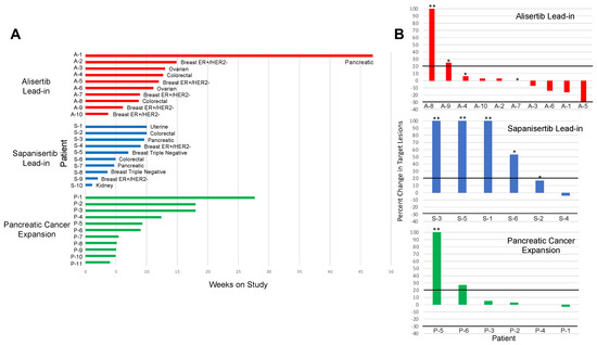
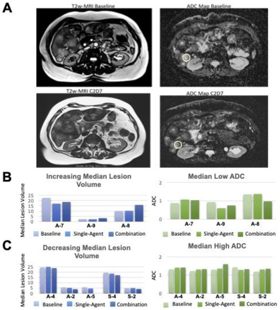
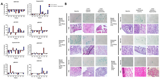
Background: This study further evaluated the safety and efficacy of the combination of alisertib and sapanisertib in an expansion cohort of patients, including a subset of patients with refractory pancreatic adenocarcinoma, with further evaluation of the pharmacodynamic characteristics of combination therapy. Methods: Twenty patients with refractory solid tumors and 11 patients with pancreatic adenocarcinoma were treated at the recommended phase 2 dose of alisertib and sapanisertib. Adverse events and disease response were assessed. Patients in the expansion cohort were treated with a 7-day lead-in of either alisertib or sapanisertib prior to combination therapy, with tumor tissue biopsy and serial functional imaging performed for correlative analysis. Results: Toxicity across treatment groups was overall similar to prior studies. One partial response to treatment was observed in a patient with ER positive breast cancer, and a patient with pancreatic cancer experienced prolonged stable disease. In an additional cohort of pancreatic cancer patients, treatment response was modest. Correlative analysis revealed variability in markers of apoptosis and immune cell infiltrate according to lead-in therapy and response. Conclusions: Dual targeting of Aurora A kinase and mTOR resulted in marginal clinical benefit in a population of patients with refractory solid tumors, including pancreatic adenocarcinoma, though individual patients experienced significant response to therapy. Correlatives indicate apoptotic response and tumor immune cell infiltrate may affect clinical outcomes.
Full article
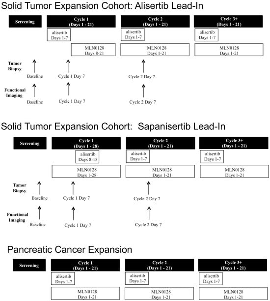
Figure 1


