Journal Description
Veterinary Sciences
Veterinary Sciences
is an international, peer-reviewed, open access journal on veterinary sciences published monthly online by MDPI.
- Open Access— free for readers, with article processing charges (APC) paid by authors or their institutions.
- High visibility: indexed within Scopus, SCIE (Web of Science), PubMed, PMC, Embase, PubAg, AGRIS, and other databases.
- Journal Rank: JCR - Q2 (Veterinary Sciences) / CiteScore - Q2 (General Veterinary)
- Rapid Publication: manuscripts are peer-reviewed and a first decision is provided to authors approximately 21.2 days after submission; acceptance to publication is undertaken in 2.7 days (median values for papers published in this journal in the second half of 2024).
- Recognition of Reviewers: reviewers who provide timely, thorough peer-review reports receive vouchers entitling them to a discount on the APC of their next publication in any MDPI journal, in appreciation of the work done.
Impact Factor:
2.0 (2023);
5-Year Impact Factor:
2.2 (2023)
Latest Articles
Fecal Microbiota and Performance of Dairy Cattle from a West Mexican Family Dairy Farm Supplemented with a Fiber-Degrading Enzymatic Complex
Vet. Sci. 2025, 12(6), 518; https://doi.org/10.3390/vetsci12060518 - 25 May 2025
Abstract
►
Show Figures
Non-starch polysaccharide-degrading enzymes are widely used as feed additives in monogastric and ruminant species, with positive effects reported. In this study, the commercial, fiber-degrading enzyme complex Hostazym® X, derived from Trichoderma citrinoviride (DSM34663), was included in the total mixed rations of 17
[...] Read more.
Non-starch polysaccharide-degrading enzymes are widely used as feed additives in monogastric and ruminant species, with positive effects reported. In this study, the commercial, fiber-degrading enzyme complex Hostazym® X, derived from Trichoderma citrinoviride (DSM34663), was included in the total mixed rations of 17 mid-lactating (135 ± 61 days in milk) Holstein cows for 10 weeks. A control group (n = 17) was included. Dry matter intake (DMI), milk yield, 4% fat-corrected milk, solid yield, and milk fatty acid profile were assessed. The structure and composition of fecal bacterial communities, as well as PICRUSt2-based functional prediction of bacterial communities, were also evaluated. Higher DMI and milk yield scores were observed in the supplemented group (27.20 vs. 26.59 kgDM/cow/d; and 39.01 vs. 36.70 L/cow/d, respectively). No effects were observed in fat yield, contrary to lactose and protein, which were greater in the supplemented group compared to the control group (1.18 vs. 1.13 and 1.83 vs. 1.75 kg/cow/d, respectively; p < 0.05). Palmitic and oleic acids, in addition to monounsaturated fat in milk, were increased in the supplemented group (p > 0.05). Enzyme supplementation increased the Patescibacteria (p < 0.5) and Actinobacteriota (p > 0.05) in feces, but slightly reduced the Bacteroidota and Firmicutes. The Turicibacter genus remained at a lower relative abundance after supplementation but Candidatus_Saccharimonas, Clostridioides, Prevotellaceae UCG 003, Corynebacterium, Akkermansia, Syntrophococcus, Erysipelotrichaceae UCG 008, other Lachnospiraceae, other members of the Eubacterium_coprostanoligenes_group, Bifidobacterium, Rumminococcus, Akkermansia, and other Spirochaetaceae increased, modifying the functional predicted profile of bacterial communities. In conclusion, a positive effect on performance and milk composition were observed through modulation of microbiota induced by enzyme supplementation. The enzyme complex could be a viable supplement alternative in the feeding of dairy cows in semi-intensive productive systems, mainly when an ad libitum feeding scheme is used.
Full article
Open AccessArticle
Morphological and Ultrastructural Insights into the Goldfish (Carassius auratus) Spleen: Immune Organization and Cellular Composition
by
Doaa M. Mokhtar, Giacomo Zaccone and Manal T. Hussein
Vet. Sci. 2025, 12(6), 517; https://doi.org/10.3390/vetsci12060517 - 25 May 2025
Abstract
The spleen plays a critical role in the immune and hematopoietic systems of teleost fish, functioning as a major secondary lymphoid organ. This study provides a detailed morphological and ultrastructural assessment of the spleen in goldfish (Carassius auratus), focusing on its
[...] Read more.
The spleen plays a critical role in the immune and hematopoietic systems of teleost fish, functioning as a major secondary lymphoid organ. This study provides a detailed morphological and ultrastructural assessment of the spleen in goldfish (Carassius auratus), focusing on its immunological organization and cellular diversity. Through light and transmission electron microscopy, we examined red and white pulps, identifying key features such as melanomacrophage centers (MMCs), ellipsoids, and various immune cell types. The red pulp was rich in sinusoidal capillaries and splenic cords, whereas the white pulp housed lymphocytes, dendritic cells, macrophages, and telocytes, all contributing to immune regulation. Notably, ellipsoids were surrounded by reticular and macrophage sheaths, forming a filtration barrier against pathogens. Ultrastructural analysis revealed diverse immune cells with active morphological traits, including macrophages with pseudopodia and pigment granules, dendritic cells with dendrite-like extensions, and epithelial reticular cells involved in forming the blood–spleen barrier. These findings highlight the complex immunological microarchitecture of the goldfish spleen and its functional relevance in teleost immune responses.
Full article
(This article belongs to the Section Anatomy, Histology and Pathology)
►▼
Show Figures
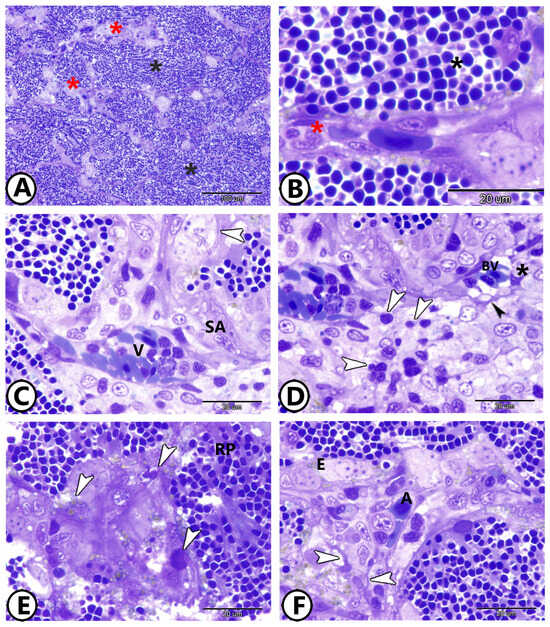
Figure 1
Open AccessArticle
Exploring Antimicrobial Resistance in Bacteria from Fecal Samples of Insectivorous Bats: A Preliminary Study
by
Santina Di Bella, Delia Gambino, Maria Foti, Bianca Maria Orlandella, Vittorio Fisichella, Francesca Gucciardi, Francesco Mira, Rosario Grasso, Maria Teresa Spena, Giuseppa Purpari and Annalisa Guercio
Vet. Sci. 2025, 12(6), 516; https://doi.org/10.3390/vetsci12060516 - 25 May 2025
Abstract
Bats (order Chiroptera) are increasingly recognized as important reservoirs and potential vectors of pathogenic and antimicrobial-resistant bacteria (ARB), with potential implications for human, animal, and environmental health. This study aimed to assess the presence and antimicrobial resistance profiles of bacterial isolates from bat
[...] Read more.
Bats (order Chiroptera) are increasingly recognized as important reservoirs and potential vectors of pathogenic and antimicrobial-resistant bacteria (ARB), with potential implications for human, animal, and environmental health. This study aimed to assess the presence and antimicrobial resistance profiles of bacterial isolates from bat populations in Sicily, an area for which data are currently limited. A total of 132 samples (120 rectal swabs and 12 guano samples) were collected at four sites in the provinces of Catania, Siracusa, and Ragusa. Bacteriological analysis yielded 213 isolates, including 161 Gram-negative and 52 Gram-positive strains, representing 55 different species. Among Gram-negative isolates, Escherichia coli, Citrobacter freundii, and Morganella morganii were most frequently detected, while Bacillus licheniformis and Staphylococcus xylosus were predominant among Gram-positive bacteria. Antimicrobial susceptibility testing revealed high resistance rates to colistin, amoxicillin, and ampicillin in Gram-negative strains, and to oxacillin, ceftazidime, and lincomycin in Gram-positive strains. Notably, 84.5% of isolates exhibited multidrug resistance. These findings highlight the potential role of bats as reservoirs of ARB and underline the importance of ongoing monitoring within a One Health framework to mitigate risks to public and animal health.
Full article
(This article belongs to the Special Issue Monitoring Wildlife Health: Surveillance and Management of Infectious Diseases)
►▼
Show Figures
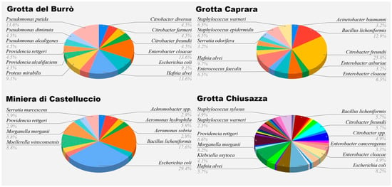
Figure 1
Open AccessArticle
Genetic Diversity of Canine Circovirus Detected in Wild Carnivores in Serbia
by
Damir Benković, Jakov Nišavić, Nenad Milić, Dejan Krnjaić, Isidora Prošić, Vladimir Gajdov, Nataša Stević, Ratko Sukara, Martina Balać and Andrea Radalj
Vet. Sci. 2025, 12(6), 515; https://doi.org/10.3390/vetsci12060515 - 24 May 2025
Abstract
Canine circovirus (CanineCV) is an emerging virus of interest in both domestic and wild carnivores that is scarcely reported in southeastern Europe. This study examined the presence, genetic diversity, and evolutionary characteristics of CanineCV in red foxes (Vulpes vulpes) and golden
[...] Read more.
Canine circovirus (CanineCV) is an emerging virus of interest in both domestic and wild carnivores that is scarcely reported in southeastern Europe. This study examined the presence, genetic diversity, and evolutionary characteristics of CanineCV in red foxes (Vulpes vulpes) and golden jackals (Canis aureus) from northwestern Serbia, a region marked by expanding mesopredator populations overlapping with human habitats. Out of 98 sampled animals, circoviral DNA was detected in 31.6%. Jackals were mostly positive for CanineCV genotype 4, while genotype 5, associated with wild carnivores, was dominant in foxes. Mixed genotype 4/genotype 5 infections were only found in jackals. Phylogenetic and haplotype analyses indicated that most jackal-derived CanineCV strains clustered along sequences from Europe, Africa, and the Americas, while genotype 5 sequences grouped separately from other genotype representatives. A recombinant strain was identified as a divergent lineage, and several sequences showed evidence of recombination between Rep and Cap genes. Despite Cap protein amino acid differences, purifying selection dominated, suggesting functional constraints on viral evolution. The results indicate that jackals may act as recombination hotspots and bridging hosts between viral lineages. This study provides insight into the molecular epidemiology of CanineCV in the Balkans, highlighting the importance of ongoing surveillance.
Full article
(This article belongs to the Special Issue Monitoring Wildlife Health: Surveillance and Management of Infectious Diseases)
Open AccessFeature PaperReview
Ovotransferrin as a Multifunctional Bioactive Protein: Unlocking Its Potential in Animal Health and Wellness
by
Sahdeo Prasad, Bhaumik Patel, Prafulla Kumar, Jeffrey Kaufman and Rajiv Lall
Vet. Sci. 2025, 12(6), 514; https://doi.org/10.3390/vetsci12060514 - 24 May 2025
Abstract
►▼
Show Figures
Ovotransferrin (OVT) is one of the major proteins of egg white and is known to bind and transport irons in animals. OVT exerts bacteriostatic and bactericidal activities due to its iron-binding capacity. OVT effectively controls the growth of various pathogenic microorganisms, including Pseudomonas,
[...] Read more.
Ovotransferrin (OVT) is one of the major proteins of egg white and is known to bind and transport irons in animals. OVT exerts bacteriostatic and bactericidal activities due to its iron-binding capacity. OVT effectively controls the growth of various pathogenic microorganisms, including Pseudomonas, E. coli, Staphylococcus, Proteus, and Klebsiella species, as well as inhibiting the replication of viruses. OVT also has antioxidant, anti-inflammatory, anti-hypertensive, anticancer, and immuno-stimulating properties. For instances, OVT quenches free radicals, induces antioxidant enzymes, suppresses pro-inflammatory cytokines, increases immune cells, reduces angiotensin-converting enzymes, and inhibits the proliferation of cancer cells. In this review, the beneficial effects of OVT in both in vitro and in vivo, particularly livestock, are described. Because of its antimicrobial properties, OVT supplementation in livestock feed would be an excellent alternative to antibiotics, which reducing the development of antibiotic-resistant pathogenic microorganisms. Thus, OVT could be a game-changing protein for the growth, performance, and healthy life of animals.
Full article
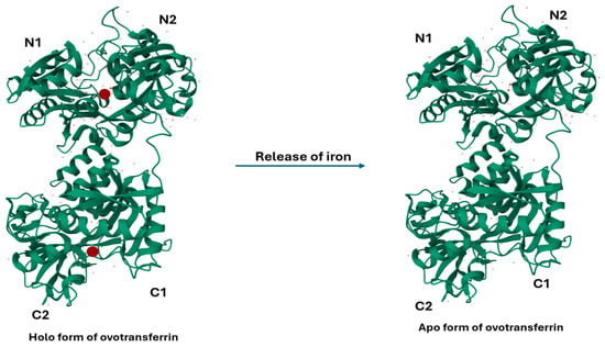
Figure 1
Open AccessArticle
Mining Porcine Blood Whole-DNA Sequencing Datasets to Uncover Pig Viromes: An Exploratory Application to Identify Potential Infecting Agents of an Undefined Disease Outbreak
by
Samuele Bovo, Anisa Ribani, Giuseppina Schiavo, Valeria Taurisano, Matteo Bolner, Francesca Bertolini and Luca Fontanesi
Vet. Sci. 2025, 12(6), 513; https://doi.org/10.3390/vetsci12060513 - 24 May 2025
Abstract
Pigs are affected by a variety of pathogenic agents that need to be identified correctly and diagnosed even when co-infections may complicate the application of specific and targeted assays. Next-generation sequencing can provide new perspective to monitor viruses infecting or co-infecting diseased pigs.
[...] Read more.
Pigs are affected by a variety of pathogenic agents that need to be identified correctly and diagnosed even when co-infections may complicate the application of specific and targeted assays. Next-generation sequencing can provide new perspective to monitor viruses infecting or co-infecting diseased pigs. In this study, we tested, for the first time for diagnostic purposes in a livestock species, a new method based on whole-genome sequencing of all the DNAs extracted from the blood of nine pigs sampled from a farm where there was a suspected outbreak of Post-weaning Multisystemic Wasting Syndrome. We then used unmapped reads on the porcine reference genome to mine for viral sequences using a specifically designed bioinformatic pipeline. Within this fraction of reads, viral sequences ranged from 0.002% to 4.4% of the total unmapped reads and were derived from twelve different viruses known to infect pigs, where three were herpesviruses, eight were parvoviruses, and one was a circovirus. All pig sequencing datasets were positive for one or more viruses, with various potential viral loads. Suid betaherpesvirus 2, also known as Porcine cytomegalovirus (PCMV), was the most frequently identified virus as five out of the nine pig sequencing datasets contained viral sequences from this virus. The results may suggest a heterogeneous viral profile of the diseased pigs that may be derived from potential secondary infections or co-infections. This pilot application demonstrated that a whole-genome sequencing approach can complement other routine diagnostic assays in veterinary virology. Other studies and improvements are needed to validate the results and apply this approach in routine monitoring applications.
Full article
(This article belongs to the Section Veterinary Biomedical Sciences)
►▼
Show Figures
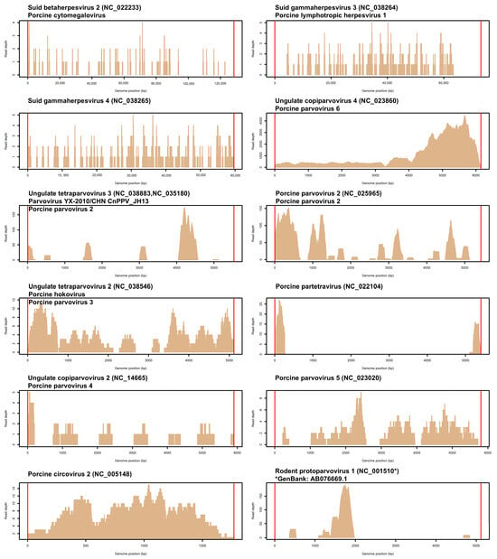
Figure 1
Open AccessArticle
Assessing Q Fever Exposure in Veterinary Professionals: A Study on Seroprevalence and Awareness in Portugal, 2024
by
Guilherme Moreira, Mário Ribeiro, Miguel Martins, José Maria Cardoso, Fernando Esteves, Sofia Anastácio, Sofia Duarte, Helena Vala, Rita Cruz and João R. Mesquita
Vet. Sci. 2025, 12(6), 512; https://doi.org/10.3390/vetsci12060512 - 23 May 2025
Abstract
►▼
Show Figures
Due to their frequent contact with animals, veterinarians may be at preferential risk of Coxiella burnetii exposure due to occupational contact with livestock. This study assesses the seroprevalence and risk factors associated with C. burnetii seropositivity in Portuguese veterinarians. A cross-sectional study compared
[...] Read more.
Due to their frequent contact with animals, veterinarians may be at preferential risk of Coxiella burnetii exposure due to occupational contact with livestock. This study assesses the seroprevalence and risk factors associated with C. burnetii seropositivity in Portuguese veterinarians. A cross-sectional study compared IgG anti-C. burnetii in veterinarians’ sera to a demographically matched control group. Univariate and multivariate logistic regression analyses evaluated associations between the demographic, occupational, and biosecurity factors and seropositivity. Seroprevalence among veterinarians was 33.7%, significantly higher (p = 0.0023) than in the controls (17.39%). Univariate analysis identified higher seropositivity in the northern region (p = 0.03), though this association was not significant after adjustment (p = 0.07). Protective measures, including isolating aborting animals from the rest of the herd (adjusted OR [aOR]: 0.35, p = 0.03) and wearing gloves during sample collection (OR: 0.28, p = 0.009), were significantly associated with lower infection risk. Veterinarians face increased C. burnetii exposure, but specific biosecurity practices reduce risk. Strengthening preventive measures, including personal protective equipment (PPE) use and biosecurity training, is essential to mitigate occupational and public health risks. Further research should explore vaccination strategies and molecular epidemiology to improve risk reduction efforts.
Full article
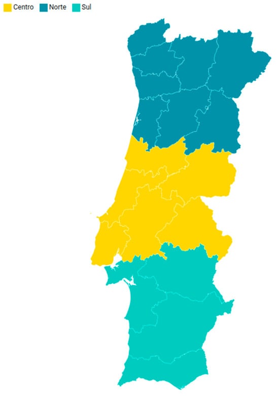
Figure 1
Open AccessArticle
Computed Tomography of the Hyoid Apparatus in Equine Headshaking Syndrome
by
Ralph A. Lloyd-Edwards, Eva Mulders, Marianne M. Sloet van Oldruitenborgh-Oosterbaan and Stefanie Veraa
Vet. Sci. 2025, 12(6), 511; https://doi.org/10.3390/vetsci12060511 - 23 May 2025
Abstract
►▼
Show Figures
Introduction: Headshaking is a common condition in horses, most cases are presumed idiopathic/trigeminal-nerve mediated. Diagnostic work-up of a headshaking horse may involve computed tomography (CT) of the head to exclude causative structural pathology. The relevance of the presence and severity of hyoid apparatus
[...] Read more.
Introduction: Headshaking is a common condition in horses, most cases are presumed idiopathic/trigeminal-nerve mediated. Diagnostic work-up of a headshaking horse may involve computed tomography (CT) of the head to exclude causative structural pathology. The relevance of the presence and severity of hyoid apparatus findings at CT to headshaking is unknown. Materials and methods: A retrospective analysis of CT changes in the hyoid apparatus in horses was carried out. Comparisons were performed between horses with signs of headshaking and a control population and a subgroup of horses with signs of headshaking and no other ‘likely relevant findings’ to headshaking and the control population. Results: The grade of temporohyoid joint sheath ossification, mineralisation of the tympanohyoid cartilage, and widening and narrowing of the temporohyoid joint all showed significant correlation with age. Findings of the remaining hyoid apparatus (fracture, deformation, or arthropathy) showed significant correlation with temporohyoid joint grade. Centres of ossification of the epihyoid, thyrohyoid, and lingual processes were described. No consistent association of headshaking to hyoid changes was found. Odds ratios were increased in many cases, particularly in comparisons of the subgroup with no ‘likely relevant findings’; however, statistical significance was not reached. Conclusions: CT findings of the temporohyoid joint are not consistently associated with clinical signs of headshaking.
Full article
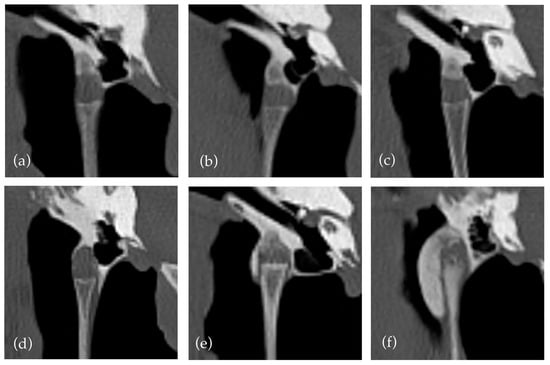
Figure 1
Open AccessArticle
The Protein Encoded by the UL3.5 Gene of the Duck Plague Virus Affects Viral Secondary Envelopment, Release, and Cell-to-Cell Spread
by
Huanhuan Cao, Bin Tian, Yanming Tian, Dongjie Cai, Mingshu Wang, Renyong Jia, Shun Chen and Anchun Cheng
Vet. Sci. 2025, 12(6), 510; https://doi.org/10.3390/vetsci12060510 - 23 May 2025
Abstract
Duck plague (DP), caused by duck plague virus (DPV), is a highly contagious and fatal disease among waterfowl. UL3.5, an unconserved gene belonging to the Herpesviridae family, Alphaherpesvirinae subfamily, and Mardivirus genus, is located downstream of UL3 and exhibits high variability in size
[...] Read more.
Duck plague (DP), caused by duck plague virus (DPV), is a highly contagious and fatal disease among waterfowl. UL3.5, an unconserved gene belonging to the Herpesviridae family, Alphaherpesvirinae subfamily, and Mardivirus genus, is located downstream of UL3 and exhibits high variability in size and sequence, with an absence in herpes simplex virus (HSV). Currently, there is little understanding of DPV UL3.5. In this study, we determined that DPV pUL3.5 is distributed within the cytoplasm and co-located with multiple organelles. In addition, we investigated the genetic type of DPV UL3.5 and found that it is an early gene encoding an early viral protein. To further explore the function of DPV UL3.5, we constructed DPV-BAC-δUL3.5 and discovered that the deletion of UL3.5 significantly impacts the viral secondary envelopment and release processes. Furthermore, the UL3.5-deleted virus shows defects in cell-to-cell spread. In conclusion, our findings demonstrate, for the first time, that the early viral protein encoded by DPV UL3.5 plays a crucial role in promoting viral replication. This offers fundamental insights for further investigations into the function of DPV UL3.5.
Full article
(This article belongs to the Special Issue Molecular Characterization of Various Viruses and Bacteria in Small Animals)
►▼
Show Figures

Figure 1
Open AccessArticle
Development of an Immunochromatographic Test with Recombinant MIC2-MIC3 Fusion Protein for Serological Detection of Toxoplasma gondii
by
Jianzhong Wang, Yi Zhao, Jicheng Qiu, Jing Liu, Rui Zhou, Xialin Ma, Xiaojie Wu, Xiaoguang Li, Wei Mao, Yiduo Liu and Heng Zhang
Vet. Sci. 2025, 12(6), 509; https://doi.org/10.3390/vetsci12060509 - 22 May 2025
Abstract
Toxoplasma gondii is a globally significant zoonotic pathogen responsible for severe parasitic diseases in humans and animals. This study aimed to design, develop, and evaluate a novel immunochromatographic test (ICT) using a recombinant MIC2-MIC3 fusion protein (rMIC2-MIC3) for detecting specific antibodies against T.
[...] Read more.
Toxoplasma gondii is a globally significant zoonotic pathogen responsible for severe parasitic diseases in humans and animals. This study aimed to design, develop, and evaluate a novel immunochromatographic test (ICT) using a recombinant MIC2-MIC3 fusion protein (rMIC2-MIC3) for detecting specific antibodies against T. gondii. The ICT demonstrated exceptional sensitivity, capable of detecting T. gondii-specific antibodies in sera diluted up to 1:8. Specificity evaluation confirmed no cross-reactivity with antibodies against other parasites, such as Neospora caninum, Cryptosporidium suis, Eimeria tenella, and Sarcocystis tenella. Stability tests revealed the test strips maintained full functionality after 12 weeks of storage at 24 °C. The coincidence rate of the colloidal gold test strips prepared in this study with a commercial ELISA kit was 94.59%. Comparisons with advanced serodiagnostic tools, such as chimeric antigen-based ELISAs and recombinant protein diagnostics, further highlighted its robustness and applicability. These findings underscore the potential of the rMIC2-MIC3-based ICT as a reliable, economical, and accessible diagnostic tool for toxoplasmosis in veterinary and human medicine.
Full article
(This article belongs to the Section Veterinary Microbiology, Parasitology and Immunology)
►▼
Show Figures
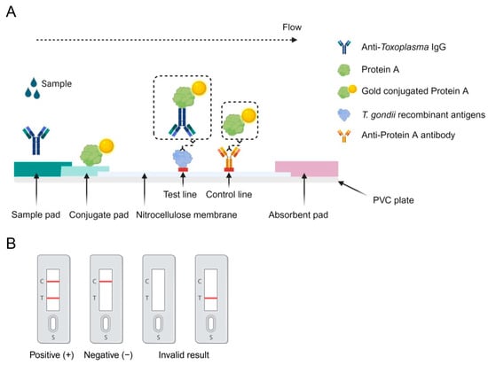
Figure 1
Open AccessArticle
Fine Mapping Identifies Candidate Genes Associated with Swine Inflammation and Necrosis Syndrome
by
Katharina Gerhards, Sabrina Becker, Josef Kühling, Joel Mickan, Mirjam Lechner, Hermann Willems and Gerald Reiner
Vet. Sci. 2025, 12(5), 508; https://doi.org/10.3390/vetsci12050508 - 21 May 2025
Abstract
►▼
Show Figures
Swine inflammation and necrosis syndrome (SINS) is a widespread disease in pigs, causing pain, suffering, and damage. Inflammation is documented at different levels based on clinical signs, histopathology, clinical chemistry, metabolomics and transcriptomics. The influence of sow and boar, as well as a
[...] Read more.
Swine inflammation and necrosis syndrome (SINS) is a widespread disease in pigs, causing pain, suffering, and damage. Inflammation is documented at different levels based on clinical signs, histopathology, clinical chemistry, metabolomics and transcriptomics. The influence of sow and boar, as well as a heritability of around 0.3, suggest a genetic component to the disease. The aim of the present study was to identify functional single nucleotide polymorphisms (SNPs) in the vicinity of gene markers previously mapped using GWAS. DNA samples were available from 234 already phenotyped piglets. These animals were re-sequenced with additional prior enrichment. The nine selected chromosomal regions cover a total length of 22 Mbp. The genome-wide association study (GWAS) revealed two series with a total of 15 significant missense polymorphisms on chromosomes 11, 14, and 15. The homozygous genotypes of the most discriminating SNPs in series 1 resulted in SINS scores of 3.5 and 17.9, respectively. Despite the partial linkage of the SNPs, interesting candidate genes were defined. The results allow a significant narrowing of the possible candidate genes for understanding the pathogenesis of SINS and for future use in selection breeding to overcome the syndrome. Further studies should be carried out on larger animal populations.
Full article
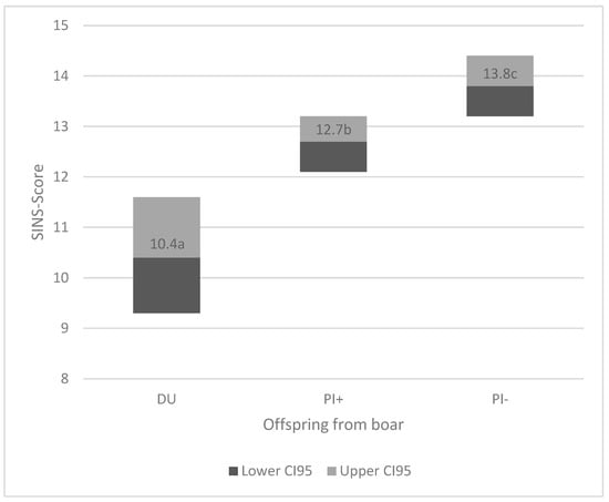
Figure 1
Open AccessArticle
Polyhexamethylene Biguanide Nanoparticles Inhibit Biofilm Formation by Mastitis-Causing Staphylococcus aureus
by
Renata de Freitas Leite, Breno Luis Nery Garcia, Kristian da Silva Barbosa, Thatiane Mendes Mitsunaga, Carlos Eduardo Fidelis, Bruna Juliana Moreira Dias, Renata Rank de Miranda, Valtencir Zucolotto, Liam Good and Marcos Veiga dos Santos
Vet. Sci. 2025, 12(5), 507; https://doi.org/10.3390/vetsci12050507 - 21 May 2025
Abstract
Staphylococcus aureus is a mastitis pathogen that compromises cow health and causes significant economic losses in the dairy industry. High antimicrobial resistance and biofilm formation by S. aureus limit the efficacy of conventional treatments. This study evaluated the potential of polyhexamethylene biguanide nanoparticles
[...] Read more.
Staphylococcus aureus is a mastitis pathogen that compromises cow health and causes significant economic losses in the dairy industry. High antimicrobial resistance and biofilm formation by S. aureus limit the efficacy of conventional treatments. This study evaluated the potential of polyhexamethylene biguanide nanoparticles (PHMB NPs) against mastitis-causing S. aureus. PHMB NPs showed low toxicity to bovine mammary epithelial cells (MAC-T cells) at concentrations up to four times higher than the minimum inhibitory concentration (1 µg/mL) against S. aureus. In Experiment 1, PHMB NPs significantly reduced biofilm formation by S. aureus by 50% at concentrations ≥1 µg/mL, though they showed limited efficacy against preformed biofilms. In Experiment 2, using an excised teat model, PHMB NPs reduced S. aureus concentrations by 37.57% compared to conventional disinfectants (chlorhexidine gluconate, povidone–iodine, and sodium dichloroisocyanurate), though limited by short contact time. These findings highlight the potential of PHMB NPs for the control of S. aureus growth and biofilm formation.
Full article
(This article belongs to the Special Issue Advancements in Livestock Staphylococcus sp.)
►▼
Show Figures
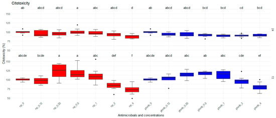
Figure 1
Open AccessArticle
Environment-Associated Variations in Blood Metabolism of Mongolian Cattle Grazing in the Alxa Desert of China
by
Chao Hai, Dongchao Pei, Yuqing Yang, Lishuang Song, Xuefei Liu, Chunling Bai, Guanghua Su, Lei Yang and Guangpeng Li
Vet. Sci. 2025, 12(5), 506; https://doi.org/10.3390/vetsci12050506 - 21 May 2025
Abstract
Desert environments pose severe challenges to livestock survival. This study examined climate-driven physiological and metabolic adaptations in 258 Mongolian cattle from six regions of the Alxa Desert, China. Serum biochemical indices were measured and analyzed using linear models to assess the effects of
[...] Read more.
Desert environments pose severe challenges to livestock survival. This study examined climate-driven physiological and metabolic adaptations in 258 Mongolian cattle from six regions of the Alxa Desert, China. Serum biochemical indices were measured and analyzed using linear models to assess the effects of climate, sex, and age. Climate significantly affected key blood parameters, including glucose (p < 0.001), creatinine (p < 0.001), alkaline phosphatase (p < 0.001), and lactate (p = 0.034). Additionally, sex significantly influenced lactate dehydrogenase (p = 0.049), bicarbonate (p = 0.0061), urea (p = 0.0055), and triglycerides (p = 0.039), while age affected total protein (p = 0.020), LDL-C (p = 0.0097), and cholesterol (p < 0.001). Glucose levels were negatively correlated with body size traits. Metabolomic profiling showed that cattle in arid, high-radiation areas exhibited reduced TCA cycle and fatty acid metabolism, with concurrent carbohydrate accumulation, including glucose, fructose, and mannose. Enhanced amino acid metabolism increased proline, valine, tyrosine, and tryptophan levels, potentially supporting physiological stability under heat and drought stress. These findings reveal how Mongolian cattle modulate metabolism in response to desert climates, offering insights into livestock adaptation and informing breeding strategies for resilience in harsh environments.
Full article
(This article belongs to the Section Nutritional and Metabolic Diseases in Veterinary Medicine)
►▼
Show Figures

Figure 1
Open AccessReview
Bluetongue’s New Frontier—Are Dogs at Risk?
by
Rita Payan-Carreira and Margarida Simões
Vet. Sci. 2025, 12(5), 505; https://doi.org/10.3390/vetsci12050505 - 20 May 2025
Abstract
Bluetongue virus (BTV), traditionally considered a pathogen of ruminants, has recently been documented in dogs, challenging conventional understanding of its epidemiology. This narrative review synthesizes emerging evidence regarding BTV infections in domestic and wild carnivores, examining transmission dynamics, pathogenesis, clinical manifestations, and diagnostic
[...] Read more.
Bluetongue virus (BTV), traditionally considered a pathogen of ruminants, has recently been documented in dogs, challenging conventional understanding of its epidemiology. This narrative review synthesizes emerging evidence regarding BTV infections in domestic and wild carnivores, examining transmission dynamics, pathogenesis, clinical manifestations, and diagnostic challenges. Carnivores can become infected through vector transmission and oral ingestion of infected material. While some infected carnivores remain subclinical, others develop severe clinical manifestations including hemorrhagic syndromes. BTV infection in carnivores is likely underdiagnosed due to limited awareness, nonspecific clinical signs, and absence of established diagnostic protocols for non-ruminant species. The potential role of carnivores in BTV epidemiology remains largely unexplored, raising questions about their function as reservoirs or dead-end hosts. Additionally, carnivores may contribute to alternative transmission pathways and overwintering mechanisms that impact disease ecology. Current biosecurity frameworks and surveillance systems, primarily focused on ruminants, require expansion to incorporate carnivores in viral maintenance and transmission. This review identifies significant knowledge gaps regarding BTV in carnivores and proposes future research directions, including serological surveys, transmission studies, and investigation of viral tropism in carnivore tissues. A comprehensive One Health approach integrating diverse host species, vector ecology, human interference, and environmental factors is crucial for effective BTV control and impact mitigation on human, animals, and environment.
Full article
(This article belongs to the Section Veterinary Biomedical Sciences)
►▼
Show Figures
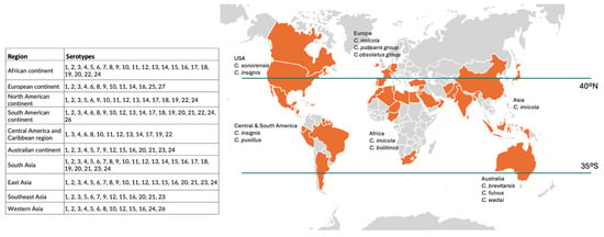
Figure 1
Open AccessArticle
Epidemiological Exploration of Cryptosporidium spp., Giardia intestinalis, Enterocytozoon bieneusi, and Blastocystis spp. in Yaks: Investigating Ecological and Zoonotic Dynamics in Lhasa, Xizang
by
Yaru Ji, Munwar Ali, Chang Xu, Jia Wang, Md. F. Kulyar, Shah Nawaz, Khalid Mehmood, Mingming Liu and Kun Li
Vet. Sci. 2025, 12(5), 504; https://doi.org/10.3390/vetsci12050504 - 20 May 2025
Abstract
►▼
Show Figures
The yak (Bos grunniens), prevalent at an altitude between 3000 and 5000 m above sea level, provides the local inhabitants with meat, milk, leather, fuel (dung), and transport. However, intestinal zoonotic parasites seriously endanger its holistic well-being. The prime concern of
[...] Read more.
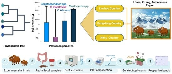
The yak (Bos grunniens), prevalent at an altitude between 3000 and 5000 m above sea level, provides the local inhabitants with meat, milk, leather, fuel (dung), and transport. However, intestinal zoonotic parasites seriously endanger its holistic well-being. The prime concern of this study is to investigate the prevalence of four globally ubiquitous zoonotic enteric protozoans, namely Cryptosporidium spp., G. intestinalis, Blastocystis spp., and E. bieneusi in yaks from different areas of Lhasa, Xizang. In the given study, 377 yak fecal samples from various regions in Lhasa were obtained, including 161 samples from Linzhou County, 66 samples from Dangxiong County, and 150 samples from the Nimu County cattle farms. Molecular identification of these protozoans was done after amplification using PCR and sequencing of PCR-positive samples, and further phylogenetic analysis was performed. The results indicated that the prevalence of Cryptosporidium spp., G. intestinalis, E. bieneusi, and Blastocystis spp. in yak farms in Linzhou County was 48.5, 22.9, 47.8, and 90.7%; 65.2, 13.6, 72.7, and 87.9% in Dangxiong County; and 56.0, 29.3, 58.0, and 80.0%, respectively, in Nimu County. The results of this study provide a basic reference for preventing and controlling intestinal parasites in yaks in Lhasa, Xizang.
Full article
Graphical abstract
Open AccessArticle
Generation and Immunogenicity of Virus-like Particles Based on the Capsid Protein of a Chinese Epidemic Strain of Feline Panleukopenia Virus
by
Erkai Feng, Guoliang Luo, Chunxia Wang, Wei Liu, Ruxun Yan, Xue Bai and Yuening Cheng
Vet. Sci. 2025, 12(5), 503; https://doi.org/10.3390/vetsci12050503 - 20 May 2025
Abstract
Feline panleukopenia (FPL), caused by the feline panleukopenia virus (FPLV), is a severe and highly contagious viral disease with high morbidity and mortality. Vaccination remains the gold standard for preventing and controlling this debilitating condition. The viral protein VP2 serves as the major
[...] Read more.
Feline panleukopenia (FPL), caused by the feline panleukopenia virus (FPLV), is a severe and highly contagious viral disease with high morbidity and mortality. Vaccination remains the gold standard for preventing and controlling this debilitating condition. The viral protein VP2 serves as the major immunogen of FPLV and represents the key target antigen in the development of a novel FPLV vaccine. Virus-like particle (VLP)-based vaccines have emerged as next-generation vaccine candidates due to their high immunogenicity and safe profile. In this study, a baculovirus expression vector system (BEVS) was employed to generate FPLV-VLPs through recombinant expression of the VP2 protein of a Chinese epidemic strain (Ala91Ser, Ile101Thr) of FPLV. The resulting FPLV-VLPs demonstrated markedly enhanced antigenicity and hemagglutination activity, achieving a hemagglutination titer of up to 1:216. Following vaccination, immunized cats developed high titers of anti-FPLV hemagglutination inhibition (HI) antibodies (1:216) and exhibited 100% protection against challenge with a virulent epidemic FPLV variant (Ala91Ser, Ile101Thr). These findings demonstrate that FPLV-VLPs hold strong potential as candidates for a novel subunit vaccine against FPLV infection.
Full article
(This article belongs to the Special Issue Gastrointestinal Disease and Health in Pets)
►▼
Show Figures

Graphical abstract
Open AccessArticle
Detection and Comparison of Sow Serum Samples from Herds Regularly Mass Vaccinated with Porcine Reproductive and Respiratory Syndrome Modified Live Virus Using Four Commercial Enzyme-Linked Immunosorbent Assays and Neutralizing Tests
by
Chaosi Li, Gang Wang, Zhicheng Liu, Shuhe Fang, Aihua Fan, Kai Chen and Jianfeng Zhang
Vet. Sci. 2025, 12(5), 502; https://doi.org/10.3390/vetsci12050502 - 20 May 2025
Abstract
Porcine reproductive and respiratory syndrome virus (PRRSV) modified live virus (MLV) vaccination is used to control PRRSV. In China, farms conduct random sampling from sow herds every 4 to 6 months. They use the enzyme-linked immunosorbent assay (ELISA) method to monitor the immune
[...] Read more.
Porcine reproductive and respiratory syndrome virus (PRRSV) modified live virus (MLV) vaccination is used to control PRRSV. In China, farms conduct random sampling from sow herds every 4 to 6 months. They use the enzyme-linked immunosorbent assay (ELISA) method to monitor the immune status of the herd by tracking the positive rate or the sample-to-positive ratio. However, in farms that implement mass vaccination and have stable production, the positive rate of ELISA antibodies has decreased, especially in high-parity sows. This poses a considerable challenge to the current monitoring approach of PRRSV immunity. It remains unclear whether this reflects insufficient sensitivity of the kits for these special scenarios or the fact that the sows have truly lost immunity. In this study, 233 samples from four farms (A–D) across different regions of China were acquired. They were tested using four representative ELISA kits, two targeting the nucleocapsid protein (N) and two targeting the glycoprotein (GP) to evaluate PRRS immune status. The respective sample positive rates in A–D were 57.1–100%, 50.9–100%, 50–100%, and 75.7–100% using the kits. The positive rates using the four ELISA kits were 50.0–75.7%, 70.0–75.7%, 82.5–97.1%, and 100%, respectively, with poor agreement among them. The positive rates and humoral antibody levels for parity 1 and 2 sows were significantly lower than those with higher parities (>4). Eighty-eight ELISA-negative samples identified using ELISA kit A were verified using a viral neutralizing test (VNT), with only 15.9% of the samples testing negative. In conclusion, the ELISA antibody negativity issue existed, mostly occurring in specific farms tested using a specific kit. However, the low correlation with the VNT results and the poor agreements among the kits suggest that relying on one ELISA test is insufficient to monitor the immune status of PRRSV MLV-vaccinated herds.
Full article
(This article belongs to the Special Issue Exploring Innovative Approaches in Veterinary Health)
►▼
Show Figures
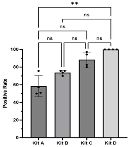
Figure 1
Open AccessCase Report
Vanishing Lung Syndrome in a Dog: Giant Pneumatocele or Giant Pulmonary Bulla Mimicking Tension Pneumothorax—First Report
by
Jack-Yves Deschamps, Nour Abboud, Pierre Penaud and Françoise A. Roux
Vet. Sci. 2025, 12(5), 501; https://doi.org/10.3390/vetsci12050501 - 20 May 2025
Abstract
A 6-month-old neutered male Belgian Malinois dog living in a kennel was presented to a veterinary emergency service for the management of severe respiratory distress that had developed within the past 24 h. Thoracic radiographs performed by a referring veterinarian showed abnormalities identified
[...] Read more.
A 6-month-old neutered male Belgian Malinois dog living in a kennel was presented to a veterinary emergency service for the management of severe respiratory distress that had developed within the past 24 h. Thoracic radiographs performed by a referring veterinarian showed abnormalities identified as a pneumothorax. Upon admission to the emergency service, the striking anomalies turned out to be a large intrathoracic air-filled cavity and countless smaller ones causing mechanical compression of the adjacent pulmonary parenchyma and mimicking tension pneumothorax. Emergency management included thoracocentesis followed by placement of a thoracostomy tube. The dog exhibited rapid clinical improvement and recovered completely within a few days, without requiring surgical intervention. Serial follow-up radiographs showed progressive and complete resolution of all lesions. Based on the complete resolution without resection, the main lesion—initially interpreted as a giant pulmonary bulla—was ultimately considered consistent with an acquired pneumatocele. To the authors’ knowledge, this is the first report in veterinary medicine of a vanishing lung syndrome presentation in a dog.
Full article
(This article belongs to the Special Issue Advancements in Small Animal Internal Medicine)
►▼
Show Figures
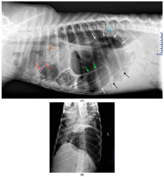
Figure 1
Open AccessArticle
Case Study on the Genetic Parameters and Possibilities of Selecting Gilts for Traits Monitored in the Performance Test
by
Nenad Stojiljković, Čedomir Radović, Marija Gogić, Vladimir Živković, Aleksandra Petrović, Krstina Zeljić Stojiljković and Dubravko Škorput
Vet. Sci. 2025, 12(5), 500; https://doi.org/10.3390/vetsci12050500 - 20 May 2025
Abstract
This research examined the phenotypic and genotypic variability of traits assessed in the gilt performance test and their subsequent impact on gilt selection. The traits evaluated in the gilt performance test were analyzed on two pig farms over a period of 3 consecutive
[...] Read more.
This research examined the phenotypic and genotypic variability of traits assessed in the gilt performance test and their subsequent impact on gilt selection. The traits evaluated in the gilt performance test were analyzed on two pig farms over a period of 3 consecutive years. A total of 3664 gilts were included in the research. At the end of the test, body weight, backfat thickness (BF1 and BF2), and longissimus dorsi muscle depth (MLD) were measured using an ultrasound device. The following breeds were evaluated on the farms: Landrace (L)–1981 gilts, Large White (LW)–1344 gilts, and Duroc (D)–339 gilts. In the analyzed population, direct genetic effects accounted for 0.2647 of the total variation in age at the end of the test (AET). Heritability coefficients of 0.37 for BF1 and 0.35 for BF2 indicate that these traits are highly heritable in the studied population. On the other hand, the heritability coefficient for the depth of MLD, which is 0.23, places this trait in the group of medium heritable traits. High heritability coefficients of these traits indicate great potential for genetic improvement through selection. The use of well-designed selection programs aimed at these traits can significantly accelerate the genetic improvement of the population and have an impact on the economic profit of pork production.
Full article
(This article belongs to the Special Issue Genetic Improvement and Reproductive Biotechnologies)
►▼
Show Figures
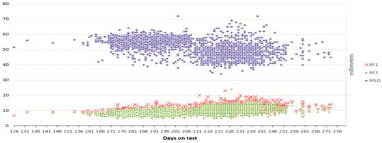
Figure 1
Open AccessArticle
Investigation of Galectin-3 and Cardiotrophin-1 Concentrations as Biomarkers in Dogs with Neurological Distemper
by
Alper Erturk, Aliye Sagkan Ozturk and Atakan Ozturk
Vet. Sci. 2025, 12(5), 499; https://doi.org/10.3390/vetsci12050499 - 20 May 2025
Abstract
Canine distemper, caused by Morbillivirus canis, is a highly morbid and lethal disease characterized by multiple systemic and neurological signs. In recent years, biomarkers, such as Galectin-3 and Cardiotrophin-1, have been investigated in inflammatory and degenerative diseases. However, the role of these
[...] Read more.
Canine distemper, caused by Morbillivirus canis, is a highly morbid and lethal disease characterized by multiple systemic and neurological signs. In recent years, biomarkers, such as Galectin-3 and Cardiotrophin-1, have been investigated in inflammatory and degenerative diseases. However, the role of these biomarkers in neurological distemper has not been investigated. The aim of this study is to compare blood serum Galectin-3 and Cardiotrophin-1 concentrations between the neurological distemper and control group, and to evaluate the correlations of these biomarkers with hematobiochemical parameters in dogs with neurological distemper. Nineteen owned dogs (13 diagnosed with neurological distemper and 6 controls) were included in the study. Hematobiochemical analyses were performed in all dogs, and Galectin-3 and Cardiotrophin-1 concentrations were measured using ELISA. Serum concentrations of Galectin-3 and Cardiotrophin-1 were markedly elevated in dogs with neurological distemper compared to the control group (p < 0.05). A negative correlation between Galectin-3 and monocytes (p < 0.05) and a positive correlation between Galectin-3 and platelet and platelecrit levels (p < 0.05) were observed. There was negative correlation with Cardiotrophin-1 and lymphocyte percentage (p < 0.01) and a positive correlation with Cardiotrophin-1 and granulocyte percentage (p < 0.01). Galectin-3 and Cardiotrophin-1 may serve as biomarkers for the diagnosis and understanding of neurological distemper pathogenesis. Elevated serum concentrations of these biomarkers may indicate underlying neuroinflammation. This may contribute to the pathogenesis of neurological distemper.
Full article
(This article belongs to the Section Veterinary Internal Medicine)
►▼
Show Figures
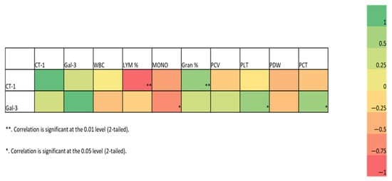
Figure 1

Journal Menu
► ▼ Journal Menu-
- Veterinary Sciences Home
- Aims & Scope
- Editorial Board
- Reviewer Board
- Topical Advisory Panel
- Instructions for Authors
- Special Issues
- Topics
- Sections & Collections
- Article Processing Charge
- Indexing & Archiving
- Editor’s Choice Articles
- Most Cited & Viewed
- Journal Statistics
- Journal History
- Journal Awards
- Conferences
- Editorial Office
Journal Browser
► ▼ Journal BrowserHighly Accessed Articles
Latest Books
E-Mail Alert
News
Topics
Topic in
Animals, Antioxidants, Veterinary Sciences, Agriculture
Feeding Livestock for Health Improvement
Topic Editors: Hui Yan, Xiao XuDeadline: 30 May 2025
Topic in
Animals, Dairy, Microorganisms, Veterinary Sciences, Metabolites, Life, Parasitologia
The Complexity of Parasites in Animals: Impacts, Innovation, and Interventions
Topic Editors: Kun Li, Rongjun Wang, Ningbo Xia, Md. F. KulyarDeadline: 31 August 2025
Topic in
Animals, Fishes, Veterinary Sciences
Application of the 3Rs to Promote the Welfare of Animals Used in Scientific Research and Testing
Topic Editors: Johnny Roughan, Laura CalvilloDeadline: 20 September 2025
Topic in
Agriculture, Animals, Veterinary Sciences, Antibiotics, Zoonotic Diseases
Animal Diseases in Agricultural Production Systems: Their Veterinary, Zoonotic, and One Health Importance, 2nd Edition
Topic Editors: Ewa Tomaszewska, Beata Łebkowska-Wieruszewska, Tomasz Szponder, Joanna Wessely-SzponderDeadline: 30 September 2025

Conferences
Special Issues
Special Issue in
Veterinary Sciences
Spotlight on Health Management in Calves
Guest Editors: Claire Windeyer, Lisa GamsjaegerDeadline: 30 May 2025
Special Issue in
Veterinary Sciences
Prevention, Diagnosis, and Management of Bovine Respiratory Diseases—2nd Edition
Guest Editor: Brad J. WhiteDeadline: 30 May 2025
Special Issue in
Veterinary Sciences
Diagnosis, Epidemiology and Vaccine Development of Infectious Diseases in Small Ruminants
Guest Editors: Wenliang Li, Li Mao, Zhentao ChengDeadline: 30 May 2025
Special Issue in
Veterinary Sciences
Genetic Diversity, Distribution and Conservation of Wild Animals in Captivity
Guest Editors: Peter Hristov, Georgi Radoslavov, Boiko NeovDeadline: 30 May 2025
Topical Collections
Topical Collection in
Veterinary Sciences
One-Health Approach to Bee Health
Collection Editors: Giovanni Cilia, Antonio Nanetti


