


Neurol. Int. 2026, 18(4), 69; https://doi.org/10.3390/neurolint18040069 - 13 Apr 2026
Abstract
►
Show Figures
Chronic prostatitis/chronic pelvic pain syndrome (CP/CPPS) is a prevalent urological disorder characterized by persistent pelvic pain, urinary symptoms, and significant impact on quality of life. In addition to its clinical symptoms, CP/CPPS is frequently associated with psychiatric comorbidities, such as anxiety and depression,
[...] Read more.
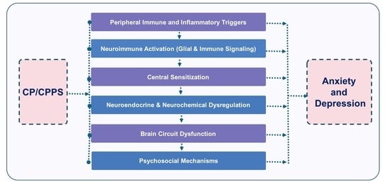
Chronic prostatitis/chronic pelvic pain syndrome (CP/CPPS) is a prevalent urological disorder characterized by persistent pelvic pain, urinary symptoms, and significant impact on quality of life. In addition to its clinical symptoms, CP/CPPS is frequently associated with psychiatric comorbidities, such as anxiety and depression, indicating complex neurobiological mechanisms. This review explores the mechanisms linking CP/CPPS with affective disorders, emphasizing central nervous system alterations, dysregulation of the hypothalamic–pituitary–adrenal (HPA) axis, and neuroimmune interactions. Evidence in-dicates that central sensitization, microglial and astrocytic activation, and elevated proinflammatory cytokines (IL-1β, IL-6, TNF-α) contribute to maladaptive painemotion network interactions. Additionally, dysregulation of hormones and neurotransmitters may exacerbate both pain perception and mood disorders. Psychosocial factors, including stress, coping strategies, and cognitive-emotional processes, further modulate symptom severity and treatment outcomes, highlighting the importance of a biopsychosocial approach. Gaining a deeper understanding of the neurobiological and psychosocial mechanisms behind anxiety and depression in CP/CPPS can lead to more effective, multidimensional management strategies and enhance patient-centered care.
Full article
Graphical abstract


