Feature Papers in Biophysics

Editors


E-Mail Website
Collection Editor
Curtin Medical School, Curtin University, Perth, WA 6845, Australia
Interests: molecular dynamics simulation; biological membranes; protein structure and dynamics; protein-protein and protein-ligand interactions; structure-based drug design
Special Issues, Collections and Topics in MDPI journals

E-Mail Website
Collection Editor
Department of Physics, Center for Theoretical Biological Physics, Northeastern University, Boston, MA 02115, USA
Interests: dynamics of large-scale molecular machines; molecular simulation; biomolecular folding; ribosome

E-Mail Website
Collection Editor
Department of Chemical and Biomedical Engineering, Cleveland State University, 2121 Euclid Avenue, Cleveland, OH 44236, USA
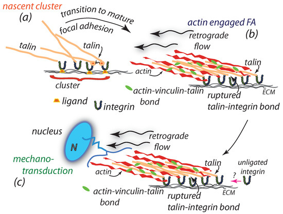
Interests: cell and tissue mechanics; mechanotransduction; molecular biophysics; mechanotoxicology; atomic force microscopy; cell-matrix interactions; stem cell fate

Topical Collection Information

Dear Colleagues,

This Topical Collection “Feature Papers in Biophysics” aims to collect high quality research articles, short communications, and review articles in all the fields of biophysics. We encourage Editorial Board Members of the Biophysica Journal to contribute papers reflecting the latest progress in their research field, or to invite relevant experts and colleagues to do so. Topics include, but are not limited to structure, dynamics and interactions of biomolecular systems (experimental, theoretical and simulation), single molecule biophysics, cell biophysics, biophysical methods (experimental and computational), bionanomaterials, molecular machines, synthetic biology, quantum biology and bionanotechnology.

Prof. Dr. Ricardo L. Mancera
Dr. Paul C. Whitford
Dr. Chandra Kothapalli
Collection Editors

Manuscript Submission Information

Manuscripts should be submitted online at www.mdpi.com by registering and logging in to this website. Once you are registered, click here to go to the submission form. Manuscripts can be submitted until the deadline. All submissions that pass pre-check are peer-reviewed. Accepted papers will be published continuously in the journal (as soon as accepted) and will be listed together on the collection website. Research articles, review articles as well as short communications are invited. For planned papers, a title and short abstract (about 100 words) can be sent to the Editorial Office for announcement on this website.

Submitted manuscripts should not have been published previously, nor be under consideration for publication elsewhere (except conference proceedings papers). All manuscripts are thoroughly refereed through a single-blind peer-review process. A guide for authors and other relevant information for submission of manuscripts is available on the Instructions for Authors page. Biophysica is an international peer-reviewed open access quarterly journal published by MDPI.

Please visit the Instructions for Authors page before submitting a manuscript. The Article Processing Charge (APC) for publication in this open access journal is 1000 CHF (Swiss Francs). Submitted papers should be well formatted and use good English. Authors may use MDPI's English editing service prior to publication or during author revisions.

Keywords

  • biomolecular structure, dynamics and interactions
  • protein structure, dynamics and interactions
  • nucleic acid structure, dynamics and interactions
  • membrane structure and dynamics
  • computational biophysics
  • molecular dynamics simulation
  • molecular modelling
  • mathematical modelling
  • single molecule biophysics
  • molecular biophysics
  • biophysical techniques in the study of biomolecular structure, function and interactions
  • X-ray and synchrotron methods in biomolecular structure
  • structural biology
  • synthetic biology
  • protein engineering
  • molecular machines
  • molecular motors and cell motility
  • cell and tissue mechanics
  • mechanotransduction
  • physics of biological systems
  • complex biological systems
  • bionanomaterials
  • enzymology

Published Papers (48 papers)

2024

Jump to: 2023, 2022

16 pages, 8969 KiB  
Article
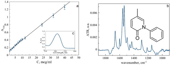
The Contrasting Effect of Sodium Alginate on Lysozyme and Albumin Denaturation and Fibril Formation
by Diliara R. Khaibrakhmanova, Polina R. Kuzivanova, Bulat R. Gainutdinov, Timur I. Magsumov, Alena A. Nikiforova and Igor A. Sedov
Biophysica 2024, 4(4), 651-666; https://doi.org/10.3390/biophysica4040043 - 12 Dec 2024
Viewed by 198
Abstract
The effect of sodium alginate on the denaturation and aggregation behavior of bovine serum albumin and hen egg-white lysozyme was studied. Large amounts of polysaccharide increase the thermal stability of albumin due to the weak binding interactions. At the same time, sodium alginate [...] Read more.
The effect of sodium alginate on the denaturation and aggregation behavior of bovine serum albumin and hen egg-white lysozyme was studied. Large amounts of polysaccharide increase the thermal stability of albumin due to the weak binding interactions. At the same time, sodium alginate can reduce the quantity of amyloid fibrils formed by albumin under denaturing conditions, which is a consequence of the stabilization of the native protein form by glycan binding. In the case of lysozyme, the polysaccharide has no influence on the thermal stability of the protein in 2 M guanidinium hydrochloride. However, the inhibition of fibril formation with an increase in the lag time was observed, which is explained by the binding of sodium alginate to lysozyme fibrils, but not to the protein monomer. The molecular nature of the binding interactions between alginate and the studied proteins was elucidated using molecular docking and known experimental structures of glycan–protein complexes. Full article
Show Figures

Figure 1

12 pages, 2191 KiB  
Article
Exploring Chemokine Homodimer Stability: Structural Insights into CXC and CC Interfaces
by David Budean, Yasser Almeida-Hernández, Jitendra Pandey, Joel Mieres Pérez, Elsa Sánchez García and Ellinor Haglund
Biophysica 2024, 4(4), 561-572; https://doi.org/10.3390/biophysica4040037 - 27 Nov 2024
Viewed by 523
Abstract
Chemokine ligands play a pivotal role in immune response by mediating cell migration and coordinating cellular processes through interactions with chemokine receptors. Understanding their sequence and structural integrity is crucial for elucidating their biological functions and potential therapeutic applications. In this study, we [...] Read more.
Chemokine ligands play a pivotal role in immune response by mediating cell migration and coordinating cellular processes through interactions with chemokine receptors. Understanding their sequence and structural integrity is crucial for elucidating their biological functions and potential therapeutic applications. In this study, we investigate the dimer interface between two distinct homodimer topologies: CXC and CC homodimers. Despite nearly identical monomeric structures, the rigid CXC interface is characterized by interactions between the N-loop/β-sheet regions, while the more flexible CC interface involves interactions through the unstructured N-terminal regions. Our structural and biophysical analyses indicate no significant differences in the free energy of folding (2–8 kcal/mol) and binding (10–14 kcal/mol) between the two homodimer topologies, showing that their free energy is primarily driven by sequence. We hypothesize that the biological signal is driven by the malleability of the dimer, depending on the binding interface. Understanding these structural dynamics opens new possibilities for designing chemokine-based therapeutics to modulate immune responses in diseases such as cancer, inflammation, and autoimmune disorders. Full article
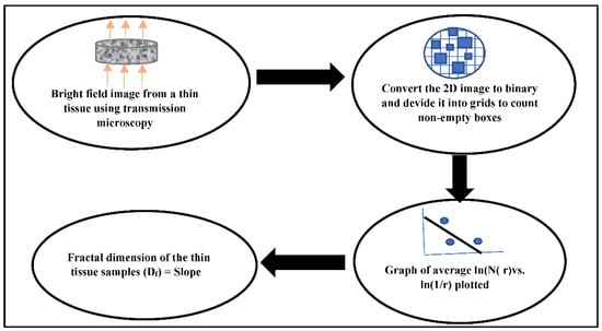
Show Figures

Figure 1

16 pages, 3654 KiB  
Article
Re-Examination of the Sel’kov Model of Glycolysis and Its Symmetry-Breaking Instability Due to the Impact of Diffusion with Implications for Cancer Imitation Caused by the Warburg Effect
by Miljko V. Satarić, Tomas Nemeš and Jack A. Tuszynski
Biophysica 2024, 4(4), 545-560; https://doi.org/10.3390/biophysica4040036 - 6 Nov 2024
Viewed by 631
Abstract
We revisit the seminal model of glycolysis first proposed by Sel’kov more than fifty years ago. We investigate the onset of instabilities in biological systems described by the Sel’kov model in order to determine the conditions of the model parameters that lead to [...] Read more.
We revisit the seminal model of glycolysis first proposed by Sel’kov more than fifty years ago. We investigate the onset of instabilities in biological systems described by the Sel’kov model in order to determine the conditions of the model parameters that lead to bifurcations. We analyze the glycolysis reaction under the circumstances when the diffusivity of both ATP and ADP reactants are taken into account. We estimate the critical value of the model’s single compact dimensionless parameter, which is responsible for the onset of reaction instability and the system’s symmetry breaking. It appears that it leads to spatial inhomogeneities of reactants, leading to the formation of standing waves instead of a homogeneous distribution of ATP molecules. The consequences of this model and its results are discussed in the context of the Warburg effect, which signifies a transition from oxidative phosphorylation to glycolysis that is correlated with the initiation and progression of cancer. Our analysis may lead to the selection of therapeutic interventions in order to prevent the symmetry-breaking phenomenon described in our work. Full article
Show Figures

Figure 1

10 pages, 1608 KiB  
Article
Is Water the Engine of Protein Folding?
by Marco Campanile and Giuseppe Graziano
Biophysica 2024, 4(4), 507-516; https://doi.org/10.3390/biophysica4040033 - 16 Oct 2024
Viewed by 811
Abstract
No one can dismiss the fundamental role played by water in several important biochemical processes, including the folding of globular proteins. The so-called hydrophobic effect is the theoretical construct to rationalize how water molecules stabilize the folded state. However, over the years, analyses [...] Read more.
No one can dismiss the fundamental role played by water in several important biochemical processes, including the folding of globular proteins. The so-called hydrophobic effect is the theoretical construct to rationalize how water molecules stabilize the folded state. However, over the years, analyses have been published that lead to the conclusion that water destabilizes the folded state. The aim of the present work is to state that the gain in translational entropy of water molecules (due to the decrease in water-accessible surface area associated with folding) is the driving force behind protein folding. Full article
Show Figures

Figure 1

11 pages, 1102 KiB  
Article
Modeling Thermal Effects of Pulsating Currents in Human Tissues: How to Prevent Necrosis
by Angiolo Farina, Antonio Fasano and Fabio Rosso
Biophysica 2024, 4(4), 477-487; https://doi.org/10.3390/biophysica4040031 - 27 Sep 2024
Viewed by 437
Abstract
In certain clinical applications, pulsating currents are applied to specific body regions for therapeutic purposes. In this paper, we analyze the resulting thermal field to determine the optimal amplitude, period, and duration of these stimuli, ensuring that the temperature in the targeted tissue [...] Read more.
In certain clinical applications, pulsating currents are applied to specific body regions for therapeutic purposes. In this paper, we analyze the resulting thermal field to determine the optimal amplitude, period, and duration of these stimuli, ensuring that the temperature in the targeted tissue remains below the necrosis threshold. Full article
Show Figures

Figure 1

13 pages, 1734 KiB  
Article
From Endogenous Quasi-Pathogens to Endogenous Smart Medicine: Understanding the Cellular Mechanisms and Implications of Benign and Malignant Cell Dynamics
by Jean-Marc Sabatier and Farzan Amini
Biophysica 2024, 4(3), 453-465; https://doi.org/10.3390/biophysica4030029 - 20 Sep 2024
Viewed by 1369
Abstract
This study investigates the formation and impact of Endogenous Quasi-Pathogens (EQPs) within cellular environments, focusing on the role of Endogenous Smart Medicine (ESM) as a therapeutic intervention. This work elucidates how induced vibrations facilitate new molecular and atomic connections between adjacent cells, leading [...] Read more.
This study investigates the formation and impact of Endogenous Quasi-Pathogens (EQPs) within cellular environments, focusing on the role of Endogenous Smart Medicine (ESM) as a therapeutic intervention. This work elucidates how induced vibrations facilitate new molecular and atomic connections between adjacent cells, leading to endobiotic bond formation and significantly altered DNA behavior. These vibrations, which dominate cellular processes, induce both temporary and permanent changes in cellular dynamics. The resulting increase in extracellular impedance triggers the emergence of new EQP sources, potentially initiating divergent pathological cycles. Cells experiencing moderate impedance changes are classified as benign, while those with substantial alterations are considered malignant. This study highlights the medical diagnostic implications of EQPs and positions ESM as a precise method for modulating cellular impedance, reducing the effects of EQPs, and potentially treating diseases where disruptions in cellular dynamics and stiffness are critical. Additionally, the integration of ChronoBit Storage (CBS) within ESM introduces a novel energy management mechanism, enhancing therapeutic precision by synchronizing energy distribution with cellular needs. The ChronoVital Index (CVI), a temporal model for assessing time dynamics across biological systems from individual cells to whole organs further refines this approach. By advancing the CVI and CBS, this research paves the way for more sophisticated therapeutic strategies, offering promising applications in the fields of disease management and cellular restoration within the framework of Endogenous Smart Medicine. Full article
Show Figures

Figure 1

17 pages, 6082 KiB  
Article
Intermolecular FRET Pairs as An Approach to Visualize Specific Enzyme Activity in Model Biomembranes and Living Cells
by Igor D. Zlotnikov, Alexander A. Ezhov and Elena V. Kudryashova
Biophysica 2024, 4(3), 340-356; https://doi.org/10.3390/biophysica4030024 - 1 Jul 2024
Cited by 1 | Viewed by 920
Abstract
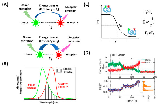
Herein, we propose an analytical approach based on intermolecular fluorescent resonant energy transfer (FRET) pairs for the visualization of specific enzyme activity in model biomembranes and in living cells. Cell visualizations with fluorescent confocal laser microscopy usually rely on fluorescent probes, such as [...] Read more.
Herein, we propose an analytical approach based on intermolecular fluorescent resonant energy transfer (FRET) pairs for the visualization of specific enzyme activity in model biomembranes and in living cells. Cell visualizations with fluorescent confocal laser microscopy usually rely on fluorescent probes, such as Fluorescein isothiocyanate (FITC), Alexa488, Tetramethylrhodamine isothiocyanate (TRITC) and many others. However, for more specific tasks, such as the detection of certain enzymatic activity inside the living cell, the toolbox is quite limited. In the case of enzyme-hydrolases for example, the choice is limited to organic molecules comprising a fluorescent dye (typically, 4-methylumbelliferone (MUmb) or 7-amino-4-methylcoumarin (AMC) derivatives) and a fluorescence quencher, bound via an enzyme-sensitive linker—so that when the linker is degraded, the fluorescent signal increases. Unfortunately, both MUmb and AMC are quenched and have a relatively low quantum yield in cells, and their excitation and emission ranges overlap with that of intracellular fluorophores, often producing a strong background noise. R6G, on the other hand, has excellent quantum yield apart from intracellular fluorophores, but there are no efficient quenchers that could be chemically linked to R6G. Herein, we show that R6G is able to form intermolecular FRET pairs with MUmb or AMC, with the latter serving as fluorescence donors. This yields a combination of R6G’s excellent fluorescence properties with a possibility to use an enzyme-sensitive linker in MUTMAC or AMC derivatives. This phenomenon was initially discovered in a model system, reversed micelles, where the donor, the acceptor, and the enzyme are forced to be in close proximity to each other, so that proximity could serve as an explanation for the intermolecular FRET effect. Surprisingly enough, the phenomenon has been reproduced in living cells. Moreover, we were able to create working intermolecular donor–acceptor FRET pairs for several different enzymes, including chymotrypsin, phosphatase, and asparaginase. This appears counterintuitive, as besides the overlap of the emission spectra of the donor and the absorption spectra of the acceptor, there are other criteria for the FRET effect, including the convergence of two fluorophores at a distance of about 1–10 nm, and the orientation of their dipoles at a certain angle, which is difficult to imagine in a bulk system like a living cell. We hypothesize that FRET-enabling donor–acceptor interaction may be taking place at the inner surface of the lipid bilayer, to which both donor and acceptor molecules would likely have an affinity. This hypothesis would require a more detailed investigation. Therefore, we have shown that the method suggested has good potential in the visualization of enzyme functioning inside living cells, which is often a challenging task. Shifting of the fluorescence signal to the long-wavelength region would increase the signal selectivity, making it easily distinguishable from autofluorescence. Full article
Show Figures

Figure 1

11 pages, 2880 KiB  
Article
Enhanced Adsorption of Cage-Shaped Proteins on Carbon Surfaces by Carbon Nanotube (CNT)-Binding Peptide Aptamers
by Narangerel Ganbaatar, Ting-Chieh Chu, Naofumi Okamoto, Kenji Iwahori, Masakazu Nakamura and Ichiro Yamashita
Biophysica 2024, 4(2), 256-266; https://doi.org/10.3390/biophysica4020018 - 24 May 2024
Viewed by 729
Abstract
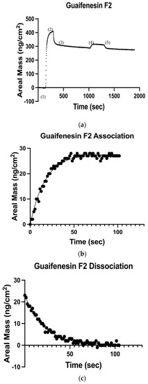
The adsorption behavior of recombinant cage-shaped proteins with carbon nanotube (CNT)-binding peptides on carbon surfaces was quantitatively and dynamically analyzed using a highly stable quartz crystal microbalance (QCM). Two types of CNT-binding peptide aptamers obtained by the phage display method were attached to [...] Read more.
The adsorption behavior of recombinant cage-shaped proteins with carbon nanotube (CNT)-binding peptides on carbon surfaces was quantitatively and dynamically analyzed using a highly stable quartz crystal microbalance (QCM). Two types of CNT-binding peptide aptamers obtained by the phage display method were attached to the N- and C-termini of the Dps (DNA-binding protein derived from starved cells) to produce carbonaceous material-binding Dps. The carbon adsorption ability of the mutant Dps was studied by QCM measurement using a carbon-coated QCM sensor. The produced peptide aptamer-modified Dps showed higher affinity than a wild Dps and also showed higher adsorption capacity than a previously used Dps with carbon nanohorn-binding peptides. The newly obtained peptide aptamers were proven to provide Dps with high adsorption affinity on carbon surfaces. Furthermore, the aptamer modified to the N-terminus of the Dps subunit showed more efficient adsorption than the aptamers attached to the C-terminus of the Dp, and the linker was found to improve the adsorption ability. Full article
Show Figures

Figure 1

11 pages, 364 KiB  
Article
Differential Scanning Calorimetry of Proteins and the Two-State Model: Comparison of Two Formulas
by Knarik Yeritsyan and Artem Badasyan
Biophysica 2024, 4(2), 227-237; https://doi.org/10.3390/biophysica4020016 - 13 May 2024
Viewed by 991
Abstract
Differential Scanning Calorimetry (DSC) is a regular and powerful tool to measure the specific heat profile of various materials. In order to connect the measured profile to the properties of a particular protein, a model is required to fit. We discuss here the [...] Read more.
Differential Scanning Calorimetry (DSC) is a regular and powerful tool to measure the specific heat profile of various materials. In order to connect the measured profile to the properties of a particular protein, a model is required to fit. We discuss here the application of an exact two-state formula with its approximation and process the DSC experimental data on protein folding in water. The approximate formula relies on the smallness of the transition interval, which is different for each protein. With an example of the set of 33 different proteins, we show the practical validity of the approximation and the equivalence of exact and approximate two-state formulas for processing DSC data. Full article
Show Figures

Figure 1

15 pages, 2179 KiB  
Article
The Signature of Fluctuations of the Hydrogen Bond Network Formed by Water Molecules in the Interfacial Layer of Anionic Lipids
by Ana-Marija Pavlek, Barbara Pem and Danijela Bakarić
Biophysica 2024, 4(1), 92-106; https://doi.org/10.3390/biophysica4010007 - 21 Feb 2024
Cited by 1 | Viewed by 1014
Abstract
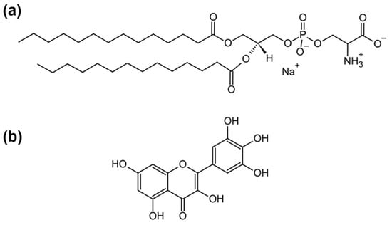
As the water molecules found at the interface of lipid bilayers exhibit distinct structural and reorientation dynamics compared to water molecules found in bulk, the fluctuations in their hydrogen bond (HB) network are expected to be different from those generated by the bulk [...] Read more.
As the water molecules found at the interface of lipid bilayers exhibit distinct structural and reorientation dynamics compared to water molecules found in bulk, the fluctuations in their hydrogen bond (HB) network are expected to be different from those generated by the bulk water molecules. The research presented here aims to gain an insight into temperature-dependent fluctuations of a HB network of water molecules found in an interfacial layer of multilamellar liposomes (MLVs) composed of anionic 1,2-dimyristoyl-sn-glycero-3-phospho-L-serine (DMPS) lipids. Besides suspending DMPS lipids in phosphate buffer saline (PBS) of different pH values (6.0, 7.4, and 8.0), the changes in HB network fluctuations were altered by the incorporation of a non-polar flavonoid molecule myricetin (MCE) within the hydrocarbon chain region. By performing a multivariate analysis on the water combination band observed in temperature-dependent FTIR spectra, the results of which were further mathematically analyzed, the temperature-dependent fluctuations of interfacial water molecules were captured; the latter were the greatest for DMPS in PBS with a pH value of 7.4 and in general were greater for DMPS multibilayers in the absence of MCE. The presence of MCE made DMPS lipids more separated, allowing deeper penetration of water molecules towards the non-polar region and their restricted motion that resulted in decreased fluctuations. The experimentally observed results were supported by MD simulations of DMPS (+MCE) lipid bilayers. Full article
Show Figures

Figure 1

2023

Jump to: 2024, 2022

16 pages, 3023 KiB  
Article
Elucidating the Influence of Lipid Composition on Bilayer Perturbations Induced by the N-Terminal Region of the Huntingtin Protein
by Yasith Indigahawela Gamage and Jianjun Pan
Biophysica 2023, 3(4), 582-597; https://doi.org/10.3390/biophysica3040040 - 28 Oct 2023
Cited by 2 | Viewed by 1456
Abstract
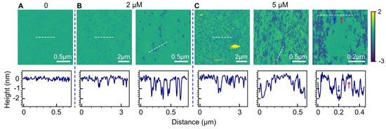
Understanding the membrane interactions of the N-terminal 17 residues of the huntingtin protein (HttN) is essential for unraveling its role in cellular processes and its impact on huntingtin misfolding. In this study, we used atomic force microscopy (AFM) to examine the effects of [...] Read more.
Understanding the membrane interactions of the N-terminal 17 residues of the huntingtin protein (HttN) is essential for unraveling its role in cellular processes and its impact on huntingtin misfolding. In this study, we used atomic force microscopy (AFM) to examine the effects of lipid specificity in mediating bilayer perturbations induced by HttN. Across various lipid environments, the peptide consistently induced bilayer disruptions in the form of holes. Notably, our results unveiled that cholesterol enhanced bilayer perturbation induced by HttN, while phosphatidylethanolamine (PE) lipids suppressed hole formation. Furthermore, anionic phosphatidylglycerol (PG) and cardiolipin lipids, along with cholesterol at high concentrations, promoted the formation of double-bilayer patches. This unique structure suggests that the synergy among HttN, anionic lipids, and cholesterol can enhance bilayer fusion, potentially by facilitating lipid intermixing between adjacent bilayers. Additionally, our AFM-based force spectroscopy revealed that HttN enhanced the mechanical stability of lipid bilayers, as evidenced by an elevated bilayer puncture force. These findings illuminate the complex interplay between HttN and lipid membranes and provide useful insights into the role of lipid composition in modulating membrane interactions with the huntingtin protein. Full article
Show Figures

Figure 1

13 pages, 3267 KiB  
Article
Exploring the Dynamics of Holo-Shikimate Kinase through Molecular Mechanics
by Pedro Ojeda-May
Biophysica 2023, 3(3), 463-475; https://doi.org/10.3390/biophysica3030030 - 21 Jul 2023
Cited by 1 | Viewed by 1760
Abstract
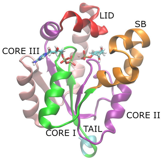
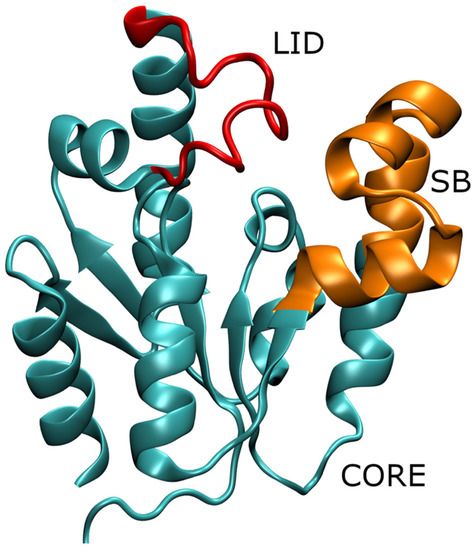
Understanding the connection between local and global dynamics can provide valuable insights into enzymatic function and may contribute to the development of novel strategies for enzyme modulation. In this work, we investigated the dynamics at both the global and local (active site) levels [...] Read more.
Understanding the connection between local and global dynamics can provide valuable insights into enzymatic function and may contribute to the development of novel strategies for enzyme modulation. In this work, we investigated the dynamics at both the global and local (active site) levels of Shikimate Kinase (SK) through microsecond time-scale molecular dynamics (MD) simulations of the holoenzyme in the product state. Our focus was on the wild-type (WT) enzyme and two mutants (R116A and R116K) which are known for their reduced catalytic activity. Through exploring the dynamics of these variants, we gained insights into the role of residue R116 and its contribution to overall SK dynamics. We argue that the connection between local and global dynamics can be attributed to local frustration near the mutated residue which perturbs the global protein dynamics. Full article
Show Figures

Figure 1

17 pages, 4600 KiB  
Article
A Structure-Guided Designed Small Molecule Is an Anticancer Agent and Inhibits the Apoptosis-Related MCL-1 Protein
by Ingrid V. Machado, Luiz F. N. Naves, Jean M. F. Custodio, Hérika D. A. Vidal, Jaqueline E. Queiroz, Allen G. Oliver, Joyce V. B. Borba, Bruno J. Neves, Lucas M. Brito, Claudia Pessoa, Hamilton B. Napolitano and Gilberto L. B. de Aquino
Biophysica 2023, 3(3), 446-462; https://doi.org/10.3390/biophysica3030029 - 7 Jul 2023
Viewed by 1517
Abstract
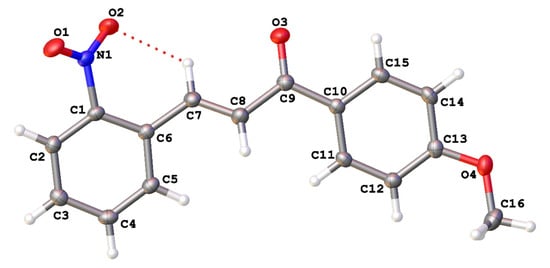
Cancer resistance to chemotherapy and radiation therapies presents significant challenges, necessitating the exploration of alternative approaches. Targeting specific proteins at the molecular level, particularly their active sites, holds promise in addressing this issue. We investigated the potential of 4′-methoxy-2-nitrochalcone (MNC) as an MCL-1 [...] Read more.
Cancer resistance to chemotherapy and radiation therapies presents significant challenges, necessitating the exploration of alternative approaches. Targeting specific proteins at the molecular level, particularly their active sites, holds promise in addressing this issue. We investigated the potential of 4′-methoxy-2-nitrochalcone (MNC) as an MCL-1 inhibitor, examining its chemical and structural characteristics to elucidate its biological activity and guide the selection of potential candidates. We conducted a docking study, followed by synthesis, structural characterization, theoretical calculations, and in vitro experiments to comprehensively evaluate MNC. The docking results revealed MNC’s excellent binding within the active site of MCL-1. At 50 µM, MNC demonstrated 99% inhibition of HCT116 cell proliferation, with an IC50 value of 15.18 µM after 24 h. Treatment with MNC at 30.36 and 15.18 µM resulted in reduced cell density. Notably, MNC exhibited marked cytotoxicity at concentrations of 15.58 µM and 7.79 µM, inducing high frequencies of plasma membrane rupture and apoptosis, respectively. Our findings highlight the significant biological potential of MNC as an MCL-1 inhibitor. Furthermore, we propose exploring chalcones with hydrogen bond acceptor substituents as promising candidates for studying inhibitors targeting this protein. In conclusion, our study addresses the challenge of cancer resistance by investigating MNC as an MCL-1 inhibitor. Through detailed characterization and experimental validation, we establish the efficacof MNC in inhibiting cell proliferation and inducing cytotoxic effects. These results underscore the potential of MNC as a valuable therapeutic agent and suggest the use of chalcones with hydrogen bond acceptor substituents as a basis for developing novel MCL-1 inhibitors. Full article
Show Figures

Figure 1

14 pages, 2990 KiB  
Article
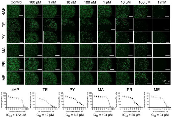
Phenotypic and Biomechanical Characteristics of Human Fetal Neural Progenitor Cells Exposed to Pesticide Compounds
by Marissa C. Sarsfield, Jennifer Vasu, Sabreen M. Abuoun, Nischal Allena and Chandrasekhar R. Kothapalli
Biophysica 2023, 3(2), 348-361; https://doi.org/10.3390/biophysica3020023 - 18 May 2023
Viewed by 1769
Abstract
Various forms of pesticides have been reported to be among the environmental toxicants, which are detrimental to human health. The active ingredients of these formulations can enter the human body through air, food, or water. Epidemiological studies suggest that these compounds strongly affect [...] Read more.
Various forms of pesticides have been reported to be among the environmental toxicants, which are detrimental to human health. The active ingredients of these formulations can enter the human body through air, food, or water. Epidemiological studies suggest that these compounds strongly affect the developing brain in fetal and infant stages due to their ability to breach the underdeveloped blood–brain barrier. Since neural progenitor stem cells (NPCs) in the developing brain are the most vulnerable to these compounds, the mechanisms by which NPCs experience toxicity upon exposure to these chemicals must be investigated. Here, we assessed the viability of human fetal NPCs in 2D cultures in the presence of the active ingredients of six widely used pesticides using Live/Dead® and Hoechst staining. The IC50 values ranged from 4.1–201 μM. A significant drop in cell viability with increasing toxicant concentration (p < 0.01) was noted, with the order of toxicity being malathion < 4-aminopyridine < methoprene < prallethrin < temephos < pyriproxyfen. Changes in cellular biomechanical characteristics (Young’s modulus, tether force, membrane tension, and tether radius) were quantified using atomic force microscopy, whereas cell migration was elucidated over 48 h using a customized wound-healing assay. The Young’s modulus of fetal NPCs exposed to IC50/2 doses of these compounds was reduced by 38–70% and that of those exposed to IC50 doses was reduced by 71–80% (p < 0.001 vs. controls for both; p < 0.01 for IC50 vs. IC50/2 for each compound). Similar patterns were noted for tether forces and membrane tension in fetal NPCs. NPC migration was found to be compound type- and dose-dependent. These results attest to the significant detrimental effects of these compounds on various aspects of the human fetal NPC phenotype, and the utility of cell mechanics as a marker to assess developmental neurotoxicity. Full article
Show Figures

Figure 1

17 pages, 3197 KiB  
Article
Insights into Chemical Interactions and Related Toxicities of Deep Eutectic Solvents with Mammalian Cells Observed Using Synchrotron Macro–ATR–FTIR Microspectroscopy
by Saffron J. Bryant, Zo L. Shaw, Louisa Z. Y. Huang, Aaron Elbourne, Amanda N. Abraham, Jitraporn Vongsvivut, Stephen A. Holt, Tamar L. Greaves and Gary Bryant
Biophysica 2023, 3(2), 318-334; https://doi.org/10.3390/biophysica3020021 - 4 May 2023
Cited by 4 | Viewed by 2153
Abstract
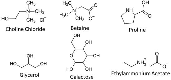
Deep eutectic solvents (DESs) and ionic liquids (ILs) are highly tailorable solvents that have shown a lot of promise for a variety of applications including cryopreservation, drug delivery, and protein stabilisation. However, to date, there is very limited information on the detailed interactions [...] Read more.
Deep eutectic solvents (DESs) and ionic liquids (ILs) are highly tailorable solvents that have shown a lot of promise for a variety of applications including cryopreservation, drug delivery, and protein stabilisation. However, to date, there is very limited information on the detailed interactions of these solvents with mammalian cells. In this work, we studied six DESs and one IL that show promise as cryoprotective agents, applying synchrotron macro–ATR–FTIR to examine their effects on key biochemical components of HaCat mammalian cells. These data were paired with resazurin metabolic assays and neutron reflectivity experiments to correlate cellular interactions with cellular toxicity. Stark differences were observed even between solvents that shared similar components. In particular, it was found that solvents that are effective cryoprotective agents consistently showed interactions with cellular membranes, while high toxicity correlated with strong interactions of the DES/IL with nucleic acids and proteins. This work sheds new light on the interactions between novel solvents and cells that may underpin future biomedical applications. Full article
Show Figures

Figure 1

19 pages, 4108 KiB  
Article
Discovery of the Universal tRNA Binding Mode for the TsaD-like Components of the t6A tRNA Modification Pathway
by Boguslaw Stec
Biophysica 2023, 3(2), 288-306; https://doi.org/10.3390/biophysica3020019 - 12 Apr 2023
Viewed by 1782
Abstract
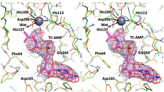
Covalent addition of the threonylcarbamoyl group to N(6) of adenosine 37 (t6A modification) within the anticodon loop of several tRNAs is central to the translational fidelity in all known organisms. Structures for each of the enzyme components in the Tsa (t [...] Read more.
Covalent addition of the threonylcarbamoyl group to N(6) of adenosine 37 (t6A modification) within the anticodon loop of several tRNAs is central to the translational fidelity in all known organisms. Structures for each of the enzyme components in the Tsa (t6A) pathway from all three kingdoms of life have been determined previously. In order to shed light on the poorly defined final step of t6A tRNA modification by TsaD-like components, we performed modeling studies. By docking a tRNA substrate molecule onto reanalyzed complete models of three TsaD-like proteins—TsaD from T. maritima, Qri7 from bacteria, and Kae1 from yeast—we identified a binding site that is common to all of them. An apparently universal binding mode has perfectly oriented tRNA for catalysis by TsaD. Furthermore, it suggests how the conformational changes in TsaD, in response to the binding of the additional regulatory subunits, control enzymatic activity. Re-refinement of the X-ray structure of the TsaBDE complex from T. maritima tentatively suggests that the moiety bound at the active site of the TsaD component is threonylcarbamoyl-AMP (TC-AMP). These findings suggest a detailed model for the mechanism of the catalytic reaction carried out by the TsaD-like components that explains the transfer of unstable TC-AMP from TsaC to TsaD proteins in the t6A modification pathway. Full article
Show Figures

Figure 1

12 pages, 6749 KiB  
Article
Silica In Silico: A Molecular Dynamics Characterization of the Early Stages of Protein Embedding for Atom Probe Tomography
by Giovanni Novi Inverardi, Francesco Carnovale, Lorenzo Petrolli, Simone Taioli and Gianluca Lattanzi
Biophysica 2023, 3(2), 276-287; https://doi.org/10.3390/biophysica3020018 - 11 Apr 2023
Cited by 1 | Viewed by 2159
Abstract
A novel procedure for the application of atom probe tomography (APT) to the structural analysis of biological systems, has been recently proposed, whereby the specimen is embedded by a silica matrix and ablated by a pulsed laser source. Such a technique, requires that [...] Read more.
A novel procedure for the application of atom probe tomography (APT) to the structural analysis of biological systems, has been recently proposed, whereby the specimen is embedded by a silica matrix and ablated by a pulsed laser source. Such a technique, requires that the silica primer be properly inert and bio-compatible, keeping the native structural features of the system at hand, while condensing into an amorphous, glass-like coating. In this work, we propose a molecular dynamics protocol, aimed at depicting and characterizing the earliest stages of the embedding process of small biomolecules in a solution of water and orthosilicic acid, here, taken as a precursor of the silica matrix. Overall, we observe a negligible influence of orthosilicic acid on the behavior of stable folded systems (such as ubiquitin). Conversely, intrinsically disordered and unstable peptides are affected by the coating, the latter seemingly inhibiting the fluctuations of flexible moieties. While further scrutiny is in order, our assessment offers a first mechanistic insight of the effects of orthosilicic acid, thereby validating its use in the proposed innovative application of APT to the structural resolution of protein molecules. Full article
Show Figures

Graphical abstract

11 pages, 3386 KiB  
Article
Computational Modeling of the Interaction of Molecular Oxygen with the miniSOG Protein—A Light Induced Source of Singlet Oxygen
by Igor Polyakov, Anna Kulakova and Alexander Nemukhin
Biophysica 2023, 3(2), 252-262; https://doi.org/10.3390/biophysica3020016 - 2 Apr 2023
Cited by 3 | Viewed by 1815
Abstract
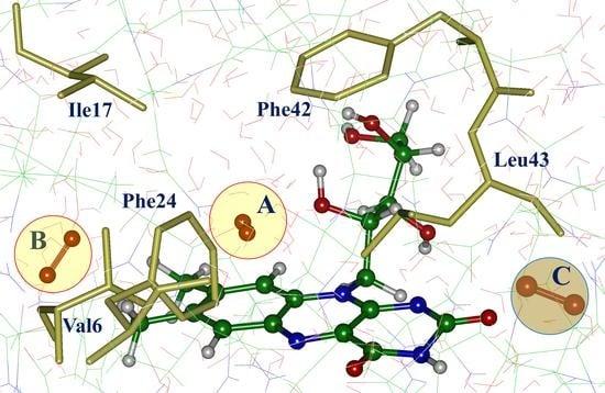
Interaction of molecular oxygen 3O2 with the flavin-dependent protein miniSOG after light illumination results in creation of singlet oxygen 1O2 and superoxide O2●−. Despite the recently resolved crystal structures of miniSOG variants, oxygen-binding sites near the [...] Read more.
Interaction of molecular oxygen 3O2 with the flavin-dependent protein miniSOG after light illumination results in creation of singlet oxygen 1O2 and superoxide O2●−. Despite the recently resolved crystal structures of miniSOG variants, oxygen-binding sites near the flavin chromophore are poorly characterized. We report the results of computational studies of the protein−oxygen systems using molecular dynamics (MD) simulations with force-field interaction potentials and quantum mechanics/molecular mechanics (QM/MM) potentials for the original miniSOG and the mutated protein. We found several oxygen-binding pockets and pointed out possible tunnels bridging the bulk solvent and the isoalloxazine ring of the chromophore. These findings provide an essential step toward understanding photophysical properties of miniSOG—an important singlet oxygen photosensitizer. Full article
Show Figures

Graphical abstract

21 pages, 3101 KiB  
Article
Complex Physical Properties of an Adaptive, Self-Organizing Biological System
by József Prechl
Biophysica 2023, 3(2), 231-251; https://doi.org/10.3390/biophysica3020015 - 31 Mar 2023
Viewed by 1643
Abstract
Physical modeling of the functioning of the adaptive immune system, which has been thoroughly characterized on genetic and molecular levels, provides a unique opportunity to define an adaptive, self-organizing biological system in its entirety. This paper describes a configuration space model of immune [...] Read more.
Physical modeling of the functioning of the adaptive immune system, which has been thoroughly characterized on genetic and molecular levels, provides a unique opportunity to define an adaptive, self-organizing biological system in its entirety. This paper describes a configuration space model of immune function, where directed chemical potentials of the system constitute a space of interactions. A mathematical approach is used to define the system that couples the variance of Gaussian distributed interaction energies in its interaction space to the exponentially distributed chemical potentials of its effector molecules to maintain its steady state. The model is validated by identifying the thermodynamic and network variables analogous to the mathematical parameters and by applying the model to the humoral immune system. Overall, this statistical thermodynamics model of adaptive immunity describes how adaptive biological self-organization arises from the maintenance of a scale-free, directed molecular interaction network with fractal topology. Full article
Show Figures

Figure 1

17 pages, 10180 KiB  
Article
Comparison of Empirical Zn2+ Models in Protein–DNA Complexes
by Senta Volkenandt and Petra Imhof
Biophysica 2023, 3(1), 214-230; https://doi.org/10.3390/biophysica3010014 - 20 Mar 2023
Viewed by 2453
Abstract
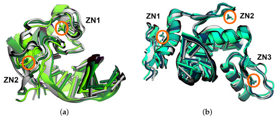
Zinc ions are the second most abundant ions found in humans. Their role in proteins can be merely structural but also catalytic, owing to their transition metal character. Modelling their geometric–coordination versatility by empirical force fields is, thus, a challenging task. In this [...] Read more.
Zinc ions are the second most abundant ions found in humans. Their role in proteins can be merely structural but also catalytic, owing to their transition metal character. Modelling their geometric–coordination versatility by empirical force fields is, thus, a challenging task. In this work, we evaluated three popular models, specifically designed to represent zinc ions with regard to their capability of preserving structural integrity. To this end, we performed molecular dynamics simulations of two zinc-containing protein–DNA complexes, which differed in their zinc coordination, i.e., four cysteines or two cysteines and two histidines. The most flexible non-bonded 12-6-4 Lennard–Jones-type model shows a preference for six-fold coordination of the Zn2+-ions in contradiction to the crystal structure. The cationic dummy atom model favours tetrahedral geometry, whereas the bonded extended zinc AMBER force field model, by construction, best preserves the initial geometry of a regular or slightly distorted tetrahedron. Our data renders the extended zinc AMBER force field the best model for structural zinc ions in a given geometry. In more complicated cases, though, more flexible models may be advantageous. Full article
Show Figures

Figure 1

16 pages, 1855 KiB  
Review
Contribution of smFRET to Chromatin Research
by Bhaswati Sengupta and Mai Huynh
Biophysica 2023, 3(1), 93-108; https://doi.org/10.3390/biophysica3010007 - 8 Feb 2023
Viewed by 2274
Abstract
Chromatins are structural components of chromosomes and consist of DNA and histone proteins. The structure, dynamics, and function of chromatins are important in regulating genetic processes. Several different experimental and theoretical tools have been employed to understand chromatins better. In this review, we [...] Read more.
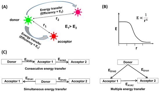
Chromatins are structural components of chromosomes and consist of DNA and histone proteins. The structure, dynamics, and function of chromatins are important in regulating genetic processes. Several different experimental and theoretical tools have been employed to understand chromatins better. In this review, we will focus on the literatures engrossed in understanding of chromatins using single-molecule Förster resonance energy transfer (smFRET). smFRET is a single-molecule fluorescence microscopic technique that can furnish information regarding the distance between two points in space. This has been utilized to efficiently unveil the structural details of chromatins. Full article
Show Figures

Figure 1

23 pages, 3505 KiB  
Article
Effect of the Neck Linker on Processive Stepping of Kinesin Motor
by Ping Xie
Biophysica 2023, 3(1), 46-68; https://doi.org/10.3390/biophysica3010004 - 28 Jan 2023
Cited by 4 | Viewed by 3113
Abstract
Kinesin motor protein, which is composed of two catalytic domains connected together by a long coiled-coil stalk via two flexible neck linkers (NLs), can step processively on a microtubule towards the plus end by hydrolyzing adenosine triphosphate (ATP) molecules. To understand what the [...] Read more.
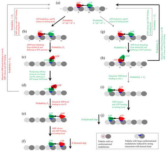
Kinesin motor protein, which is composed of two catalytic domains connected together by a long coiled-coil stalk via two flexible neck linkers (NLs), can step processively on a microtubule towards the plus end by hydrolyzing adenosine triphosphate (ATP) molecules. To understand what the role is that the NL plays in the processive stepping, the dynamics of the kinesin motor are studied theoretically here by considering the mutation or deletion of an N-terminal cover strand that contributes to the docking of the NL in kinesin-1, the extension of the NL in kinesin-1, the mutation of the NL in kinesin-1, the swapping of the NL of kinesin-2 with that of kinesin-1, the joining of the stalk and neck of Ncd that moves towards the minus end of MT to the catalytic domain of kinesin-1, the replacement of catalytic domain of kinesin-1 with that of Ncd, and so on. The theoretical results give a consistent and quantitative explanation of various available experimental results about the effects of these mutations on motor dynamics and, moreover, provide predicted results. Additionally, the processive motility of kinesin-6 MKLP2 without NL docking is also explained. The available experimental data about the effect of NL mutations on the dynamics of the bi-directional kinesin-5 Cin8 are also explained. The studies are critically implicative to the mechanism of the stepping of the kinesin motor. Full article
Show Figures

Figure 1

2022

Jump to: 2024, 2023

10 pages, 4266 KiB  
Article
Plasmonic Biosensors Based on Deformed Graphene
by Vahid Faramarzi, Mohsen Heidari, Nik Humaidi bin Nik Zulkarnine and Michael Taeyoung Hwang
Biophysica 2022, 2(4), 538-547; https://doi.org/10.3390/biophysica2040045 - 29 Nov 2022
Cited by 11 | Viewed by 2269
Abstract
Rapid, accurate, and label-free detection of biomolecules and chemical substances remains a challenge in healthcare. Optical biosensors have been considered as biomedical diagnostic tools required in numerous areas including the detection of viruses, food monitoring, diagnosing pollutants in the environment, global personalized medicine, [...] Read more.
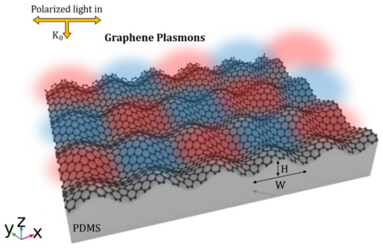
Rapid, accurate, and label-free detection of biomolecules and chemical substances remains a challenge in healthcare. Optical biosensors have been considered as biomedical diagnostic tools required in numerous areas including the detection of viruses, food monitoring, diagnosing pollutants in the environment, global personalized medicine, and molecular diagnostics. In particular, the broadly emerging and promising technique of surface plasmon resonance has established to provide real-time and label-free detection when used in biosensing applications in a highly sensitive, specific, and cost-effective manner with small footprint platform. In this study we propose a novel plasmonic biosensor based on biaxially crumpled graphene structures, wherein plasmon resonances in graphene are utilized to detect variations in the refractive index of the sample medium. Shifts in the resonance wavelength of the plasmon modes for a given change in the RI of the surrounding analyte are calculated by investigating the optical response of crumpled graphene structures on different substrates using theoretical computations based on the finite element method combined with the semiclassical Drude model. The results reveal a high sensitivity of 4990 nm/RIU, corresponding to a large figure-of-merit of 20 for biaxially crumpled graphene structures on polystyrene substrates. We demonstrate that biaxially crumpled graphene exhibits superior sensing performance compared with a uniaxial structure. According to the results, crumpled graphene structures on a titanium oxide substrate can improve the sensor sensitivity by avoiding the damping effects of polydimethylsiloxane substrates. The enhanced sensitivity and broadband mechanical tunability of the biaxially crumpled graphene render it a promising platform for biosensing applications. Full article
Show Figures

Figure 1

13 pages, 2537 KiB  
Article
Adherent Moving of Polymers in Spherical Confined Binary Semiflexible Ring Polymer Mixtures
by Xiaolin Zhou and Wan Wei
Biophysica 2022, 2(4), 525-537; https://doi.org/10.3390/biophysica2040044 - 24 Nov 2022
Viewed by 1950
Abstract
Based on the coarse-grained model, we used molecular dynamics methods to calculate and simulate a semiflexible long ring–semiflexible short ring blended polymer system confined in a hard sphere. We systematically studied the distribution and motion characteristics of the long ring chain. The results [...] Read more.
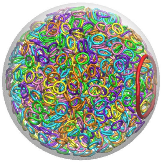
Based on the coarse-grained model, we used molecular dynamics methods to calculate and simulate a semiflexible long ring–semiflexible short ring blended polymer system confined in a hard sphere. We systematically studied the distribution and motion characteristics of the long ring chain. The results show that when the short ring is short enough (Lshort < 20), the long ring (Llong = 50) is separated from the blend system and then distributed against the inner wall. As the length of the short ring increases (Lshort ≥ 20), the long ring can no longer be separated from the blending system. Moreover, we found that the long ring demonstrates a random direction of adherent walking behavior on the inner surface of the hard sphere. The velocity of the long ring decreases with the increase in the short ring length Lshort. Specifically for Lshort ≥ 20, the system does not undergo phase separation and the speed of the long ring decreases sharply along with the long ring distributed inside the confined bulk. This is related to the inner wall layer moving faster than the inside bulk of the restricted system. Our simulation results can help us to understand the distribution of macromolecules in biological systems in confined systems, including the restricted chromosome partitioning distribution and packing structure of circular DNA molecules. Full article
Show Figures

Figure 1

12 pages, 3845 KiB  
Article
RBL-2H3 Mast Cell Receptor Dynamics in the Immunological Synapse
by Ming Chih Tsai and Kathrin Spendier
Biophysica 2022, 2(4), 428-439; https://doi.org/10.3390/biophysica2040038 - 7 Nov 2022
Viewed by 1761
Abstract
The RBL-2H3 mast cell immunological synapse dynamics is often simulated with reaction–diffusion and Fokker–Planck equations. The equations focus on how the cell synapse captures receptors following an immune response, where the receptor capture at the immunological site appears to be a delayed process. [...] Read more.
The RBL-2H3 mast cell immunological synapse dynamics is often simulated with reaction–diffusion and Fokker–Planck equations. The equations focus on how the cell synapse captures receptors following an immune response, where the receptor capture at the immunological site appears to be a delayed process. This article investigates the physical nature and mathematics behind such time-dependent delays. Using signal processing methods, convolution and cross-correlation-type delay capture simulations give a χ-squared range of 22 to 60, in good agreement with experimental results. The cell polarization event is offered as a possible explanation for these capture delays, where polarizing rates measure how fast the cell polarization event occurs. In the case of RBL-2H3 mast cells, polarization appears to be associated with cytoskeletal rearrangement; thus, both cytoskeletal and diffusional components are considered. From these simulations, a maximum polarizing rate ranging from 0.0057 s−2 to 0.031 s−2 is obtained. These results indicate that RBL-2H3 mast cells possess both temporal and spatial memory, and cell polarization is possibly linked to a Turing-type pattern formation. Full article
Show Figures

Figure 1

11 pages, 3027 KiB  
Article
The Modification of Polyvinyl Alcohol for Ice Nucleation Based upon the Structures of Antifreeze Glycoproteins Found in Antarctic Fish
by Monika Bleszynski
Biophysica 2022, 2(4), 417-427; https://doi.org/10.3390/biophysica2040037 - 3 Nov 2022
Cited by 1 | Viewed by 1918
Abstract
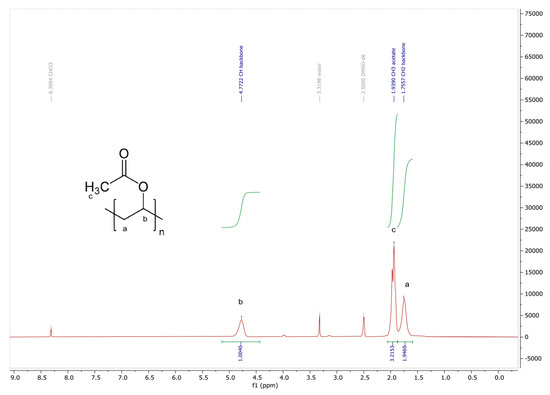
Various alternative compounds have been investigated to prevent icing, one of which includes poly(vinyl) alcohol (PVA), which has shown promising anti-freeze effects. However, determining the optimal structures and formulations of PVA for anti-icing applications has remained a challenge. Building upon our previous work, [...] Read more.
Various alternative compounds have been investigated to prevent icing, one of which includes poly(vinyl) alcohol (PVA), which has shown promising anti-freeze effects. However, determining the optimal structures and formulations of PVA for anti-icing applications has remained a challenge. Building upon our previous work, which used molecular dynamics simulations to assess the effects of hydroxyl group separation distance on ice nucleation, in this work, PVA was modified based upon the structures of antifreeze glycoproteins (AFGPs) found in Antarctic fish, and examined as a potential antifreeze compound. Four different PVA samples with different degrees of hydrolysis were fabricated and subsequently examined for their effects on ice crystallization. The results showed that the modified PVA samples with degrees of hydrolysis of 76% and 66% had an effect on ice crystallization, delaying ice crystallization by an average of approximately 20 min, and even preventing ice crystallization altogether in a small portion of the sample. Meanwhile, other samples with degrees of hydrolysis of 100% and 34% either showed no effect on ice crystallization, shortened the ice crystallization time, and appeared to promote ice nucleation. Full article
Show Figures

Figure 1

5 pages, 541 KiB  
Article
Noninvasive Digital Method for Determining Inflammation after Dental Implantation
by Diana V. Prikule, Vladimir I. Kukushkin and Vladislav F. Prikuls
Biophysica 2022, 2(4), 412-416; https://doi.org/10.3390/biophysica2040036 - 1 Nov 2022
Viewed by 1608
Abstract
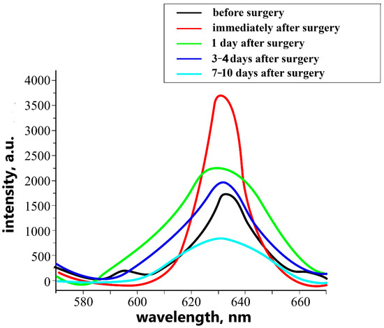
This study shows that the luminescent diagnostic of oral fluid allows the determination of the severity of inflammatory markers after implantation. The noninvasive diagnostic method, which is used, allows the rapid detection of the stages of development of the inflammatory process after intraosseous [...] Read more.
This study shows that the luminescent diagnostic of oral fluid allows the determination of the severity of inflammatory markers after implantation. The noninvasive diagnostic method, which is used, allows the rapid detection of the stages of development of the inflammatory process after intraosseous implantation and prevents the development of complications in the postoperative period. Full article
Show Figures

Figure 1

12 pages, 1782 KiB  
Article
Optimizing Mineralization of Bioprinted Bone Utilizing Type-2 Fuzzy Systems
by Ashkan Sedigh, Mohammad-R. Akbarzadeh-T. and Ryan E. Tomlinson
Biophysica 2022, 2(4), 400-411; https://doi.org/10.3390/biophysica2040035 - 28 Oct 2022
Cited by 2 | Viewed by 1428
Abstract
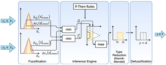
Bioprinting is an emerging tissue engineering method used to generate cell-laden scaffolds with high spatial resolution. Bioprinting parameters, such as pressure, nozzle size, and speed, highly influence the quality of the bioprinted construct. Moreover, cell suspension density and other critical biological parameters directly [...] Read more.
Bioprinting is an emerging tissue engineering method used to generate cell-laden scaffolds with high spatial resolution. Bioprinting parameters, such as pressure, nozzle size, and speed, highly influence the quality of the bioprinted construct. Moreover, cell suspension density and other critical biological parameters directly impact the biological function. Therefore, an approximation model that can be used to find the optimal bioprinting parameter settings for bioprinted constructs is highly desirable. Here, we propose a type-2 fuzzy model to handle the uncertainty and imprecision in the approximation model. Specifically, we focus on the biological parameters, such as the culture period, that can be used to maximize the output value (mineralization volume 21.8 mm3 with the same culture period of 21 days). We have also implemented a type-1 fuzzy model and compared the results with the proposed type-2 fuzzy model using two levels of uncertainty. We hypothesize that the type-2 fuzzy model may be preferred in biological systems due to the inherent vagueness and imprecision of the input data. Our numerical results confirm this hypothesis. More specifically, the type-2 fuzzy model with a high uncertainty boundary (30%) is superior to type-1 and type-2 fuzzy systems with low uncertainty boundaries in the overall output approximation error for bone bioprinting inputs. Full article
Show Figures

Figure 1

13 pages, 1548 KiB  
Article
Drug–Membrane Interaction as Revealed by Spectroscopic Methods: The Role of Drug Structure in the Example of Rifampicin, Levofloxacin and Rapamycin
by Irina M. Le-Deygen, Anastasia S. Safronova, Polina V. Mamaeva, Ilya M. Kolmogorov, Anna A. Skuredina and Elena V. Kudryashova
Biophysica 2022, 2(4), 353-365; https://doi.org/10.3390/biophysica2040032 - 17 Oct 2022
Cited by 11 | Viewed by 2556
Abstract
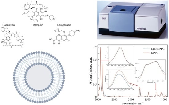
We have investigated the nature of the interaction of small organic drug molecules with lipid membranes of various compositions. Using infrared spectroscopy and differential scanning calorimetry methods, we studied the role of the structure of the active molecule in interaction with the membrane [...] Read more.
We have investigated the nature of the interaction of small organic drug molecules with lipid membranes of various compositions. Using infrared spectroscopy and differential scanning calorimetry methods, we studied the role of the structure of the active molecule in interaction with the membrane using the example of dipalmitoylphosphatidylcholine (DPPC) and dipalmitoylphosphatidylcholine:cardiolipin (DPPC:CL) liposomes. We discovered the key role of the heterocycle in interaction with the polar part of the bilayer and the network of unsaturated bonds in interaction with the hydrophobic part. For rifampicin and levofloxacin, the main binding sites were phosphate and carbonyl groups of lipids, and in the case of anionic liposomes we found a slight penetration of rifampicin into the hydrophobic part of the bilayer. For rapamycin, experimental confirmation of the localization of the molecule in the region of fatty acid chains was obtained, and perturbation in the region of phosphate groups was demonstrated for the first time. The process of phase transition of liposomal forms of rifampicin and levofloxacin was studied. DPPC liposomes accelerate the phase transition when loaded with a drug. DPPC:CL liposomes are less susceptible to changes in the phase transition rate. Full article
Show Figures

Graphical abstract

13 pages, 801 KiB  
Article
Data Structures and Algorithms for k-th Nearest Neighbours Conformational Entropy Estimation
by Roberto Borelli, Agostino Dovier and Federico Fogolari
Biophysica 2022, 2(4), 340-352; https://doi.org/10.3390/biophysica2040031 - 13 Oct 2022
Cited by 2 | Viewed by 2443
Abstract
Entropy of multivariate distributions may be estimated based on the distances of nearest neighbours from each sample from a statistical ensemble. This technique has been applied on biomolecular systems for estimating both conformational and translational/rotational entropy. The degrees of freedom which mostly define [...] Read more.
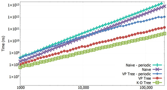
Entropy of multivariate distributions may be estimated based on the distances of nearest neighbours from each sample from a statistical ensemble. This technique has been applied on biomolecular systems for estimating both conformational and translational/rotational entropy. The degrees of freedom which mostly define conformational entropy are torsion angles with their periodicity. In this work, tree structures and algorithms to quickly generate lists of nearest neighbours for periodic and non-periodic data are reviewed and applied to biomolecular conformations as described by torsion angles. The effect of dimensionality, number of samples, and number of neighbours on the computational time is assessed. The main conclusion is that using proper data structures and algorithms can greatly reduce the complexity of nearest neighbours lists generation, which is the bottleneck step in nearest neighbours entropy estimation. Full article
Show Figures

Figure 1

7 pages, 1221 KiB  
Article
Comparative Characteristics of Biomaterials from Juvenile Dentin and Brefomatrix Using Raman Spectroscopy
by Elena V. Timchenko, Irina V. Bazhutova, Pavel Е. Timchenko, Oleg О. Frolov and Larisa Т. Volova
Biophysica 2022, 2(4), 308-314; https://doi.org/10.3390/biophysica2040028 - 26 Sep 2022
Viewed by 1578
Abstract
The results of studies on the assessment of new biomaterials from juvenile teeth for further use in surgical dentistry for bone tissue repair are presented in this work. The comparative assessment of these materials and brefomatrices used in dentistry was carried out. It [...] Read more.
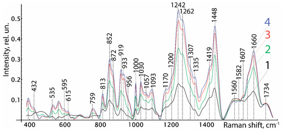
The results of studies on the assessment of new biomaterials from juvenile teeth for further use in surgical dentistry for bone tissue repair are presented in this work. The comparative assessment of these materials and brefomatrices used in dentistry was carried out. It was shown that spectral properties of new biomaterials from juvenile dentin were similar to the spectral properties of brefomatrices from cortical tissue according to the developed discriminant model of the characteristic changes of Raman line intensities. The calculated accuracy of the discriminant model was 82.7 ± 3.2%. Full article
Show Figures

Figure 1

16 pages, 1460 KiB  
Review
Fibonacci Sequences, Symmetry and Order in Biological Patterns, Their Sources, Information Origin and the Landauer Principle
by Edward Bormashenko
Biophysica 2022, 2(3), 292-307; https://doi.org/10.3390/biophysica2030027 - 16 Sep 2022
Cited by 7 | Viewed by 5950
Abstract
Physical roots, exemplifications and consequences of periodic and aperiodic ordering (represented by Fibonacci series) in biological systems are discussed. The physical and biological roots and role of symmetry and asymmetry appearing in biological patterns are addressed. A generalization of the Curie–Neumann principle as [...] Read more.
Physical roots, exemplifications and consequences of periodic and aperiodic ordering (represented by Fibonacci series) in biological systems are discussed. The physical and biological roots and role of symmetry and asymmetry appearing in biological patterns are addressed. A generalization of the Curie–Neumann principle as applied to biological objects is presented, briefly summarized as: “asymmetry is what creates a biological phenomenon”. The “top-down” and “bottom-up” approaches to the explanation of symmetry in organisms are presented and discussed in detail. The “top-down” approach implies that the symmetry of the biological structure follows the symmetry of the media in which this structure is functioning; the “bottom-up” approach, in turn, accepts that the symmetry of biological structures emerges from the symmetry of molecules constituting the structure. A diversity of mathematical measures applicable for quantification of order in biological patterns is introduced. The continuous, Shannon and Voronoi measures of symmetry/ordering and their application to biological objects are addressed. The fine structure of the notion of “order” is discussed. Informational/algorithmic roots of order inherent in the biological systems are considered. Ordered/symmetrical patterns provide an economy of biological information, necessary for the algorithmic description of a biological entity. The application of the Landauer principle bridging physics and theory of information to the biological systems is discussed. Full article
Show Figures

Figure 1

15 pages, 2198 KiB  
Article
Immunological Effects of Cold Atmospheric Plasma-Treated Cells in Comparison with Those of Cells Treated with Lactaptin-Based Anticancer Drugs
by Olga Troitskaya, Diana Novak, Mikhail Varlamov, Mikhail Biryukov, Anna Nushtaeva, Galina Kochneva, Dmitriy Zakrevsky, Irina Schweigert, Vladimir Richter and Olga Koval
Biophysica 2022, 2(3), 266-280; https://doi.org/10.3390/biophysica2030025 - 1 Sep 2022
Cited by 3 | Viewed by 2218
Abstract
The ability of dying cancer cells to induce an anticancer immune response can increase the effectiveness of anticancer therapies, and such type of death is termed immunogenic cell death (ICD). Cells can die along the ICD pathway when exposed not only to chemo- [...] Read more.
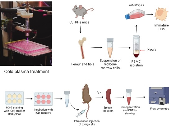
The ability of dying cancer cells to induce an anticancer immune response can increase the effectiveness of anticancer therapies, and such type of death is termed immunogenic cell death (ICD). Cells can die along the ICD pathway when exposed not only to chemo- and immunotherapeutics, but also to various types of radiation, such as ionizing radiation and cold atmospheric plasma jets (CAP). We have previously shown that CAP, lactaptin, and a recombinant vaccinia virus encoding lactaptin induce in vitro molecular changes typical of ICD in cancer cells. In the current work, we treated MX-7 rhabdomyosarcoma cells with CAP and lactaptin-based anticancer drugs and evaluated the immunological effects of the treated cells. We showed that dendritic cells (DCs) captured cells treated with various ICD inducers with different efficiency. CAP-treated cells were weakly potent in inducing the maturation of DCs according to MHC II externalization. Moreover, CAP-treated cells were worse in the stimulation of IFN-α release in vitro and were poorly captured by spleen DCs in vivo. Under the irradiation conditions used, CAP was not capable of activating a significant immunological anti-tumor effect in vivo. It is possible that modifications of the CAP irradiation regimen will enhance the activation of the immune system. Full article
Show Figures

Graphical abstract

18 pages, 5397 KiB  
Article
The Counteraction of Cultivated Cistus creticus L. (Rock Rose) Plants to the Strain Imposed by a Long-Term Exposure to Non-Ionizing Radiation and the Role of DDC
by Aikaterina L. Stefi, Georgia Kalouda, Aikaterini S. Skouroliakou, Dido Vassilacopoulou and Nikolaos S. Christodoulakis
Biophysica 2022, 2(3), 248-265; https://doi.org/10.3390/biophysica2030024 - 1 Sep 2022
Cited by 1 | Viewed by 2862
Abstract
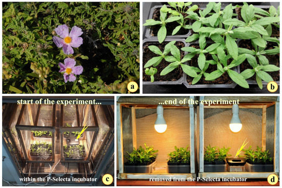
Two groups of Cistus creticus seedlings were grown in two chambers under controlled environmental conditions. In one of the chambers, a continuously emitting base unit of a wireless telephone was placed. After fifty days of culture, the two groups of plants were removed [...] Read more.
Two groups of Cistus creticus seedlings were grown in two chambers under controlled environmental conditions. In one of the chambers, a continuously emitting base unit of a wireless telephone was placed. After fifty days of culture, the two groups of plants were removed and thoroughly investigated and compared. The aboveground parts of the exposed plants were retarded in development while their roots exhibited increased biomass, compared to the controls. There was a minor decrease in the absorbance of the photosynthetic pigments in exposed plants, while an overproduction of Reactive Oxygen Species (ROS) ROS in their leaves and roots was detected. The expression of the L-Dopa decarboxylase (DDC) seemed to “erupt” following the exposure to radiation in both shoots and roots of the stressed plants, and their roots slow down their secondary development; strangely, the phenolic content is reduced in their leaves, the external topography of which indicates a rather xeromorphic response. We may suggest that Cistus creticus plants, forced by the radiation stress, can finely tune their metabolic pathways in a way that can be useful in the pharmaceutical industry. Full article
Show Figures

Figure 1

8 pages, 835 KiB  
Article
On the Molecular Driving Force of Protein–Protein Association
by Roberta Rapuano and Giuseppe Graziano
Biophysica 2022, 2(3), 240-247; https://doi.org/10.3390/biophysica2030023 - 25 Aug 2022
Cited by 1 | Viewed by 1572
Abstract
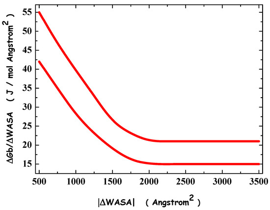
The amount of water-accessible-surface-area, WASA, buried upon protein–protein association is a good measure of the non-covalent complex stability in water; however, the dependence of the binding Gibbs free energy change upon buried WASA proves to be not trivial. We assign a precise physicochemical [...] Read more.
The amount of water-accessible-surface-area, WASA, buried upon protein–protein association is a good measure of the non-covalent complex stability in water; however, the dependence of the binding Gibbs free energy change upon buried WASA proves to be not trivial. We assign a precise physicochemical role to buried WASA in the thermodynamics of non-covalent association and perform close scrutiny of the contributions favoring and those contrasting protein–protein association. The analysis indicates that the decrease in solvent-excluded volume, an entropic effect, described by means of buried WASA, is the molecular driving force of non-covalent association in water. Full article
Show Figures

Figure 1

19 pages, 4623 KiB  
Article
Physicochemical Characterization of the Catalytic Unit of Hammerhead Ribozyme and Its Relationship with the Catalytic Activity
by Yoshiyuki Tanaka, Daichi Yamanaka, Saori Morioka, Taishi Yamaguchi, Masayuki Morikawa, Takashi S. Kodama, Vladimír Sychrovský, Chojiro Kojima and Yoshikazu Hattori
Biophysica 2022, 2(3), 221-239; https://doi.org/10.3390/biophysica2030022 - 25 Aug 2022
Viewed by 2106
Abstract
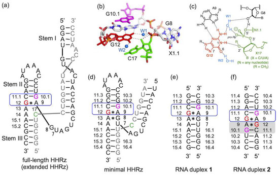
The catalytic mechanism of hammerhead ribozymes (HHRzs) attracted great attention in relation to the chemical origin of life. However, the basicity (pKa) of the catalytic sites of HHRzs has not been studied so far. As a result, the investigation of [...] Read more.
The catalytic mechanism of hammerhead ribozymes (HHRzs) attracted great attention in relation to the chemical origin of life. However, the basicity (pKa) of the catalytic sites of HHRzs has not been studied so far. As a result, the investigation of the currently assumed mechanism from an experimentally derived pKa value has been impossible. In HHRzs, there exists a highly functionalized structural unit (A9-G10.1 site) with a catalytic residue (G12) for the nucleophile activation and metal ion-binding residue (G10.1). As inferred from this fact, there might be a possibility that HHRzs may utilize specific functions of the A9-G10.1 motif for the catalytic reaction. Therefore, here we studied the basicity of G12/G10.1-corresponding residues using RNA duplexes including the A9-G10.1 motif without other conserved residues of HHRzs. From the pH-titration experiments with NMR spectra, we have obtained the intrinsic basicity of the G12/G10.1-corresponding residues in the motif, with pKa > 11.5 (N1 of G12) and pKa 4.5 (N7 of G10.1) for the first time. Based on the derived irregular basicity, their correlation with a catalytic activity and a metal affinity were investigated. In total, the derived thermodynamic properties are an intrinsic nature of the exclusive catalytic unit of HHRzs, which will be an outstanding pivot point for the mechanistic analyses. Full article
Show Figures

Figure 1

18 pages, 4063 KiB  
Article
Peptide-Functionalized Nanoparticles for the Targeted Delivery of Cytotoxins to MMP-14-Expressing Cancer Cells
by Jillian Cathcart, Giulia Suarato, Weiyi Li, Jian Cao and Yizhi Meng
Biophysica 2022, 2(3), 203-220; https://doi.org/10.3390/biophysica2030021 - 24 Aug 2022
Cited by 1 | Viewed by 2519
Abstract
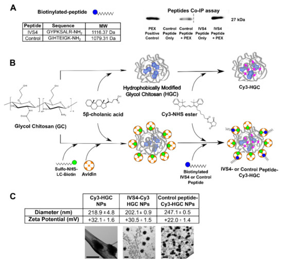
As 90% of cancer-patient deaths are due to metastasis, novel therapeutics that selectively target and kill metastatic cells are desperately needed. Matrix metalloproteinase-14 (MMP-14), which plays a critical role in digesting the basement membrane and in inducing cancer cell migration, has been found [...] Read more.
As 90% of cancer-patient deaths are due to metastasis, novel therapeutics that selectively target and kill metastatic cells are desperately needed. Matrix metalloproteinase-14 (MMP-14), which plays a critical role in digesting the basement membrane and in inducing cancer cell migration, has been found to be expressed at the cell surface of circulating and metastasized tumor cells in various human cancers. We have recently shown that the IVS4 peptide, which mimics the minimal binding motif of the hemopexin-like (PEX) domain of MMP-14, interrupts MMP-14 dimerization and decreases MMP-14-mediated cell invasion. In this study, cancer-homing nanocarriers were assembled by linking IVS4 to polysaccharide-based nanoparticles (NPs), followed by the encapsulation of a pharmaceutical agent. IVS4-NPs efficiently prevented MMP-14-mediated cell migration and conferred an uptake advantage compared to the control peptide in an MMP-14-dependent manner. While the IVS4-NPs alone were not cytotoxic, drug-encapsulated NPs were shown to effectively target MMP-14-expressing cancer cells. This novel nanotherapeutic is capable of inhibiting MMP-14-mediated functions and efficiently killing MMP-14-expressing cancer cells, without affecting the viability of non-cancer cells. Full article
Show Figures

Figure 1

9 pages, 2034 KiB  
Article
Exploring the Dynamics of Shikimate Kinase through Molecular Mechanics
by Pedro Ojeda-May
Biophysica 2022, 2(3), 194-202; https://doi.org/10.3390/biophysica2030020 - 23 Aug 2022
Cited by 2 | Viewed by 2778
Abstract
Shikimate kinase (SK) enzyme is a suitable target for antimicrobial drugs as it is present in pathogenic microorganisms and absent in mammals. A complete understanding of the functioning of this enzyme can unveil novel methods to inactivate it. To do this, a clear [...] Read more.
Shikimate kinase (SK) enzyme is a suitable target for antimicrobial drugs as it is present in pathogenic microorganisms and absent in mammals. A complete understanding of the functioning of this enzyme can unveil novel methods to inactivate it. To do this, a clear understanding of SK performance is needed. Previously, the chemical step of SK was studied in detail, but a study of longer-term scale simulations is still missing. In the present work, we performed molecular dynamics (MD) simulations in the μs time scale that allowed us to explore further regions of the SK energy landscape than previously. Simulations were conducted on the wild-type (WT) enzyme and the R116A and R116K mutants. We analyzed the dynamics of the enzymes through standard MD tools, and we found that the global motions in the mutants were perturbed. These motions can be linked to the observed undetectable binding affinity of the WT enzyme and the R116A and R116K mutants. Full article
Show Figures

Figure 1

10 pages, 1609 KiB  
Review
Sodium Channels Involved in the Initiation of Action Potentials in Invertebrate and Mammalian Neurons
by Daria Y Romanova, Pavel M Balaban and Evgeny S Nikitin
Biophysica 2022, 2(3), 184-193; https://doi.org/10.3390/biophysica2030019 - 11 Aug 2022
Cited by 4 | Viewed by 4242
Abstract
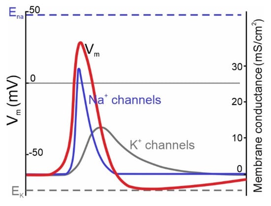
Living organisms react to external stimuli to adapt their activity to the environment for survival. Acquired information is encoded by neurons by action potentials (APs) in a series of discrete electrical events. Rapid initiation of the AP is critical for fast reactions and [...] Read more.
Living organisms react to external stimuli to adapt their activity to the environment for survival. Acquired information is encoded by neurons by action potentials (APs) in a series of discrete electrical events. Rapid initiation of the AP is critical for fast reactions and strongly relies on voltage-activated Na+-selective channels (NaVs), which are widely expressed by both invertebrate and vertebrate neurons. Intuitively, NaVs of higher mammals should be activated faster than those of any other species. In addition to improved NaV channel structure, central mammalian neurons also demonstrate a patterned distribution of specific types of NaV1 channels at and near the site of AP initiation within the axonal initial segment (AIS). The AIS has different types of fast Nav1 channels and is thought to provide the biological basis for efficient frequency coding of information. In the present work, we review data related to the channels underlying fast initiation of action potentials in vertebrates and invertebrates, along with their evolution, distribution, and known specific roles. Current research has established that all mammalian NaV1 (1.1–1.9) channels share a similar structure, with 4 conservative transmembrane D-domains with a highly homologous sequence, but significant differences in the length of the functional cytoplasmic linkers. Similarly, the structure of NaV1 channels in invertebrates is generally similar to that of mammals, but it shows high variability across the evolutionary tree in the length of the linkers. AP initiation in mammalian cortical neurons is mediated by NaV1.2 and NaV1.6 channels, whereas interneurons mostly rely on NaV1.1 channels in their firing. Although invertebrate NaV1 channels normally display relatively slow kinetics, their activation is fast enough to produce APs, even in simple animals such as Placozoa. Remarkably, fast sodium-based excitability is not limited to animals. Recently, a photosynthetic prokaryote has been found to show rapidly activated sodium currents provided by their independently evolved single D-domain EuKatB sodium channels. Full article
Show Figures

Figure 1

10 pages, 526 KiB  
Perspective
Present and Future Opportunities in Imaging the Ubiquitin System (Ub-System)
by Leonardo Mortati, Barbara Pergolizzi, Cristina Panuzzo and Enrico Bracco
Biophysica 2022, 2(3), 174-183; https://doi.org/10.3390/biophysica2030018 - 28 Jul 2022
Viewed by 2029
Abstract
From yeast to mammalian cells, ubiquitination is one of the most conserved, and reversible, eukaryotic post-translational modifications (PTMs) responsible for controlling nearly all cellular processes. Potentially, every single eukaryotic cell can accomplish different ubiquitination processes at once, which in turn control the execution [...] Read more.
From yeast to mammalian cells, ubiquitination is one of the most conserved, and reversible, eukaryotic post-translational modifications (PTMs) responsible for controlling nearly all cellular processes. Potentially, every single eukaryotic cell can accomplish different ubiquitination processes at once, which in turn control the execution of specific cellular events in time and space with different biological significance (e.g., protein degradation or protein–protein interaction). Overall, all these signals are highly dynamic and need to be finely integrated to achieve a proper cellular response. Altogether, ubiquitination appears to be an extremely complex process, likely more than any other PTMs. Until a few years ago, the prevailing experimental approaches to investigate the different aspects of the ubiquitin system entailed genetic and biochemical analysis. However, recently, reagents and technologies have been developed enabling microscopy-based imaging of ubiquitination to enter the scene. In this paper, we discuss the progress made with conventional (confocal fluorescence microscopy) and non-conventional non-linear microscopy (Atomic Force Microscopy—AFM, Coherent Anti-Stokes Raman Scattering—CARS, Stimulated Raman Scattering—SRS) and we speculate on future developments. Full article
Show Figures

Figure 1

6 pages, 11884 KiB  
Article
Blue Light Enhances Fluoride Anticariogenic Activity against Streptococcus mutans
by Uziel Jeffet, Shiri Livne, Shir Dviker and Nir Sterer
Biophysica 2022, 2(3), 168-173; https://doi.org/10.3390/biophysica2030017 - 7 Jul 2022
Viewed by 1967
Abstract
Previous studies have shown that sub-lethal exposure of blue light caused increased bacterial cell membrane permeability. We hypothesized that combining blue light exposure with other antibacterial agents may increase their efficacy. The aim of the present study was to test the combined effect [...] Read more.
Previous studies have shown that sub-lethal exposure of blue light caused increased bacterial cell membrane permeability. We hypothesized that combining blue light exposure with other antibacterial agents may increase their efficacy. The aim of the present study was to test the combined effect of blue light and sodium fluoride against dental caries pathogen Streptococcus mutans. Sm biofilms were exposed to blue light (400–500 nm) with or without sodium fluoride. Exposed and non-exposed samples were studied for acid production (lactate assay kit), acid tolerance (ATPase assay kit) and bacterial cell membrane damage (fluorescence microscopy). Results showed that the combined treatment significantly reduced the virulence of Sm concomitant with an increase in bacterial cell membrane permeability. Taken together, these results suggest that adjacent blue light exposure may increase fluoride caries preventive properties. Full article
Show Figures

Figure 1

14 pages, 1035 KiB  
Review
Application of Non-Destructive Testing Techniques (NDTT) to Characterize Nanocarriers Used for Drug Delivery: A Mini Review
by Rahul Islam Barbhuiya, Saipriya Ramalingam, Harsimran Kaur Kalra, Abdallah Elsayed, Winny Routray, Manickavasagan Annamalai and Ashutosh Singh
Biophysica 2022, 2(3), 154-167; https://doi.org/10.3390/biophysica2030016 - 24 Jun 2022
Viewed by 2754
Abstract
The synthesis of tailored and highly engineered multifunctional pharmaceutical nanocarriers is an emerging field of study in drug delivery applications. They have a high surface-area-to-volume ratio, aiding the targeted drug’s biodistribution and pharmacokinetic properties. Therefore, the characterization of nanocarriers is critical for understanding [...] Read more.
The synthesis of tailored and highly engineered multifunctional pharmaceutical nanocarriers is an emerging field of study in drug delivery applications. They have a high surface-area-to-volume ratio, aiding the targeted drug’s biodistribution and pharmacokinetic properties. Therefore, the characterization of nanocarriers is critical for understanding their physicochemical properties, which significantly impact their molecular and systemic functioning. To achieve specific goals, particle size, surface characteristics, and drug release properties of nanocarriers must be managed. This mini review provides an overview of the applications of non-destructive testing techniques (NDTT) to reveal the characteristics of nanocarriers, considering their surface charge, porosity, size, morphology, and crystalline organization. The compositional and microstructural characterization of nanocarriers through NDTT, such as dynamic light scattering, X-ray diffraction, confocal laser scanning microscopy, ultraviolet-visible spectroscopy, scanning electron microscopy, atomic force microscopy, and nuclear magnetic resonance spectroscopy, have been comprehensively reviewed. Furthermore, NDTT is only used to characterize physicochemical parameters related to the physiological performance of nanocarriers but does not account for nanocarrier toxicity. Hence, it is highly recommended that in the future, NDTT be developed to assess the toxicity of nanocarriers. In addition, by developing more advanced, effective, and precise techniques, such as machine vision techniques using artificial intelligence, the future of using NDTT for nanocarrier characterization will improve the evaluation of internal quality parameters. Full article
Show Figures

Figure 1

19 pages, 6878 KiB  
Article
The Amyloidogenic Peptide Amyloid Beta(16–22) Displays Facet Dependent Conformation on Metal Surfaces
by Kieran P. Somers and David L. Cheung
Biophysica 2022, 2(2), 135-153; https://doi.org/10.3390/biophysica2020015 - 9 Jun 2022
Cited by 2 | Viewed by 2517
Abstract
Currently, it is not understood how metal nanoparticles influence the formation of protein fibrils, although recent literature highlights that the shape and chemical composition of such nanoparticles can strongly influence the process. Understanding this process at a fundamental level can potentially unlock routes [...] Read more.
Currently, it is not understood how metal nanoparticles influence the formation of protein fibrils, although recent literature highlights that the shape and chemical composition of such nanoparticles can strongly influence the process. Understanding this process at a fundamental level can potentially unlock routes to the development of new therapeutics, as well as novel materials for technological applications. This requires a microscopic picture of the behaviour of amyloidogenic proteins on metal surfaces. Using replica exchange molecular dynamics simulations, we investigate the conformation of the model amyloidogenic peptide, Aβ(16–22), on different gold and silver surfaces. The conformation of the peptide on gold surfaces also shows a strong facet dependence, with fibril-like conformations being promoted in the 100 surface and inhibited on the 111 surface. A smaller degree of facet dependence is seen for silver with the peptide behaving similar on both of these. The difference in the facet dependence can be related to the difference between direct adsorption onto the gold 111 surface, with a preference towards indirect (water mediated) adsorption onto the other surfaces. This new information on the behaviour of an amyloidogenic peptide on metal surfaces can give insight into the size-dependent effect of nanoparticles on fibril formation and the use of surfaces to control fibrillation. Full article
Show Figures

Figure 1

12 pages, 1630 KiB  
Review
Analysis of Enzyme Conformation Dynamics Using Single-Molecule Förster Resonance Energy Transfer (smFRET)
by Mai Huynh and Bhaswati Sengupta
Biophysica 2022, 2(2), 123-134; https://doi.org/10.3390/biophysica2020014 - 6 Jun 2022
Cited by 2 | Viewed by 2761
Abstract
Single-molecule Förster resonance energy transfer (smFRET) enables the deconvolution of various conformational substates of biomolecules. Over the past two decades, it has been widely used to understand the conformational dynamics of enzymes. Commonly, enzymes undergo reversible transitions between active and inactive states in [...] Read more.
Single-molecule Förster resonance energy transfer (smFRET) enables the deconvolution of various conformational substates of biomolecules. Over the past two decades, it has been widely used to understand the conformational dynamics of enzymes. Commonly, enzymes undergo reversible transitions between active and inactive states in solution. Using smFRET, the details of these transitions and the effect of ligands on these dynamics have been determined. In this mini-review, we discuss the various works focused on the investigation of enzyme conformational dynamics using smFRET. Full article
Show Figures

Figure 1

8 pages, 836 KiB  
Article
The Relationship between Hydrophobicity and Drug-Protein Binding in Human Serum Albumin: A Quartz Crystal Microbalance Study
by Ahmad R. Alhankawi, Jacob K. Al-Husseini, Archie Spindler, Clark Baker, Tonderai T. Shoniwa, Mohammed Ahmed, Peter A. Chiarelli and Malkiat S. Johal
Biophysica 2022, 2(2), 113-120; https://doi.org/10.3390/biophysica2020012 - 23 May 2022
Cited by 8 | Viewed by 4184
Abstract
In this paper, the quartz crystal microbalance with dissipation monitoring (QCM-D) was used to investigate hydrophobicity and binding strength (KD) for 10 different drugs interacting with human serum albumin (HSA). Quantitative structure activity relationship (QSAR) analysis was used to determine the [...] Read more.
In this paper, the quartz crystal microbalance with dissipation monitoring (QCM-D) was used to investigate hydrophobicity and binding strength (KD) for 10 different drugs interacting with human serum albumin (HSA). Quantitative structure activity relationship (QSAR) analysis was used to determine the relationship between drug hydrophobicity (ClogP) and HSA binding strength log(1/KD). The results are compared to prior knowledge on bovine serum albumin (BSA) binding. We demonstrate a positive correlation between drug hydrophobicity and the strength of ligand-protein binding to HSA and show a statistically significant similarity with the trend reported in BSA. The findings presented in this work provide insight into the role that bound water plays in ligand-protein interactions. Further, the comparison between HSA and BSA provides quantitative justification for the use of these proteins interchangeably in the analysis of drug-based binding kinetics. Full article
Show Figures

Figure 1

10 pages, 861 KiB  
Article
Cholesterol Significantly Affects the Interactions between Pirfenidone and DPPC Liposomes: Spectroscopic Studies
by Irina M. Le-Deygen, Anastasia S. Safronova, Polina V. Mamaeva, Anna A. Skuredina and Elena V. Kudryashova
Biophysica 2022, 2(1), 79-88; https://doi.org/10.3390/biophysica2010008 - 16 Feb 2022
Cited by 4 | Viewed by 3855
Abstract
In this work, we studied the effect of as on the interaction of membrane DPPC with the key antifibrotic drug pirfenidone. Liposomal forms of pirfenidone were obtained using passive loading. The addition of cholesterol reduces the loading efficiency of pirfenidone by 10%. The [...] Read more.
In this work, we studied the effect of as on the interaction of membrane DPPC with the key antifibrotic drug pirfenidone. Liposomal forms of pirfenidone were obtained using passive loading. The addition of cholesterol reduces the loading efficiency of pirfenidone by 10%. The main binding site of pirfenidone in DPPC liposomes is the carbonyl group: the interaction with PF significantly increases the proportion of low-hydrated carbonyl groups as revealed by ATR-FTIR spectroscopy. The phosphate group acts as an additional binding site; however, due to shielding by the choline group, this interaction is weak. The hydrophobic part of the bilayer is not involved in PF binding at room temperature. Cholesterol changes the way of interaction between carbonyl groups and pirfenidone probably because of the formation of two subpopulations of DPPC and causes a dramatic redistribution of carbonyl groups onto the degrees of hydration. The proportion of moderately hydrated carbonyl groups increases, apparently due to the deepening of pirfenidone into the circumpolar region of the bilayer. For the first time, a change in the microenvironment of pirfenidone upon binding to liposomes was shown: aromatic moiety interacts with the bilayer. Full article
Show Figures

Figure 1

11 pages, 4162 KiB  
Article
Fractal Dimension Analysis to Detect the Progress of Cancer Using Transmission Optical Microscopy
by Liam Elkington, Prakash Adhikari and Prabhakar Pradhan
Biophysica 2022, 2(1), 59-69; https://doi.org/10.3390/biophysica2010005 - 7 Jan 2022
Cited by 17 | Viewed by 7098
Abstract
Fractal dimension, a measure of self-similarity in a structure, is a powerful physical parameter for the characterization of structural property of many partially filled disordered materials. Biological tissues are fractal in nature and reports show a change in self-similarity associated with the progress [...] Read more.
Fractal dimension, a measure of self-similarity in a structure, is a powerful physical parameter for the characterization of structural property of many partially filled disordered materials. Biological tissues are fractal in nature and reports show a change in self-similarity associated with the progress of cancer, resulting in changes in their fractal dimensions. Here, we report that fractal dimension measurement is a potential technique for the detection of different stages of cancer using transmission optical microscopy. Transmission optical microscopy of a thin tissue sample produces intensity distribution patterns proportional to its refractive index pattern, representing its mass density distribution. We measure fractal dimension detection of different cancer stages and find its universal feature. Many deadly cancers are difficult to detect in their early to different stages due to the hard-to-reach location of the organ and/or lack of symptoms until very late stages. To study these deadly cancers, tissue microarray (TMA) samples containing different stages of cancers are analyzed for pancreatic, breast, colon, and prostate cancers. The fractal dimension method correctly differentiates cancer stages in progressive cancer, raising possibilities for a physics-based accurate diagnosis method for cancer detection. Full article
Show Figures

Figure 1

25 pages, 15952 KiB  
Article
Nascent Adhesion Clustering: Integrin-Integrin and Integrin-Substrate Interactions
by Kuanpo Lin and Robert J. Asaro
Biophysica 2022, 2(1), 34-58; https://doi.org/10.3390/biophysica2010004 - 7 Jan 2022
Cited by 3 | Viewed by 4002
Abstract
Nascent adhesions (NAs) are a general precursor to the formation of focal adhesions (FAs) that provide a fundamental mechanism for cell adhesion that is, in turn, involved in cell proliferation, migration, and mechanotransduction. Nascent adhesions form when cells come into contact with substrates [...] Read more.
Nascent adhesions (NAs) are a general precursor to the formation of focal adhesions (FAs) that provide a fundamental mechanism for cell adhesion that is, in turn, involved in cell proliferation, migration, and mechanotransduction. Nascent adhesions form when cells come into contact with substrates at all rigidities and generally involve the clustering of ligated integrins that may recruit un-ligated integrins. Nascent adhesions tend to take on characteristic sizes in the range of O(100nm150nm) in diameter and tend to contain integrin numbers of O(2060). The flexible, adaptable model we present provides and clear explanation of how these conserved cluster features come about. Our model is based on the interaction among ligated and un-ligated integrins that arise due to deformations that are induced in the cell membrane-cell glycocalyx and substrate system due to integrin activation and ligation. This model produces a clearly based interaction potential, and from it an explicit interaction force among integrins, that our stochastic diffusion-interaction simulations then show will produce nascent clusters with experimentally observed characteristics. Our simulations reveal effects of various key parameters related to integrin activation and ligation as well as some unexpected and previously unappreciated effects of parameters including integrin mobility and substrate rigidity. Moreover, the model’s structure is such that refinements are readily incorporated and specific suggestions are made as to what is required for further progress in understanding nascent clustering and the development of mature focal adhesions in a truly predictive manner. Full article
Show Figures

Figure 1

Back to TopTop