Article

The Contrasting Effect of Sodium Alginate on Lysozyme and Albumin Denaturation and Fibril Formation

by
Diliara R. Khaibrakhmanova
1,
Polina R. Kuzivanova
1,
Bulat R. Gainutdinov
1,
Timur I. Magsumov
1,
Alena A. Nikiforova
1 and
Igor A. Sedov
1,2,*
1
Chemical Institute, Kazan Federal University, Kremlevskaya 18, 420008 Kazan, Russia
2
Kazan Institute of Biochemistry and Biophysics, FRC Kazan Scientific Center of RAS, 420111 Kazan, Russia
*
Author to whom correspondence should be addressed.
Biophysica 2024, 4(4), 651-666; https://doi.org/10.3390/biophysica4040043
Submission received: 6 November 2024 / Revised: 4 December 2024 / Accepted: 10 December 2024 / Published: 12 December 2024
(This article belongs to the Collection Feature Papers in Biophysics)

Abstract

:
The effect of sodium alginate on the denaturation and aggregation behavior of bovine serum albumin and hen egg-white lysozyme was studied. Large amounts of polysaccharide increase the thermal stability of albumin due to the weak binding interactions. At the same time, sodium alginate can reduce the quantity of amyloid fibrils formed by albumin under denaturing conditions, which is a consequence of the stabilization of the native protein form by glycan binding. In the case of lysozyme, the polysaccharide has no influence on the thermal stability of the protein in 2 M guanidinium hydrochloride. However, the inhibition of fibril formation with an increase in the lag time was observed, which is explained by the binding of sodium alginate to lysozyme fibrils, but not to the protein monomer. The molecular nature of the binding interactions between alginate and the studied proteins was elucidated using molecular docking and known experimental structures of glycan–protein complexes.

1. Introduction

Polysaccharides are natural compounds with promising medical applications. They can be used as drug carriers [1,2] as well as pharmacologically active substances [3,4,5], potentially showing activity against neurodegenerative diseases caused by abnormal protein aggregation. One of the examples of this is sodium oligomannate, which passed two phases of clinical trials for Alzheimer’s disease treatment [6]. Wozniak et al. [7] showed that sulfonated fucan derivatives from brown algae can prevent the formation of amyloid fibrils in cell cultures. Negatively charged polysaccharide ulvan inhibits aggregation and disrupts mature fibrils of α-synuclein and β-amyloid [8]. The fibril formation process turns out to be particularly sensitive to the presence of polyanionic compounds of various natures. Montgomery et al. [9] tested the effect of anionic polysaccharides, nucleic acids, polypeptides, polysulfonates, and other polyanions on the amyloid aggregation of tau protein. It was shown that most of the tested molecules promote fibril formation, which was attributed to the minimization of the electrostatic repulsion between positively charged tau molecules and the scaffolding of the fibril assembly. The strength of induction and the resulting fibril morphology depend on the structure of the polyanion.
Sodium alginate is a negatively charged polysaccharide composed of uronic acids which interacts with positively charged regions of proteins due to electrostatic forces [10,11]. Its effect on protein aggregation and denaturation has been relatively poorly studied. Motomura and co-authors [12] showed that sodium alginate can act as a cryoprotectant for fish myofibrillar protein by retaining water in a liquid state while freezing protein molecules. Zhao et al. [13] used FT-IR and SE-HPLC to investigate the interactions of the native and denatured states of bovine serum albumin with sodium alginate. Denatured protein has shown stronger interactions with the polysaccharide due to the presence of positively charged amino acids exposed to the protein surface. Chang et al. [14] found that sodium alginate strongly stabilizes native phosphoglycerate kinase, increasing its denaturation temperature by 14.5 °C, while for the much smaller hPin1 WW domain, only a 3.5 °C increase was observed. Surprisingly, the stabilization decreased at higher alginate concentrations. Alginate was also observed to suppress the aggregation of both proteins at high temperatures.
Bovine and human serum albumins (BSA and HSA, respectively) and hen egg-white lysozyme (HEWL) are among the most popular model globular proteins. They easily form fibrils under the denaturing conditions in vitro [15,16,17]. It was previously shown that various low- and high-molecular-weight substances can inhibit the fibrillation of these proteins [18,19,20,21,22]. In the present work, we aim to establish the effects of sodium alginate on the fibril formation of the two proteins and study the interactions in the corresponding systems. We expect that these effects and the patterns of interactions with alginate and other anionic polysaccharides are shared by a variety of fibril-forming proteins.

2. Materials and Methods

2.1. Materials

BSA (PanEko, Russia, purity > 98%), HEWL (Sigma-Aldrich, St. Louis, MO, USA, purity > 98%), sodium alginate from brown algae (Sigma-Aldrich, St. Louis, MO, USA), thioflavin T (ThT, Sigma-Aldrich, St. Louis, MO, USA, purity > 99%), guanidinium hydrochloride (GdHCl, Dia-M, Moscow, Russia, purity > 99.5%), and Tris buffer (Trizma base, Trizma hydrochloride, 25 mM, pH 7.4, Sigma-Aldrich, St. Louis, MO, USA, purity > 99%) were used without preliminary purification.
The molecular weight of the sample of sodium alginate used in our study was previously determined by Kolotova et al. [23] using HPLC and equals 507 kDa. Sodium alginate solution was prepared by soaking a polysaccharide sample in a Tris buffer, followed by shaking until complete dissolution at 60 °C. Solutions of proteins and ThT were prepared by dissolving a sample in a Tris buffer (BSA) or in 2 M GdHCl (HEWL). All solutions used for differential scanning calorimetry (DSC) experiments were preliminarily degassed in vacuum.

2.2. Fibril Preparation

HEWL fibrils were prepared by incubating a solution of HEWL monomer (2 mg·mL−1, 140 μM unless otherwise mentioned explicitly) in 2 M GdHCl solution without buffer (pH 6.5) in the absence or the presence of sodium alginate in a thermostated shaker at 60 °C with a shaking frequency of 1100 rpm. At certain times, 50 μL aliquots of the incubated solution were taken, cooled to room temperature, and used in the ThT assay.
Albumin fibrils were prepared by incubating BSA monomer solution at concentrations of 2 to 20 mg·mL−1 during different experiments in Tris buffer solution at pH 7.4 in the absence or the presence of sodium alginate in a thermostat without stirring at 65 °C. At certain times, 100 μL aliquots of the incubated solution was taken, cooled to room temperature, and used in the ThT assay.

2.3. ThT Assay

The kinetics of fibril formation were studied using a standard method based on recording the fluorescence spectrum of ThT bound to fibrils [24].
The samples of incubated protein were added to cuvettes containing ThT solution and a buffer, adding up to the final concentrations of 3 and 10.4 μM of protein and ThT, respectively. The cuvettes with the samples were kept for 10 min at 25 °C to allow for the binding of the dye to the fibrils, after which the fluorescence spectra were recorded on a Cary Eclipse fluorescence spectrophotometer (Agilent) in the wavelength range from 475 to 495 nm at excitation wavelength λex = 450 nm. All the experiments were replicated 3–5 times taking the samples at the same moments of time t from the beginning. The average ThT fluorescence signal F at a wavelength of 485 nm as a function of time was used to obtain the kinetic curves. These curves were fitted with the following equation:
F = F 1 1 k b e k a t 1 + 1 ,
where F is the plateau ThT fluorescence, and ka and kb are the kinetic constants of the fibril formation process determined from the fit. This equation was previously used to describe both sigmoidal fibril formation curves with the values kb < 0.5 as in the case of lysozyme and lag-free hyperbolic curves typical for albumin aggregation, for which kb > 0.5 [25]. The values of the half-time of fibril formation τ50 corresponding to the time at which half of the plateau fluorescence is achieved can be expressed in terms of ka and kb:
τ 50 = 1 k a ln 1 k b + 1

2.4. Differential Scanning Calorimetry

DSC thermograms of denaturation of BSA and HEWL in solutions in the absence and presence of sodium alginate with the same composition as in fibril formation studies were obtained using a NanoDSC capillary differential scanning calorimeter (TA Instruments, New Castle, DE, USA). The protein solution was heated from 275 K to 373 K at a constant rate of 2 K·min−1 in a 300 μL capillary calorimeter cell. The reference cell contained all components of the sample except protein. The analysis and integration of denaturation peaks were performed after the subtraction of a sigmoidal baseline. The measurements were repeated three times with freshly prepared samples, resulting in almost identical thermograms.

2.5. Molecular Docking

Two distinct molecular docking protocols, the scoring function-based AutoDock Vina 1.2.5 [26] and the recently proposed DiffDock based on a diffusion generative model [27], were employed to ascertain the binding poses of the alginate fragment to HEWL and BSA. The alginate fragment L-GulA(a1–4)ManA(b1–4)ManA(b1–4)L-GulA(a1–4)L-GulA(a1–4)ManA(a1- comprising three L-guluronate and three L-mannuronate residues (KEGG GLYCAN: G00316) with negatively charged carboxyl groups was energy-minimized with molecular mechanics (MMFF94) and used as a ligand. The experimental protein structures of HEWL (7BR5) and BSA (4JK4) were taken from the PDB database and protonated at pH 7. Only one chain of BSA was used for docking in our experiments, as it is predominantly monomeric at micromolar concentrations [28].
Docking with AutoDock Vina was blind with the protein kept rigid and the ligand flexible. The exhaustiveness parameter was set to 64. DiffDock was used with the default configuration (19 inference steps, no final step noise). In this protocol, the protein is rigid, and the random poses of the ligand are denoised through reverse diffusion over translations, rotations, and dihedral angles torsions.

3. Results

3.1. DSC Thermograms

3.1.1. Denaturation of BSA

DSC thermograms allow to analyze the effect of sodium alginate on the denaturation behavior of BSA and HEWL. When the ligand binds to the native but not to the denatured form of protein (or at least binds stronger to the native than to the denatured form), its denaturation temperature increases [29,30,31]. The denaturation of pure small globular proteins usually produces a single, almost symmetric peak in the DSC curves in agreement with the two-state model of denaturation. In the presence of the protein-binding ligand, an asymmetric peak or two overlapping peaks may be observed in the DSC thermogram [28,32].
In the first series of experiments, DSC thermograms were recorded in the presence of increasing amounts of alginate at 5 mg·mL−1 BSA concentration. However, the use of concentrations of alginate higher than 10 mg·mL−1 in the capillary DSC method is impossible due to the high viscosity of the resulting solutions. In the second series of experiments, the BSA concentration varied at the constant alginate concentration (10 mg·mL−1). The addition of sodium alginate to BSA results in a slight shift of the maximum of the denaturation peak Td and an appearance at the right shoulder. For the higher alginate–protein ratio, more notable changes in denaturation temperatures and the peak shape are observed (Table 1, Figure 1).
It was previously shown that the equilibrium concentration of the denatured form of a protein during the fibrillation process determines the rate and final yield in the process of albumin fibril formation [19]. In Table 1, the values of the total denaturation peak area ΔHd, the integral of this peak from the onset to the protein incubation temperature (65 °C) ΔH65, and the estimate of the fraction of denatured BSA at this temperature equal to their ratio α65 = ΔH65Hd are shown. It can be noted that a significant change in the content of the denatured form occurs only when a large excess of polysaccharide is added, which is likely due to the stoichiometry of binding.

3.1.2. Denaturation of HEWL

We use GdHCl as a denaturing agent that accelerates HEWL fibril formation at relatively low temperatures and neutral pH values [33,34]. We found that using a 2 M solution is optimal for experiment reproducibility and leads to the high yield of the fibrils at 60 °C. Larger concentrations of GdHCl reduce their yield. The concentrations above 4–5 M lead to disruption of the fibrils and completely stop the process of fibril formation [33].
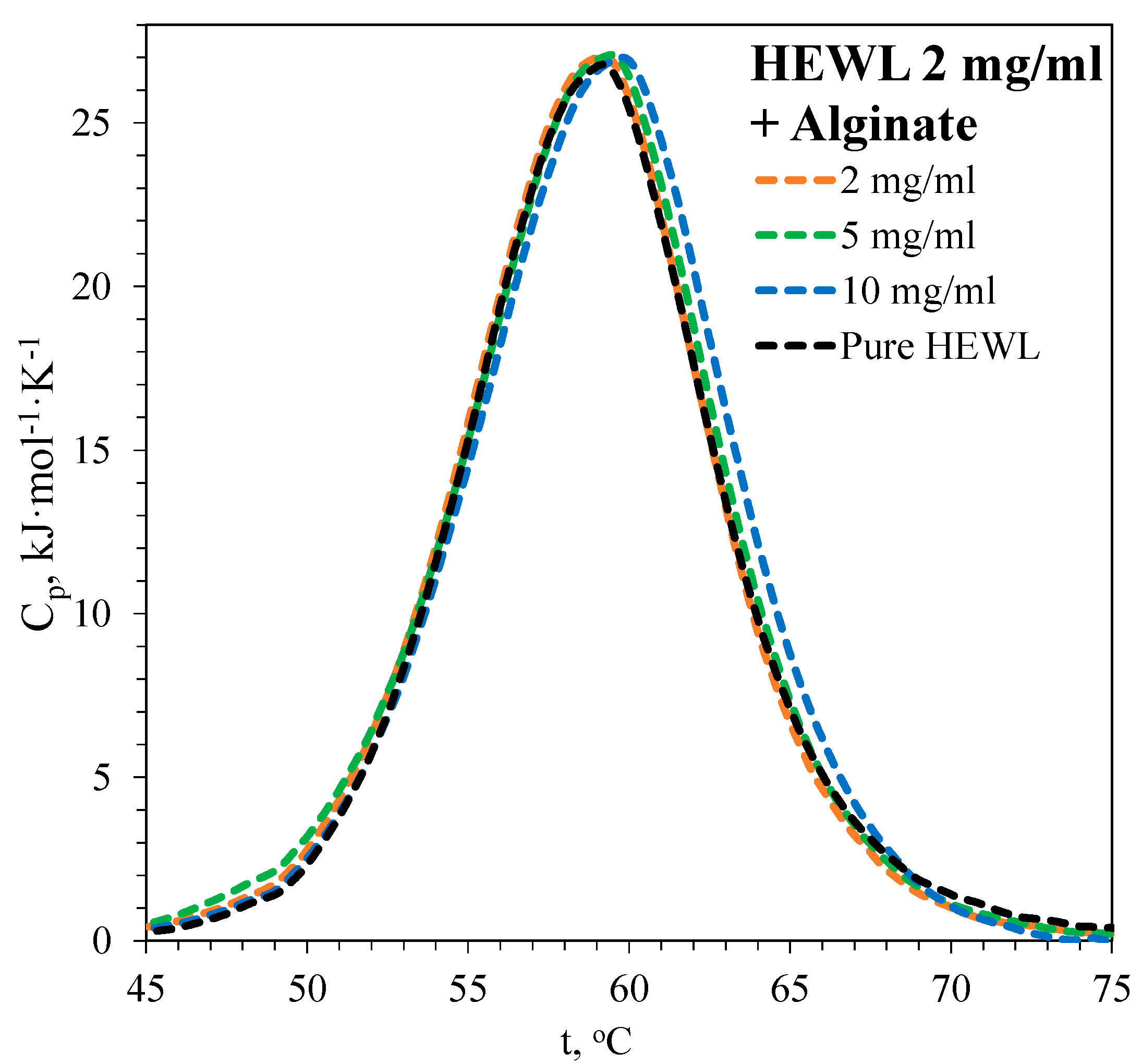
In the presence of various concentrations of sodium alginate, the denaturation peak of HEWL shows almost no shift or change in enthalpy (Figure 2, Table 2), indicating no significant binding with the native or unfolded form of the protein. As a result, the fraction of the denatured form of HEWL under the experimental conditions remains almost the same, independent of the concentration of alginate.

3.2. Kinetics of Fibril Formation

3.2.1. Fibril Formation of BSA

BSA fibril formation occurs without a significant lag time [19]. The ThT signal reaches a maximum and remains constant after about two hours of incubation at the conditions of our experiments. We have previously shown [19] that the initial rate of fibril formation is proportional to albumin concentration. In addition, for the pure protein, the magnitude of the fluorescence signal at the plateau after the end of the process is proportional to the initial monomer concentration in the solution.
The process of BSA fibril formation was studied at different protein–alginate mass ratios. At the highest ratio, 1:5, the fraction of the unfolded BSA form according to the DSC data (Table 1) was significantly lower than that for pure protein.
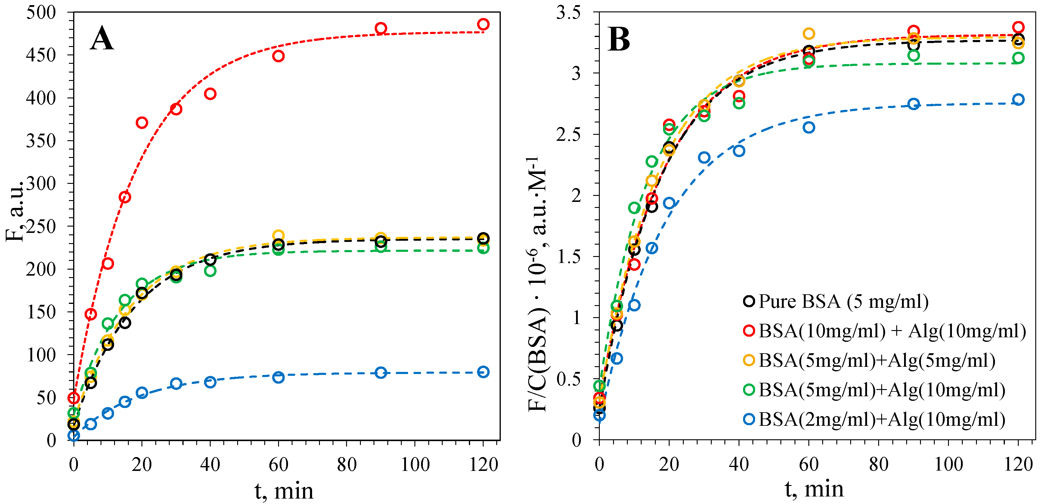
The addition of up to 10 mg·mL−1 of sodium alginate to a solution containing 5 mg·mL−1 BSA does not lead to a significant change in the rate of fibril growth, nor to changes in the final yield of fibrils (Figure 3A). At these concentrations, the denaturation curve of BSA also changed insignificantly according to DSC. As shown above, the DSC peak shifts, and the fraction of the denatured form markedly decreases only in the mixture of 2 mg·mL−1 BSA with 10 mg·mL−1 sodium alginate. In order to compare the kinetic curves at different concentrations of BSA, the fluorescence values were divided by the BSA concentration (Figure 3B). Indeed, a noticeable decrease in the initial fibril formation rate and their final yield was observed only at the highest alginate and the lowest BSA concentration. The value of the half-time of fibril formation τ50 does not change, even in this case.

3.2.2. Fibril Formation of HEWL

The fibril formation of HEWL in GdHCl solution was previously studied in a number of works [33,35,36]. However, the dependence of the yield and the rate of fibril formation on the monomer concentration had not been considered in these works.
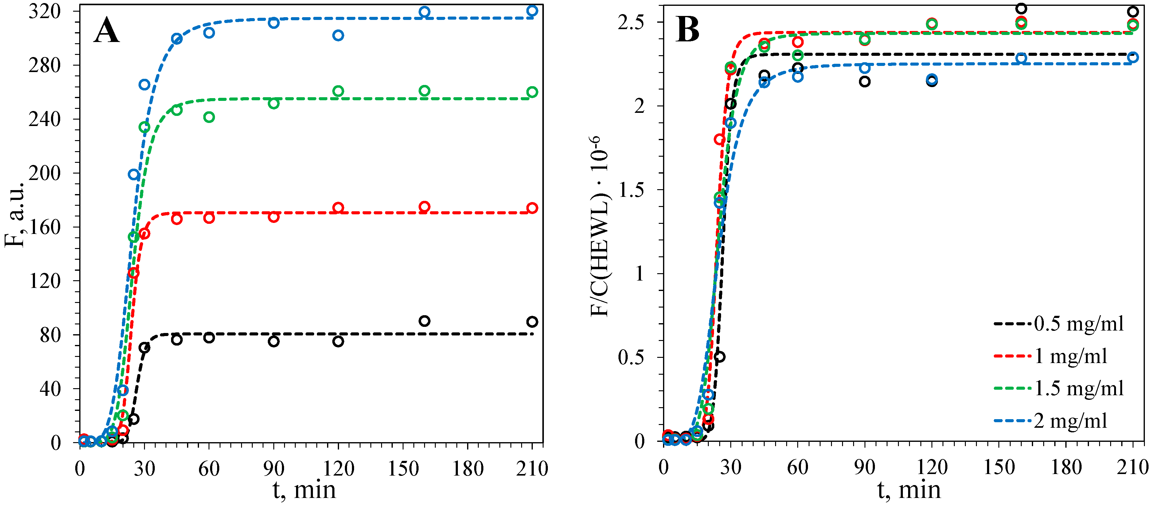
We obtained kinetic curves of fibril formation for various HEWL monomer concentrations (Figure 4A). The final fibril yield increases proportionally to the initial monomer concentration (Figure 5), but both the lag time τlag (the onset point of the curve jump) and the half-time of fibril formation τ50 remain almost constant over a wide range of concentrations (Figure 4B).
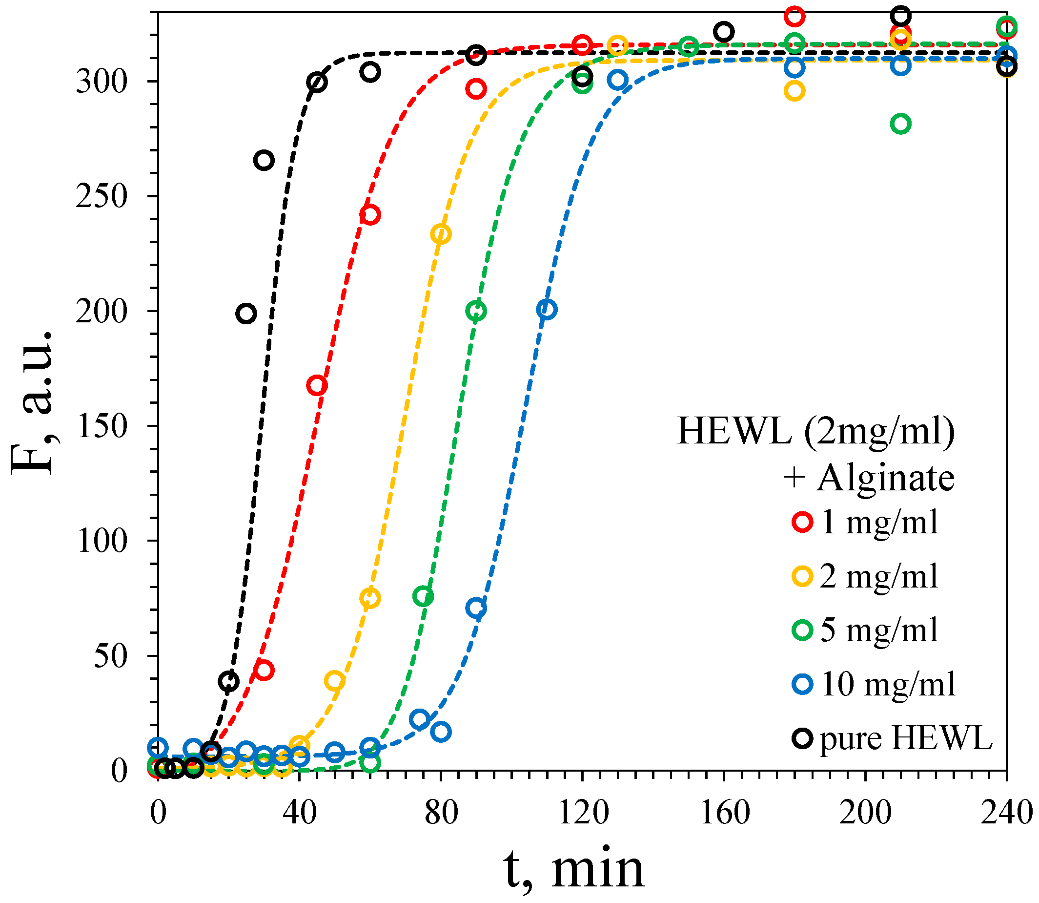
In the presence of sodium alginate, both the lag and fibril formation half-time significantly increase, but the final yield of fibrils does not change (Figure 6, Table 3).

4. Discussion

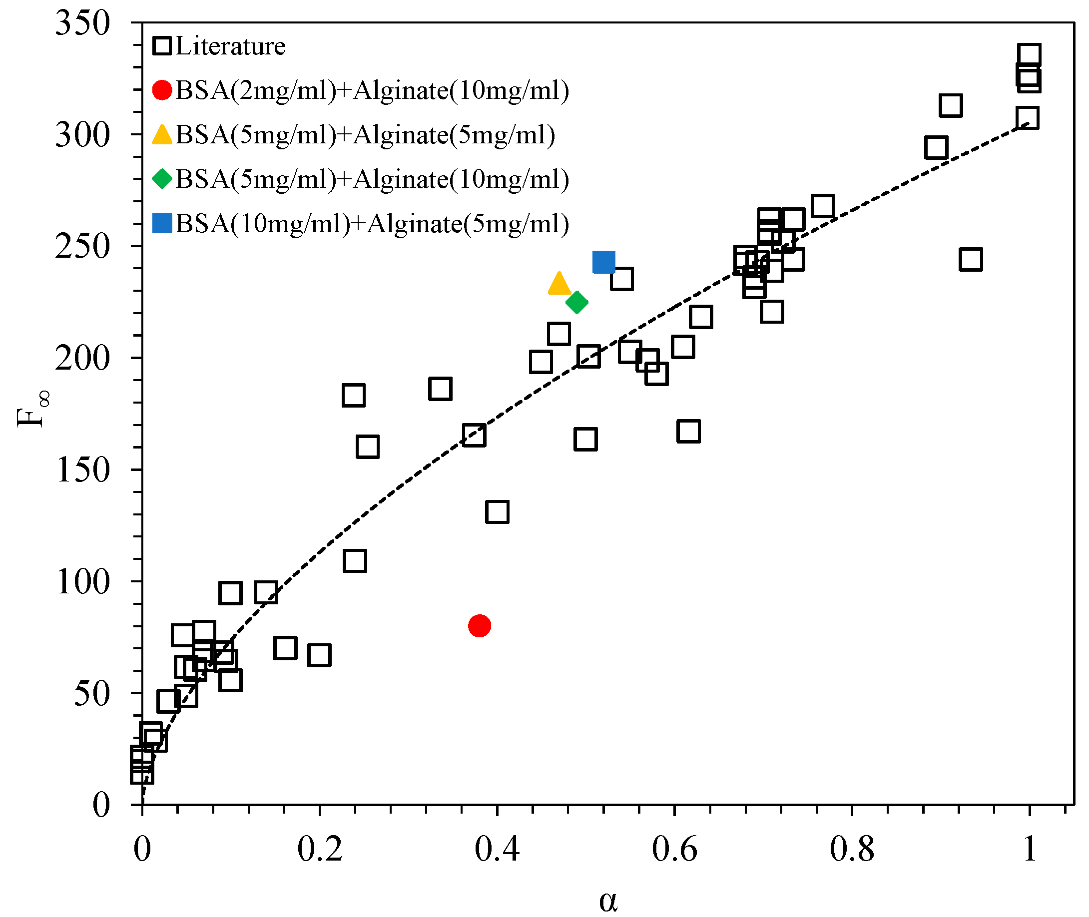
For BSA and HSA, a general dependence of the fibril yield (as measured by the ThT fluorescence intensity at the plateau) in the presence of different low-molecular ligands (warfarin, naproxen, flurbiprofen, ibuprofen, isoniazid, glucuronate, lactobionate, and ranitidine) on the equilibrium fraction of the protein denatured form (α) was revealed in our previous works [18,19]. Ligands that bind to the native form of protein, increasing its thermal stability and decreasing the value of α, tend to lower the fibril yield (Figure 7). This is caused by the equilibrium between the native protein monomer and the fibrils, which is established after prolonged incubation, being shifted towards the former due to its binding with the ligand. In the present work, both fluorescence and DSC curves were recorded at the same experimental conditions as in our previous works (25 mM Tris buffer, pH = 7.4, incubation at 65 °C) [18,19], which allows us to compare the results directly. Figure 7 shows that fibril formation in the presence of sodium alginate falls into the general trend. Despite the total negative charge of BSA at neutral pH, DSC curves suggest its binding with alginate at the high alginate–BSA mass ratio. At the same time, the half-time of fibril formation τ50 remained constant in the presence of any small-molecule binders as well as after the addition of alginate.
In the case of HEWL, the polysaccharide had no effect on the DSC curves, which indicates the absence of binding with the protein monomer. Hence, it should be suggested that interactions occur between the fibrils and alginate. The slowdown of fibril formation can be explained by the binding of alginate to the growing fibril surface, which may inhibit the fibril growth rate and/or secondary nucleation processes [37]. Previous experimental studies [38,39] showed that sodium alginate can bind to HEWL fibrils. Binding with fibrils prepared from different proteins was also reported for other polysaccharides such as hyaluronic acid [40] or chitin [41].
An alternative explanation of inhibition of fibril formation could be related to the viscosity of alginate solutions, leading to a lower rate of collisions between protein chains. However, this hypothesis cannot explain the absence of inhibition at a high BSA concentration and a decrease in the rate and yield of fibril formation at the constant alginate concentration with decreasing BSA concentration. Moreover, the viscous solutions of high-molecular compounds that are not prone to interactions with proteins often accelerate fibril formation, which is attributed to the excluded volume effects [42].
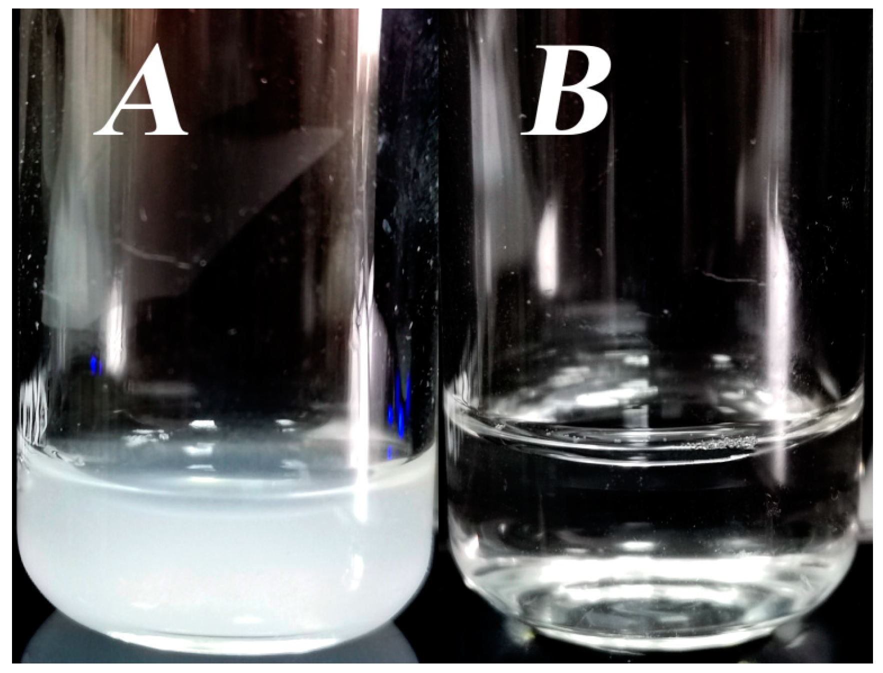
When comparing the obtained results for both proteins, it may seem surprising that the positively charged monomer of HEWL only has a weak interaction with a negatively charged molecule of alginate. This is caused by the presence of 2 M GdHCl. When solutions of HEWL and alginate are mixed in the absence of GdHCl, a white precipitate forms immediately (Figure 8A). This fact suggests a strong binding between the protein and polysaccharide [43]. No fibrils are formed when the insoluble complex is heated, even when heated for a long time. After the addition of GdHCl, the precipitate dissolves (Figure 8B).
GdHCl is known to weaken intra- and intermolecular electrostatic interactions, which leads to the disruption of the attractive forces between HEWL and alginate. Moreover, it was shown that guanidinium cations can bind to HEWL. In the recent study of Raskar et al. [44], X-ray crystallography was used to obtain the structures of HEWL crystals soaked in 2.5 M GdHCl. After soaking, guanidinium cations were found at several different sites (Figure 9).
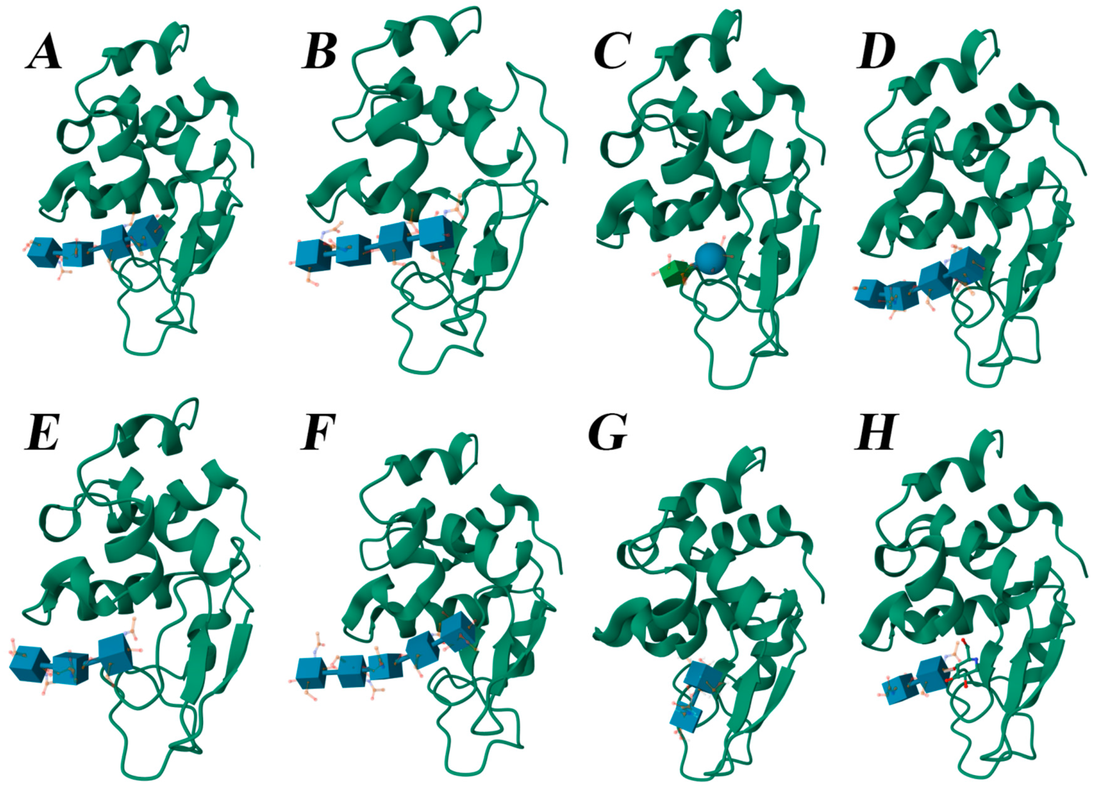
In order to have an insight about the possible structure of the complexes of proteins with sodium alginate, the blind molecular docking of the alginate fragment was conducted using two very different approaches: AutoDock Vina with an empirical scoring describing the pairwise interactions between individual atoms and DiffDock based on machine learning trained on the experimental structures of protein complexes. In the case of HEWL, all top scoring poses obtained using both methods contain an alginate fragment bound in the active site cleft of lysozyme between its two domains (Figure 10). Moreover, this binding mode is similar to that observed in the experimental structures of lysozyme complexes with various glycans (Figure 11). Such complexes are stabilized by an extensive network of hydrogen bonds between the glycan and protein residues.
Apparently, guanidinium chloride can easily disrupt these complexes by screening charge–charge interactions as it does in proteins [45,46]. The Debye length in 2 M GdHCl at 60 °C is only about 2 Å, which is less than a typical N+—O distance in a NH3+—COO salt bridge (3.5–4.0 Å [47]). Moreover, as can be seen from Figure 9, in the structure of HEWL soaked in GdHCl, bound guanidinium cations can be found in the cleft of the HEWL molecule involved in carbohydrate binding.
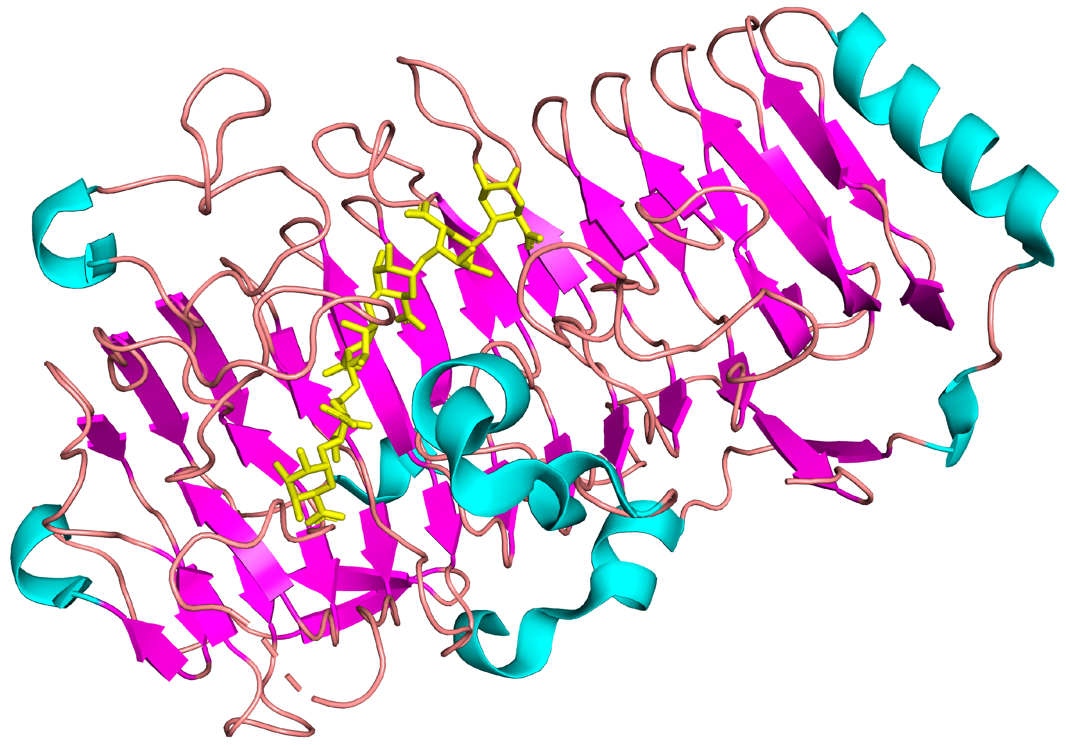
At the same time, the β-strands in the HEWL structure located in the cleft and participating in the interactions with oligosaccharides were shown to serve as the amyloidogenic core of the protein [48]. Frare et al. [49] argued that the 49–101 fragment is a key region for HEWL fibrillation. This fragment includes two β-strands involved in glycan binding. It is possible to hypothesize that the residues from these strands may also be prone to alginate binding in the fibrillar aggregates of HEWL. Moreover, since fibrils comprise lined up positively charged residues, it is advantageous for the negatively charged alginate chain to be oriented along the fibril axis, forming strong multiple electrostatic contacts with a fibril which are more stable to the action of GdHCl. Bound glycan can alter the rate of further fibril growth by impeding the approach of protein monomers to the end of the fibrils or can prevent secondary nucleation occurring at the surface of already formed fibrils, making a part of this surface inaccessible for the monomers. Due to the polymorphism of fibrils and the multistage nature of the aggregation process involving protofibrils or “flexible” fibrils [50,51], it is not possible to reliably predict the atomic-level structure of glycan complexes with lysozyme fibrils, e.g., using the docking method. Nevertheless, it is certainly true that the β-sheets are known as effective scaffolds for the binding of glycans [52,53]. In particular, the natural alginate-binding enzyme, alginate lyase, comprises a cleft formed by a long β-sheet structure on one side (Figure 12) [54].
Furthermore, there is experimental evidence of the binding of alginate to HEWL fibrils, already mentioned above. The interactions between alginate and biotinylated HEWL and its fibrils immobilized on the sensor were studied using bio-layer interferometry (BLI) [39]. The binding constant of sodium alginate to fibrils was found to be at least an order of magnitude higher than to the HEWL monomer, which is in agreement with our results.
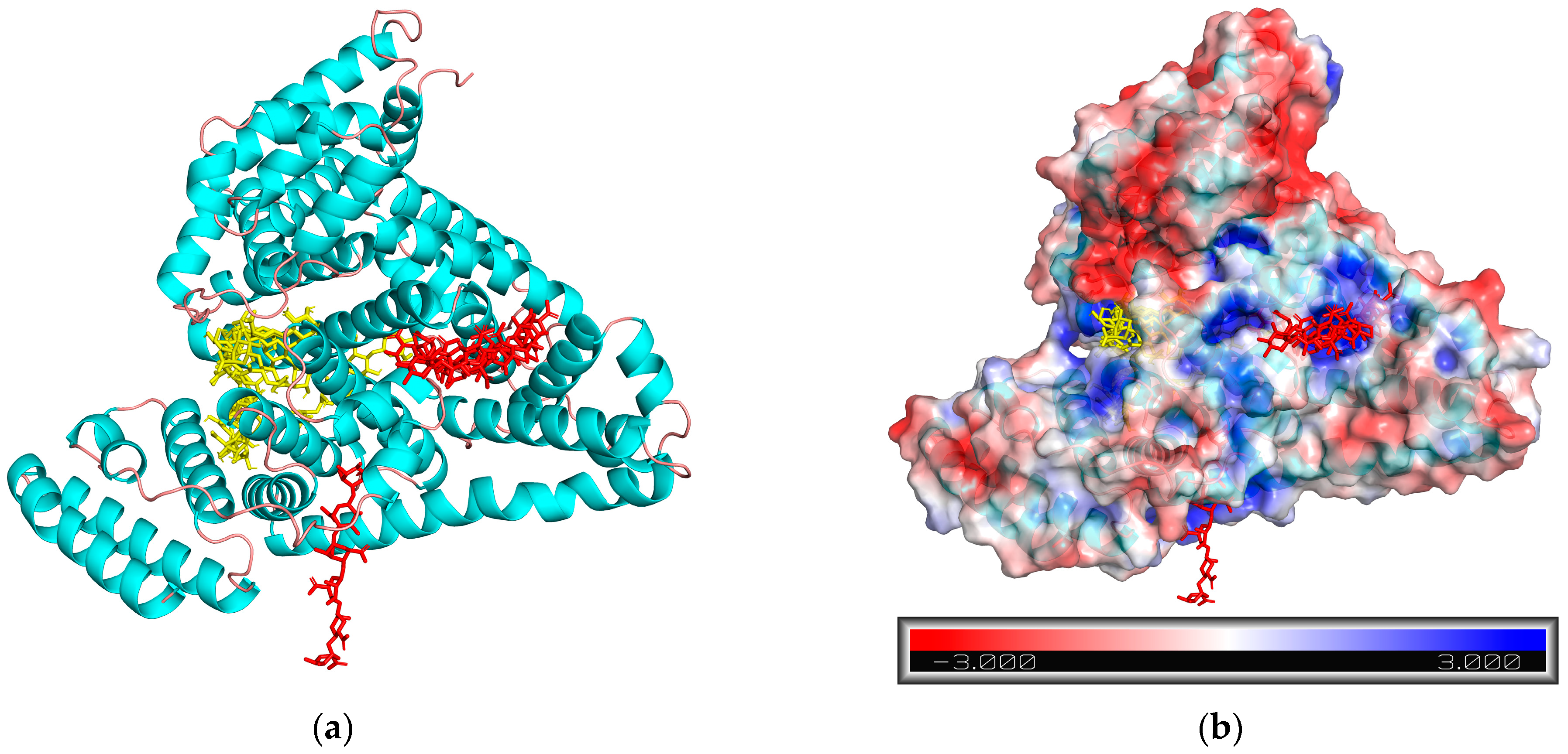
The docking of the alginate fragment to BSA resulted in diverse poses with close docking scores in different runs (Figure 13a). However, all these poses comprise the carbohydrate rings bound in the clefts between different domains of BSA. These clefts have a positive surface electrostatic potential [55,56]. The electrostatic potential map of BSA at pH 7 was calculated using an adaptive Poisson–Boltzmann solver (APBS) and is shown in Figure 13b. It is likely that the binding of alginate to BSA is not site-specific and can occur at any part of the protein surface rich in positively charged residues.
We have shown that alginate is able to bind both monomeric and fibrillar forms of different proteins and can be employed in different strategies of amyloid formation inhibition. Moreover, Bi et al. [57] found that oligomannuronates obtained by hydrolysis from alginate can inhibit the aggregation of tau-K18 oligomer in vitro and suppress the levels of pathogenic phosphorylated tau protein in cell cultures.
As mentioned above, HEWL and BSA are popular model proteins for studying fibril formation. In the human organism, albumin is not known to form fibrils, but lysozyme amyloidosis happens as a rare form of hereditary amyloidosis [58]. However, the fibrils of both model and pathology-related proteins, including both intrinsically disordered and ordered proteins, share the same beta-sheet structural features. Furthermore, the influence of different chemical species on the amyloid aggregation of various disease-related proteins can be studied using the same experimental techniques as for model proteins, including DSC. The latter method is not commonly used in amyloid research. Nevertheless, it provides robust information on ligand binding to globular proteins and can be used to screen for high-affinity ligands, which are likely to inhibit the amyloid aggregation of such proteins and therefore hold promise as anti-amyloid agents.

5. Conclusions

The effect of sodium alginate on the thermal and aggregation stability of BSA and HEWL was studied. It was shown that native BSA can bind to the polysaccharide, which leads to some decrease in the fraction of the denatured form of BSA at the temperature of fibril preparation. In the protein–glycan complex, the areas of the BSA surface rich in positively charged residues are likely to serve as anchors for the negatively charged alginate units. As a consequence, the equilibrium between the native BSA monomer and the fibrils shifts towards the former, and the fibril yield slightly diminishes. This is consistent with the general trend observed for different low-molecular ligands in our previous studies.
In the case of HEWL, very different behavior is observed: alginate does not bind lysozyme in 2 M GdHCl. However, the presence of alginate affects the lag time and the rate of fibril growth. This allows us to suggest that the polysaccharide binds HEWL fibrils, which leads to the retardation of fibril growth or secondary nucleation processes by blocking the HEWL monomers from accessing the fibril ends or surface. Alginate binding to HEWL fibrils was previously observed in BLI studies and can be stabilized by multiple salt contacts between the repeating negatively charged alginate units and lined up positively charged residues of the fibril. In the absence of GdHCl, the complex of HEWL with alginate precipitates, which is not prone to fibril formation. Hence, the results of the work underline the sensitivity of the fibril formation process to the nature of both proteins and ligands as well as other compounds present in protein solution.

Author Contributions

D.R.K.—investigation, formal analysis, writing—original draft; P.R.K.—investigation; B.R.G.—investigation, formal analysis; T.I.M.—investigation, formal analysis; A.A.N.—investigation, formal analysis; I.A.S.—conceptualization, investigation, writing—original draft, supervision. All authors have read and agreed to the published version of the manuscript.

Funding

This work was supported by a grant from the Russian Science Foundation and the Academy of Sciences of the Republic of Tatarstan under project No. 23-23-10084.

Data Availability Statement

Data available within the article.

Conflicts of Interest

The authors declare no conflict of interest.

References

  1. Barclay, T.G.; Day, C.M.; Petrovsky, N.; Garg, S. Review of polysaccharide particle-based functional drug delivery. Carbohydr. Polym. 2019, 221, 94–112. [Google Scholar] [CrossRef] [PubMed]
  2. Kirschning, A.; Dibbert, N.; Dräger, G. Chemical functionalization of polysaccharides—Towards biocompatible hydrogels for biomedical applications. Chem.–A Eur. J. 2018, 24, 1231–1240. [Google Scholar] [CrossRef] [PubMed]
  3. Mohammed, A.S.A.; Naveed, M.; Jost, N. Polysaccharides; Classification, Chemical Properties, and Future Perspective Applications in Fields of Pharmacology and Biological Medicine (A Review of Current Applications and Upcoming Potentialities). J. Polym. Environ. 2021, 29, 2359–2371. [Google Scholar] [CrossRef] [PubMed]
  4. Yu, Y.; Shen, M.; Song, Q.; Xie, J. Biological activities and pharmaceutical applications of polysaccharide from natural resources: A review. Carbohydr. Polym. 2018, 183, 91–101. [Google Scholar] [CrossRef] [PubMed]
  5. Wang, X.-F.; Chen, X.; Tang, Y.; Wu, J.-M.; Qin, D.-L.; Yu, L.; Yu, C.-L.; Zhou, X.-G.; Wu, A.-G. The Therapeutic Potential of Plant Polysaccharides in Metabolic Diseases. Pharmaceuticals 2022, 15, 1329. [Google Scholar] [CrossRef]
  6. Elrosasy, A.; Zeid, M.A.; Negida, A. Sodium Oligomannate (GV-971) Safety and Efficacy in the Treatment of Alzheimer’s Disease: A Systematic Review and Meta-analysis of Randomized Controlled Trials (P11-9.005). Neurology 2024, 102, 5352. [Google Scholar] [CrossRef]
  7. Wozniak, M.; Bell, T.; Dénes, Á.; Falshaw, R.; Itzhaki, R. Anti-HSV1 activity of brown algal polysaccharides and possible relevance to the treatment of Alzheimer’s disease. Int. J. Biol. Macromol. 2015, 74, 530–540. [Google Scholar] [CrossRef]
  8. Wang, W.; Wang, X.; Gao, W.; Cui, Z.; Zhang, H.; Lu, F.; Liu, F. Ulvan inhibits α-synuclein fibrillation and disrupts the mature fibrils: In vitro and in vivo studies. Int. J. Biol. Macromol. 2022, 211, 580–591. [Google Scholar] [CrossRef]
  9. Montgomery, K.M.; Carroll, E.C.; Thwin, A.C.; Quddus, A.Y.; Hodges, P.; Southworth, D.R.; Gestwicki, J.E. Chemical Features of Polyanions Modulate Tau Aggregation and Conformational States. J. Am. Chem. Soc. 2023, 145, 3926–3936. [Google Scholar] [CrossRef]
  10. Braia, M.; Loureiro, D.; Tubio, G.; Lienqueo, M.E.; Romanini, D. Interaction between trypsin and alginate: An ITC and DLS approach to the formation of insoluble complexes. Colloids Surf. B Biointerfaces 2017, 155, 507–511. [Google Scholar] [CrossRef]
  11. Gorji, E.G.; Waheed, A.; Ludwig, R.; Toca-Herrera, J.L.; Schleining, G.; Gorji, S.G. Complex Coacervation of Milk Proteins with Sodium Alginate. J. Agric. Food Chem. 2018, 66, 3210–3220. [Google Scholar] [CrossRef] [PubMed]
  12. Motomura, H.; Somjit, K.; Nozaki, Y. Effect of sodium alginate on changes in the denaturation of fish myofibrillar protein and the state of water during frozen storage. Fish. Sci. 2002, 68, 1633–1634. [Google Scholar] [CrossRef] [PubMed]
  13. Zhao, Y.; Li, F.; Carvajal, M.T.; Harris, M.T. Interactions between bovine serum albumin and alginate: An evaluation of alginate as protein carrier. J. Colloid Interface Sci. 2009, 332, 345–353. [Google Scholar] [CrossRef]
  14. Chang, R.; Gruebele, M.; Leckband, D.E. Protein Stabilization by Alginate Binding and Suppression of Thermal Aggregation. Biomacromolecules 2022, 23, 4063–4073. [Google Scholar] [CrossRef]
  15. Juárez, J.; Taboada, P.; Mosquera, V. Existence of Different Structural Intermediates on the Fibrillation Pathway of Human Serum Albumin. Biophys. J. 2009, 96, 2353–2370. [Google Scholar] [CrossRef]
  16. Maciążek-Jurczyk, M.; Janas, K.; Pożycka, J.; Szkudlarek, A.; Rogóż, W.; Owczarzy, A.; Kulig, K. Human Serum Albumin Aggregation/Fibrillation and its Abilities to Drugs Binding. Molecules 2020, 25, 618. [Google Scholar] [CrossRef]
  17. Swaminathan, R.; Ravi, V.K.; Kumar, S.; Kumar, M.V.S.; Chandra, N. Lysozyme, In Advances in Protein Chemistry and Structural Biology; Elsevier: Amstardam, The Netherlands, 2011; pp. 63–111. [Google Scholar] [CrossRef]
  18. Khaibrakhmanova, D.R.; Nikifirova, A.A.; Li, Z.; Sedov, I.A. Human Serum Albumin Fibril Formation in the Presence of Ligands with Different Affinity. Russ. J. Gen. Chem. 2024, 94, 395–405. [Google Scholar] [CrossRef]
  19. Khaibrakhmanova, D.; Nikiforova, A.; Li, Z.; Sedov, I. Effect of ligands with different affinity on albumin fibril formation. Int. J. Biol. Macromol. 2022, 204, 709–717. [Google Scholar] [CrossRef]
  20. Mondal, U.S.; Paul, S. Inhibition of hen egg white lysozyme fibrillation by a self-assembled nanostructured lysozyme and graphene oxide conjugate. New J. Chem. 2023, 47, 17666–17678. [Google Scholar] [CrossRef]
  21. Basu, A.; Mahammad, A.; Das, A. Inhibition of the formation of lysozyme fibrillar assemblies by the isoquinoline alkaloid coralyne. New J. Chem. 2022, 46, 3258–3269. [Google Scholar] [CrossRef]
  22. Morshedi, D.; Rezaei-Ghaleh, N.; Ebrahim-Habibi, A.; Ahmadian, S.; Nemat-Gorgani, M. Inhibition of amyloid fibrillation of lysozyme by indole derivatives − possible mechanism of action. FEBS J. 2007, 274, 6415–6425. [Google Scholar] [CrossRef] [PubMed]
  23. Kolotova, D.S.; Borovinskaya, E.V.; Bordiyan, V.V.; Zuev, Y.F.; Salnikov, V.V.; Zueva, O.S.; Derkach, S.R. Phase Behavior of Aqueous Mixtures of Sodium Alginate with Fish Gelatin: Effects of pH and Ionic Strength. Polymers 2023, 15, 2253. [Google Scholar] [CrossRef] [PubMed]
  24. Xue, C.; Lin, T.Y.; Chang, D.; Guo, Z. Thioflavin T as an amyloid dye: Fibril quantification, optimal concentration and effect on aggregation. R. Soc. Open Sci. 2017, 4, 160696. [Google Scholar] [CrossRef] [PubMed]
  25. Crespo, R.; Rocha, F.A.; Damas, A.M.; Martins, P.M. A Generic Crystallization-like Model That Describes the Kinetics of Amyloid Fibril Formation. J. Biol. Chem. 2012, 287, 30585–30594. [Google Scholar] [CrossRef]
  26. Eberhardt, J.; Santos-Martins, D.; Tillack, A.F.; Forli, S. AutoDock Vina 1.2.0: New Docking Methods, Expanded Force Field, and Python Bindings. J. Chem. Inf. Model. 2021, 61, 3891–3898. [Google Scholar] [CrossRef]
  27. Corso, G.; Stärk, H.; Jing, B.; Barzilay, R.; Jaakkola, T. DiffDock: Diffusion Steps, Twists, and Turns for Molecular Docking. arXiv 2022. [Google Scholar] [CrossRef]
  28. Sedov, I.; Nikiforova, A.; Khaibrakhmanova, D. Evaluation of the binding properties of drugs to albumin from DSC thermograms. Int. J. Pharm. 2020, 583, 119362. [Google Scholar] [CrossRef]
  29. Giancola, C.; De Sena, C.; Fessas, D.; Graziano, G.; Barone, G. DSC studies on bovine serum albumin denaturation Effects of ionic strength and SDS concentration. Int. J. Biol. Macromol. 1997, 20, 193–204. [Google Scholar] [CrossRef]
  30. Burgos, M.I.; Fernández, R.A.; Celej, M.S.; Rossi, L.I.; Fidelio, G.D.; Dassie, S.A. Binding of the Highly Toxic Tetracycline Derivative, Anhydrotetracycline, to Bovine Serum Albumin. Biol. Pharm. Bull. 2011, 34, 1301–1306. [Google Scholar] [CrossRef]
  31. Celej, M.S.; Dassie, S.A.; Freire, E.; Bianconi, M.L.; Fidelio, G.D. Ligand-induced thermostability in proteins: Thermodynamic analysis of ANS–albumin interaction. Biochim. Biophys. Acta (BBA)—Proteins Proteom. 2005, 1750, 122–133. [Google Scholar] [CrossRef]
  32. Brandts, J.F.; Lin, L.N. Study of strong to ultratight protein interactions using differential scanning calorimetry. Biochemistry 1990, 29, 6927–6940. [Google Scholar] [CrossRef]
  33. Vernaglia, B.A.; Huang, J.; Clark, E.D. Guanidine Hydrochloride Can Induce Amyloid Fibril Formation from Hen Egg-White Lysozyme. Biomacromolecules 2004, 5, 1362–1370. [Google Scholar] [CrossRef]
  34. Sulatskaya, A.I.; Sulatsky, M.I.; Stepanenko, O.V.; Povarova, O.I.; Kuznetsova, I.M.; Turoverov, K.K. Denaturing Effect of Guanidine Hydrohloride on Amyloid Fibrils. Biophys. J. 2020, 118, 509a. [Google Scholar] [CrossRef]
  35. Hédoux, A.; Krenzlin, S.; Paccou, L.; Guinet, Y.; Flament, M.-P.; Siepmann, J. Influence of urea and guanidine hydrochloride on lysozyme stability and thermal denaturation; a correlation between activity, protein dynamics and conformational changes. Phys. Chem. Chem. Phys. 2010, 12, 13189–13196. [Google Scholar] [CrossRef]
  36. Emadi, S.; Behzadi, M. A comparative study on the aggregating effects of guanidine thiocyanate, guanidine hydrochloride and urea on lysozyme aggregation. Biochem. Biophys. Res. Commun. 2014, 450, 1339–1344. [Google Scholar] [CrossRef]
  37. Sedov, I.; Khaibrakhmanova, D. Molecular Mechanisms of Inhibition of Protein Amyloid Fibril Formation: Evidence and Perspectives Based on Kinetic Models. Int. J. Mol. Sci. 2022, 23, 13428. [Google Scholar] [CrossRef]
  38. Makshakova, O.; Bogdanova, L.; Faizullin, D.; Khaibrakhmanova, D.; Ziganshina, S.; Ermakova, E.; Zuev, Y.; Sedov, I. The Ability of Some Polysaccharides to Disaggregate Lysozyme Amyloid Fibrils and Renature the Protein. Pharmaceutics 2023, 15, 624. [Google Scholar] [CrossRef]
  39. Wang, X.; Nian, Y.; Zhang, Z.; Chen, Q.; Zeng, X.; Hu, B. High internal phase emulsions stabilized with amyloid fibrils and their polysaccharide complexes for encapsulation and protection of β-carotene. Colloids Surf. B Biointerfaces 2019, 183, 110459. [Google Scholar] [CrossRef]
  40. Peydayesh, M.; Kistler, S.; Zhou, J.; Lutz-Bueno, V.; Victorelli, F.D.; Meneguin, A.B.; Spósito, L.; Bauab, T.M.; Chorilli, M.; Mezzenga, R. Amyloid-polysaccharide interfacial coacervates as therapeutic materials. Nat. Commun. 2023, 14, 1848. [Google Scholar] [CrossRef]
  41. Wei, S.; Li, Y.; Li, K.; Zhong, C. Biofilm-inspired Amyloid-Polysaccharide Composite Materials. Appl. Mater. Today 2022, 27, 101497. [Google Scholar] [CrossRef]
  42. Gorensek-Benitez, A.H.; Kirk, B.; Myers, J.K. Protein Fibrillation under Crowded Conditions. Biomolecules 2022, 12, 950. [Google Scholar] [CrossRef] [PubMed]
  43. Amara, C.B.; Eghbal, N.; Oulahal, N.; Degraeve, P.; Gharsallaoui, A. Properties of lysozyme/sodium alginate complexes for the development of antimicrobial films. Food Res. Int. 2016, 89, 272–280. [Google Scholar] [CrossRef] [PubMed]
  44. Raskar, T.; Koh, C.Y.; Niebling, S.; Kini, R.M.; Hosur, M.V. X-ray crystallographic analysis of time-dependent binding of guanidine hydrochloride to HEWL: First steps during protein unfolding. Int. J. Biol. Macromol. 2019, 122, 903–913. [Google Scholar] [CrossRef] [PubMed]
  45. Perez-Jimenez, R.; Godoy-Ruiz, R.; Ibarra-Molero, B.; Sanchez-Ruiz, J.M. The Efficiency of Different Salts to Screen Charge Interactions in Proteins: A Hofmeister Effect? Biophys. J. 2004, 86, 2414–2429. [Google Scholar] [CrossRef] [PubMed]
  46. Monera, O.D.; Kay, C.M.; Hodges, R.S. Protein denaturation with guanidine hydrochloride or urea provides a different estimate of stability depending on the contributions of electrostatic interactions. Protein Sci. 1994, 3, 1984–1991. [Google Scholar] [CrossRef]
  47. Ban, X.; Lahiri, P.; Dhoble, A.S.; Li, D.; Gu, Z.; Li, C.; Cheng, L.; Hong, Y.; Li, Z.; Kaustubh, B. Evolutionary Stability of Salt Bridges Hints Its Contribution to Stability of Proteins. Comput. Struct. Biotechnol. J. 2019, 17, 895–903. [Google Scholar] [CrossRef]
  48. Tokunaga, Y.; Sakakibara, Y.; Kamada, Y.; Watanabe, K.; Sugimoto, Y. Analysis of Core Region from Egg White Lysozyme Forming Amyloid Fibrils. Int. J. Biol. Sci. 2013, 9, 219–227. [Google Scholar] [CrossRef]
  49. Frare, E.; De Laureto, P.P.; Zurdo, J.; Dobson, C.M.A. Fontana, A Highly Amyloidogenic Region of Hen Lysozyme. J. Mol. Biol. 2004, 340, 1153–1165. [Google Scholar] [CrossRef]
  50. Fändrich, M.; Nyström, S.; Nilsson, K.P.R.; Böckmann, A.; LeVine, H.; Hammarström, P. Amyloid fibril polymorphism: A challenge for molecular imaging and therapy. J. Intern. Med. 2018, 283, 218–237. [Google Scholar] [CrossRef]
  51. Frey, L.; Zhou, J.; Cereghetti, G.; Weber, M.E.; Rhyner, D.; Pokharna, A.; Wenchel, L.; Kadavath, H.; Cao, Y.; Meier, B.H.; et al. A structural rationale for reversible vs irreversible amyloid fibril formation from a single protein. Nat. Commun. 2024, 15, 8448. [Google Scholar] [CrossRef]
  52. Ward, E.M.; Zamora, C.Y.; Schocker, N.S.; Ghosh, S.; Kizer, M.E.; Imperiali, B. Engineered Glycan-Binding Proteins for Recognition of the Thomsen–Friedenreich Antigen and Structurally Related Disaccharides. ACS Chem. Biol. 2023, 18, 70–80. [Google Scholar] [CrossRef] [PubMed]
  53. Boraston, A.B.; Bolam, D.N.; Gilbert, H.J.; Davies, G.J. Carbohydrate-binding modules: Fine-tuning polysaccharide recognition. Biochem. J. 2004, 382, 769–781. [Google Scholar] [CrossRef] [PubMed]
  54. Zhang, K.; Liu, T.; Liu, W.; Lyu, Q. Structural insights into the substrate-binding cleft of AlyF reveal the first long-chain alginate-binding mode. Acta Crystallogr. Sect. D Struct. Biol. 2021, 77, 336–346. [Google Scholar] [CrossRef] [PubMed]
  55. Adamczyk, O.; Szota, M.; Rakowski, K.; Prochownik, M.; Doveiko, D.; Chen, Y.; Jachimska, B. Bovine Serum Albumin as a Platform for Designing Biologically Active Nanocarriers—Experimental and Computational Studies. Int. J. Mol. Sci. 2023, 25, 37. [Google Scholar] [CrossRef]
  56. Bujacz, A.; Talaj, J.A.; Zielinski, K.; Pietrzyk-Brzezinska, A.J.; Neumann, P. Crystal structures of serum albumins from domesticated ruminants and their complexes with 3,5-diiodosalicylic acid. Acta Crystallogr. Sect. D Struct. Biol. 2017, 73, 896–909. [Google Scholar] [CrossRef]
  57. Bi, D.; Xiao, S.; Lin, Z.; Yao, L.; Fang, W.; Wu, Y.; Xu, H.; Lu, J.; Xu, X. Alginate-Derived Mannuronate Oligosaccharide Attenuates Tauopathy through Enhancing Autophagy. J. Agric. Food Chem. 2021, 69, 4438–4445. [Google Scholar] [CrossRef]
  58. Pleyer, C.; Flesche, J.; Saeed, F. Lysozyme amyloidosis—A case report and review of the literature. Clin. Nephrol. Case Stud. 2015, 3, 42–45. [Google Scholar] [CrossRef]
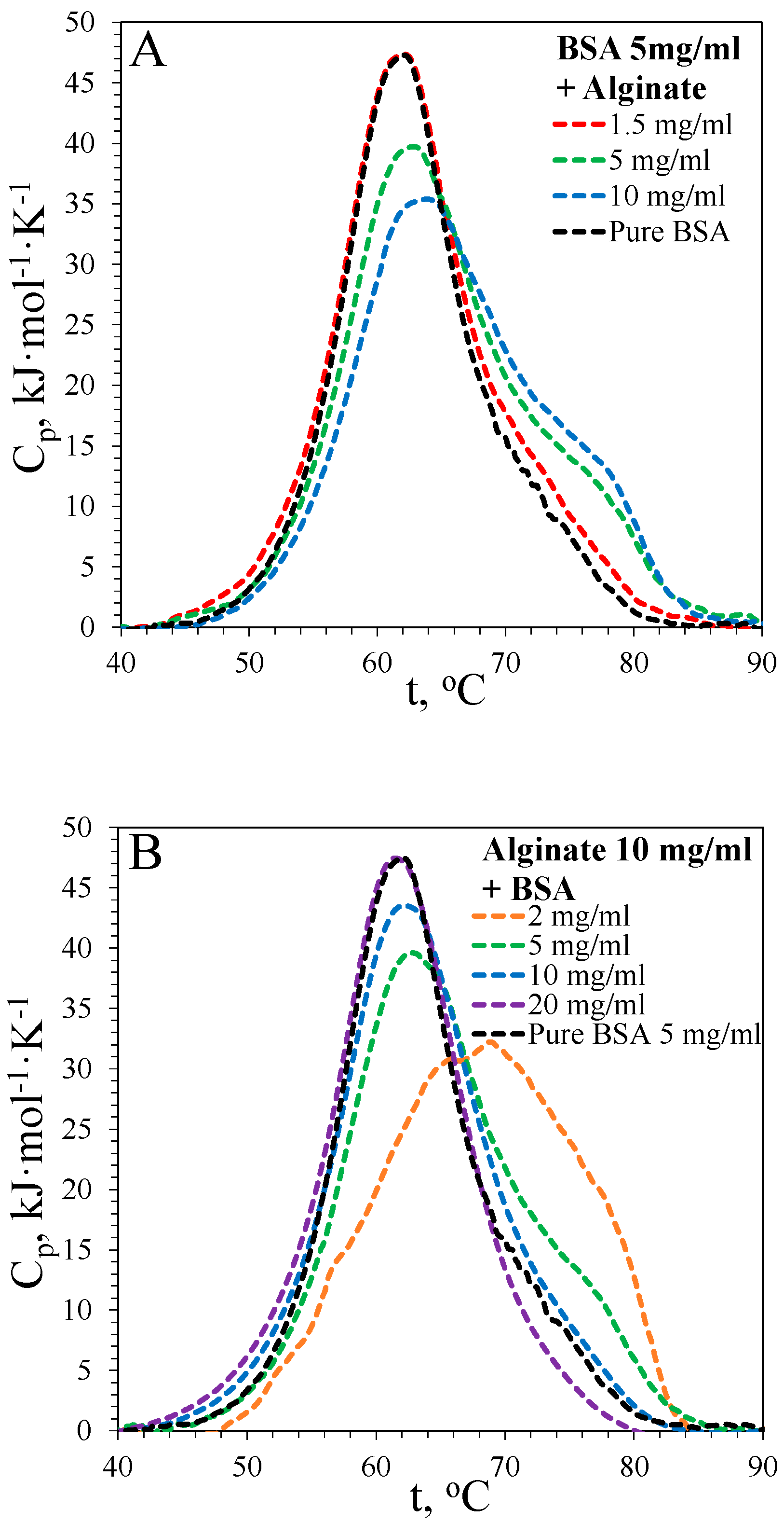
Figure 1. DSC thermograms of bovine serum albumin (BSA) denaturation in the presence of sodium alginate. (A) 5 mg·mL−1 BSA concentration, alginate concentration varies; (B) 10 mg·mL−1 alginate concentration, BSA concentration varies.
Biophysica 04 00043 g001
Figure 2. DSC thermograms of hen egg-white lysozyme (HEWL, 2 mg·mL−1) denaturation in the presence of 2 M guanidinium chloride and different concentrations of sodium alginate.
Biophysica 04 00043 g002
Figure 3. Kinetic curves of BSA fibril formation. (A)—time dependence of ThT fluorescence signal intensity for BSA fibrils formed at 65 °C in the absence or presence of sodium alginate, the legend is the same as in (B); (B)—fluorescence intensity from (A) divided by BSA concentration.
Biophysica 04 00043 g003
Figure 4. Kinetic curves of lysozyme fibril formation at different monomer concentrations. (A)—time dependence of ThT fluorescence signal intensity for HEWL fibrils formed at 60 °C at different monomer concentrations in 2 M guanidinium chloride, the legend is the same as in (B); (B)—fluorescence intensity from (A) divided by the initial HEWL concentration.
Biophysica 04 00043 g004
Figure 5. Dependence of the plateau ThT fluorescence after HEWL fibril formation on the initial concentration of the protein monomer.
Biophysica 04 00043 g005
Figure 6. Kinetic curves (time dependence of ThT fluorescence signal intensity) of HEWL fibril formation at 60 °C in 2 M guanidinium chloride in the absence or presence of sodium alginate.
Biophysica 04 00043 g006
Figure 7. The dependence of the plateau ThT fluorescence after fibril formation on the fraction α of the denatured form of BSA. Dashed squares correspond to the results for aggregation of BSA or HSA in the presence of different substances (warfarin, flurbiprofen, ibuprofen, isoniazid, glucuronate, lactobionate, naproxen, and ranitidine) from our previous works [18,19].
Biophysica 04 00043 g007
Figure 8. Photos of the mixture containing HEWL and sodium alginate: (A) after mixing; (B) after addition of 2 M GdHCl.
Biophysica 04 00043 g008
Figure 9. X-ray structure of HEWL soaked for 8 min in 2.5 M GdHCl from the work of Raskar et al. [44] (PDB code 6A4N).
Biophysica 04 00043 g009
Figure 10. Five best docking poses of the alginate fragment to HEWL obtained using AutoDock Vina (yellow molecules) and DiffDock (red molecules).
Biophysica 04 00043 g010
Figure 11. Experimental structures of lysozyme with bound carbohydrates and their derivatives. (A) tetramer of N-acetyl-D-glucosamine, PDB code 7BR5; (B) tetra-N-acetylchitotetraose PDB code 1SF7; (C) sucrose, PDB code 3T6U; (D) PDB code 1LSZ; (E) trimer of N-acetyl-D-glucosamine, PDB code 1UIB; (F) chitopentaose, PDB code 2WAR; (G) N,N’-diacetylchitobiose, PDB code 8CWC; (H) 2-acetamido-2-deoxy-beta-D-glucopyranose-(1–4)-2-acetamido-2-deoxy-beta-D-glucopyranose, PDB code 4HPI.
Biophysica 04 00043 g011
Figure 12. Experimental structures of alginate lyase AlyF-OU02 in the complex with alginate fragment, PDB code 7BZ0.
Biophysica 04 00043 g012
Figure 13. (a) The five best docking poses of the alginate fragment to BSA obtained using AutoDock Vina (yellow molecules) and DiffDock (red molecules). (b) The same poses and the surface electrostatic potential map of BSA at pH 7.
Biophysica 04 00043 g013
Table 1. BSA denaturation parameters obtained from DSC thermograms.
[BSA], mg·mL−1[Alginate], mg·mL−1Td, °CΔHd, kJ·mol−1ΔH65, kJ·mol−1α65
5062.1611 ± 22300 ± 120.49
51.562.2620 ± 16320 ± 100.52
5562.9616 ± 25290 ± 80.47
51063.9613 ± 22303 ± 130.49
21068.8659 ± 18254 ± 230.38
101062.2635 ± 12336 ± 70.52
201062.1639 ± 11344 ± 100.53
Table 2. Denaturation parameters obtained from thermograms of HEWL in the presence of 2 M guanidinium chloride and different concentrations of sodium alginate.
[HEWL], mg·mL−1[Alginate], mg·mL−1Td, °CΔHd, kJ·mol−1
2059.1260 ± 12
2259.2262 ± 9
2559.4266 ± 8
21059.8268 ± 8
Table 3. Kinetic parameters of the aggregation of HEWL (2 mg·mL−1) in the presence of 2 M guanidinium chloride and different concentrations of sodium alginate at 60 °C.
[Alginate], mg·mL−1τlag, minτ50, min
18.624.1
122.745.7
248.369.8
567.185.5
1081.9103.3
Disclaimer/Publisher’s Note: The statements, opinions and data contained in all publications are solely those of the individual author(s) and contributor(s) and not of MDPI and/or the editor(s). MDPI and/or the editor(s) disclaim responsibility for any injury to people or property resulting from any ideas, methods, instructions or products referred to in the content.