Article

The Amyloidogenic Peptide Amyloid Beta(16–22) Displays Facet Dependent Conformation on Metal Surfaces

School of Chemistry, National University of Ireland Galway, H91 TK33 Galway, Ireland
*
Author to whom correspondence should be addressed.
Biophysica 2022, 2(2), 135-153; https://doi.org/10.3390/biophysica2020015
Submission received: 29 April 2022 / Revised: 23 May 2022 / Accepted: 31 May 2022 / Published: 9 June 2022
(This article belongs to the Collection Feature Papers in Biophysics)

Abstract

:
Currently, it is not understood how metal nanoparticles influence the formation of protein fibrils, although recent literature highlights that the shape and chemical composition of such nanoparticles can strongly influence the process. Understanding this process at a fundamental level can potentially unlock routes to the development of new therapeutics, as well as novel materials for technological applications. This requires a microscopic picture of the behaviour of amyloidogenic proteins on metal surfaces. Using replica exchange molecular dynamics simulations, we investigate the conformation of the model amyloidogenic peptide, A β (16–22), on different gold and silver surfaces. The conformation of the peptide on gold surfaces also shows a strong facet dependence, with fibril-like conformations being promoted in the 100 surface and inhibited on the 111 surface. A smaller degree of facet dependence is seen for silver with the peptide behaving similar on both of these. The difference in the facet dependence can be related to the difference between direct adsorption onto the gold 111 surface, with a preference towards indirect (water mediated) adsorption onto the other surfaces. This new information on the behaviour of an amyloidogenic peptide on metal surfaces can give insight into the size-dependent effect of nanoparticles on fibril formation and the use of surfaces to control fibrillation.

1. Introduction

Research on fibril formation via protein agglomeration into amyloid fibrils [1,2] is of great importance in a wide variety of fields, ranging from microbiology to biochemistry to materials science and medicine. Whilst fibril formation is involved in many normal biological functions, the abnormal aggregation of proteins into amyloids and subsequently fibrils, or “plaques”, has been implicated in the pathology of numerous human diseases, and, specifically, in the onset of a group of diseases eponymously encompassed by the term “amyloidosis”. These include degenerative diseases, such as Alzheimer’s and Parkinson’s disease, which due to increasing lifespans are becoming more prevalent [3]. As well as pathogenic fibrils, so-called functional fibrils have been identified; these play a role in many processes such as surface adhesion [4] and biofilm formation [5]. Outside the field of human pathology, many are trying to exploit protein fibrillation to develop novel materials [6,7].
While protein fibrils can form under a range of conditions, the formation of protein fibrils is known to be influenced by surfaces and interfaces [8,9], both natural and synthetic. In vivo, fibrils are often associated with the cell membrane [10], which has been implicated in amyloid-diseases, such as Alzheimers [11,12,13,14] and type-II diabetes [15]. The presence of synthetic surfaces and interfaces can also impact fibril formation [16]. This can be exploited in the creation of amyloid fibrils for materials applications [6,17,18,19] and can also influence the interaction of man-made materials with synthetic systems [20,21]. Recently, particular interest has focused on the interaction of nanomaterials with proteins and the potential for nanoparticles to influence fibril formation [22,23,24].
Nanoparticles consisting of noble metals, such as gold and silver, have attracted significant attention for biomedical applications, such as in drug delivery or medical imaging. This is driven by their relatively low-reactivity, which makes them largely inert. Studying the interactions of proteins with noble metal nanoparticles and surfaces is thus important for understanding their behaviour in the body, with obvious consequences for their medical applications. Understanding this is also important as noble metal surfaces are also commonly used as templates for the directed assembly of biomolecular structures. The effect of gold nanoparticles and nanoclusters on the formation of amyloid fibrils has been investigated [25]. This previous work found a size dependent effect with nanoclusters and small gold nanoparticles inhibiting fibrillation with larger ones promoting this [24]. Nanoparticles consisting of other noble metals, such as silver or platinum, have also been investigated for biomedical applications [26,27,28,29,30,31]. Their effect on fibrillation behaviour is less clear so it is of interest to determine the how different metal surfaces interact with amyloidogenic proteins [22]. As well as depending on the metal, the interactions between proteins and surfaces can depend on the exposed crystal face, with facet dependent adsorption being seen for metal [32,33] and oxide surfaces [34,35]. Differential adsorption of proteins onto surfaces can be used to control the formation of nanoparticles [36,37] and lead to the size dependent effect of nanoparticles on protein fibrillation [38].
While the link between surface properties and fibrillation is of significant interest, our understanding of this is still developing. The effect of surfaces on the formation of amyloid fibrils depends on the interplay between protein and surface properties [9]. Commonly, changes to the surface hydrophobicity have been investigated. For amyloid-beta, fibrillation is enhanced on hydrophobic surfaces and inhibited in hydrophilic surfaces [39], while the opposite behaviour is seen for amylin [40,41,42]. For both proteins, however, the concentration on the hydrophobic surfaces is higher than on the hydrophilic ones, suggesting that changes in fibrillation are not simply due to changes in protein concentration, with surface induced conformational change and protein mobility on surfaces [39] also being important factors [9]. A wide range of experimental methods have been used to investigate protein conformation and fibrillation on surfaces [43,44].
Experimental studies, however, can have difficulty in resolving the structure of single proteins or peptides or small oligomers of these on surfaces. This limits their ability to give information on the early stages of fibril formation or to delineate between different factors that affect this (e.g., increased concentration, surface induced conformational change). Molecular dynamics simulation has emerged as a powerful tool for the investigation of protein behaviour on surfaces [45,46]. As it is able to directly access the molecular level, it can provide information on protein structure that complements experimental investigations. In particular, recent work has applied it to the investigation of a number of amyloidogenic proteins, including amyloid beta [47,48,49], amylin [38,50], and beta-microglobulin [51] on solid surfaces. This previous work has shown that the structure of proteins on surfaces can be related to their experimentally observed fibrillation behaviour. These simulations have been enabled through advances in simulation methodology, such as replica exchange [52] or metadynamics [53] simulations that allow for the sampling of the diverse conformational ensemble of these proteins and accurately describe the protein–surface interaction. Due to the importance of electrostatic effects, metal surfaces have proven to be particularly challenging for force field based modelling [54]. Recent work has developed simulation models for noble metals including polarization, which can account for these. In particular, the GolP [55,56] and related models introduce polarization through the inclusion of rigid dipoles on the metal atoms, which provides a computationally efficient method for approximating electrostatic effects into modelling biomolecular-interface interactions. These models have been used to investigate the adsorption of proteins onto the effect of gold and silver surfaces, including fibril forming proteins [38,47,48,51] and gold and silver binding peptides [57,58].
In this paper, we use molecular dynamics simulations to investigate the behaviour of a model amyloidogenic peptide, the A β (16–22) fragment of amyloid-beta [59], on metal surfaces. The main goal is to investigate how changes to the surface can affect conformation of A β (16–22) and its fibrillation behaviour. Specifically, the 111 and 100 gold and silver surfaces are investigated, which allows for comparison between different metals and facets. Note that metal nanoparticles commonly have ligands molecules on their surfaces, which affects their interaction with biomolecules, depending on the ligand density interactions between the underlying metal and proteins being possible. This is particularly the case where ligands are non-uniformly distributed on the nanoparticle surface [60]. Interaction with metal surfaces is also likely in materials and biotechnology applications.

2. Materials and Methods

All simulated systems contained a single A β (16–22) peptide (KLVFFAE). The termini and polarizable residues (K and E) were charged, as appropriate for pH 7. For bulk simulations, this was solvated in a water box with water molecules overlapping with the peptide removed, giving a total number of water molecules of 2117.
To examine the effect of crystal facet on adsorption, both 111 and 100 surfaces were investigated. The two surfaces differ in the arrangement of surface atoms, with the 111 and 100 surfaces having hexagonal and square arrangements, respectively. These have been found to be the lowest energy surfaces for the noble metals. The 111 crystal faces consisted of a 20 × 12 unit cells, while the 100 faces consisted of 20 × 20 unit cells. In both cases, the surfaces consisted of five layers of atoms. Lattice parameters of 4.14 Å and 4.16 Å were used for Au and Ag surface, respectively, consistent with previous work [61,62]. Positions of the surface atoms (except for the mobile charge sites) were held fixed in the simulations. Note that, while surface reconstruction can change the structure of metal surfaces, previous work has shown that allowing the surface to change its structure has only a minor effect on the simulation results [58]. The peptide was placed approximately 10 Å from the surface, and the system was solvated, with water molecules overlapping with the peptide and surface removed. Each system contained approximately 7800 water molecules.
The peptide was modelled using the charmm22* force field [63,64,65], with the modified charmm-TIP3P [66] water model. This combination has been shown to give a good description of the A β (16–22) peptide [67]. Gold and silver surfaces were modelled using the GolP-Charmm [61,68] and AgP-Charmm [62] models, respectively.
For all cases, the initial structure was energy minimised, with a tolerance (maximum force on an atom) of 10 3 kJ mol 1 nm 1 . For the surface simulations, the z-box length was adjusted so the water density far from the surface was equal to the bulk water density. Simulations in the NVT ensemble (for surfaces) and NPT ensemble (for bulk simulations) were then performed for 100 ns to equilibrate the systems.
In order to enhance sampling of protein conformations, replica exchange with solute tempering (REST) was employed [69,70]. This is a variation on replica exchange molecular dynamics [71], where the temperature varies only for a subset of the system, in this case the protein. The temperature scaling was performed by scaling the protein–protein and protein–solvent interactions by a factor depending on the effective temperature. Specifically, the potential energy was given by [70]
E i = β i E p p + β i 1 / 2 E p s + E s s
where E p p , E p s , and E s s are the protein–protein, protein–solvent, and solvent–solvent interaction and the scaling factor β i = T 0 / T i . For all systems, the effective temperature was in the range 300 K to 440 K; six replicas with scaling factors (effective temperatures) 1 (300 K), 0.926 (324 K), 0.858 (350 K), 0.795 (378 K), 0.736 (408 K), and 0.682 (440 K). This range of temperatures was found to be sufficient to allow for efficient sampling of peptide conformations in previous studies [72]. Acceptance rates (Table A1) and replica trajectories (Figure A1) show that this gave efficient exchanges between replicas.
Surface simulations were performed in the NVT ensemble at a temperature of 300 K, using the velocity-rescaling algorithm of Bussi et al. [73] with a relaxation time of 0.2 ps. For bulk solutions, simulations were performed in the NPT ensemble with a temperature of 300 K and pressure of 1 atmosphere. The pressure was controlled using the Parinello–Rahman barostat [74] with relaxation time of 2 ps. All simulations were performed with periodic boundary conditions in all dimensions. The Gromacs molecular dynamics package [75] (version 4.6.7) was used for all the simulations, with a modified version of the PLUMED library [76,77] used to implement REST.
Simulations were performed for 200 ns (surface) and 400 ns (solution). These simulation times were found to be sufficient for the cluster entropy
S c l u s t e r = i p i ln p i
to plateau (Figure A2). In Equation (2), the sum runs over the unique peptide conformations and p i is the probability of the ith conformation. The unique conformations were determined using the method of Daura [78], with two conformations being in the same cluster if the C α RMSD is less than 1 Å. The simulation length was also long enough to ensure that the ratio between adsorbed and desorbed conformations become constant (Figure A3).
Analysis of the simulations was performed using a combination of standard gromacs utilities and custom-written Python scripts using the MDAnalysis library [79]. Simulation snapshots were generated using VMD (visual molecular dynamics) [80]. Unless otherwise stated, analysis was performed over the last 100 ns of each simulation and, for the surface simulations, was performed only on conformations where the peptide is in contact with the surface. This was taken to be when the closest distance between the surface and a heavy (non-hydrogen) atom in the peptide is less than 7 Å. This cut-off was large enough to encompass both direct contacts between the peptide and surface and cases where adsorption is mediated by water molecules.
The peptide–surface interaction energies ( E i n t ) was determined from
E i n t = E p e p t i d e s u r f a c e E p e p t i d e E s u r f a c e
where E p e p t i d e s u r f a c e is the potential energy of the peptide–surface system and E p e p t i d e and E s u r f a c e are the potential energies of the peptide and surface on their own. Average values were calculated across the simulation trajectories using the gromacs rerun option.
The conformational ensemble of the peptide in the different simulations was determined using a cluster analysis based on the C α RMSD, using the method of Daura et al. with a cut off of 1 Å. To analyze the differences between the conformational ensemble on the metal surfaces and in bulk solution, a combined cluster analysis was preformed. In this, conformations from the last 100 ns of each simulation (for surface simulations this included only conformations where the peptide is in contact with the surface) were grouped together and a single cluster analysis was performed.

3. Results

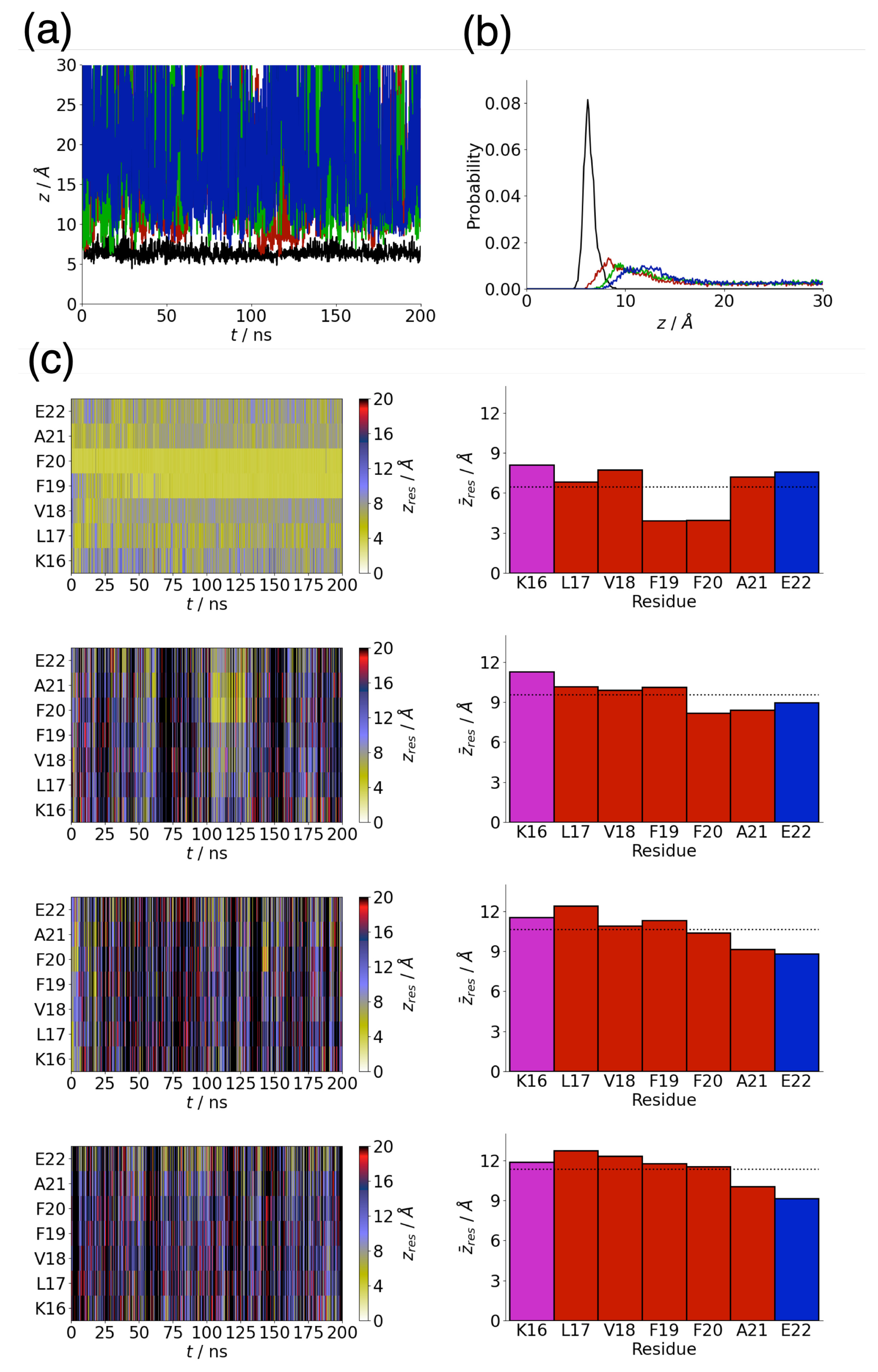
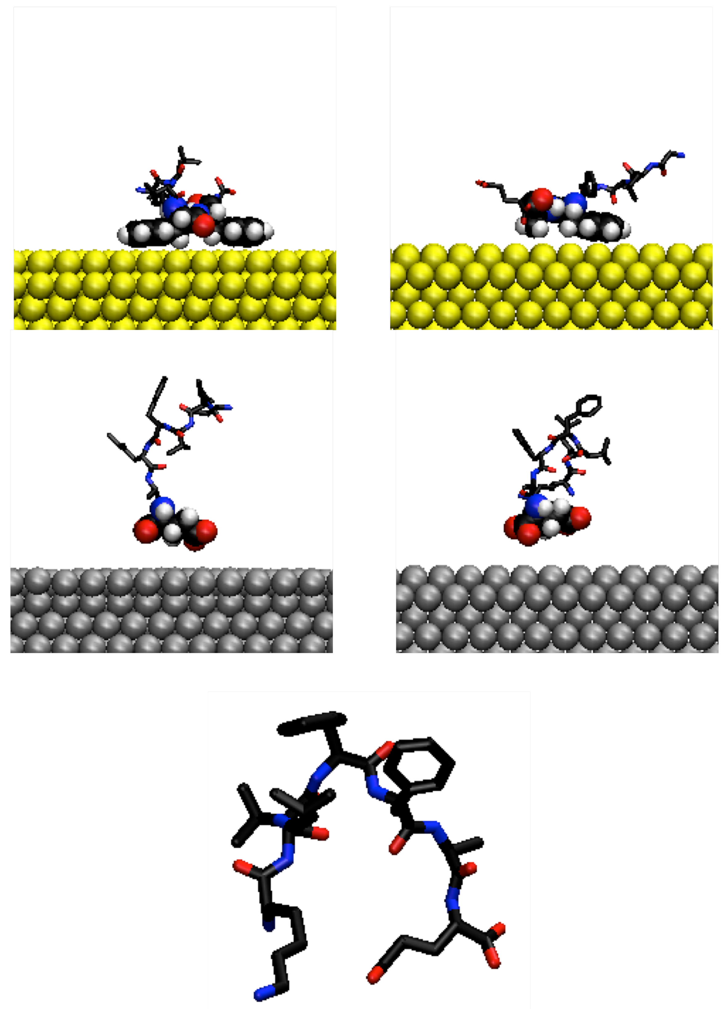
3.1. Adsorption Behaviour of Aβ(16–22) on Gold and Silver Surfaces

The adsorption of A β (16–22) onto different metal surface depends on both the metal and exposed facet. For the Au111 surface, the peptide is strongly bound to the surface, with the separation between the peptide centre-of-mass and surface being between 5 and 10 Å throughout the simulation (Figure 1a). This strong adsorption is consistent with previous simulations of A β (16–22)on Au111 surfaces [47]. In contrast to the Au111, surface adsorption onto the other surfaces is weaker, with the peptide able to desorb from the surface (Figure 1a). The weaker adsorption can be seen by considering the P ( z ) histogram of peptide–surface separations (Figure 1b). For the Au111 surface, this is tightly peaked, with the peptide only found within 10 Å of the surface, while the distribution for the other surfaces is broader. Notably for the silver surfaces, the peak is further away from the surface, indicative of water mediated adsorption onto the surface, similar to that seen for single amino acids onto silver [57].
The average peptide–surface interaction energy, E i n t , was calculated for each of the surfaces (Table 1). A significantly stronger interaction was found with the Au111 surface compared to the other surfaces.
To gain more insight into the behaviour of the peptide on the different surfaces, the separation between the centre-of-mass of each residue and surface are shown in Figure 1c. For the Au111 surface, the two phenylalanine residues (F19 and F20) are consistently in contact with the surface. This is in line with previous simulations of A β (16–22) on the Au111 surface [47] and with the strong adsorption of phenylalanine onto Au111 surfaces [61]. The importance of the phenylalanine residues on the adsorption can also be seen from the average separation between each residue and the surface. Both F19 and F20 have z ¯ 3 4 Å with the other residues further from the surface, driven by the affinity of the charged termini for water.
While adsorption onto the other surfaces is weaker, marked by frequent desorption from the surface, specific residues that mediate adsorption can also be identified for these surfaces. On the Au100 surface, adsorption is mediated through F20 and A21 residues. Involvement of only one of the phenylalanine residues may be due to the weaker adsorption of phenylalanine onto Au100 not being sufficient to overcome the unfavourable rotation around the peptide backbone for both phenylalanine residues to contact the surface. Less difference is seen for the two silver surfaces, with the protein termini, primarily the C-terminus, being typically closer to the surface.
The different residues involved in adsorption can be seen from considering the probability of each residue being in contact with the surface (Figure 2). These are largely higher for the Au111 surface due to the strong adsorption, with the contact probabilities for F19 and F20 being close to one. For the Au111 surface, the terminal residues (K16 and E22) and the L17 also have a significant probability of contact with the surface. The contact probabilities for the other surfaces are lower and are typically larger for residues towards the C-terminus.

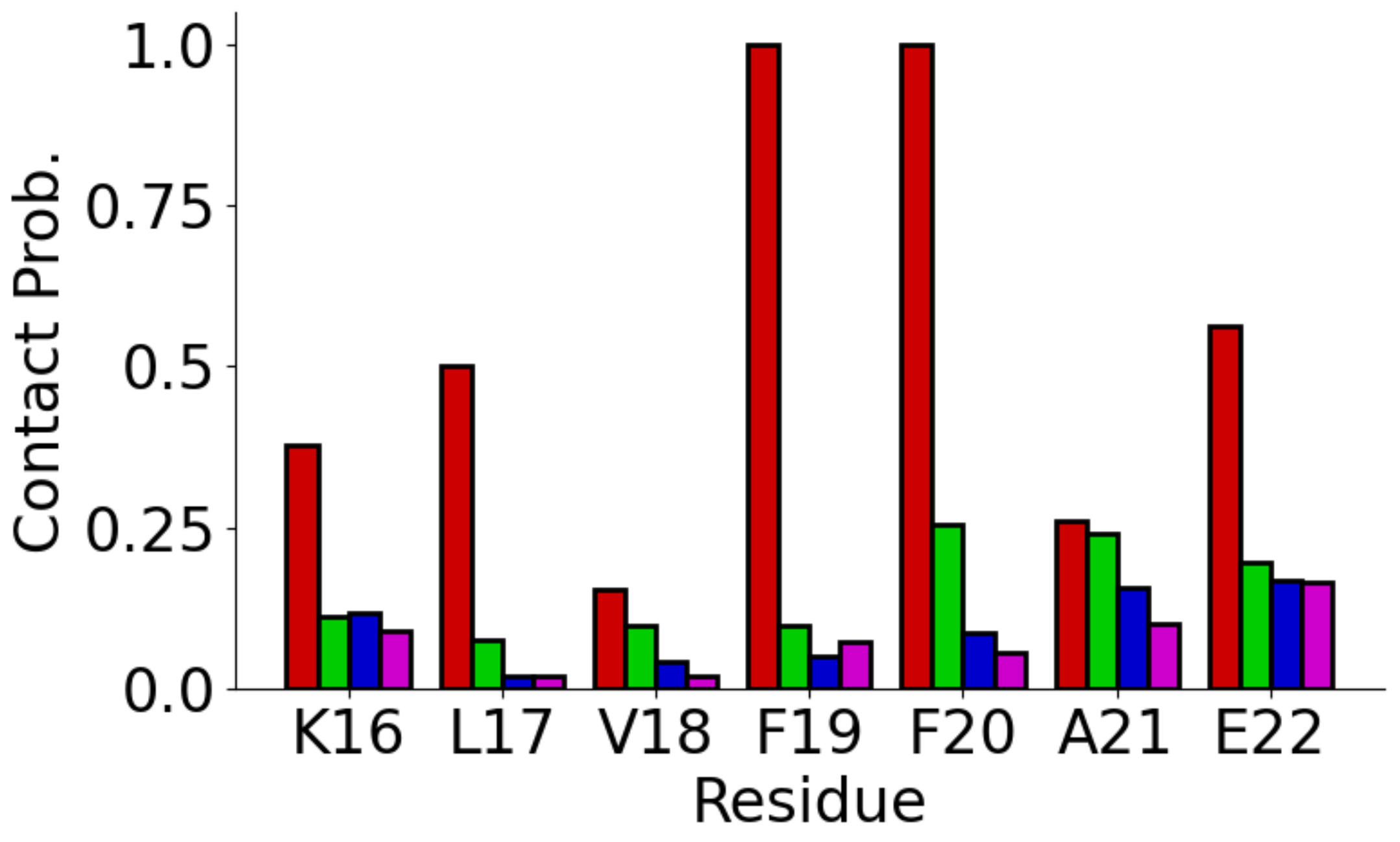
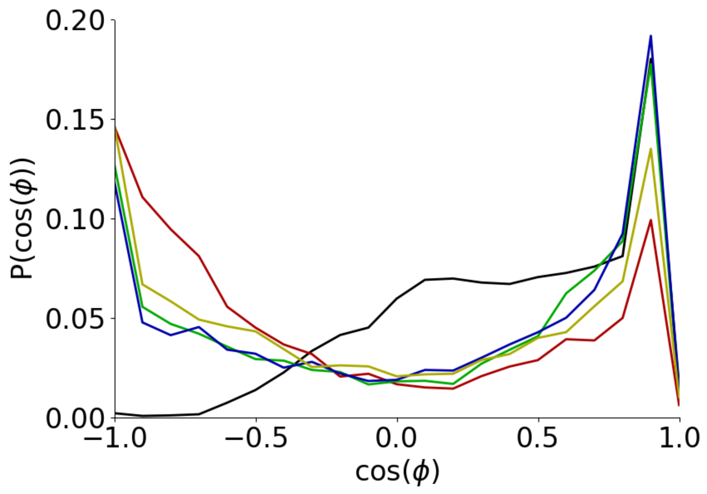
3.2. Conformation of Aβ(16–22) on Metal Surfaces

The binding of specific residues to the surfaces affects the conformations A β (16–22) can adopt. Qualitatively, this can be seen in representative snapshots (Figure 3). For the gold surfaces, the peptide lies flatter on the surface, with two (Au111) or one (Au100) phenylalanine residue in contact with the surface, in line with prior simulations [47]. On silver surfaces, fewer residues, typically consisting of the terminal ones, are in contact with the surface (Figure 2). Similar to bulk solution, this also allows for contacts between the two termini of the peptide.
The strong adsorption of the two phenylalanine residues onto the Au111 surface enhances the formation of cis-conformations compared to other surfaces and bulk solutions. This can be seen from the histogram of the angle ϕ between the phenylalanine side chains (Figure 4). All the systems have a peak in this near cos ϕ = 1 , but this is largest for the Au111 surface and the probability of cos ϕ > 0 (corresponding to the cis-state) is higher for the Au111 surface. For cos ϕ < 0 , which corresponds to the trans-state, the probability is lower for the Au111 compared to the other systems with P ( cos ϕ ) going to zero at cos ϕ = 1 . This enhancement of cis-conformers has been seen in previous studies of A β (16–22) on interfaces [47,72] and of the diphenylalanine dipeptide [81] at the air–water interface.
While the adsorption of the phenylalanine residues to the silver surfaces is lower, these also have a higher probability of forming the cis-conformers. In these cases, it may be driven by the tendency of the two terminal residues to adsorb to the surface (Figure 1c).
The preponderance of the cis-conformer leads the peptide to adopt U-shaped conformations, which brings the ends of the peptide closer together. This can be seen by the end-to-end length (Table 2), which is lower for the Au111 surface than in bulk solution. By contrast, A β (16–22) has a higher average end-to-end length on the Au100 surface indicating a preference for more extended conformations, which are favoured by when the phenylalanine residues adopt the trans-conformation. For the silver surfaces, R e e is also on average lower than in bulk solution, driven by the adsorption of the terminal residues to the surface.
The less linear conformations adopted on some of the surfaces can also change the interactions between different residues in the peptide. To examine this, the hydrogen bonds between different residues have been determined (Table 2). For all systems, hydrogen bonding between the two terminal residues (K16 and E22) is found, although only for the Au111 surface is this present in the majority of the simulation. The persistence of this is likely driven by nonlinear conformations caused by cis-conformations of the diphenylalanine motif. This is the only hydrogen bond seen for the two gold surfaces, while other hydrogen bonds are found on the other systems.
As well as hydrogen bonding, contacts between the two terminal residues are driven by attractive electrostatic interactions. Shown in Figure 5 are the histograms for the separation between the two terminal residues ( R t e r m ), defined as being the minimum distance between either of the positively charged nitrogen atoms in K16 and any of the negatively charged oxygen atoms in E22. These have a peak at R t e r m 2.5 Å corresponding to close contacts. This peak is smaller for Au100 and in bulk solution, indicating a lower probability of contacts in these cases. The second peak at R t e r m 5 Å is also weaker for Au100 and solution. Notably, in all cases, contacts between nitrogen and oxygen atoms in the Lys and Glu side chains are more common than between the nitrogen and oxygen atoms in the charged end groups, suggesting that the effect of changes to the charge state of the termini (e.g., through neutral capping groups) is likely to be small. This is a consequence of having two residues with charged side chains at the end of the peptide; for other cases, where the side chains of the terminal residues are uncharged, changes to the charge states of the termini (e.g., through capping groups) would potentially have a larger effect [82].
On the Au100 surface, A β (16–22) adopts more extended conformations with a higher probability of trans-conformers so it may be expected that it forms more fibril-like structures. To quantify this, we examine the C α -C α distance RMSD (DRMSD) between the simulation structure and experimental fibril structure (from PDB code 2y29 [83]). This is calculated using
D R M S D = 1 N C α C α i r C α C α , i s i m r C α C α , i e x p 2
where the sum is over all the C α C α pairs in the peptide. This suggests that the gold surfaces have a differing effect on the formation of fibril-like structures (Figure 6), with fibril-like structures being more common on the Au100 surface. By contrast, fibril-like conformations are unlikely on the Au111 surface, consistent with previous simulation work [47]. While both gold surfaces differ substantially from bulk solution for the two silver surfaces, there is less difference to solution. The formation of fibril-like structures is also similar for the Ag111 and Ag100 surfaces, suggesting a smaller facet dependence compared to gold.

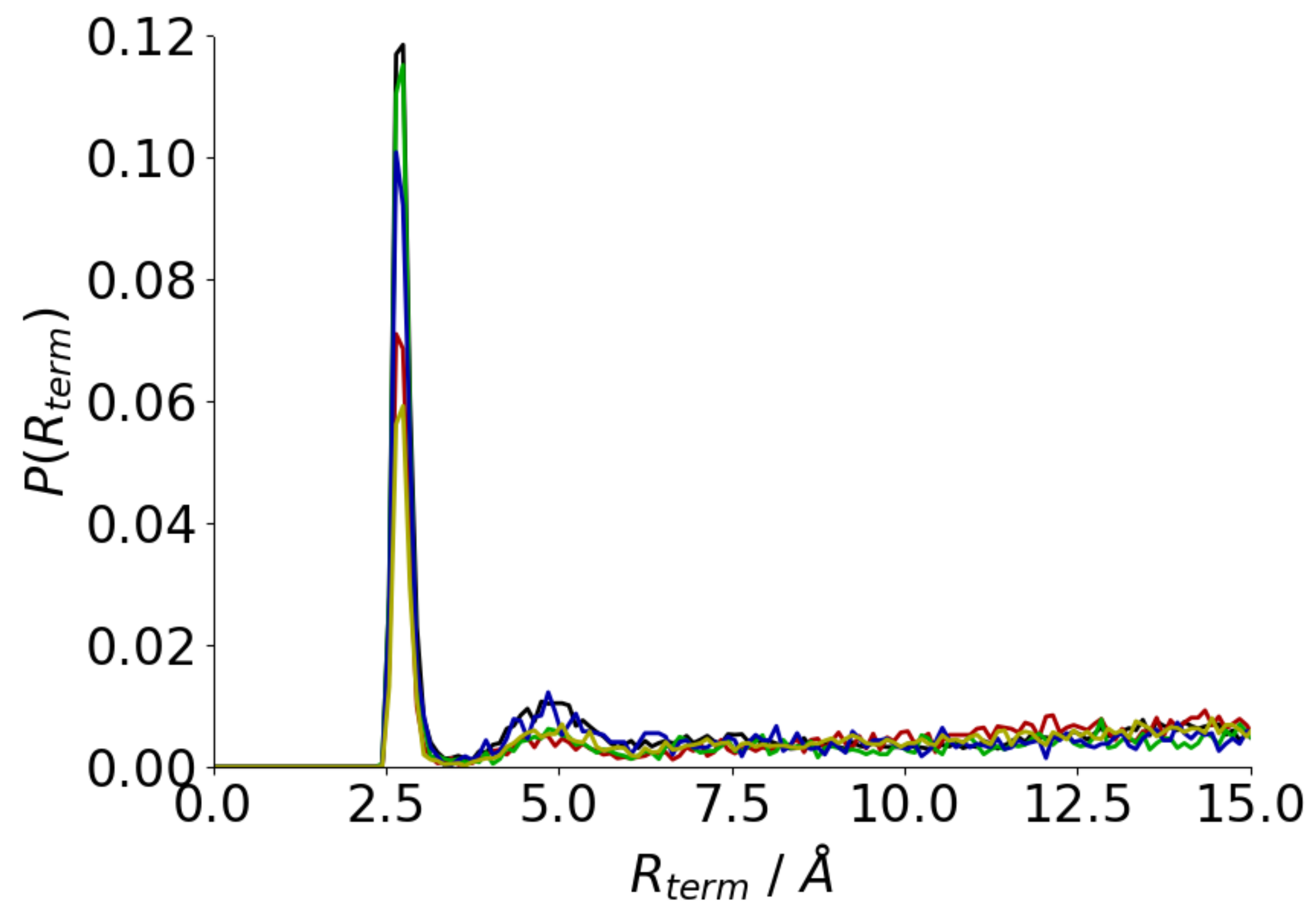
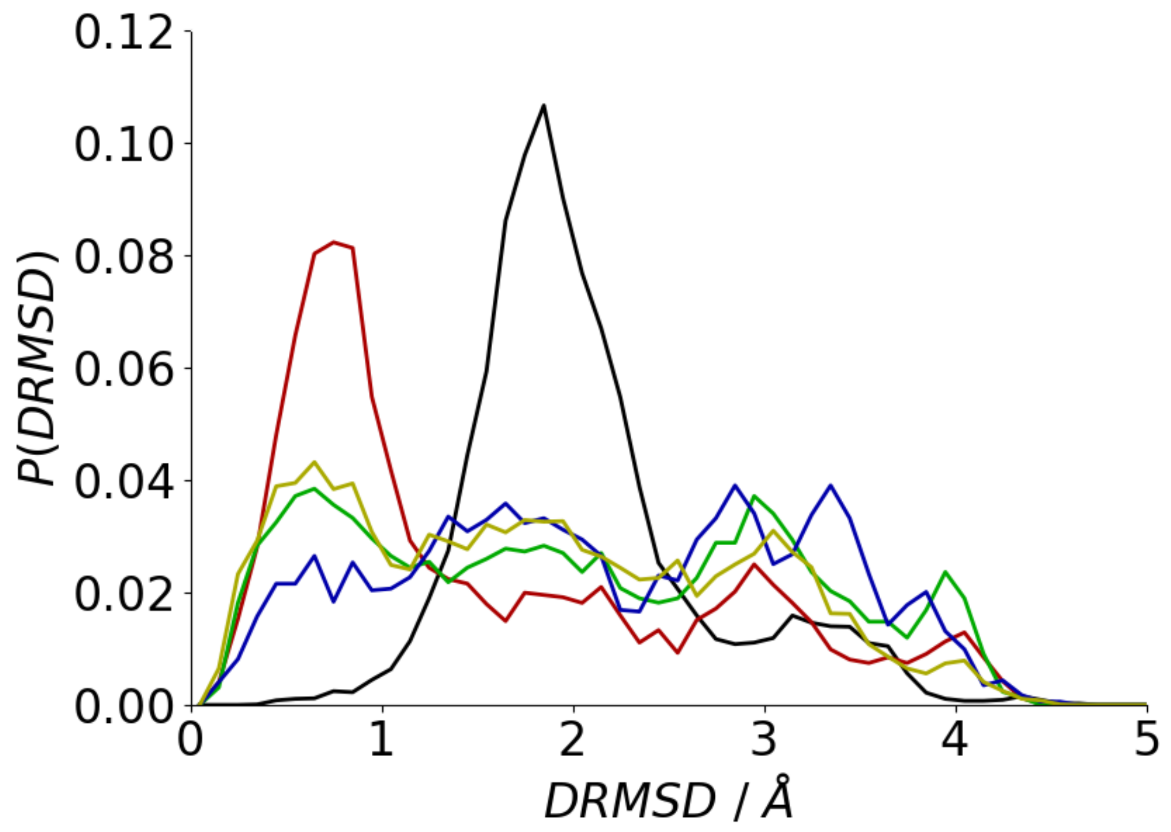
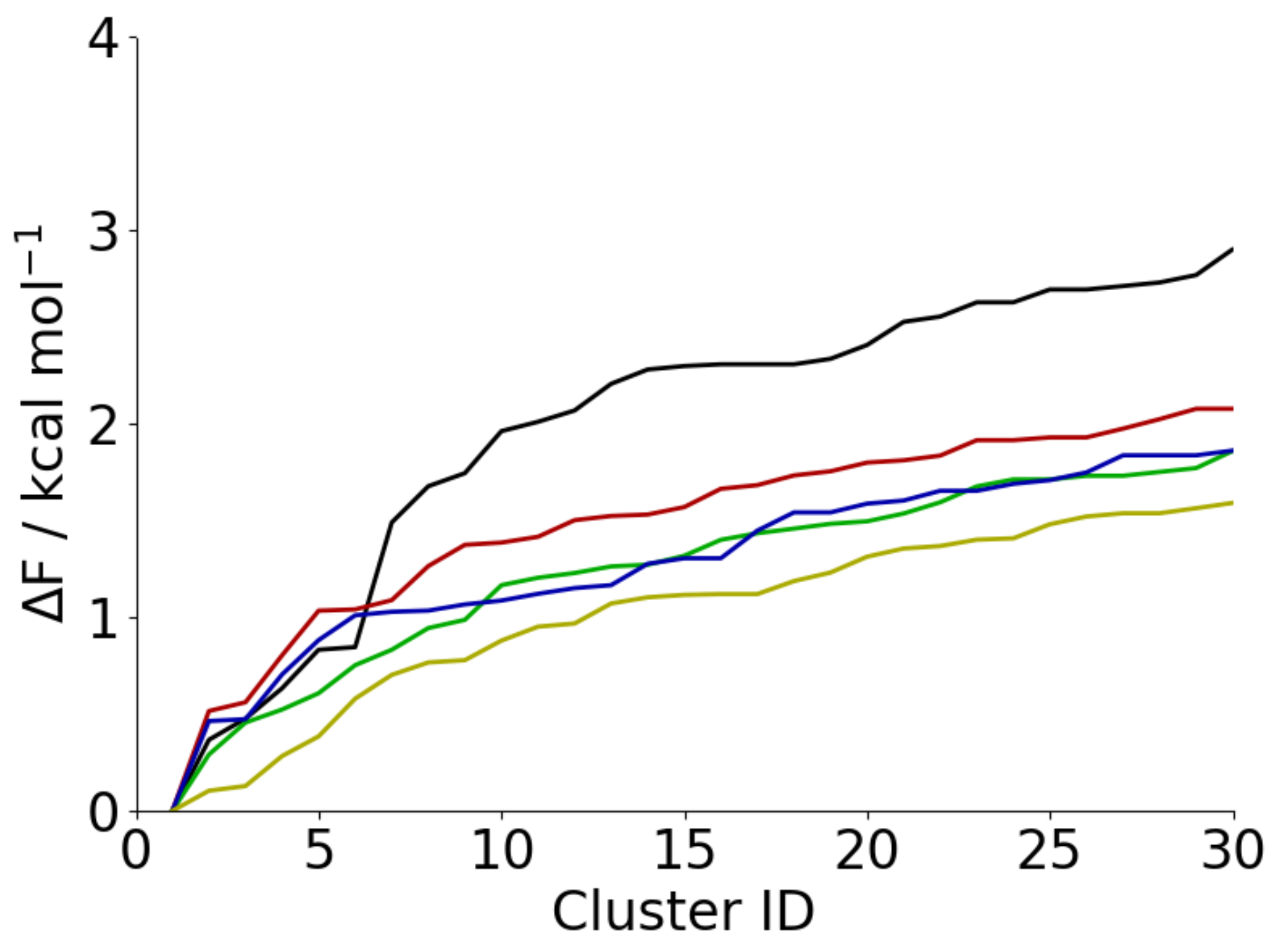
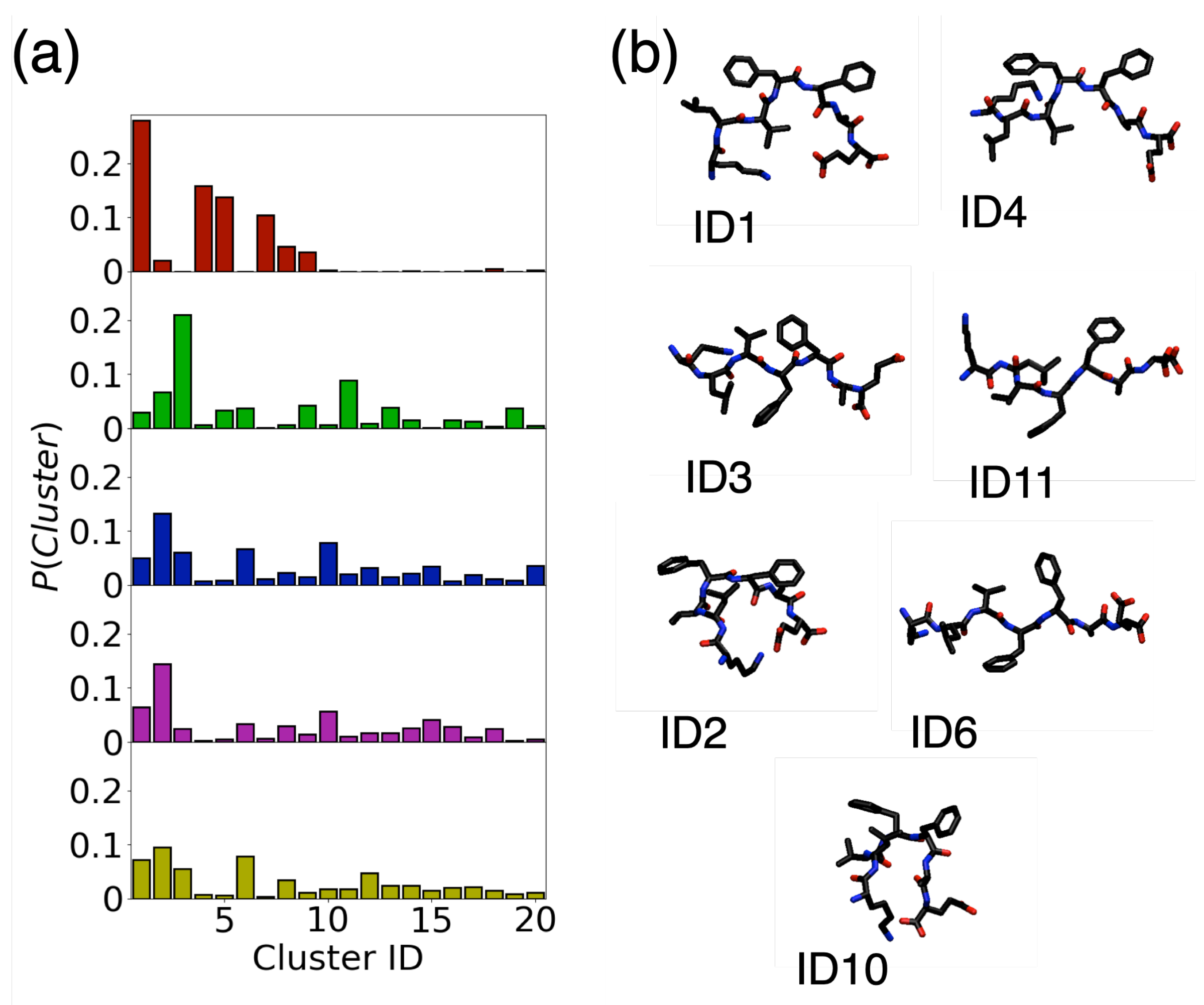
3.3. Adsorption onto Metal Surfaces Affects the Conformational Ensemble of Aβ(16–22)

As an intrinsically disordered peptide, A β (16–22) forms an ensemble of different conformations. Both the set of conformations and the number of distinct conformations that a peptide can adopt can be affected by surfaces. For the different systems, the conformational ensemble has been determined using a cluster analysis. Shown in Table 3 are the number of distinct clusters found for the different systems. Notably, N c l u s t e r s is smaller for all surfaces compared to bulk solution, due to the binding of specific residues to the surfaces (Figure 1c) which limits the possible conformations, which has been seen in previous simulations of intrinsically disordered proteins on surfaces [50,72]. This is particularly noticeable for the Au111 surface due to the strong binding of the two Phe residues to the surface. The number of conformations is higher for the other surfaces, which may be reflective of the weaker binding of A β (16–22) to these surfaces. While the cluster analysis is restricted to the conformations where the peptide is adsorbed to the surface, the ability of the peptide to desorb from the surface may also give A β (16–22) greater conformational freedom on these surfaces compared to the Au111 surface. Again, a smaller difference is seen between the two silver surfaces.
Changes in the conformational ensemble can also be seen in the conformational entropy (Table 3). This is lower for all the surfaces compared to bulk solution and is considerably lower for Au111 compared to the other surfaces. The higher conformational entropy on silver surfaces compared to gold and the higher values on 100 compared to 111 surfaces is consistent with prior simulations of gold and silver binding peptides [57].
The probability of each cluster in the simulations can be used to determine the free energy of each cluster
Δ F = k B T ln P i P 1
where Δ F is the free energy difference between the ith and 1st cluster, and P i is the probability of the ith cluster (Figure 7). For the silver surfaces and bulk solution, the free energy varies similarly with cluster ID, showing a relatively slow increase consistent with a number of conformations with similar free energies. On the gold surfaces, there are fewer low energy states, suggesting that there is a greater restriction on the different conformations.
The cumulative probability of different conformations has been used to classify peptides in terms of their binding entropy [84]. For the Au100, Ag111, and Ag100 surfaces, the cumulative probability of the top five clusters is under 60% classifying A β (16–22) as a high entropy binder on these surfaces. By the same scheme, A β (16–22) is a medium entropy binder on the Au111 (the cumulative probability of the top five clusters is over 60%, but, for the top three clusters, it is under 75%. In combination with the difference with the interaction energies between the surfaces, this suggests a qualitative difference in the behaviour of A β (16–22) on the Au111 surface.
To examine how similar the conformational ensembles are for the different systems, we perform a cluster analysis on the combination of the different systems. Comparing the probability of the different conformations in each simulation (Figure 8a) shows that the conformational ensembles on the two gold surfaces are significantly different to the other systems. Notably on the Au111 surface, there are fewer conformations that are significantly populated, consistent with the lower conformational entropy. The most populated conformations for the Au111 and Au100 surfaces are also typically less likely on the silver surfaces and in bulk solution.
The observation that P ( C l u s t e r ) for the two gold surfaces differ from each other and the other surfaces suggests that we can characterise the adsorption of A β (16–22) onto gold as an example of induced fit-like behaviour [85], where the surface causes the peptide to adopt different conformations. In the case of A β (16–22) on gold, this set of conformations also depends on the particular exposed facet. The close similarity between the distributions on the silver surfaces and bulk solution suggests that the adsorption of A β (16–22) onto silver is characterised by conformational selection [86], where similar conformations are found with, in this case minor, changes in likelihood of the different conformations.
The structures commonly found for the different systems (Figure 8b) can be related to the typical interaction between the peptide and surface. For the Au111 surface, the most common conformations (ID 1, 4) have the two phenylalanine residues in the cis-conformation, with the two rings in the same plane, which maximises their ability to contact the gold surface. The most common conformations for the Au100 surface (e.g., 3, 11) include more extended, fibril-like structures. On the silver surfaces and in bulk solution, both extended and more compact conformations are found. Many of the conformations found on the silver surfaces and in bulk solution typically have contacts between the terminal residues, driven by electrostatic attraction between these.

4. Conclusions

Using replica exchange molecular dynamics simulation, we have investigated the conformation of the model amyloidogenic peptide, A β (16–22) on gold and silver surfaces. We found significant differences in behaviour between the Au111 and Au100 surfaces, with the peptide adsorption significantly stronger on the Au111 surface. This is due to the strong interaction between phenylalanine residues and the Au111 surface, with the weaker interaction between these and the Au100 surface leading to weaker adsorption. By contrast, a smaller difference between the two silver surfaces (Ag111 and Ag100) was seen, suggesting that adsorption onto silver is less dependent on the exposed facet. The contrasting facet dependence of adsorption is similar to that seen for gold and silver binding peptides.
Comparison of the peptide–surface interaction energies and conformational entropy suggests that, despite the structural similarities between gold and silver surfaces, the binding of A β (16–22) to these have different driving forces. For the Au111 surface, the low interaction energy and the decrease in the peptide conformational entropy suggest that this is largely driven by enthalpy. On the other surfaces, the entropy may be expected to play a larger role. This is in line with previous simulation studies of gold and silver binding peptides [57]. The difference between these is smaller for the 100 facets compared to the 111 facets.
Adsorption onto surfaces affects the conformations adopted by A β (16–22). As an intrinsically disordered peptide, it adopts a range of different conformations (conformational ensemble). The size of this ensemble is smaller on surfaces compared to bulk solution, as the surface restricts the possible conformations of the peptide. The Au111 surface has the smallest number of conformations, as the strong adsorption of the phenylalanine residues effectively pins these to the surface. Comparison of the conformational ensembles for the different surfaces and bulk solution shows that the gold surfaces induce different sets of conformations to the silver surfaces and bulk solution. This suggests that we can describe adsorption onto gold surfaces as induced fit-like while adsorption onto silver surfaces can be thought of as conformational selection. It may be argued that induced fit-like behaviour is more likely for cases with strong adsorption, consistent with previous simulations of IDPs on hydrophobic and hydrophilic self-assembled monolayers [50].
The conformations of A β (16–22) adopted on different surfaces has implications for how they affect peptide fibrillation. On the Au111 surface, A β (16–22) adopts structures that are quite different to those of A β (16–22) in amyloid fibrils, while it is more likely to adopt fibril-like conformations on the Au100 surface. This suggests that, even for the same material, different surface faces may have differing effects on fibrillation. As the proportion of 111 and 100 faces on nanoparticles varies with their size [87], this may contribute to the size dependent effect of gold nanoparticles on the formation of amyloid fibrils [24]. The smaller difference between the two silver surfaces suggests that nanoparticle size may have less of an effect for silver compared to gold.
While the results of this are consistent with previous simulation results, the behaviour of peptides and proteins on surfaces depends on both the surface properties and on the peptide sequence. It would also be interesting to extend this to the examination of other metal surfaces to consider how changes to these structurally similar systems affect the behaviour of amyloidogenic peptides. The simulation results are also amenable to experimental verification. Quartz crystal microbalance and surface plasmon resonance measurements can be used to determine the adsorbed amount on different surfaces, which may be correlated to the stronger binding observed on gold surfaces compared to silver. The conformation of the peptide on surfaces can be investigated using sum frequency generation or PM-IRRAS measurements. While it can not directly image the structure of such small peptides, high-speed AFM can be used to study fibril formation on different surfaces, which can be related to peptide conformation.

Author Contributions

Conceptualization, K.P.S. and D.L.C.; methodology, D.L.C.; software, K.P.S. and D.L.C.; investigation, K.P.S. and D.L.C.; writing—original draft preparation, D.L.C.; writing—review and editing, K.P.S. and D.L.C. All authors have read and agreed to the published version of the manuscript.

Funding

Computational facilities for this work were provided by the SFI/HEA funded Irish Centre for High End Computing (project code ngche092b). K.P.S. was supported by the Irish Research Council (GOPID/2019/372).

Data Availability Statement

The data presented in this study are available in this article.

Acknowledgments

Gromacs files containing the AgP-charmm force field were kindly provided by Tiffany Walsh (Deakin University).

Conflicts of Interest

The authors declare no conflict of interest. The funders had no role in the design of the study; in the collection, analyses, or interpretation of data; in the writing of the manuscript, or in the decision to publish the results.

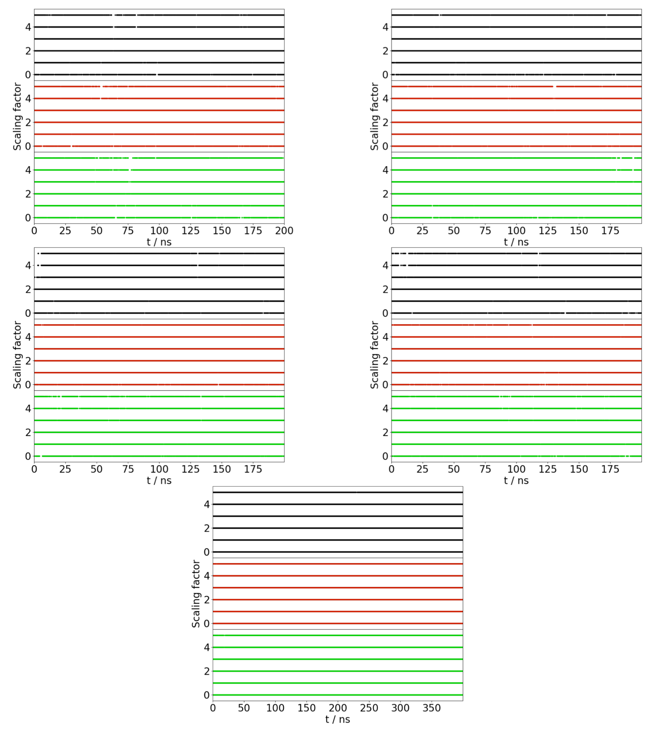
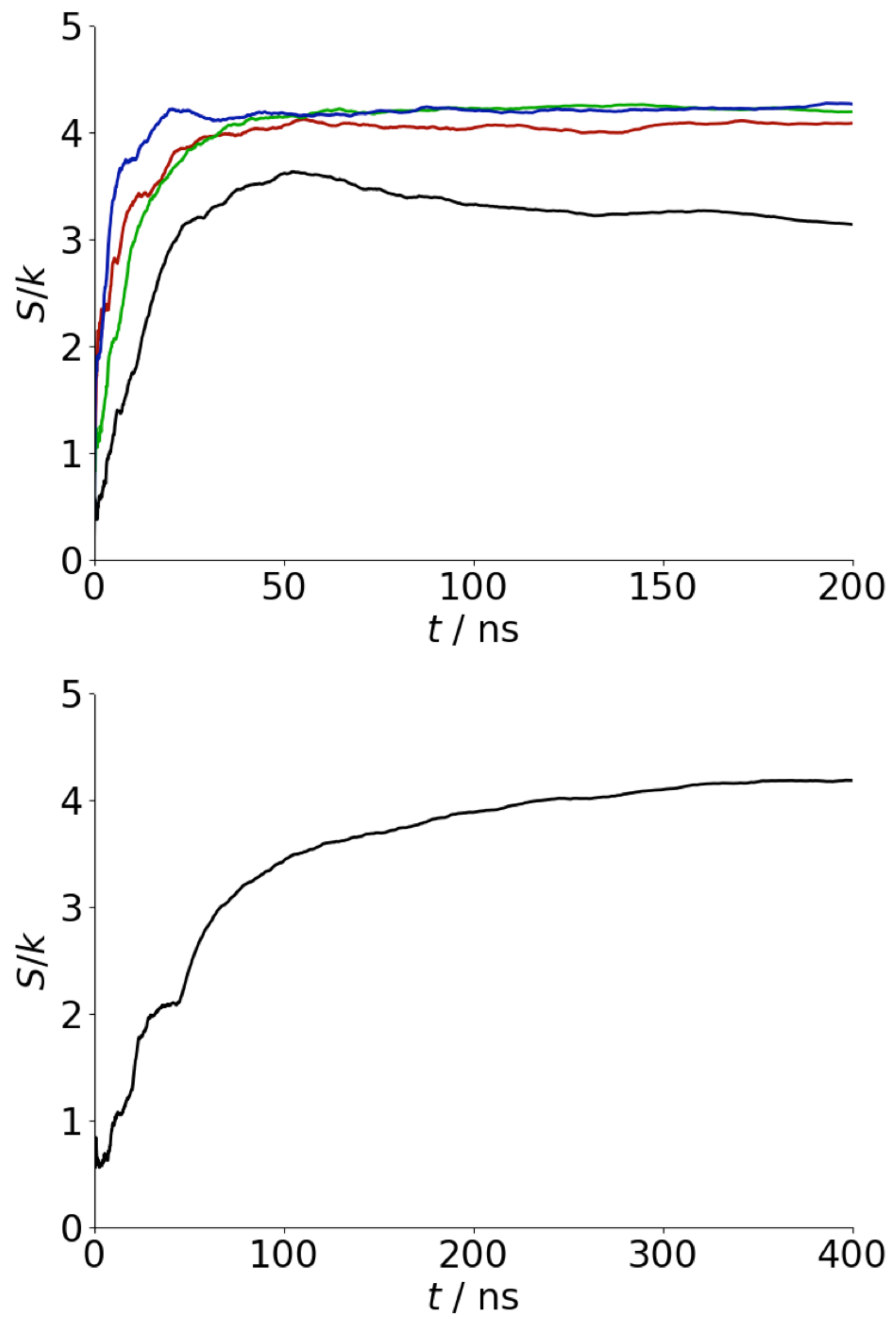
Appendix A. Convergence of REST Simulations

To test the efficiency of the replica exchange simulations, the acceptance rates (Table A1) and trajectories of the replicas (Figure A1) have been determined. For all cases, the acceptance rates are above 25%, indicating exchanges between replicas occur at a sufficient rate. Similarly in all cases, the different replicas explore all values of scaling factor, indicating the absence of significant bottlenecks.
Table A1. Acceptance rates for REST simulations.
System0 ↔ 11 ↔ 22 ↔ 33 ↔ 44 ↔ 5
Au1110.2620.2830.3300.3260.309
Au1000.2980.3090.3310.3610.355
Ag1110.3080.3070.3280.3560.357
Ag1000.2840.2970.2950.3240.339
Solution0.3120.3000.3170.3630.359
Figure A1. Plot of scaling factor (i denotes β i ) for REST simulations. Graphs show Au111 (top left), Au100 (top right), Ag111 (middle left), Ag100 (middle right), and bulk solution (bottom). Graphs show replicas with i = 0 (black), 3 (red), and 5 (green) at t = 0.
Biophysica 02 00015 g0a1
To test that the simulations are sufficiently long, the peptide conformational entropy was calculated as a function of simulation time (Figure A2). For all cases, this is approximately constant across the final 100 ns of the simulations, suggesting that the peptide conformational has been sufficiently sampled.
The ratio between adsorbed and desorbed conformation for the surface simulations (with the exception of the Au111 surface where only adsorbed conformations were found) was examined to ensure that an equilibrium between adsorbed and desorbed conformations was found. As shown in Figure A3, this remains approximately constant for the last 100 ns of the simulations, suggesting that this was the case.
Figure A2. Variation in peptide conformational entropy with simulation time for surface (top) and solution (bottom) simulations. For surface simulations black, red, green, and blue denote Au111, Au100, Ag111, and Ag100, respectively.
Biophysica 02 00015 g0a2
Figure A3. Ratio of adsorbed and desorbed conformations for REST simulations on Au100 (red), Ag111 (green), and Ag100 (blue) surfaces.
Biophysica 02 00015 g0a3

References

  1. Iadanza, M.G.; Jackson, M.P.; Hewitt, E.W.; Ranson, N.A.; Radford, S.E. A new era for understanding amyloid structures and disease. Nat. Rev. Mol. Cell. Biol. 2018, 19, 755–773. [Google Scholar] [CrossRef] [PubMed]
  2. Ke, P.C.; Zhou, R.; Serpell, L.C.; Riek, R.; Knowles, T.P.J.; Lashuel, H.A.; Gazit, E.; Hamley, I.W.; Davis, T.P.; Fandrich, M.; et al. Half a century of amyloids: Past, present and future. Chem. Soc. Rev. 2020, 49, 5473–5509. [Google Scholar] [CrossRef] [PubMed]
  3. Maresova, P.; Mohelska, H.; Dolejs, J.; Kuca, K. Socio-economic Aspects of Alzheimer’s Disease. Curr. Alzheimer Res. 2015, 12, 903–911. [Google Scholar] [CrossRef] [PubMed]
  4. Zhong, C.; Gurry, T.; Cheng, A.A.; Downey, J.; Deng, Z.; Stultz, C.M.; Lu, T.K. Strong underwater adhesives made by self-assembling multi-protein nanofibres. Nat. Nanotech. 2014, 9, 858–866. [Google Scholar] [CrossRef] [PubMed] [Green Version]
  5. Erskine, E.E.; MacPhee, C.E.; Stanley-Wall, N.R. Functional Amyloid and Other Protein Fibres in the Biofilm Matrix. J. Mol. Biol. 2018, 430, 3642–3656. [Google Scholar] [CrossRef] [PubMed]
  6. Knowles, T.P.J.; Mezzenga, R. Amyloid Fibrils as Building Blocks for Natural and Artificial Functional Materials. Adv. Mater. 2016, 28, 6546–6561. [Google Scholar] [CrossRef] [PubMed]
  7. Li, C.; Qin, R.; Liu, R.; Miao, S.; Yang, P. Functional amyloid materials at surfaces/interfaces. Biomater. Sci. 2018, 6, 462–472. [Google Scholar] [CrossRef] [PubMed]
  8. Burke, K.A.; Yates, E.A.; Legleiter, J. Biophysical Insights into How Surfaces, Including Lipid Membranes, Modulate Protein Aggregation Related to Neurodegeneration. Front. Neurol. 2013, 4, 17. [Google Scholar] [CrossRef] [PubMed] [Green Version]
  9. Keller, A.; Grundmeier, G. Amyloid aggregation at solid-liquid interfaces: Perspectives of studies using model surfaces. Appl. Surf. Sci. 2020, 506, 144991. [Google Scholar] [CrossRef]
  10. Stefani, M. Generic Cell Dysfunction in Neurodegenerative Disorders: Role of Surfaces in Early Protein Misfolding, Aggregation, and Aggregate Cytotoxicity. Neuroscientist 2007, 13, 519–531. [Google Scholar] [CrossRef]
  11. Terzi, E.; Hozlemann, G.; Seelig, J. Interaction of Alzheimer beta-Amyloid Peptide(1-40) with Lipid Membranes. Biochemistry 1997, 36, 14845–14852. [Google Scholar] [CrossRef]
  12. Koppaka, V.; Axelsen, P.H. Accelerated Accumulation of Amyloid-beta Proteins on Oxidatively Damaged Lipid Membranes. Biochemistry 2000, 39, 10011–10016. [Google Scholar] [CrossRef]
  13. Bokvist, M.; Lindstrom, F.; Watts, A.; Grabner, G. Two Types of Alzheimer’s Amyloid (1-40) Peptide Membrane Interactions: Aggregation Preventing Transmembrane Anchoring Versus Accelerated Surface Fibril Formation. J. Mol. Biol. 2004, 335, 1039–1049. [Google Scholar] [CrossRef]
  14. Verdier, Y.; Zarandi, M.; Penke, B. Amyloid-beta peptide interactions with neuronal and glial cell plasma membrane: Binding sites and implications for Alzheimer’s disease. J. Pept. Sci. 2004, 10, 229–248. [Google Scholar] [CrossRef]
  15. Jayasinghe, S.A.; Langen, R. Lipid Membranes Modulate the Structure of Islet Amyloid Polypeptide. Biochemistry 2005, 44, 12113–12119. [Google Scholar] [CrossRef]
  16. Jean, L.; Lee, C.F.; Lee, C.; Shaw, M.; Vaux, D.J. Competing discrete interfacial effects are critical for amyloidogenesis. FASEB J. 2010, 24, 309–317. [Google Scholar] [CrossRef]
  17. Bolisetty, S.; Mezzenga, R. Amyloid-beta-carbon hybrid membranes for universal water purification. Nat. Nanotechnol. 2016, 11, 365–371. [Google Scholar] [CrossRef]
  18. Hauser, C.A.E.; Maurer-Stroh, S.; Martins, I.C. Amyloid-based nanosensors and nanodevices. Chem. Soc. Rev. 2014, 43, 5326–5345. [Google Scholar] [CrossRef] [Green Version]
  19. Zhang, Q.; Bolisetty, S.; Cao, Y.; Handschin, S.; Adamcik, J.; Peng, Q.; Mezzenga, R. Selective and Efficient Removal of Fluoride from Water: In Situ Engineered Amyloid Fibril/ZrO2 Hybrid Membranes. Angew. Chem. Int. Ed. 2019, 58, 6012–6016. [Google Scholar] [CrossRef]
  20. Cedervall, T.; Lynch, I.; Lindman, S.; Berggard, T.; Thulin, E.; Nilsson, H.; Dawson, K.a.; Linse, S. Understanding the nanoparticle-protein corona using methods to quantify exchange rates and affinities of proteins for nanoparticles. Proc. Natl. Acad. Sci. USA 2007, 104, 2050–2055. [Google Scholar] [CrossRef] [Green Version]
  21. Cabaleiro-Lago, C.; Quinlan-Pluck, F.; Lynch, I.; Dawson, K.A.; Linse, S. Dual effect of amino modified polystyrene nanoparticles on amyloid-beta protein fibrillation. ACS Chem. Neurosci. 2010, 1, 279–287. [Google Scholar] [CrossRef]
  22. Gladytz, A.; Wagner, M.; Haupl, T.; Elsner, C.; Abel, B. Structure-Making Effects of Metal Nanoparticles in Amyloid Peptide Fibrillation. Part. Part. Syst. Charact. 2015, 32, 573–582. [Google Scholar] [CrossRef]
  23. Gladytz, A.; Abel, B.; Risselada, H.J. Gold-Induced Fibril Growth: The Mechanism of Surface-Facilitated Amyloid Aggregation. Angew. Chem. Int. Ed. 2016, 55, 11242–11246. [Google Scholar] [CrossRef]
  24. Gao, G.; Zhang, M.; Gong, D.; Chen, R.; Hu, X.; Sun, T. The size-effect of gold nanoparticles and nanoclusters in the inhibition of amyloid-beta fibrillation. Nanoscale 2017, 9, 4107–4113. [Google Scholar] [CrossRef]
  25. John, T.; Gladytz, A.; Kubeil, C.; Martin, L.L.; Risselada, H.J.; Abel, B. Impact of nanoparticles on amyloid peptide and protein aggregation: A review with a focus on gold nanoparticles. Nanoscale 2018, 10, 20894–20913. [Google Scholar] [CrossRef]
  26. Arvizo, R.R.; Bhattacharyya, S.; Kudgus, R.A.; Giri, K.; Bhattacharya, R.; Mukherjee, P. Intrinsic therapeutic applications of noble metal nanoparticles: Past, present and future. Chem. Soc. Rev. 2012, 41, 2943–2970. [Google Scholar] [CrossRef] [Green Version]
  27. He, Y.; Du, Z.; Ma, S.; Liu, Y.; Li, D.; Huang, H.; Jiang, S.; Cheng, S.; Wu, W.; Zhang, K.; et al. Effects of green-synthesized silver nanoparticles on lung cancer cells in vitro and grown as xenograft tumors in vivo. Int. J. Nanomed. 2016, 11, 1879–1887. [Google Scholar] [CrossRef] [Green Version]
  28. Cheng, Q.; Liu, Y. Multifunctional platinum-based nanoparticles for biomedical applications. WIREs Nanomed. Nanobiotechnol. 2017, 9, e1410. [Google Scholar] [CrossRef]
  29. Pedone, D.; Moglianetti, M.; Luca, E.D.; Bardi, G.; Paolo-Pompa, P. Platinum nanoparticles in nanobiomedicine. Chem. Soc. Rev. 2017, 46, 4951–4975. [Google Scholar] [CrossRef]
  30. Puja, P.; Kumar, P. A perspective on biogenic synthesis of platinum nanoparticles and their biomedical applications. Spectrochim. Acta Part A Mol. Biomol. Spectrosc. 2019, 211, 94–99. [Google Scholar] [CrossRef]
  31. Sonawane, S.K.; Ahmad, A.; Chinnathambi, S. Protein-Capped Metal Nanoparticles Inhibit Tau Aggregation in Alzheimer‚ Äôs Disease. ACS Omega 2019, 4, 12833–12840. [Google Scholar] [CrossRef] [PubMed] [Green Version]
  32. Coppage, R.; Slocik, J.M.; Briggs, B.D.; Frenkel, A.I.; Heinz, H.; Naik, R.R.; Knecht, M.R. Crystallographic Recognition Controls Peptide Binding for Bio-Based Nanomaterials. J. Am. Chem. Soc. 2011, 133, 12346–12349. [Google Scholar] [CrossRef] [PubMed]
  33. Hughes, Z.E.; Kochandra, R.; Walsh, T.R. Facet-Specific Adsorption of Tripeptides at Aqueous Au Interfaces: Open Questions in Reconciling Experiment and Simulation. Langmuir 2017, 33, 3742–3754. [Google Scholar] [CrossRef] [PubMed] [Green Version]
  34. Schneider, J.; Colombi Ciacchi, L. Specific material recognition by small peptides mediated by the interfacial solvent structure. J. Am. Chem. Soc. 2012, 134, 2407–2413. [Google Scholar] [CrossRef]
  35. Dong, L.; Luo, Q.; Cheng, K.; Shi, H.; Wang, Q.; Weng, W.; Han, W.Q. Facet-Specific Assembly of Proteins on SrTiO3 Polyhedral Nanocrystals. Sci. Rep. 2014, 4, 5084. [Google Scholar] [CrossRef] [Green Version]
  36. Li, Y.; Huang, Y. Morphology-Controlled Synthesis of Platinum Nanocrystals with Specific Peptides. Adv. Mater. 2010, 22, 1921–1925. [Google Scholar] [CrossRef]
  37. Chiu, C.Y.; Li, Y.; Ruan, L.; Ye, X.; Murray, C.B.; Huang, Y. Platinum nanocrystals selectively shaped using facet-specific peptide sequences. Nat. Chem. 2011, 3, 393–399. [Google Scholar] [CrossRef]
  38. Wang, S.T.; Lin, Y.; Todorova, N.; Xu, Y.; Mazo, M.; Rana, S.; Leonardo, V.; Amdursky, N.; Spicer, C.D.; Alexander, B.D.; et al. Facet-Dependent Interactions of Islet Amyloid Polypeptide with Gold Nanoparticles: Implications for Fibril Formation and Peptide-Induced Lipid Membrane Disruption. Chem. Mater. 2017, 29, 1550–1560. [Google Scholar] [CrossRef]
  39. Shen, L.; Adachi, T.; Vanden Bout, D.; Zhu, X.Y. A Mobile Precursor Determines Amyloid-beta Peptide Fibril Formation at Interfaces. J. Am. Chem. Soc. 2012, 134, 14172–14178. [Google Scholar] [CrossRef]
  40. Keller, A.; Fritzsche, M.; Yu, Y.P.; Liu, Q.; Li, Y.M.; Dong, M.; Besenbacher, F. Influence of hydrophobicity on the surface-catalyzed assembly of the islet amyloid polypeptide. ACS Nano 2011, 5, 2770–2778. [Google Scholar] [CrossRef]
  41. Hajiraissi, R.; Giner, I.; Grundmeier, G.; Keller, A. Self-Assembly, Dynamics, and Polymorphism of hIAPP(20-29) Aggregates at Solid-Liquid Interfaces. Langmuir 2016, 33, 372–381. [Google Scholar] [CrossRef]
  42. Hajiraissi, R.; Hanke, M.; Yang, Y.; Duderija, B.; Gonzalez Orive, A.; Grundmeier, G.; Keller, A. Adsorption and Fibrillization of Islet Amyloid Polypeptide at Self-Assembled Monolayers Studied by QCM-D, AFM, and PM-IRRAS. Langmuir 2018, 34, 3517–3524. [Google Scholar] [CrossRef]
  43. Ando, T. High-speed AFM imaging. Curr. Opin. Struct. Biol. 2014, 28, 63–68. [Google Scholar] [CrossRef]
  44. Hosseinpour, S.; Roeters, S.J.; Bonn, M.; Peukert, W.; Woutersen, S.; Weidner, T. Structure and Dynamics of Interfacial Peptides and Proteins from Vibrational Sum-Frequency Generation Spectroscopy. Chem. Rev. 2020, 120, 3420–3465. [Google Scholar] [CrossRef]
  45. Ozboyaci, M.; Kokh, D.B.; Corni, S.; Wade, R.C. Modeling and simulation of protein-surface interactions: Achievements and challenges. Q. Rev. Biophys. 2016, 49, 1–45. [Google Scholar] [CrossRef] [Green Version]
  46. Walsh, T.R. Pathways to Structure-Property Relationships of Peptide-Materials Interfaces: Challenges in Predicting Molecular Structures. Acc. Chem. Res. 2017, 50, 1617–1624. [Google Scholar] [CrossRef] [Green Version]
  47. Bellucci, L.; Ardèvol, A.; Parrinello, M.; Lutz, H.; Lu, H.; Weidner, T.; Corni, S. The interaction with gold suppresses fiber-like conformations of the amyloid beta (16–22) peptide. Nanoscale 2016, 8, 8737–8748. [Google Scholar] [CrossRef] [Green Version]
  48. Bellucci, L.; Bussi, G.; Di Felice, R.; Corni, S. Fibrillation-prone conformations of the amyloid-beta-42 peptide at the gold/water interface. Nanoscale 2017, 9, 2279–2290. [Google Scholar] [CrossRef]
  49. Tavanti, F.; Pedone, A.; Menziani, M.C. Disclosing the Interaction of Gold Nanoparticles with Abeta(1-40) Monomers through Replica Exchange Molecular Dynamics Simulations. Int. J. Mol. Sci. 2020, 22, 26. [Google Scholar] [CrossRef]
  50. Cheung, D.L. Effect of surface chemistry on islet amyloid polypeptide conformation. Biointerphases 2020, 15, 051001. [Google Scholar] [CrossRef]
  51. Brancolini, G.; Corazza, A.; Vuano, M.; Fogolari, F.; Mimmi, M.C.; Bellotti, V.; Stoppini, M.; Corni, S.; Esposito, G. Probing the Influence of Citrate-Capped Gold Nanoparticles on an Amyloidogenic Protein. ACS Nano 2015, 9, 2600–2613. [Google Scholar] [CrossRef]
  52. Earl, D.J.; Deem, M.W. Parallel tempering: Theory, applications, and new perspectives. Phys. Chem. Chem. Phys. 2005, 7, 3910–3916. [Google Scholar] [CrossRef] [Green Version]
  53. Laio, A.; Gervasio, F.L. Metadynamics: A method to simulate rare events and reconstruct the free energy in biophysics, chemistry and material science. Rep. Prog. Phys. 2008, 71, 126601. [Google Scholar] [CrossRef]
  54. Heinz, H.; Jha, K.C.; Luettmer-Strathmann, J.; Farmer, B.L.; Naik, R.R. Polarization at metal‚ Äìbiomolecular interfaces in solution. J. R. Soc. Interface 2011, 8, 220–232. [Google Scholar] [CrossRef]
  55. Iori, F.; Corni, S. Including image charge effects in the molecular dynamics simulations of molecules on metal surfaces. J. Comput. Chem. 2008, 29, 1656–1666. [Google Scholar] [CrossRef]
  56. Corni, S. Reply to ‘Molecular mechanics models for the image charge’. J. Comput. Chem. 2017, 38, 2130–2133. [Google Scholar] [CrossRef] [Green Version]
  57. Palafox-Hernandez, J.P.; Tang, Z.; Hughes, Z.E.; Li, Y.; Swihart, M.T.; Prasad, P.N.; Walsh, T.R.; Knecht, M.R. Comparative Study of Materials-Binding Peptide Interactions with Gold and Silver Surfaces and Nanostructures: A Thermodynamic Basis for Biological Selectivity of Inorganic Materials. Chem. Mater. 2014, 26, 4960–4969. [Google Scholar] [CrossRef]
  58. Wright, L.B.; Palafox-Hernandez, J.P.; Rodger, P.M.; Corni, S.; Walsh, T.R. Facet selectivity in gold binding peptides: Exploiting interfacial water structure. Chem. Sci. 2015, 6, 5204–5214. [Google Scholar] [CrossRef] [Green Version]
  59. Balbach, J.J.; Ishii, Y.; Antzutkin, O.N.; Leapman, R.D.; Rizzo, N.W.; Dyda, F.; Reed, J.; Tycko, R. Amyloid Fibril Formation by Amyloid Beta-22, a Seven-Residue Fragment of the Alzheimer’s Beta-Amyloid Peptide, and Structural Characterization by Solid State NMR. Biochemistry 2000, 39, 13748–13759. [Google Scholar] [CrossRef]
  60. Perfilieva, O.A.; Pyshnyi, D.V.; Lomzov, A.A. Molecular Dynamics Simulation of Polarizable Gold Nanoparticles Interacting with Sodium Citrate. J. Chem. Theory Comput. 2019, 15, 1278–1292. [Google Scholar] [CrossRef]
  61. Wright, L.B.; Rodger, P.M.; Walsh, T.R.; Corni, S. First-principles-based force field for the interaction of proteins with Au(100)(5 × 1): An extension of GolP-CHARMM. J. Phys. Chem. C 2013, 117, 24292–24306. [Google Scholar] [CrossRef]
  62. Hughes, Z.E.; Wright, L.B.; Walsh, T.R. Biomolecular Adsorption at Aqueous Silver Interfaces: First-Principles Calculations, Polarizable Force-Field Simulations, and Comparisons with Gold. Langmuir 2013, 29, 13217–13229. [Google Scholar] [CrossRef] [PubMed]
  63. MacKerell, A.D.; Bashford, D.; Dunbrack, R.L.; Evanseck, J.D.; Field, M.J.; Fischer, S.; Gao, J.; Guo, H.; Ha, S.; Joseph-McCarthy, D.; et al. All-Atom Empirical Potential for Molecular Modeling and Dynamics Studies of Proteins. J. Phys. Chem. B 1998, 102, 3586–3616. [Google Scholar] [CrossRef] [PubMed]
  64. Bjelkmar, P.; Larsson, P.; Cuendet, M.A.; Hess, B.; Lindahl, E. Implementation of the {CHARMM} Force Field in {GROMACS}: Analysis of Protein Stability Effects from Correction Maps, Virtual Interaction Sites, and Water Models. J. Chem. Theory Comput. 2010, 6, 459–466. [Google Scholar] [CrossRef]
  65. Piana, S.; Lindorff-Larsen, K.; Shaw, D. How Robust Are Protein Folding Simulations with Respect to Force Field Parameterization? Biophys. J. 2011, 100, L47–L49. [Google Scholar] [CrossRef] [Green Version]
  66. Jorgensen, W.L.; Chandrasekhar, J.; Madura, J.D. Comparison of simple potential functions for simulating liquid water. J. Chem. Phys. 1983, 79, 926–935. [Google Scholar] [CrossRef]
  67. Man, V.H.; He, X.; Derreumaux, P.; Ji, B.; Xie, X.Q.; Nguyen, P.H.; Wang, J. Effects of All-Atom Molecular Mechanics Force Fields on Amyloid Peptide Assembly: The Case of Abeta 16-22 Dimer. J. Chem. Theory Comput. 2019, 15, 1440–1452. [Google Scholar] [CrossRef]
  68. Wright, L.B.; Rodger, P.M.; Corni, S.; Walsh, T.R. GolP-CHARMM: First-principles based force fields for the interaction of proteins with Au(111) and Au(100). J. Chem. Theory Comput. 2013, 9, 1616–1630. [Google Scholar] [CrossRef]
  69. Liu, P.; Kim, B.; Friesner, R.A.; Berne, B.J. Replica exchange with solute tempering: A method for sampling biological systems in explicit water. Proc. Natl. Acad. Sci. USA 2005, 102, 13749–13754. [Google Scholar] [CrossRef] [Green Version]
  70. Wang, L.; Friesner, R.A.; Berne, B.J. Replica exchange with solute scaling: A more efficient version of replica exchange with solute tempering (REST2). J. Phys. Chem. B 2011, 115, 9431–9438. [Google Scholar] [CrossRef] [Green Version]
  71. Sugita, Y.; Okamoto, Y. Replica exchange molecular dynamics method for protein folding simulation. Chem. Phys. Lett. 1999, 314, 141–151. [Google Scholar] [CrossRef]
  72. Samantray, S.; Cheung, D.L. Effect of the air-water interface on the conformation of amyloid beta. Biointerphases 2020, 15, 061011. [Google Scholar] [CrossRef]
  73. Bussi, G.; Donadio, D.; Parrinello, M. Canonical sampling through velocity rescaling. J. Chem. Phys. 2007, 126, 014101. [Google Scholar] [CrossRef] [Green Version]
  74. Parrinello, M.; Rahman, A. Polymorphic transitions in single crystals: A new molecular dynamics method. J. Appl. Phys. 1981, 52, 7182–7190. [Google Scholar] [CrossRef]
  75. Abraham, M.J.; Murtola, T.; Schulz, R.; Páll, S.; Smith, J.C.; Hess, B.; Lindahl, E. GROMACS: High performance molecular simulations through multi-level parallelism from laptops to supercomputers. SoftwareX 2015, 1–2, 19–25. [Google Scholar] [CrossRef] [Green Version]
  76. Tribello, G.A.; Bonomi, M.; Branduardi, D.; Camilloni, C.; Bussi, G. PLUMED 2: New feathers for an old bird. Comput. Phys. Commun. 2014, 185, 604–613. [Google Scholar] [CrossRef] [Green Version]
  77. Bussi, G. Hamiltonian replica exchange in GROMACS: A flexible implementation. Mol. Phys. 2013, 112, 379–384. [Google Scholar] [CrossRef] [Green Version]
  78. Daura, X.; Gademann, K.; Jaun, B.; Seebach, D.; Van Gunsteren, W.F.; Mark, A.E. Peptide Folding: When Simulation Meets Experiment. Angew. Chem. Int. Ed. 1999, 38, 236–240. [Google Scholar] [CrossRef]
  79. Michaud-Agrawal, N.; Denning, E.J.; Woolf, T.B.; Beckstein, O. MDAnalysis: A Toolkit for the Analysis of MolecularDynamics Simulations. J. Comput. Chem. 2011, 32, 2319–2327. [Google Scholar] [CrossRef] [Green Version]
  80. Humphrey, W.; Dalke, A.; Schulten, K. VMD: Visual molecular dynamics. J. Mol. Graph. 1996, 14, 33–38. [Google Scholar] [CrossRef]
  81. Dalgicdir, C.; Sensoy, O.; Peter, C.; Sayar, M. A transferable coarse-grained model for diphenylalanine: How to represent an environment driven conformational transition. J. Chem. Phys. 2013, 139, 234115. [Google Scholar] [CrossRef] [Green Version]
  82. Hajiraissi, R.; Hanke, M.; Gonzalez Orive, A.; Duderija, B.; Hofmann, U.; Zhang, Y.; Grundmeier, G.; Keller, A. Effect of Terminal Modifications on the Adsorption and Assembly of hIAPP(20–29). ACS Omega 2019, 4, 2649–2660. [Google Scholar] [CrossRef]
  83. Colletier, J.P.; Laganowsky, A.; Landau, M.; Zhao, M.; Soriaga, A.B.; Goldschmidt, L.; Flot, D.; Cascio, D.; Sawaya, M.R.; Eisenberg, D. Molecular basis for amyloid- polymorphism. Proc. Natl. Acad. Sci. USA 2011, 108, 16938–16943. [Google Scholar] [CrossRef] [Green Version]
  84. Tang, Z.; Palafox-Hernandez, J.P.; Law, W.C.; Hughes, Z.E.; Swihart, M.T.; Prasad, P.N.; Knecht, M.R.; Walsh, T.R. Biomolecular Recognition Principles for Bionanocombinatorics: An Integrated Approach To Elucidate Enthalpic and Entropic Factors. ACS Nano 2013, 7, 9632–9646. [Google Scholar] [CrossRef]
  85. Koshland, D.E. Application of a Theory of Enzyme Specificity to Protein Synthesis. Proc. Natl. Acad. Sci. USA 1958, 44, 98–104. [Google Scholar] [CrossRef] [Green Version]
  86. Kumar, S.; Ma, B.; Tsai, C.J.; Sinha, N.; Nussinov, R. Folding and binding cascades: Dynamic landscapes and population shifts. Protein Sci. 2008, 9, 10–19. [Google Scholar] [CrossRef]
  87. Barnard, A.S.; Chen, Y. Kinetic modelling of the shape-dependent evolution of faceted gold nanoparticles. J. Mater. Chem. 2011, 21, 12239. [Google Scholar] [CrossRef]
Figure 1. (a) A β (16–22) centre-of-mass position. Black, red, green, and blue lines denote Au111, Au100, Ag111, and Ag100, respectively. (b) probability histograms (averaged over last 100 ns of simulations) for A β (16–22) on Au111 (black), Au100 (red), Ag111 (green), and Ag100 (blue); (c) residue centre-of-mass-surface separations (left) and average residue centre-of-mass-surface separations (right). From top-to-bottom, Au111, Au100, Ag111, and Ag100. In the left hand panel, colours denote separation (as denoted in the colour bar). In the right hand panel, red, blue, and magenta denote hydrophobic, negatively-charged, and positively-charged residues.
Biophysica 02 00015 g001
Figure 2. Residue–surface contact probabilities for A β (16–22) on Au111 (red), Au100 (green), Ag111 (blue), and Ag100 (magenta) surfaces.
Biophysica 02 00015 g002
Figure 3. Snapshots showing typical adsorbed conformations for A β (16–22) on Au111 (top left), Au100 (top right), Ag111(middle left), and Ag100 (middle right) surfaces and typical conformation in bulk solution (bottom). Gold and silver spheres show locations of metal atoms. For the surface simulations, residues in contact with the surface are shown as spheres. These are F19 and F20 (Au111), F20 and A21 (Au100), E22 (Ag111), and E22 (Ag100).
Biophysica 02 00015 g003
Figure 4. Probability distribution of angle between phenylalanine side chains for Au111 (black), Au100 (red), Ag111 (green), Ag100 (blue) surfaces, and bulk solution (yellow).
Biophysica 02 00015 g004
Figure 5. Histogram of separation between termini ( R t e r m ) for Au111 (black), Au100 (red), Ag111 (green), Ag100 (blue) surfaces, and bulk solution (yellow).
Biophysica 02 00015 g005
Figure 6. Histogram of DRMSD relative to experimental fibril structure for Au111 (black), Au100 (red), Ag111 (green), Ag100 (blue) surfaces, and bulk solution (yellow).
Biophysica 02 00015 g006
Figure 7. Free energy of clusters for Au111 (black), Au100 (red), Ag111 (green), Ag100 (blue), and solution (yellow).
Biophysica 02 00015 g007
Figure 8. (a) Probability of highest ranked clusters for (top to bottom) Au111, Au100, Ag111, Ag100, and bulk solution; (b) snapshots of selected conformations found from the combined cluster analysis.
Biophysica 02 00015 g008
Table 1. Peptide–surface interaction energies calculated from MD simulations, uncertainties estimated from standard deviation.
Surface E int /kJ mol 1
Au111−188 ± 28
Au100−57 ± 48
Ag111−26 ± 26
Ag100−16 ± 10
Table 2. Average end-to-end lengths and common hydrogen bonds. Numbers in parentheses give probabilities of each hydrogen bond.
R ee Hydrogen Bonds
Au11113.74 ± 3.3K16-E22 (0.693)
Au10015.4 ± 4.1K16-E22 (0.211)
Ag11114.2 ± 4.4K16-E22 (0.231), L17-F20 (0.202)
Ag10013.6 ± 4.2K16-E22 (0.296), L17-F20 (0.228),
V18-F20 (0.178)
Solution15.2 ± 4.1K16-E22 (0.128), L17-F20 (0.109)
Table 3. Number of clusters and conformational entropy determined from cluster analysis over the last 100 ns of simulations.
N clusters S conf / k B
Au111932.54 ± 0.02
Au1001573.54 ± 0.15
Ag1111563.72 ± 0.05
Ag1001633.84 ± 0.08
Solution2604.18 ± 0.02
Publisher’s Note: MDPI stays neutral with regard to jurisdictional claims in published maps and institutional affiliations.

