- Article

Structural and Functional Characterization of Ultrasonically Treated PLA-PEDOT:PSS Nonwoven Composites for Soft Implantable Bioelectronics
- Anastasiia D. Tsareva,
- Sergey V. Kravchenko and
- Dimitri A. Ivanov
- + 4 authors
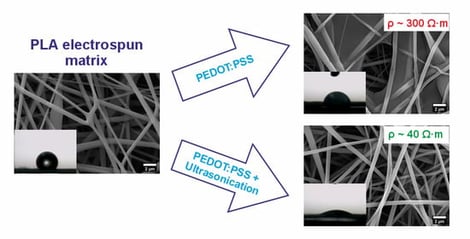
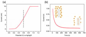
Flexible implantable electrodes require biocompatibility, mechanical stability, and sufficient electrical conductivity for effective neural interfacing. This work examines ultrasonic treatment during poly(3,4-ethylenedioxythiophene):poly(styrenesulfonate) (PEDOT:PSS) impregnation of electrospun poly(D,L)-lactide (PLA) nonwoven scaffolds as a route to improve filler distribution and functional performance. Four sample types were studied: pristine PLA (untreated and sonicated) and PLA–PEDOT:PSS composites prepared with and without ultrasonication. Scanning electron microscopy shows that ultrasonic treatment suppresses the formation of continuous surface films and promotes homogeneous three-dimensional penetration of PEDOT:PSS throughout the fibrous network. As a result, electrical resistivity decreases by a factor of 7.3, from 294.4 to 40.2 Ω·m. Contact-angle measurements reveal markedly enhanced wettability, with sonicated composites exhibiting rapid water uptake (5–13 s), unlike non-sonicated controls. These findings demonstrate that ultrasound-assisted PEDOT:PSS impregnation yields conductive, highly wettable, and structurally stable scaffolds, highlighting their potential for flexible implantable neural electrodes.
29 May 2026
![Schematic apparatus of a PLD and RPLD system. MS: mass spectrometer, L: lens; T: target; S: substrate; W: laser window; M: mirror; O: optical window (reproduced with permission of ref. [5]).](https://mdpi-res.com/cdn-cgi/image/width=281%2Cheight=192/https://mdpi-res.com/surfaces/surfaces-09-00044/article_deploy/html/images/surfaces-09-00044-g001-550.jpg)