


Biosensors 2026, 16(6), 336; https://doi.org/10.3390/bios16060336 (registering DOI) - 14 Jun 2026
Abstract
Chronic diseases such as cardiovascular disorders, diabetes, neurological conditions, and kidney disease continue to rise worldwide. These conditions create a growing demand for continuous, non-invasive, and personalized health monitoring technologies. Wearable biosensors meet this need by enabling real-time physiological and biochemical measurements outside
[...] Read more.
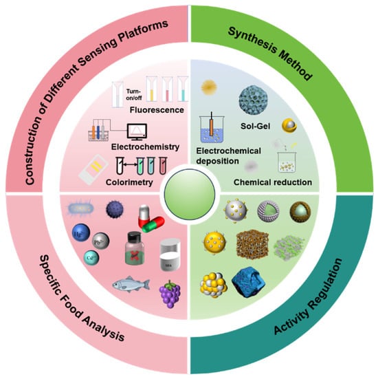
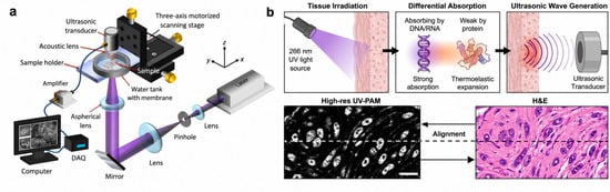
Chronic diseases such as cardiovascular disorders, diabetes, neurological conditions, and kidney disease continue to rise worldwide. These conditions create a growing demand for continuous, non-invasive, and personalized health monitoring technologies. Wearable biosensors meet this need by enabling real-time physiological and biochemical measurements outside traditional clinical settings. Among wearable biosensors, those based on biofluids like sweat, tears, and saliva provide a painless alternative to blood sampling. These fluids also grant access to metabolites, electrolytes, hormones, proteins, and disease related biomarkers that reflect systemic health status. Advanced sensing technology allow us to continuously track health status by analyzing key biomarkers in these accessible biofluids. This review summarizes recent advances in non-invasive wearable biosensors and focuses on their sensing principles which includes biorecognition elements, signal transduction mechanisms, and data acquisition strategies. We also discussed key sensing modalities, including electrochemical, optical, thermal, and piezoelectric approaches, highlighting their advantages for wearable integration and performance in biofluid sensing. Finally the review also outlines recent developments and applications of these systems in biofluid sensing. In the end we highlights existing challenges, potential solutions, and future directions toward clinically deployable, AI-assisted precision healthcare systems.
Full article
(This article belongs to the Special Issue Latest Wearable Biosensors—2nd Edition)
►
Show Figures

Figure 1


