


Dent. J., Volume 13, Issue 7 (July 2025) – 57 articles
Cover Story (view full-size image):
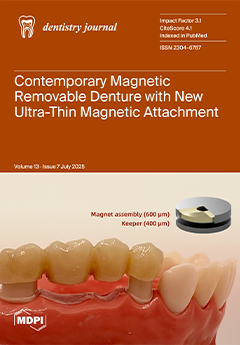
Magnetic attachments have been utilized to improve the retention of removable dentures due to their automatic positioning, easy maintenance, and other advantages. However, they require sufficient occlusal space and involve time-consuming fabrication procedures. This study evaluated the retentive force and durability of a newly developed ultra-thin magnetic attachment (UTMA), approximately 50% thinner than conventional types, applied to a magnet-retained telescopic partial denture. The prosthesis was fabricated entirely using a digital workflow, which simplified clinical and laboratory procedures and provided constant retentive force and stability after repeated insertion–removal cycles. View this paper
- Issues are regarded as officially published after their release is announced to the table of contents alert mailing list.
- You may sign up for e-mail alerts to receive table of contents of newly released issues.
- PDF is the official format for papers published in both, html and pdf forms. To view the papers in pdf format, click on the "PDF Full-text" link, and use the free Adobe Reader to open them.
Previous Issue
Next Issue