Next Issue
Volume 13, July
Previous Issue
Volume 13, May


Dent. J., Volume 13, Issue 6 (June 2025) – 49 articles

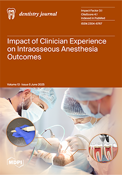
Cover Story (view full-size image): Intraosseous anesthesia (IOA) is gaining recognition as a reliable alternative to traditional mandibular techniques, yet concerns remain about its sensitivity. This retrospective cohort study explored whether clinician experience impacts IOA effectiveness, comfort, and safety during endodontic treatment. Our findings showed no significant difference between experienced endodontists and residents, supporting the broader clinical adoption of IOA regardless of practitioner experience and encouraging its integration into standard training programs. View this paper
  • Issues are regarded as officially published after their release is announced to the table of contents alert mailing list.
  • You may sign up for e-mail alerts to receive table of contents of newly released issues.
  • PDF is the official format for papers published in both, html and pdf forms. To view the papers in pdf format, click on the "PDF Full-text" link, and use the free Adobe Reader to open them.
Order results
Result details
Section
Select all
Export citation of selected articles as:
11 pages, 1189 KiB  
Article
Segmentation of Pulp and Pulp Stones with Automatic Deep Learning in Panoramic Radiographs: An Artificial Intelligence Study
by Mujgan Firincioglulari, Mehmet Boztuna, Omid Mirzaei, Tolgay Karanfiller, Nurullah Akkaya and Kaan Orhan
Dent. J. 2025, 13(6), 274; https://doi.org/10.3390/dj13060274 - 19 Jun 2025
Viewed by 403
Abstract
Background/Objectives: Different sized calcified masses called pulp stones are often detected in dental pulp and can impact dental procedures. The current research was conducted with the aim of measuring the ability of artificial intelligence algorithms to accurately diagnose pulp and pulp stone [...] Read more.
Background/Objectives: Different sized calcified masses called pulp stones are often detected in dental pulp and can impact dental procedures. The current research was conducted with the aim of measuring the ability of artificial intelligence algorithms to accurately diagnose pulp and pulp stone calcifications on panoramic radiographs. Methods: We used 713 panoramic radiographs, on which a minimum of one pulp stone was detected, identified retrospectively, and included in the study—4675 pulp stones and 5085 pulps were marked on these radiographs using CVAT v1.7.0 labeling software. Results: In the test dataset, the AI model segmented 462 panoramic radiographs for pulp stone and 220 panoramic radiographs for pulp. The dice coefficient and Intersection over Union (IoU) recorded for the Pulp Segmentation model were 0.84 and 0.758, respectively. Precision and recall were computed to be 0.858 and 0.827, respectively. The Pulp Stone Segmentation model achieved a dice coefficient of 0.759 and an IoU of 0.686, with precision and recall of 0.792 and 0.773, respectively. Conclusions: Pulp and pulp stones can successfully be identified using artificial intelligence algorithms. This study provides evidence that artificial intelligence software using deep learning algorithms can be valuable adjunct tools in aiding clinicians in radiographic diagnosis. Further research in which larger datasets are examined are needed to enhance the capability of artificial intelligence models to make accurate diagnoses. Full article
Show Figures

Figure 1

25 pages, 744 KiB  
Review
Effectiveness of Irrigation Protocols in Endodontic Therapy: An Umbrella Review
by Manuel J. Orozco-Gallego, Eliana L. Pineda-Vélez, Wilder J. Rojas-Gutiérrez, Martha L. Rincón-Rodríguez and Andrés A. Agudelo-Suárez
Dent. J. 2025, 13(6), 273; https://doi.org/10.3390/dj13060273 - 18 Jun 2025
Viewed by 859
Abstract
Background: With the inclusion of evidence-based dentistry, numerous systematic reviews (SRs) and meta-analyses (MAs) have been conducted in endodontics with the best available scientific evidence to improve diagnosis and treatment. Objective: To synthesize the scientific evidence on the effectiveness of irrigation protocols in [...] Read more.
Background: With the inclusion of evidence-based dentistry, numerous systematic reviews (SRs) and meta-analyses (MAs) have been conducted in endodontics with the best available scientific evidence to improve diagnosis and treatment. Objective: To synthesize the scientific evidence on the effectiveness of irrigation protocols in endodontic therapy. Methods: Following the umbrella review methodology (UR), a comprehensive literature search was conducted using scientific and grey literature databases. A quality evaluation and a descriptive analysis of the included SRs and MAs were conducted. Quantitative comparability between MAs was carried out. Results: Four descriptive SRs and nine MAs were included. Eight articles evidenced high methodological quality. Studies showed the effectiveness and efficacy depending on the study design, the findings of primary clinical trials, and factors related to the type of irrigant, concentration, volume, and irrigation systems. Variability between irrigants and protocols was observed. Follow-up periods extend from hours to years, and there were different study samples. SRs and MAs evidenced limitations regarding methodological aspects. Low overlap of the primary studies was found. Quantitative analyses indicated greater efficacy in microbial reduction and apical healing in favor of passive ultrasonic irrigation (PUI; RD −0.15; 95% CI −0.28, −0.01; p = 0.03; I2 = 60%; RD −0.09; 95% CI −0.16, −0.02; p = 0.01; I2 = 0%, respectively). Conclusions: This UR highlights the importance of root canal disinfection, emphasizing sodium hypochlorite (NaOCl) as the primary irrigant. Enhanced activation methods, such as PUI and lasers, improve irrigant efficiency, while alternatives like chlorhexidine (CHX) offer better biocompatibility. Standardized protocols and evidence-based clinical guidelines are needed. PROSPERO register: CRD42023409044. Full article
(This article belongs to the Special Issue Endodontics: From Technique to Regeneration)
Show Figures

Figure 1

9 pages, 859 KiB  
Article
Fourier-Transform Infrared Spectroscopy Analysis of 3D-Printed Dental Resins Reinforced with Yttria-Stabilized Zirconia Nanoparticles
by Andrea Izabella Borș
Dent. J. 2025, 13(6), 272; https://doi.org/10.3390/dj13060272 - 18 Jun 2025
Viewed by 284
Abstract
Background/Objectives: This study investigates the chemical structure and molecular interactions in 3D-printed dental resins reinforced with varying concentrations of Yttria-Stabilized Zirconia (YSZ) nanoparticles, using Fourier-Transform Infrared Spectroscopy (FTIR) to assess the compatibility and bonding behavior at the molecular level. Methods: Three groups of [...] Read more.
Background/Objectives: This study investigates the chemical structure and molecular interactions in 3D-printed dental resins reinforced with varying concentrations of Yttria-Stabilized Zirconia (YSZ) nanoparticles, using Fourier-Transform Infrared Spectroscopy (FTIR) to assess the compatibility and bonding behavior at the molecular level. Methods: Three groups of 3D-printed methacrylate-based resin discs were fabricated: a control (0% YSZ), and experimental groups reinforced with 1% and 3% YSZ nanoparticles. Samples were produced using Digital Light Processing (DLP) technology and post-processed under standardized conditions. FTIR spectra were collected via ATR mode over a wavenumber range of 4000–600 cm−1. Spectral differences at key wavenumbers (1721.16, 1237.11, and 929.62 cm−1) were statistically analyzed using one-way ANOVA and Tukey’s post hoc test. Results: FTIR spectra showed no significant shifts in the ester carbonyl band at 1721.16 cm−1, suggesting the preservation of the core resin matrix. However, a statistically significant increase in absorbance at 1237.11 cm−1 was observed in the 1% YSZ group (p = 0.034), indicating dipolar interaction. A distinct new peak at 929.62 cm−1, corresponding to Zr–O vibrations, emerged in the 3% YSZ group (p = 0.002), confirming successful nanoparticle integration. Conclusions: YSZ nanoparticles enhance specific molecular interactions within methacrylate-based dental resins without compromising structural integrity. These findings support the potential application of YSZ-reinforced 3D-printed resins in durable, biocompatible permanent dental restorations. Full article
(This article belongs to the Special Issue Feature Papers in Digital Dentistry)
Show Figures

Figure 1

14 pages, 912 KiB  
Article
Evaluation of Large Language Model Performance in Answering Clinical Questions on Periodontal Furcation Defect Management
by Georgios S. Chatzopoulos, Vasiliki P. Koidou, Lazaros Tsalikis and Eleftherios G. Kaklamanos
Dent. J. 2025, 13(6), 271; https://doi.org/10.3390/dj13060271 - 18 Jun 2025
Viewed by 408
Abstract
Background/Objectives: Large Language Models (LLMs) are artificial intelligence (AI) systems with the capacity to process vast amounts of text and generate human-like language, offering the potential for improved information retrieval in healthcare. This study aimed to assess and compare the evidence-based potential [...] Read more.
Background/Objectives: Large Language Models (LLMs) are artificial intelligence (AI) systems with the capacity to process vast amounts of text and generate human-like language, offering the potential for improved information retrieval in healthcare. This study aimed to assess and compare the evidence-based potential of answers provided by four LLMs to common clinical questions concerning the management and treatment of periodontal furcation defects. Methods: Four LLMs—ChatGPT 4.0, Google Gemini, Google Gemini Advanced, and Microsoft Copilot—were used to answer ten clinical questions related to periodontal furcation defects. The LLM-generated responses were compared against a “gold standard” derived from the European Federation of Periodontology (EFP) S3 guidelines and recent systematic reviews. Two board-certified periodontists independently evaluated the answers for comprehensiveness, scientific accuracy, clarity, and relevance using a predefined rubric and a scoring system of 0–10. Results: The study found variability in LLM performance across the evaluation criteria. Google Gemini Advanced generally achieved the highest average scores, particularly in comprehensiveness and clarity, while Google Gemini and Microsoft Copilot tended to score lower, especially in relevance. However, the Kruskal–Wallis test revealed no statistically significant differences in the overall average scores among the LLMs. Evaluator agreement and intra-evaluator reliability were high. Conclusions: While LLMs demonstrate the potential to answer clinical questions related to furcation defect management, their performance varies. LLMs showed different comprehensiveness, scientific accuracy, clarity, and relevance degrees. Dental professionals should be aware of LLMs’ capabilities and limitations when seeking clinical information. Full article
(This article belongs to the Special Issue Artificial Intelligence in Oral Rehabilitation)
Show Figures

Graphical abstract

11 pages, 1820 KiB  
Article
Establishment of Dental Pulp Cell Culture System for Analyzing Dentinogenesis in Mouse Incisors
by Yuka Kato, Insoon Chang and Satoshi Yokose
Dent. J. 2025, 13(6), 270; https://doi.org/10.3390/dj13060270 - 18 Jun 2025
Viewed by 275
Abstract
Background: The dentin–pulp complex plays a vital role in tooth health. Dentin forms the main body the tooth and continues to form throughout life to maintain homeostasis and provide protection against deleterious external stimuli. However, the detailed mechanism of dentin formation remains [...] Read more.
Background: The dentin–pulp complex plays a vital role in tooth health. Dentin forms the main body the tooth and continues to form throughout life to maintain homeostasis and provide protection against deleterious external stimuli. However, the detailed mechanism of dentin formation remains poorly understood, and there is a need for new regenerative therapies. This study therefore investigated whether primary dental pulp cells from mice could be used to establish a new culture system. Methods: Mouse mandibles were divided along the ramus to extract dental pulp tissue containing cervical loops. The extracted tissue was cultured in an incubator to promote cell out-growth and increase the number of cells available for experimentation. Results: Cultured cells formed mineralized nodules, confirmed by Alizarin red S staining. The expression levels of dentin sialo protein, bone gamma-carboxyglutamate protein, and type I collagen mRNAs in cultured dental pulp cells on day 15 were lower than those in intact mouse dental pulp tissue, and the expression of all mRNAs was confirmed through electrophoresis. Conclusions: This study established a primary culture system using dental pulp tissue extracted from mouse mandibular incisors. The results demonstrated that dental pulp cells can differentiate into odontoblast-like cells and form dentin-like mineralized nodules, thereby offering a useful system for studying dentin formation and odontoblast differentiation. Full article
Show Figures

Figure 1

21 pages, 303 KiB  
Review
Cytotoxicity and Endocrine Disruption in Materials Used for Removable Orthodontic Retainers: A Comprehensive Review
by Katarzyna Chojnacka and Marcin Mikulewicz
Dent. J. 2025, 13(6), 269; https://doi.org/10.3390/dj13060269 - 17 Jun 2025
Viewed by 510
Abstract
Objective: To evaluate the cytotoxicity and endocrine-disrupting potential of materials used in removable orthodontic retainers. Methods: A literature search (2015–2025) covered in vitro cytotoxicity, estrogenicity, in vivo tissue responses, and clinical biomarkers in PMMA plates, thermoplastic foils, 3D-printed resins, PEEK, and fiber-reinforced composites. [...] Read more.
Objective: To evaluate the cytotoxicity and endocrine-disrupting potential of materials used in removable orthodontic retainers. Methods: A literature search (2015–2025) covered in vitro cytotoxicity, estrogenicity, in vivo tissue responses, and clinical biomarkers in PMMA plates, thermoplastic foils, 3D-printed resins, PEEK, and fiber-reinforced composites. Results: Thirty-eight in vitro and ten clinical studies met inclusion criteria, identified via a structured literature search of electronic databases (2015–2025). Photopolymer resins demonstrated the highest cytotoxicity, whereas thermoplastics and PMMA exhibited predominantly mild effects, which diminished further following 24 h water storage. Bisphenol-type compound release was reported, but systemic exposure remained below regulatory limits. No statistically significant mucosal alterations or endocrine-related effects were reported in clinical studies. Conclusions: Retainer materials are generally biocompatible, though data on long-term endocrine effects are limited. Standardized biocompatibility assessment protocols are necessary to enable comparative evaluation across diverse orthodontic materials. Single-use thermoplastics contribute to microplastic release and pose end-of-life management challenges, raising concerns regarding environmental sustainability. Full article
(This article belongs to the Special Issue Dental Materials Design and Innovative Treatment Approach)
18 pages, 490 KiB  
Article
Final-Year Dental Students’ Perceived Confidence: Competencies in General Dentistry
by Navodika Yaparathna, Iresha Udayamalee, Megan Gray, Cheree He, Rachel Wu, Chelsea Taing and Menaka Abuzar
Dent. J. 2025, 13(6), 268; https://doi.org/10.3390/dj13060268 - 16 Jun 2025
Viewed by 327
Abstract
Background: Competency in providing high-quality, comprehensive patient care is essential for newly qualified dentists. Dental curricula are designed to equip graduates with necessary skills to develop competencies. Outplacement training has been incorporated into most dental curricula to provide broader clinical experience in a [...] Read more.
Background: Competency in providing high-quality, comprehensive patient care is essential for newly qualified dentists. Dental curricula are designed to equip graduates with necessary skills to develop competencies. Outplacement training has been incorporated into most dental curricula to provide broader clinical experience in a real-world situation. Methods: This cross-sectional study aimed to assess (1) the perceived confidence of final-year dental students (FYDSs) at an Australian university with reference to the Australian Dental Council (ADC) professional competencies for newly qualified dentists; (2) the association between perceived confidence and the timing of outplacement training; and (3) students’ perceptions on outplacement training in developing their competencies. Two online surveys were administered to a cohort of FYDSs at the end of the first and second trimesters. ‘Survey 1’ was based on the ADC competency requirements (2022) and assessed aims 1 and 2. The five domains assessed were (1) communication and leadership; (2) critical thinking; (3) health promotion; (4) scientific and clinical knowledge; and (5) person-centred care. ‘Survey 2’ assessed students’ perception on outplacement training and was administered towards the completion of their outplacement to assess aim 3. Results: Students’ perceived competency levels were high across all domains. Those with prior tertiary education were more confident in communication and leadership, health promotion, and scientific and clinical knowledge than students with secondary qualifications. The perceived confidence in professional competencies among FYDSs had no significant association (p > 0.05) with the location or the sequence of outplacement. The thematic analysis of survey 2 responses reported the guidance and constructive feedback received from supervisors while managing cases in a real-life setup as a significant contributor to their confidence development. Conclusions: FYDSs reported a satisfactory level of perceived confidence in professional competencies. Both in-house training and outplacement equally improve the levels of competency development. FYDSs perceive outplacement training as a positive integral component in the development of skills. Full article
(This article belongs to the Special Issue Dental Education: Innovation and Challenge)
Show Figures

Figure 1

16 pages, 3402 KiB  
Article
A New Classification of Inferior Alveolar Nerve Repositioning Procedures for Dental Implant Placement
by Fares Kablan
Dent. J. 2025, 13(6), 267; https://doi.org/10.3390/dj13060267 - 16 Jun 2025
Viewed by 404
Abstract
Background: Tooth loss significantly impacts the quality of life for adults. Inferior alveolar nerve (IAN) repositioning has garnered interest as a treatment for facilitating dental implant placement in the severely atrophic posterior mandible. However, there remains a need for standardization and classification [...] Read more.
Background: Tooth loss significantly impacts the quality of life for adults. Inferior alveolar nerve (IAN) repositioning has garnered interest as a treatment for facilitating dental implant placement in the severely atrophic posterior mandible. However, there remains a need for standardization and classification of these techniques to improve outcomes. This study aims to propose a new clinical classification system for IAN repositioning procedures based on anatomical and procedural parameters. Methods: This study retrospectively analyzed preoperative radiographic records and surgical procedure documents over a 15-year period (2008–2023) for patients who underwent implant placement combined with IAN repositioning in the posterior atrophic mandible. Cases were classified into four categories according to bone availability, nerve location, and type of surgical intervention. Results: The study analyzed 142 edentulous posterior mandibles in 105 patients (77 women, 28 men; age range: 20–75). The cases were divided into four categories: Category 1 (58 patients, 78 sites), treated with one surgery; Category 2 (15 patients, 15 sites), treated in two stages; Category 3 (20 patients, 25 sites); and Category 4 (12 patients, 24 sites), with Categories 3 and 4 treated in a single surgery. Across all 132 sites, 411 dental implants were placed and restored with implant-supported fixed prostheses. Conclusions: This proposed classification provides a structured systematic framework for assessing and planning IAN repositioning procedures. It facilitates better diagnosis, treatment planning, and prediction of surgical stages in patients needing IAN repositioning for dental implant placement. Full article
(This article belongs to the Section Oral and Maxillofacial Surgery)
Show Figures

Figure 1

16 pages, 1858 KiB  
Article
Identifying Gaps in Predoctoral Craniofacial Education
by Catherine Bingham, Linda Sangalli, Kathryn Preston, Poojan Shrestha and Caroline M. Sawicki
Dent. J. 2025, 13(6), 266; https://doi.org/10.3390/dj13060266 - 16 Jun 2025
Viewed by 284
Abstract
Background/Objectives: It is essential that dental school graduates are adequately prepared to provide care to patients with craniofacial differences (PCD). This study aimed to identify potential educational deficiencies in predoctoral dental school curricula regarding the management of PCD. Methods: An electronic survey was [...] Read more.
Background/Objectives: It is essential that dental school graduates are adequately prepared to provide care to patients with craniofacial differences (PCD). This study aimed to identify potential educational deficiencies in predoctoral dental school curricula regarding the management of PCD. Methods: An electronic survey was distributed to predoctoral dental students across the United States. The 20-item questionnaire assessed students’ educational experiences, clinical encounters, and perceived knowledge and confidence in managing PCD. Results: The most taught didactic topic was diagnostic characteristics of craniofacial differences (77.1%), followed by psychosocial challenges (43.0%) and treatment/referral (36.3%). Respondents reported low levels of understanding and confidence in managing craniofacial conditions, with the lowest confidence in providing surgical treatment (30.1 ± 27.9) and the highest in referrals and communication (41.7 ± 30.1, on a 0–100 scale). Logistic regression showed that overall understanding was a significant predictor of confidence, increasing odds by 8% (OR = 1.08, 95% CI 1.05, 1.12). Participants noted that hands-on clinical experience would most improve their confidence in managing PCD. Conclusions: Predoctoral dental students exhibit low confidence and understanding in managing PCD. Incorporating more targeted craniofacial education, particularly hands-on clinical experience, into the curriculum is essential to better prepare dental graduates for craniofacial care. Full article
(This article belongs to the Special Issue Dental Education: Innovation and Challenge)
Show Figures

Figure 1

11 pages, 661 KiB  
Article
Histological Evaluation of Oral Soft Tissue Biopsy by Dual-Wavelength Diode Laser: An Ex Vivo Study
by Daniele Pergolini, Alessandro Del Vecchio, Mohamed Mohsen, Veronica Cerullo, Cinzia Angileri, Eduardo Troiani, Paolo Visca, Barbara Antoniani, Umberto Romeo and Gaspare Palaia
Dent. J. 2025, 13(6), 265; https://doi.org/10.3390/dj13060265 - 13 Jun 2025
Viewed by 296
Abstract
Background: Diode lasers are valuable in oral surgery due to their excellent hemostasis, minimum post-operative pain, and minimally invasive procedures. A dual-wavelength diode laser in dentistry combines two distinct wavelengths, typically 450 nm and 808 nm, to provide a versatile approach to soft [...] Read more.
Background: Diode lasers are valuable in oral surgery due to their excellent hemostasis, minimum post-operative pain, and minimally invasive procedures. A dual-wavelength diode laser in dentistry combines two distinct wavelengths, typically 450 nm and 808 nm, to provide a versatile approach to soft tissue procedures. This ex vivo study investigated the quantity of thermal effects of a dual-wavelength diode laser on porcine lingual mucosa to determine the optimal laser parameters for oral soft tissue biopsies and to improve the reliability of histological evaluation. The presence of thermal damage in the prelesional margins may compromise the diagnostic accuracy, particularly in cases of suspected malignancy. Methods: Thirty-six porcine lingual mucosa samples were excised using a diode laser (Wiser 3, Doctor Smile) in continuous wave (CW) and pulsed wave (PW) modes at average powers of 2, 3, and 4 W. The samples, preserved in 5% buffered formalin, underwent histological evaluation to measure epithelial and connective tissue damage. Results: The study demonstrated variable thermal effects depending on the laser mode and power settings. Minimal epithelial damage (0.62 mm) was observed at 2 W CW, while maximum damage (3.12 mm) occurred at 4 W pulsed wave (PW). Connective tissue exhibited slightly greater damage than epithelial tissue, with minimal damage (0.53 mm) at 4 W CW and maximum damage (3.19 mm) at 4 W pulsed wave (PW). Statistical analyses were performed using t-tests and ANOVA and revealed significant differences in tissue damage between certain groups, highlighting the impact of laser parameters on thermal effects. Conclusions: The dual-wavelength diode laser seems to have good surgical properties and is suitable for managing complex clinical cases. Although the low power average showed minimal thermal damage, for the importance of the diagnosis of suspected lesions of malignancy, a 2 mm prelesional margin should be maintained. Full article
Show Figures

Figure 1

20 pages, 702 KiB  
Systematic Review
The Effectiveness and Complication Rate of Resorbable Biopolymers in Oral Surgery: A Systematic Review
by Riccardo Fabozzi, Francesco Bianchetti, Domenico Baldi, Catherine Yumang Sanchez, Francesco Bagnasco and Nicola De Angelis
Dent. J. 2025, 13(6), 264; https://doi.org/10.3390/dj13060264 - 13 Jun 2025
Viewed by 834
Abstract
Background: Resorbable biopolymers are increasingly explored for use in regenerative procedures within dental surgery. Their ability to degrade naturally, minimize surgical reinterventions, and potentially reduce immunogenicity makes them appealing in guided bone and tissue regeneration applications. However, despite these advantages, uncertainties persist [...] Read more.
Background: Resorbable biopolymers are increasingly explored for use in regenerative procedures within dental surgery. Their ability to degrade naturally, minimize surgical reinterventions, and potentially reduce immunogenicity makes them appealing in guided bone and tissue regeneration applications. However, despite these advantages, uncertainties persist regarding their comparative effectiveness and associated risks. For example, polyethylene glycol (PEG)-based membranes have shown comparable outcomes to porcine-derived collagen membranes in bone regeneration procedures, yet studies have reported a higher incidence of soft tissue healing complications associated with PEG-based materials. Similarly, while polycaprolactone (PCL) and dextrin-based hydrogels have demonstrated promising clinical handling and bone fill capabilities, their long-term performance and consistency across different anatomical sites remain under investigation. These findings highlight the need for further well-powered clinical trials to establish standardized guidelines for their safe and effective use. Methods: A systematic review protocol was registered with the PROSPERO database and developed in alignment with PRISMA guidelines. Database searches were conducted in PubMed, Medline, Scopus, and Cochrane from June to December 2024. Only randomized controlled trials (RCTs) focusing on synthetic resorbable biopolymers in bone augmentation procedures were considered. Bias was evaluated using the Cochrane Risk of Bias tool. Results: Eleven RCTs were included, totaling 188 patients. The findings suggest that materials such as polylactic acid (PLA), polycaprolactone (PCL), and polyethylene glycol (PEG) contributed effectively to new bone formation. PEG-based membranes were found to perform on par with or occasionally better than traditional collagen membranes derived from porcine sources. Additionally, the application of 3D-printable polymers demonstrated promise in site-specific healing. Conclusions: Resorbable biopolymers are effective and safe for GBR procedures, with clinical outcomes comparable to traditional materials. Advances in 3D-printing technology and bioactive coatings may further enhance their regenerative potential. However, the incidence of soft tissue healing complications suggests the need for further long-term studies to optimize material properties and clinical application. Full article
(This article belongs to the Special Issue Dental Materials Design and Innovative Treatment Approach)
Show Figures

Figure 1

17 pages, 3792 KiB  
Article
Influence of Cement Thickness, Dentine Thickness, and Intracoronal Depth on the Fracture Resistance of 3D-Printed Endocrowns: A Pilot In Vitro Study
by Osama Abuabboud, Adrian-George Marinescu, Mihai Paven, Izabella-Maria Kovacs, Luminita Maria Nica, Andrei-Bogdan Faur, Dan Ioan Stoia and Anca Jivănescu
Dent. J. 2025, 13(6), 263; https://doi.org/10.3390/dj13060263 - 12 Jun 2025
Viewed by 902
Abstract
Background/Objectives: Endodontically treated molars are structurally weakened due to internal tissue loss, increasing their risk of fracture. Endocrowns, developed as a conservative alternative to post–core systems, have gained popularity with the rise of digital dentistry, CAD/CAM workflows, and 3D-printed restorations. In this context, [...] Read more.
Background/Objectives: Endodontically treated molars are structurally weakened due to internal tissue loss, increasing their risk of fracture. Endocrowns, developed as a conservative alternative to post–core systems, have gained popularity with the rise of digital dentistry, CAD/CAM workflows, and 3D-printed restorations. In this context, the aim of the present pilot study was to investigate the influence of cement layer thickness, intracoronal depth, and dentine wall thickness on the fracture resistance of molars restored with 3D-printed endocrowns. Methods: Twelve extracted human molars were endodontically treated and restored with endocrowns fabricated from a 3D-printed resin material, SprintRay CrownTM (SprintRay Inc. Los Angeles, CA, USA), via masked stereolithography (MSLA) on a Prusa SL1 printer. Cementation was performed using RelyX Universal Resin Cement (3M, Maplewood, MN USA). Cone beam computed tomography (CBCT) was used to measure the dentine thickness and intracoronal depth before cementation and cement thickness after cementation. The fracture resistance was evaluated using a universal testing machine. For each variable (Td, Dp, Tc), the 12 specimens were divided into two groups (n = 6). Statistical analysis included Pearson correlation, a one-way ANOVA, and the non-parametric Mann–Whitney U test. Results: Within the limitations of this pilot in vitro study, cement thickness demonstrated a strong positive correlation with fracture resistance (r = 0.577) and was the only variable showing statistical significance in the ANOVA (F = 7.847, p = 0.019). In contrast, intracoronal depth and dentine wall thickness exhibited weaker and nonsignificant correlations. No significant mechanical advantage was observed from increasing the pulp chamber depth or peripheral dentine wall thickness. This result was further supported by nonparametric Mann–Whitney U testing (p = 0.015). Conclusions: Cement layer thickness is a key biomechanical factor influencing the fracture resistance of endocrown restorations. Preparation depth and dentine wall geometry appear to have a less direct impact. Full article
(This article belongs to the Special Issue Innovative Materials in Dental Prosthetics)
Show Figures

Graphical abstract

10 pages, 1087 KiB  
Article
Influence of the Adaptive Torque Control Motion on the Ability of Neolix EDMax to Reach Working Length When Used as a Single Shaping File—An In Vitro Study
by Vlad Mircea Lup, Carlo Gaeta, Ashkan Tavakkoli, Andreas Louloudiadis, Simone Grandini and Gabriela Ciavoi
Dent. J. 2025, 13(6), 262; https://doi.org/10.3390/dj13060262 - 12 Jun 2025
Viewed by 301
Abstract
Objectives: The aim of this study is to investigate how Adaptive Torque Control motion influences the shaping efficiency of Neolix EDMax (Neolix SAS, Évron, France) and its ability to reach working length with or without a pre-existing glide path. Methods: A total of [...] Read more.
Objectives: The aim of this study is to investigate how Adaptive Torque Control motion influences the shaping efficiency of Neolix EDMax (Neolix SAS, Évron, France) and its ability to reach working length with or without a pre-existing glide path. Methods: A total of 90 endo training blocks with an S-shape curvature were divided into three groups based on the kinematics and preparation phase: the control group, where the Neolix EDMax (Neolix SAS, Évron, France) was used for shaping after a glide path was established; the no glide path group, where the Neolix EDMax (Neolix SAS, Évron, France) was used for shaping without a glide path; and the Adaptive Torque Control group, where the Neolix EDMax (Neolix SAS, Évron, France) was used for shaping without a glide path but in an Adaptive Torque Control motion. The time for shaping, the instrument passes, and the ability to reach working length were recorded and analyzed using a one-way Anova and Tukey’s HSD post hoc test. Results: Establishing a glide path helped the shaping file to reach working length faster and in fewer passes when compared with the no glide path group, but the Adaptive Torque Control group was able to perform even better than the control group despite not having a pre-established glide path. Conclusions: The Adaptive Torque Control motion on continuous rotation instruments does impact their performance. Combining the efficiency of continuous rotation and the safety of reciprocation, this type of motion had a significant effect on the ability to shape the simulated root canal even in the presence of a double curvature and without a pre-established glide path. Full article
(This article belongs to the Special Issue Endodontics: From Technique to Regeneration)
Show Figures

Figure 1

14 pages, 826 KiB  
Systematic Review
Current Applications of Chatbots Powered by Large Language Models in Oral and Maxillofacial Surgery: A Systematic Review
by Vincenzo Ronsivalle, Simona Santonocito, Umberto Cammarata, Eleonora Lo Muzio and Marco Cicciù
Dent. J. 2025, 13(6), 261; https://doi.org/10.3390/dj13060261 - 11 Jun 2025
Viewed by 449
Abstract
Background/Objectives: In recent years, interest has grown in the clinical applications of artificial intelligence (AI)-based chatbots powered by large language models (LLMs) in oral and maxillofacial surgery (OMFS). However, there are conflicting opinions regarding the accuracy and reliability of the information they provide, [...] Read more.
Background/Objectives: In recent years, interest has grown in the clinical applications of artificial intelligence (AI)-based chatbots powered by large language models (LLMs) in oral and maxillofacial surgery (OMFS). However, there are conflicting opinions regarding the accuracy and reliability of the information they provide, raising questions about their potential role as support tools for both clinicians and patients. This systematic review aims to analyze the current literature on the use of conversational agents powered by LLMs in the field of OMFS. Methods: The review was conducted following PRISMA guidelines and the Cochrane Handbook for Systematic Reviews of Interventions. Original studies published between 2023 and 2024 in peer-reviewed English-language journals were included. Sources were identified through major electronic databases, including PubMed, Scopus, Google Scholar, and Web of Science. The risk of bias in the included studies was assessed using the ROBINS-I tool, which evaluates potential bias in study design and conduct. Results: A total of 49 articles were identified, of which 4 met the inclusion criteria. One study showed that ChatGPT provided the most accurate responses compared to Microsoft Copilot (ex-Bing) and Google Gemini (ex-Bard) for questions related to OMFS. Other studies highlighted that ChatGPT-4 can assist surgeons with quick and relevant information, though responses may vary depending on the quality of the questions. Conclusions: Chatbots powered by LLMs can enhance efficiency and decision-making in OMFS routine clinical cases. However, based on the limited number of studies included in this review (four), their performance remains constrained in complex clinical scenarios and in managing emotionally sensitive patient interactions. Further research on clinical validation, prompt formulation, and ethical oversight is essential to safely integrating LLM technologies into OMFS practices. Full article
(This article belongs to the Special Issue Artificial Intelligence in Oral Rehabilitation)
Show Figures

Graphical abstract

14 pages, 352 KiB  
Review
Comparative Analysis of Fully Guided and Free-Hand Orthognathic Surgery: Advancements, Precision, and Clinical Outcomes
by Sophia Tsokkou, Ioannis Konstantinidis, Antonios Keramas, Georgios Kiosis, Kanellos Skourtsidis, Danai Alexiou, Georgia-Nektaria Keskesiadou, Sofia Karachrysafi, Theodora Papamitsou and Ioannis Chatzistefanou
Dent. J. 2025, 13(6), 260; https://doi.org/10.3390/dj13060260 - 11 Jun 2025
Viewed by 407
Abstract
Background/Objectives: Orthognathic surgery has evolved with digital advancements, improving precision and predictability. Traditional free-hand techniques rely on surgeon expertise, often leading to variable outcomes. Fully guided approaches integrate computer-assisted surgery, including virtual surgical planning (VSP), CAD/CAM, and dynamic navigation, enhancing accuracy and [...] Read more.
Background/Objectives: Orthognathic surgery has evolved with digital advancements, improving precision and predictability. Traditional free-hand techniques rely on surgeon expertise, often leading to variable outcomes. Fully guided approaches integrate computer-assisted surgery, including virtual surgical planning (VSP), CAD/CAM, and dynamic navigation, enhancing accuracy and efficiency. This review compares these approaches, assessing their impact on surgical accuracy, efficiency, and patient outcomes. Methods: A scoping review was conducted across PubMed, MEDLINE, Scopus, Cochrane Library, and Embase databases, focusing on clinical trials and cohort studies. Key parameters analyzed include surgical precision, operative efficiency, complication rates, and functional/aesthetic results. Results: Fully guided techniques achieve sub-millimetric accuracy with mean length deviations ranging from 1.3 mm to 2.4 mm and mean angular deviations between 2.29° and 3.51°. Moreover, these approaches markedly reduce operative time, averaging between 34 min and 1.7 h, and postoperative complications. Digital tools streamline workflow, improving reproducibility and aesthetic outcomes. Free-hand methods remain cost-effective but require greater surgical expertise, often resulting in longer recovery periods and higher variability. Conclusions: Computer-assisted orthognathic surgery enhances precision and efficiency, outperforming free-hand techniques in accuracy and predictability. While free-hand methods remain viable for simpler cases, fully guided approaches optimize surgical execution. Future research should explore hybrid strategies combining digital precision with manual adaptability to further refine surgical techniques. Full article
(This article belongs to the Special Issue Dentistry in the 21st Century: Challenges and Opportunities)
Show Figures

Graphical abstract

24 pages, 34320 KiB  
Case Report
A 10-Year Follow-Up of an Approach to Restore a Case of Extreme Erosive Tooth Wear
by Davide Foschi, Andrea Abate, Francesca Vailati, Ignazio Loi, Cinzia Maspero and Valentina Lanteri
Dent. J. 2025, 13(6), 259; https://doi.org/10.3390/dj13060259 - 10 Jun 2025
Viewed by 1197
Abstract
Background: In recent years, thanks to the improvement of adhesive techniques, patients affected by tooth wear, related to erosion and/or parafunctional habits, can undergo restoration by adding only what has been lost of their dentition (additive approach). However, since not all clinicians are [...] Read more.
Background: In recent years, thanks to the improvement of adhesive techniques, patients affected by tooth wear, related to erosion and/or parafunctional habits, can undergo restoration by adding only what has been lost of their dentition (additive approach). However, since not all clinicians are convinced that dental rehabilitation should be proposed in the early stages of exposed dentin, several treatments are often postponed. It is important to emphasize that, in the early stages, the clinical approach should remain conservative, focusing on dietary counseling, the modification of harmful habits, fluoride application, and risk factor management. Only when these preventive and non-invasive strategies prove insufficient, and the condition continues to progress, should invasive restorative treatments be considered. Unfortunately, epidemiological studies are reporting an increase in the number of young patients affected by erosive tooth wear, and not intercepting these cases earlier could lead to a severe degradation of the affected dentition. In addition, parafunctional habits are also becoming more frequent among patients. The combination of erosion and attrition can be very destructive, and may progress rapidly once dentin is exposed and the risk factors remain unaddressed. The aim of this report was to present a conservative full-mouth rehabilitation approach for severe erosive lesions and to provide a 10-year follow-up assessing the biological, functional, and esthetic outcomes. Methods: In this article, the postponed restorative treatment of a patient, suffering from severe tooth wear, is illustrated. The patient had sought dental treatment in the past; however, due to the already very compromised dentition, a conventional but very aggressive treatment was proposed and refused. Four years later, when the patient finally accepted an alternative conservative therapy, the tooth degradation was very severe, especially at the level of the maxillary anterior teeth. The combination of three different approaches, Speed-Up Therapy, BOPT (Biologically-Oriented Preparation Technique), and the 3 Step Technique, however, improved the capacity to successfully complete the difficult therapeutic task. Results: The biological goals (maintenance of the pulp vitality of all of the teeth and the minimal removal of healthy tooth structure) were accomplished, relying only on adhesive techniques. Conclusions: The overall treatment was very comfortable for the patient and less complicated for the clinician. At 10-year follow-up, biological, functional, and esthetic success was still confirmed. Full article
Show Figures

Figure 1

15 pages, 523 KiB  
Article
Oral Health Disparities in Type 2 Diabetes: Examining the Elevated Risk for Dental Caries—A Comparative Study
by José Frias-Bulhosa, Maria Conceição Manso, Carla Lopes Mota and Paulo Melo
Dent. J. 2025, 13(6), 258; https://doi.org/10.3390/dj13060258 - 10 Jun 2025
Viewed by 486
Abstract
Background/Objectives: Dental caries (DCs) and type 2 diabetes share common risk factors. Dental caries risk in type 2 diabetics (T2DM) shows contradictory results. The aim of this study was to determine if there is a difference in DC prevalence in adults with [...] Read more.
Background/Objectives: Dental caries (DCs) and type 2 diabetes share common risk factors. Dental caries risk in type 2 diabetics (T2DM) shows contradictory results. The aim of this study was to determine if there is a difference in DC prevalence in adults with and without T2DM and whether body mass index (BMI) classes or glycated hemoglobin (HbA1c) levels interfere in that difference. Methods: A total of 666 adults (n(T2DM) = 343; n(nT2DM) = 323), from Espinho Primary Health Care Center, were interviewed by calibrated observers. Data from clinical records were collected and oral health status was registered using WHO criteria. Inference analysis was conducted using non-parametric tests (α = 0.05). Results: A similar caries prevalence was found between the T2DM (98.2%) and nT2DM (98.8%) groups, with the T2DM group showing significantly higher tooth loss (p < 0.001), higher caries experience rerted as mean ± sd (17.7 ± 8.3 vs. 15.9 ± 7.8, p = 0.005), fewer decayed teeth (p < 0.001) and filled teeth (p = 0.016) compared to nT2DM. The most frequently identified comorbidity was hypertension (53.6%). Tobacco use (12.9%) was lower in T2DM (p < 0.001). The restorative and treatment indices indicated a significantly higher proportion of use of oral care services (p < 0.001) in T2DM individuals. The prevalence of the higher classes of BMI indicative of pre-obesity or obesity shows significant differences (p < 0.001). The differences found in the DMFT or each of its components for the prevalence or for the mean in HbA1c control were not statistically significant (p = 0.368, and 0.524, respectively). Conclusions: Adults with T2DM and higher BMI classes could be associated with a greater prevalence of DCs. The glycemic control of T2DM does not significantly influence DMFT score or each of its components. Full article
(This article belongs to the Special Issue Updates and Highlights in Cariology)
Show Figures

Figure 1

10 pages, 233 KiB  
Review
The Reevaluation of Subgingival Calculus: A Narrative Review
by Stephen K. Harrel, Atsutoshi Yoshimura and Charles M. Cobb
Dent. J. 2025, 13(6), 257; https://doi.org/10.3390/dj13060257 - 9 Jun 2025
Viewed by 545
Abstract
Aim: Despite a persistent presence in periodontitis, calculus remains a paradox. This narrative review reevaluates the role of calculus in periodontitis based on in situ, ex vivo, and in vitro studies published over the last two decades. Review: Results from multiple studies argue [...] Read more.
Aim: Despite a persistent presence in periodontitis, calculus remains a paradox. This narrative review reevaluates the role of calculus in periodontitis based on in situ, ex vivo, and in vitro studies published over the last two decades. Review: Results from multiple studies argue for the reconsideration of calculus as an independent risk factor in periodontitis. The results of a human study suggest that calculus contributes more to inflammation than simply serving as a substrate for biofilm accumulation. Ultrastructure studies have revealed residual calculus embedded in cementum following scaling and root planing (SRP). In vitro studies show that calculus particles can stimulate IL-1β secretion via the NLRP3 inflammasome in human and mouse phagocytes, and the crystalline structure is partially responsible for the activation. Other studies indicate that calculus particles may promote bone resorption via IL-1β induction in patients with periodontitis. Further, heat-treated calculus particles and hydroxyapatite crystals induce cell death in epithelial cell lines, suggesting that calculus plays a role in the breakdown of pocket epithelial integrity. Conclusions: Studies have shown that particles of microscopic calculus persist following traditional SRP. In vitro studies report that sterile and calcined calculus particles free of proteinaceous material are cytotoxic to cultured oral epithelial cells. Collectively, these studies suggest that residual microscopic calculus may be a potential risk factor for the failure of periodontal therapy. Full article
(This article belongs to the Special Issue Dentistry in the 21st Century: Challenges and Opportunities)
Show Figures

Graphical abstract

11 pages, 228 KiB  
Article
Comparison of Postoperative Complications and Reoperation Rates of Le Fort I Osteotomies Using Demineralized Bone Matrix (DBM) or Autogenous Bone Grafts in Patients with Orofacial Clefts and Craniofacial Malformations
by Noémi Sipos, Junnu Leikola, Arja Heliövaara, Eeva Kormi and Juho Suojanen
Dent. J. 2025, 13(6), 256; https://doi.org/10.3390/dj13060256 - 9 Jun 2025
Viewed by 392
Abstract
Background: This study aims to evaluate surgical outcomes and compares the prevalence and severity of postoperative complications and reoperations with maxillary osteotomies, focusing on the effectiveness of fixation with demineralized bone matrix (DBM) versus autogenous bone grafts (ABG) in patients with orofacial clefts [...] Read more.
Background: This study aims to evaluate surgical outcomes and compares the prevalence and severity of postoperative complications and reoperations with maxillary osteotomies, focusing on the effectiveness of fixation with demineralized bone matrix (DBM) versus autogenous bone grafts (ABG) in patients with orofacial clefts and craniofacial malformations. Methods: This retrospective cohort study included 138 consecutive patients treated at the Cleft Palate and Craniofacial Center, Helsinki University Hospital, from 2014 to 2022. The cohort consisted of patients with clefts (n = 113), craniosynostosis, and craniofacial syndromes (n = 25). The DBM group (n = 103) received DBX® (Musculoskeletal Transplant Foundation, Edison, NJ, USA), while the ABG group (n = 35) received autogenous bone grafts. Surgical procedures included Le Fort I and bimaxillary osteotomies. Complications involving the maxilla or both jaws were included in the analysis. Both major and minor complications, as well as reoperations, were analyzed and compared. Results: The DBM group had 13.6% of patients with complications, while the ABG group had 20.0%. Reoperation rates were 6.8% for the DBM group and 5.7% for the ABG group. There were no statistically significant differences in complication or reoperation rates between the DBM and ABG groups. Conclusions: The findings suggest that using DBM or ABG in maxillary osteotomies does not significantly affect complication or reoperation rates, supporting DBM as a viable alternative for maxillary surgeries. Full article
(This article belongs to the Special Issue Bone Regeneration and Tissue Reconstruction in Dentistry)
17 pages, 749 KiB  
Article
Unveiling Drug-Drug Interactions in Dental Patients: A Retrospective Real-World Study
by Daiana Colibășanu, Sebastian Mihai Ardelean, Florina-Diana Goldiș, Maria-Medana Drăgoi, Sabina-Oana Vasii, Tamara Maksimović, Șerban Colibășanu, Codruța Șoica and Lucreția Udrescu
Dent. J. 2025, 13(6), 255; https://doi.org/10.3390/dj13060255 - 9 Jun 2025
Viewed by 511
Abstract
Background: Drug-drug interactions (DDIs) are a growing safety concern in dental care, particularly among patients with comorbidities and polypharmacy. However, real-world data (RWD) on the prevalence and severity of DDIs in dental settings remain scarce. Objectives: This study aimed to assess [...] Read more.
Background: Drug-drug interactions (DDIs) are a growing safety concern in dental care, particularly among patients with comorbidities and polypharmacy. However, real-world data (RWD) on the prevalence and severity of DDIs in dental settings remain scarce. Objectives: This study aimed to assess the frequency, severity, and clinical relevance of DDIs in dental patients and to identify age- and comorbidity-related risk patterns. Methods: This retrospective study analyzed a cohort of 105 dental patients, considering demographics, preexisting diseases, dental procedures, and prescribed medications. We examined drug-drug interactions (DDIs) employing the DrugBank Drug Interaction Checker, which yields DDI severity into major, moderate, or minor. Results: 45.7% of patients had preexisting diseases, with cardiovascular diseases most prevalent (19.0%). Higher prevalent dental diagnoses and procedures included apical lesions (47.6%) and tooth extractions (53.3%), suggesting frequent pharmacotherapy exposure. We identified 542 DDIs out of 1332 drug pairs and found 2.3% major, 25.0% moderate, and 13.4% minor, with 59.3% showing no interactions. Key high-risk DDIs included epinephrine with beta-blockers. Fifteen patients aged 31–60 years experienced the most major DDIs of 61.3%, patients ≥ 61 years faced 38.7%, and the 0–30 group had none, highlighting age-specific risks. The higher DDIs burden in the 31–60 age group may reflect better knowledge of the drugs they used and accurate reporting of them. Conclusions: Our retrospective study addresses the paucity of dental DDIs real-world data (RWD) studies, pleading for improved drug reconciliation, systematic screening, and age- and comorbidity-tailored strategies to enhance patient safety. Full article
(This article belongs to the Topic Preventive Dentistry and Public Health)
Show Figures

Figure 1

14 pages, 513 KiB  
Systematic Review
Probiotics and Oral Complications of Antineoplastic Therapy in Head and Neck Cancer: A Systematic Review and Meta-Analysis
by Tanya Pereira-Riveros, Enric Jané-Salas, José López-López, Constanza Saka-Herran, Isabel Linares-Galiana and Teresa Vinuesa-Aumedes
Dent. J. 2025, 13(6), 254; https://doi.org/10.3390/dj13060254 - 6 Jun 2025
Viewed by 1062
Abstract
Background/Objectives: Patients with head and neck cancer frequently develop oral complications such as oral mucositis, infections, necrosis, and periodontal disease among others as a consequence of antineoplastic therapy. It is mainly radiotherapy that promotes oral dysbiosis, favouring the overgrowth of opportunistic microorganisms. Identifying [...] Read more.
Background/Objectives: Patients with head and neck cancer frequently develop oral complications such as oral mucositis, infections, necrosis, and periodontal disease among others as a consequence of antineoplastic therapy. It is mainly radiotherapy that promotes oral dysbiosis, favouring the overgrowth of opportunistic microorganisms. Identifying effective adjunctive strategies to prevent or mitigate these adverse effects is crucial. Recent studies have suggested that probiotics could be used to restore microbial homeostasis and modulate inflammatory responses in the oral cavity. This systematic review and meta-analysis assessed the efficacy of probiotics in alleviating oral complications associated with antineoplastic treatments in this patient population. Methods: A comprehensive search was conducted in PubMed, LILACS, Scopus and the Cochrane Central Register of Controlled Trials, following the PRISMA 2020 guidelines. Only randomised controlled trials (RCTs) were included. Results: Nine eligible RCTs were analysed using a random-effects meta-analysis. Probiotic use was significantly associated with a reduced incidence of severe (grade 3–4) oral mucositis (RR = 0.58; 95% CI: 0.41–0.81). Moderate benefits were also observed in modulating the oral microbiota and reducing levels of pathogenic bacteria and Candida spp. However, no significant improvements were noted in periodontal parameters or plaque indices. Conclusions: Probiotics show promise in the management of oral mucositis, but further well-designed trials are needed to evaluate their broader impact on oral health during cancer therapy. This review is not registered on PROSPERO. Full article
(This article belongs to the Topic Oral Health Management and Disease Treatment)
Show Figures

Figure 1

16 pages, 1251 KiB  
Article
Exploring Global Interest in Propolis, Nanosilver, and Biomaterials: Insights and Implications for Dentistry from Big Data Analytics
by Magdalena Sycińska-Dziarnowska, Liliana Szyszka-Sommerfeld, Krzysztof Woźniak and Gianrico Spagnuolo
Dent. J. 2025, 13(6), 253; https://doi.org/10.3390/dj13060253 - 6 Jun 2025
Viewed by 345
Abstract
Background: The growing demand for innovative biomaterials with antimicrobial properties has driven research into natural and synthetic compounds, such as propolis and nanosilver, known for their antimicrobial efficacy. Methods: This study uses Google Trends data to analyze global search interest in [...] Read more.
Background: The growing demand for innovative biomaterials with antimicrobial properties has driven research into natural and synthetic compounds, such as propolis and nanosilver, known for their antimicrobial efficacy. Methods: This study uses Google Trends data to analyze global search interest in five key terms—propolis, antimicrobial, antibacterial, nanosilver, and biomaterials—over a ten-year period (starting November 2014). The objective is to evaluate temporal variations, quantify correlations between the terms, and explore how external events, such as the COVID-19 pandemic, have influenced public and clinical interest in these topics. Search data were extracted, normalized, and analyzed using multivariate time series methods, including vector autoregression (VAR) modeling, Impulse Response Function (IRF) analysis, and forecast error variance decomposition (FEVD). Stability, causality, and inter-period relationships were assessed using statistical analysis, with results visualized through time series plots and impulse response coefficients. Results: Key findings reveal significant interdependencies between search terms, with surges in one often resulting in immediate or short-term increases in others. Notable trends include a marked increase in COVID-19 interest for nanosilver, propolis, and antibacterial, followed by a return to baseline levels, while antimicrobial maintained a sustained upward trajectory. Biomaterials experienced initial declines but later stabilized at elevated levels. Conclusions: These findings underscore the oscillating nature of public interest in antimicrobial and biomaterial innovations, highlighting opportunities for targeted research and commercialization. By adapting future material development to emerging trends and clinical needs, dentistry can use these insights to develop infection control strategies, improve restorative materials, and deal with persistent challenges such as antimicrobial resistance, peri-implantitis, and tooth caries treatment. Full article
(This article belongs to the Special Issue Dental Materials Design and Innovative Treatment Approach)
Show Figures

Figure 1

18 pages, 552 KiB  
Systematic Review
The Effects of Scan Body Geometry on the Precision and the Trueness of Implant Impressions Using Intraoral Scanners: A Systematic Review
by Roksana Mohajerani, Shirin Djalalinia and Marzieh Alikhasi
Dent. J. 2025, 13(6), 252; https://doi.org/10.3390/dj13060252 - 5 Jun 2025
Viewed by 691
Abstract
Background/Objectives: Accurate implant impressions are critical for capturing the three-dimensional (3D) spatial positioning of implants. Digital workflows using intraoral scanners (IOSs) and scan bodies offer distinct advantages over conventional elastomeric techniques. However, the geometry of scan bodies may influence the precision and trueness [...] Read more.
Background/Objectives: Accurate implant impressions are critical for capturing the three-dimensional (3D) spatial positioning of implants. Digital workflows using intraoral scanners (IOSs) and scan bodies offer distinct advantages over conventional elastomeric techniques. However, the geometry of scan bodies may influence the precision and trueness of IOS-acquired data, and optimal design parameters remain undefined. This systematic review aims to evaluate the effects of scan body geometry on the trueness of digital implant impressions captured using IOSs. Methods: A systematic search was conducted across PubMed, Scopus, EMBASE, Web of Science, the Cochrane Library, and Google Scholar up to 25 February 2025. Eligible studies assessed the impact of scan body geometry on the accuracy of implant-level impressions acquired with IOSs. Study quality was assessed using the Quality Assessment Tool for In Vitro Studies of Dental Materials (QUIN). Results: Twenty-eight studies were included, of which twenty-six were in vitro. The included studies, published between 2020 and 2025, demonstrated that variations in macro- and micro-geometries influenced both linear and angular trueness. Cylindrical designs with optimal dimensions generally outperformed cuboidal or spherical forms. Structural modifications, such as rigid bar extensions and surface facets, often improved scan accuracy. Some hybrid or modified designs performed comparably to conventional scan bodies. According to QUIN, 27 studies were moderate quality and one had high quality. Conclusions: Scan body geometry affected the accuracy of intraoral implant digital impressions. Designs featuring rigid extensions or simplified geometries improve trueness and precision. Further standardized clinical studies are needed to define optimal design features and validate current in vitro findings. Full article
Show Figures

Figure 1

20 pages, 6821 KiB  
Review
Transposed Maxillary Canines: Narrative Review with Clinical Case Report
by Teresa Pinho and Rui Amaral
Dent. J. 2025, 13(6), 251; https://doi.org/10.3390/dj13060251 - 5 Jun 2025
Viewed by 449
Abstract
Permanent maxillary canines are key teeth from both functional and aesthetic perspectives. Tooth transposition, a rare anomaly where two permanent teeth exchange positions within the same quadrant, poses significant diagnostic and therapeutic challenges. This study aimed to identify the clinical conditions guiding the [...] Read more.
Permanent maxillary canines are key teeth from both functional and aesthetic perspectives. Tooth transposition, a rare anomaly where two permanent teeth exchange positions within the same quadrant, poses significant diagnostic and therapeutic challenges. This study aimed to identify the clinical conditions guiding the decision to correct or maintain maxillary canine transposition through a narrative review of the literature, complemented by a detailed clinical case. Following PRISMA guidelines, a search of PubMed, ScienceDirect, Cochrane Library, EBSCO, and Scopus databases (2014–May 2024) yielded 19 relevant studies. This review found no consensus on treatment protocols, reinforcing the need for individualized decision-making based on patient characteristics, anatomical constraints, and clinician expertise. While orthodontic correction can yield favorable aesthetic outcomes, it requires meticulous planning and biomechanical control. Conservative approaches are often indicated in early-diagnosed or anatomically complex cases. The clinical case illustrates the complexity of managing the transposition, specifically of the maxillary canine into the central incisor position, emphasizing the importance of early diagnosis, digital planning, and a multidisciplinary approach for functional and aesthetic success. Full article
(This article belongs to the Special Issue Malocclusion: Treatments and Rehabilitation)
Show Figures

Figure 1

19 pages, 1614 KiB  
Article
The Occlusal Contact Area Is Associated with the Magnitude but Not Peak Timing of Changes in Chewing-Induced Brain Blood Flow in Mandibular Prognathism
by Hiroyuki Kanzaki, Chihiro Kariya, Kana Yoshida, Yuri Inagawa, Masao Kumazawa and Hiroshi Tomonari
Dent. J. 2025, 13(6), 250; https://doi.org/10.3390/dj13060250 - 3 Jun 2025
Viewed by 1081
Abstract
Background/Objectives: Jaw deformities affect not only facial aesthetics but also various oral functions. While previous studies have demonstrated that mandibular prognathism (MP) alters masticatory-induced brain blood flow (BBF), the temporal characteristics of these hemodynamic changes have remained unclear. In this cross-sectional observational [...] Read more.
Background/Objectives: Jaw deformities affect not only facial aesthetics but also various oral functions. While previous studies have demonstrated that mandibular prognathism (MP) alters masticatory-induced brain blood flow (BBF), the temporal characteristics of these hemodynamic changes have remained unclear. In this cross-sectional observational study, we investigated the following two specific objectives: (1) whether food hardness affects not only the magnitude but also the temporal patterns of BBF changes during mastication and (2) how malocclusion is associated with these temporal hemodynamic responses. Methods: Twenty-six participants with normal occlusion (NORM) and twenty patients with MP participated in this study. BBF was measured using functional near-infrared spectroscopy, while participants chewed soft paraffin or hard gummy candy. Maximum oxygenated hemoglobin (oxy-Hb) values and time-to-peak BBF were analyzed. Results: While food hardness did not significantly affect maximum oxy-Hb within groups, the MP group showed significantly lower responses during hard gummy candy mastication compared to the NORM group. The occlusal contact area exhibited significant positive correlation with maximum oxy-Hb values, while the ANB angle, an indicator of intermaxillary skeletal relationship, showed no significant correlation with BBF parameters. The hard gummy candy/paraffin ratio of maximum oxy-Hb was significantly higher in the NORM group compared to the MP group. Time-to-peak BBF was approximately twice as long for hard gummy candy compared to paraffin in both groups, with no significant differences between groups. Conclusions: These findings reveal that while MP attenuates the magnitude of masticatory-induced BBF, particularly during hard food mastication, the temporal adaptation to increased food hardness is preserved. This dissociation between magnitude and timing effects suggests that intact basic neurovascular coupling mechanisms would be maintained even in the condition of altered masticatory function in a MP subject, which is providing new insights for rehabilitation strategies in orthognathic surgery cases. Full article
(This article belongs to the Topic Oral Health Management and Disease Treatment)
Show Figures

Figure 1

12 pages, 952 KiB  
Article
Erosive Tooth Wear in Children and Adolescents: Awareness, Knowledge, and Management: A Case-Based Questionnaire Among Greek Dentists
by Sofia Chatzimarkou, Kyriaki Seremidi, William Papaioannou, Diana Mortensen, Svante Twetman and Sotiria Gizani
Dent. J. 2025, 13(6), 249; https://doi.org/10.3390/dj13060249 - 3 Jun 2025
Viewed by 374
Abstract
Background/Objective: With a prevalence linked to modern lifestyle, erosive tooth wear (ETW) is a growing clinical problem among children and adolescents. The aim of this cross-sectional study was to record the awareness and knowledge of ETW among Greek dentists and to explore [...] Read more.
Background/Objective: With a prevalence linked to modern lifestyle, erosive tooth wear (ETW) is a growing clinical problem among children and adolescents. The aim of this cross-sectional study was to record the awareness and knowledge of ETW among Greek dentists and to explore their preferred treatment decisions. Methods: A case-based digital questionnaire was distributed to a stratified sample of dentists practising in Athens (n = 218). We collected data regarding clinical scoring, possible aetiological factors, and clinical management of ETW and used descriptive statistics, chi-square tests, and odds ratio calculations to process the outcome. Results: The response rate was 92%. The majority (71%) recorded ETW among their patients, but only 5% used an established and validated scoring system. Almost 1/3 registered only the location of the lesions. Over 70% disclosed the possible causes of ETW, with frequent consumption of fizzy soft drinks (67%) and acidic foods (56%) being the most common. Most respondents managed the ETW patients in their own clinic, while 23% referred them to another dentist or a university clinic. The respondents focused on secondary prevention (dietary advice, oral hygiene, and fluoride exposure) and preferred less invasive options for anterior teeth, with restorative care restricted to the lower molars. Conclusions: The majority of the dentists participating in this survey registered ETW and its possible causes and seem to have adopted a preventive and minimally invasive approach for the management in children and adolescents. For the case documentation, a minority took advantage of standardised scoring systems for lesions and dietary habits, and very few performed salivary diagnostics. The findings underscore the need of continuing education to offer updates on the most current guidelines and focusing on patients’ needs and expectations. Full article
(This article belongs to the Special Issue Current Advances in Pediatric Odontology)
Show Figures

Figure 1

10 pages, 212 KiB  
Article
Reliability and Validity of the Modified Dental Anxiety Scale Among Children Aged 9 to 12 Years
by Satu Lahti, Mika Kajita, Vesa Pohjola and Auli Suominen
Dent. J. 2025, 13(6), 248; https://doi.org/10.3390/dj13060248 - 30 May 2025
Viewed by 475
Abstract
Objectives: Our aim was to study whether the Modified Dental Anxiety Scale (MDAS) is reliable and valid for use in children aged 9 to 12 years. Methods: The study population was a convenient sample of Finnish comprehensive school pupils in the third and [...] Read more.
Objectives: Our aim was to study whether the Modified Dental Anxiety Scale (MDAS) is reliable and valid for use in children aged 9 to 12 years. Methods: The study population was a convenient sample of Finnish comprehensive school pupils in the third and sixth grades (N = 57 and N = 69, respectively). Dental fear and anxiety (DFA) were measured with the Finnish validated adult version of MDAS, the modified Child Fear Survey Schedule—Dental Subscale (CFSS-DS-M), and a single question. Reliability was evaluated using Cronbach’s alpha. The criterion validity of MDAS was assessed using Spearman rank correlation coefficients against CFSS-DS-M and the single question. Construct validity was assessed by examining the ability of MDAS to find differences according to gender and treatment procedures using the chi-square test for categorized and the Mann–Whitney and Jonckheere–Terpstra tests for continuous variables. Results: The Cronbach alphas were 0.841, 0.708, and 0.778 for MDAS total, anticipatory, and treatment-related DFA, respectively. Correlations between MDAS and CFSS-DS-M total and subscale scores were moderate to strong (ρ = 0.559–0.794), supporting criterion validity. Girls in third grade had lower mean MDAS anticipatory DFA (3.4, SD = 1.44) than boys (4.5, SD = 2.21, p = 0.051). In sixth graders, girls had higher mean MDAS treatment-related DFA (8.4, SD = 3.17) than boys (6.9, SD = 2.61, p = 0.067). Children reporting orthodontic treatment had lower anticipatory DFA (mean = 3.4, SD = 2.13) than children not reporting (mean = 4.0, SD = 1.83; p = 0.009), supporting construct validity. Conclusions: The Finnish version of the MDAS showed good reliability, good criterion validity, and acceptable construct validity, supporting its use in children aged 9–12 years. Full article
(This article belongs to the Special Issue Current Advances in Pediatric Odontology)
20 pages, 799 KiB  
Article
Determinants of Oral Health Outcomes and Quality of Life in Diabetic Patients from Western Romania: A Behavioral Model Approach
by Iulia Alexa, Ramona Dumitrescu, Doina Chioran, Octavia Balean, Vanessa Bolchis, Ruxandra Sava-Rosianu, Simona Popescu, Atena Galuscan and Daniela Jumanca
Dent. J. 2025, 13(6), 247; https://doi.org/10.3390/dj13060247 - 30 May 2025
Viewed by 793
Abstract
Background/Objectives: Oral health and diabetes are closely linked through shared inflammatory, behavioral, and socioeconomic factors. This study examined the determinants of oral health outcomes and oral-health-related quality of life (OHRQoL) in Romanian diabetic patients using Andersen’s Behavioral Model. Methods: A cross-sectional [...] Read more.
Background/Objectives: Oral health and diabetes are closely linked through shared inflammatory, behavioral, and socioeconomic factors. This study examined the determinants of oral health outcomes and oral-health-related quality of life (OHRQoL) in Romanian diabetic patients using Andersen’s Behavioral Model. Methods: A cross-sectional study was conducted in early 2025 among 79 diabetic patients at a public clinic in Western Romania. Data were collected through questionnaires, clinical oral exams, and the OHIP-14 instrument. Variables were analyzed using Andersen’s Behavioral Model and standard statistical tests, including regression and correlation analyses. Results: Participants had a mean age of 61.2 years; 86.1% had type 2 diabetes and 13.9% type 1. Over 49% reported gingival bleeding, and 38% experienced dental sensitivity. Regression analysis identified limited awareness (OR = 2.21, p = 0.033) and low income (OR = 1.89, p = 0.041) as significant predictors of periodontal symptoms. OHIP-14 scores were weakly correlated with glycemic control (r = 0.17) and dental sensitivity (r = 0.16) but not with objective periodontal parameters. Rural residence, lower education levels, and poor awareness were associated with reduced service utilization and poorer perceived oral health. Conclusions: This study highlights the impact of behavioral, clinical, and socioeconomic factors on oral condition and OHRQoL. Oral health should be integrated into chronic disease care, with prevention-focused strategies aimed at improving access and reducing disparities, especially in older and rural populations. Full article
(This article belongs to the Special Issue Oral Health-Related Quality of Life and Its Determinants)
Show Figures

Figure 1

21 pages, 10272 KiB  
Article
Fluoride Casein Phosphopeptide and Tri-Calcium Phosphate Treatments for Enamel Remineralization: Effects on Surface Properties and Biofilm Resistance
by Cecilia Carlota Barrera-Ortega, Sandra E. Rodil, Phaedra Silva-Bermudez, Arturo Delgado-Cardona, Argelia Almaguer-Flores and Gina Prado-Prone
Dent. J. 2025, 13(6), 246; https://doi.org/10.3390/dj13060246 - 30 May 2025
Viewed by 495
Abstract
Objectives: This study aimed to compare in vitro the protective effect of two enamel remineralizing agents, a varnish containing β-tricalcium phosphate with sodium fluoride (β-TCP-F) and a paste containing casein phosphopeptide-amorphous calcium phosphate with sodium fluoride (CPP-ACP-F), on artificially demineralized human enamel. Methods: [...] Read more.
Objectives: This study aimed to compare in vitro the protective effect of two enamel remineralizing agents, a varnish containing β-tricalcium phosphate with sodium fluoride (β-TCP-F) and a paste containing casein phosphopeptide-amorphous calcium phosphate with sodium fluoride (CPP-ACP-F), on artificially demineralized human enamel. Methods: A total of 120 human third molar enamel specimens were randomly assigned to four groups (n = 30 each): Group I (healthy enamel, control), Group II (initially demineralized, lesioned enamel), Group III (demineralized enamel and treated with β-TCP-F), and Group IV (demineralized enamel and treated with CPP-ACP-F). Groups II–IV underwent, for 15 days, a daily pH cycling regimen consisting of 21 h of demineralization under pH 4.4, followed by 3 h of remineralization under pH 7. Groups III and IV were treated with either β-TCP-F or CPP-ACP-F, prior to each 24 h demineralization–remineralization cycle. Fluoride ion release was measured after each pH cycle. Surface hardness, roughness, wettability, and Streptococcus mutans biofilm formation were assessed on days 5, 10, and 15 after a daily pH cycle. Results: CPP-ACP-F treatment showed a larger improvement in surface hardness (515.2 ± 10.7) compared to β-TCP-F (473.6 ± 12.8). Surface roughness decreased for both treatments compared to initially lesioned enamel; however, the decrease in roughness in the β-TCP-F group only reached a value of 1.193 μm after 15 days of treatment, a significantly larger value in comparison to healthy enamel. On the other hand, the decrease in roughness in the CPP-ACP-F treatment group reached a value of 0.76 μm, similar to that of healthy enamel. Contact angle measurements indicated that wettability increased in both treatment groups (β-TCP-F: 71.01°, CPP-ACP-F: 65.24°) compared to initially lesioned samples in Group II, reaching WCA values similar to or smaller than those of healthy enamel surfaces. Conclusions: Both treatments, β-TCP-F and CPP-ACP-F, demonstrated protective effects against enamel demineralization, with CPP-ACP-F showing superior enhancement of surface hardness and smoother enamel texture under in vitro pH cycling conditions. β-TCP-F varnish and CPP-ACP-F paste treatments counteracted surface modifications produced on human healthy enamel by in vitro demineralization. Full article
(This article belongs to the Special Issue Dental Materials Design and Application)
Show Figures

Figure 1

14 pages, 789 KiB  
Review
Artificial Intelligence and Dentomaxillofacial Radiology Education: Innovations and Perspectives
by Daniel Negrete, Sérgio Lúcio Pereira de Castro Lopes, Matheus Dantas de Araújo Barretto, Nicole Berton de Moura, Ana Carla Raphaelli Nahás and Andre Luiz Ferreira Costa
Dent. J. 2025, 13(6), 245; https://doi.org/10.3390/dj13060245 - 29 May 2025
Viewed by 942
Abstract
Artificial intelligence (AI) is transforming dentomaxillofacial radiology education by enabling adaptive, personalized, and data-driven learning experiences. This review critically examines the pedagogical potential of AI within dental curricula, focusing on its ability to enhance student engagement, improve diagnostic competencies, and streamline clinical decision-making [...] Read more.
Artificial intelligence (AI) is transforming dentomaxillofacial radiology education by enabling adaptive, personalized, and data-driven learning experiences. This review critically examines the pedagogical potential of AI within dental curricula, focusing on its ability to enhance student engagement, improve diagnostic competencies, and streamline clinical decision-making processes. Key innovations include real-time feedback systems, AI-guided simulations, automated assessments, and clinical decision support tools. Through these resources, AI transforms static learning into dynamic, interactive, and competency-based education. Additionally, this review discusses the integration of AI into formative assessment frameworks, such as OSCEs and mini-CEX, and its impact on student confidence, performance tracking, and educational scalability. Although primarily narrative in structure, this review synthesizes the current literature on dentomaxillofacial radiology education, supported by selected insights from medical radiology, to provide a comprehensive and up-to-date perspective on the educational applications of AI. Challenges (including ethical implications and other practical considerations) are addressed, alongside future directions for research and curriculum development. Overall, AI has the potential to significantly enhance radiology education by fostering clinically competent, ethically grounded, and technologically literate dental professionals. Full article
(This article belongs to the Section Dental Education)
Show Figures

Graphical abstract

Previous Issue
Next Issue
Back to TopTop