Next Issue
Volume 14, June
Previous Issue
Volume 14, April


Antioxidants, Volume 14, Issue 5 (May 2025) – 117 articles

Cover Story (view full-size image): 
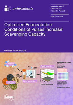
The optimized fermentation of black bean, black-eyed pea, green split pea, pinto bean, and red lentil (RL) using Lactiplantibacillus plantarum 299v enhanced antioxidant capacity and inhibited enzymes linked to type 2 diabetes (T2D). Through the Box–Behnken response surface methodology, conditions that significantly increased 2,2-diphenyl-1-picrylhydrazyl radical scavenging capacity (57–83%) were identified, with soluble protein concentrations ranging from 3–10 mg/mL. Inhibitory effects on dipeptidyl peptidase-IV (DPP-IV) and α-glucosidase activities ranged from 40–70% and 30–60%, respectively. Fermented RL notably inhibited DPP-IV activity in a differentiated Caco-2 cell model. These results highlight the potential of fermented pulses as functional food ingredients in dietary strategies for T2D management. View this paper
  • Issues are regarded as officially published after their release is announced to the table of contents alert mailing list.
  • You may sign up for e-mail alerts to receive table of contents of newly released issues.
  • PDF is the official format for papers published in both, html and pdf forms. To view the papers in pdf format, click on the "PDF Full-text" link, and use the free Adobe Reader to open them.
Order results
Result details
Section
Select all
Export citation of selected articles as:
17 pages, 3944 KiB  
Article
Rutaecarpine Protects Human Endothelial Cells from Oxidative-Stress-Induced Apoptosis via TRPV1- and AhR-Mediated Nrf2 Activation
by Chae Yeon Kim, Gi Ho Lee, Seung Yeon Lee, Anh Thi Ngoc Bui and Hye Gwang Jeong
Antioxidants 2025, 14(5), 616; https://doi.org/10.3390/antiox14050616 - 21 May 2025
Viewed by 21
Abstract
Endothelial cells play a crucial role in cardiovascular health by maintaining vascular homeostasis, regulating blood flow and vascular wall permeability, and protecting against external stressors. Oxidative stress, particularly excessive reactive oxygen species (ROS), disrupts cellular homeostasis and contributes to endothelial cell dysfunction. Rutaecarpine [...] Read more.
Endothelial cells play a crucial role in cardiovascular health by maintaining vascular homeostasis, regulating blood flow and vascular wall permeability, and protecting against external stressors. Oxidative stress, particularly excessive reactive oxygen species (ROS), disrupts cellular homeostasis and contributes to endothelial cell dysfunction. Rutaecarpine (RUT), an indolopyridoquinazolinone alkaloid isolated from Evodia rutaecarpa, has cytoprotective potential. However, the molecular mechanism underlying its cytoprotective activity in endothelial cells remains unclear. In this study, we investigated the protective effects of RUT against H2O2-induced apoptosis in human EA.hy926 endothelial cells and explored its underlying mechanism of action. RUT enhanced nuclear factor erythroid 2-related factor 2 (Nrf2) activation by increasing its expression and phosphorylation, resulting in the upregulation of antioxidant enzymes (GCLC, NQO1, and HO-1). RUT increased the level of the anti-apoptotic marker (Bcl-2) while inhibiting apoptotic markers (cleaved caspase-3 and Bax) in H2O2-induced apoptotic cells. Mechanistic analysis revealed that RUT activates Nrf2 through two pathways: TRPV1-mediated PKCδ/Akt phosphorylation and aryl hydrocarbon receptor (AhR)-dependent Nrf2 expression. These findings suggest that RUT exerts protective effects against oxidative-stress-induced apoptosis by controlling the Nrf2 signaling pathway in endothelial cells. Full article
(This article belongs to the Special Issue Antioxidant Capacity of Natural Products—2nd Edition)
Show Figures

Figure 1

22 pages, 3140 KiB  
Review
Sex-Specific Antioxidant and Anti-Inflammatory Protective Effects of AMPK in Cardiovascular Diseases
by Lea Strohm, Dominika Mihalikova, Alexander Czarnowski, Zita Schwaibold, Andreas Daiber and Paul Stamm
Antioxidants 2025, 14(5), 615; https://doi.org/10.3390/antiox14050615 - 21 May 2025
Viewed by 37
Abstract
Cardiovascular diseases such as coronary heart disease, heart failure, or stroke are the most common cause of death worldwide and are regularly based on risk factors like diabetes mellitus, hypertension, or obesity. At the same time, both diseases and risk factors are significantly [...] Read more.
Cardiovascular diseases such as coronary heart disease, heart failure, or stroke are the most common cause of death worldwide and are regularly based on risk factors like diabetes mellitus, hypertension, or obesity. At the same time, both diseases and risk factors are significantly influenced by sex hormones. In order to better understand this influence and also specifically improve the therapy of female patients, medical research has recently focused increasingly on gender-specific differences. The goal is to develop personalized, gender-specific therapy concepts for these diseases to further enhance health outcomes. The enzyme adenosine monophosphate-activated protein kinase (AMPK) is a central regulator of energy metabolism, protecting the cardiovascular system from energy depletion, thereby promoting vascular health and preventing cellular damage. AMPK confers cardioprotective effects by preventing endothelial and vascular dysfunction, and by controlling or regulating oxidative stress and inflammatory processes. For AMPK, sex-specific effects were reported, influencing metabolic and cardiovascular responses. Exercise and metabolic stress generally cause higher AMPK activity in males. At the same time, females exhibit protective mechanisms against insulin resistance or oxidative stress, particularly in conditions like obesity. Additionally, males subject to AMPK deficiency seem to experience greater cardiac and mitochondrial dysfunction. In contrast, females show improvement in cardiovascular function after pharmacological AMPK activation. These differences, influenced by hormones, body composition, and gene expression, highlight the potential to develop personalized, sex-specific AMPK-targeted therapeutic strategies for cardiovascular diseases in the future. Here, we discuss the most actual scientific background, focusing on the protective, gender-specific effects of AMPK, and highlight potential clinical applications. Full article
(This article belongs to the Special Issue Oxidative/Nitrosative Stress in Cardiovascular Diseases)
Show Figures

Graphical abstract

27 pages, 25331 KiB  
Article
Attenuating Effect of a Polyphenol Ellagic Acid on Ovarian Aging by Inhibiting the Ferroptosis Pathway in Low-Yield Laying Chickens
by Qiongyu Yuan, Xinyu Wang, Yingyu Xiao, Zhaoyu Yang, Xiangyu Cai, Wanyue Gao, Yuling Mi and Caiqiao Zhang
Antioxidants 2025, 14(5), 614; https://doi.org/10.3390/antiox14050614 - 21 May 2025
Viewed by 29
Abstract
Aging leads to ovarian degeneration in poultry, reducing egg production and quality. Ellagic acid (EA), a natural plant-derived compound, may help delay ovarian aging, though its precise mechanisms remain unclear. This study investigated the effects of EA on ovarian aging of low-yield laying [...] Read more.
Aging leads to ovarian degeneration in poultry, reducing egg production and quality. Ellagic acid (EA), a natural plant-derived compound, may help delay ovarian aging, though its precise mechanisms remain unclear. This study investigated the effects of EA on ovarian aging of low-yield laying chickens and explored its underlying mechanism. EA supplementation (100 and 500 mg/kg) significantly increased ovarian weight as well as the number and proportion of small yellow follicles in aging chickens. EA administration elevated serum antioxidant levels and upregulated the expression of glutathione peroxidase 4 (GPX4) expression to reduce oxidative stress. Importantly, EA treatment suppressed the mRNA and protein expression of ferroptosis markers transferrin receptor protein 1 (TFRC) and solute carrier family 7 member 11 (SLC7A11), increased Proliferating Cell Nuclear Antigen (PCNA) expression, and alleviated G1 phase arrest in granulosa cells (GCs), promoting cell proliferation, which improves egg quality and production. Furthermore, in vitro experiments demonstrated that EA treatment decreased reactive oxygen species production, improved mitochondrial function, inhibited ferroptosis, and attenuated GCs aging. In conclusion, this study reveals the critical role of ferroptosis in chicken ovarian aging and suggests that EA may provide a promising approach for delaying ovarian aging and enhancing productivity in low-yield poultry. Full article
Show Figures

Figure 1

27 pages, 2333 KiB  
Review
The Ferroptosis–Mitochondrial Axis in Depression: Unraveling the Feedforward Loop of Oxidative Stress, Metabolic Homeostasis Dysregulation, and Neuroinflammation
by Xu Liu, Qiang Luo, Yulong Zhao, Peng Ren, Yu Jin and Junjie Zhou
Antioxidants 2025, 14(5), 613; https://doi.org/10.3390/antiox14050613 - 20 May 2025
Viewed by 66
Abstract
Emerging evidence links ferroptosis–mitochondrial dysregulation to depression pathogenesis through an oxidative stress–energy deficit–neuroinflammation cycle driven by iron overload. This study demonstrates that iron accumulation initiates ferroptosis via Fenton reaction-mediated lipid peroxidation, compromising neuronal membrane integrity and disabling the GPx4 antioxidant system. Concurrent mitochondrial [...] Read more.
Emerging evidence links ferroptosis–mitochondrial dysregulation to depression pathogenesis through an oxidative stress–energy deficit–neuroinflammation cycle driven by iron overload. This study demonstrates that iron accumulation initiates ferroptosis via Fenton reaction-mediated lipid peroxidation, compromising neuronal membrane integrity and disabling the GPx4 antioxidant system. Concurrent mitochondrial complex I/IV dysfunction impairs ATP synthesis, creating an AMPK/mTOR signaling imbalance and calcium dyshomeostasis that synergistically impair synaptic plasticity. Bidirectional crosstalk emerges: lipid peroxidation derivatives oxidize mitochondrial cardiolipin, while mitochondrial ROS overproduction activates ACSL4 to amplify ferroptotic susceptibility, forming a self-reinforcing neurodegenerative loop. Prefrontal–hippocampal metabolomics reveal paradoxical metabolic reprogramming with glycolytic compensation suppressing mitochondrial biogenesis (via PGC-1α/TFAM downregulation), trapping neurons in bioenergetic crisis. Clinical data further show that microglial M1 polarization through cGAS-STING activation sustains neuroinflammation via IL-6/TNF-α release. We propose a “ferroptosis–mitochondrial fragmentation–metabolic maladaptation” triad as mechanistic subtyping criteria for depression. Preclinical validation shows that combinatorial therapy (iron chelators + SIRT3 agonists) rescues neuronal viability by restoring mitochondrial integrity and energy flux. This work shifts therapeutic paradigms from monoaminergic targets toward multimodal strategies addressing iron homeostasis, organelle dynamics, and metabolic vulnerability—a framework with significant implications for developing neuroprotective antidepressants. Full article
Show Figures

Figure 1

23 pages, 3535 KiB  
Article
Cardio–Renal and Systemic Effects of SGLT2i Dapagliflozin on Short-Term Anthracycline and HER-2-Blocking Agent Therapy-Induced Cardiotoxicity
by Vincenzo Quagliariello, Annabella Di Mauro, Gerardo Ferrara, Francesca Bruzzese, Giuseppe Palma, Antonio Luciano, Maria Laura Canale, Irma Bisceglia, Martina Iovine, Christian Cadeddu Dessalvi, Carlo Maurea, Matteo Barbato, Alessandro Inno, Massimiliano Berretta, Andrea Paccone, Alfredo Mauriello, Celeste Fonderico, Anna Chiara Maratea and Nicola Maurea
Antioxidants 2025, 14(5), 612; https://doi.org/10.3390/antiox14050612 - 20 May 2025
Viewed by 46
Abstract
Anthracyclines and human epidermal growth factor receptor 2 (HER-2) inhibitors are cornerstone therapies for breast cancer but are associated with significant cardiotoxicity. While sodium–glucose cotransporter 2 (SGLT2) inhibitors such as dapagliflozin have demonstrated cardio–renal protective effects during anthracycline treatment, their efficacy in preventing [...] Read more.
Anthracyclines and human epidermal growth factor receptor 2 (HER-2) inhibitors are cornerstone therapies for breast cancer but are associated with significant cardiotoxicity. While sodium–glucose cotransporter 2 (SGLT2) inhibitors such as dapagliflozin have demonstrated cardio–renal protective effects during anthracycline treatment, their efficacy in preventing cardiotoxicity from sequential anthracycline and HER-2 blockade remains poorly understood. This study investigates the cardioprotective role of dapagliflozin in a preclinical model of chemotherapy-induced cardiotoxicity. Female C57Bl/6 mice were divided into four groups and treated for 10 days as follows: (1) a normal control group receiving saline (sham); (2) a model control group receiving doxorubicin (2.17 mg/kg/day for 5 days) followed by HER-2-blocking monoclonal antibody (2.25 mg/kg/day for 5 days); (3) a dapagliflozin-only group (10 mg/kg/day via oral gavage); and (4) a treatment group receiving the combination of doxorubicin, HER-2 inhibitor, and dapagliflozin. Cardiac function was assessed using echocardiography (VEVO 2100). Biomarkers of myocardial injury and inflammation (NLRP3, MyD88, CXCR4, H-FABP, troponin-T, and cytokines) were quantified via ELISA and immunohistochemistry. Circulating markers such as mitofusin-2, cardiac myosin light chain, malondialdehyde (MDA), and 4-hydroxy-2-nonenal (4-HNE) were also measured. Dapagliflozin significantly preserved the ejection fraction and reduced both radial and longitudinal strain impairment in mice treated with the doxorubicin–HER-2 inhibitor combination (p < 0.001). Levels of myocardial NLRP3, MyD88, CXCR4, H-FABP, interleukin-1β, and troponin-T were significantly lower in the dapagliflozin-treated group compared to the chemotherapy-only group. Serum markers of oxidative stress and cardiac injury, including mitofusin-2, MDA, 4-HNE, BNP, and high-sensitivity C-reactive protein (hs-CRP), were also reduced by dapagliflozin treatment. Our findings demonstrate that dapagliflozin effectively mitigates early cardiac dysfunction and injury in a preclinical model of sequential doxorubicin and HER-2 inhibitor therapy. Full article
Show Figures

Figure 1

18 pages, 2600 KiB  
Article
c-Abl/TFEB Pathway Activation as a Common Pathogenic Mechanism in Lysosomal Storage Diseases: Therapeutic Potential of c-Abl Inhibitors
by Miguel V. Guerra, Juan Castro, Antonio Moreno, Elisa Balboa, Juan J. Marugan, Alejandra R. Alvarez and Silvana Zanlungo
Antioxidants 2025, 14(5), 611; https://doi.org/10.3390/antiox14050611 - 20 May 2025
Viewed by 53
Abstract
Lysosomal storage diseases (LSDs) are characterized by the accumulation of undegraded substrates within lysosomes, often associated with oxidative stress and impaired lysosomal function. In this study, we investigate the role of the c-Abl/TFEB pathway in different LSDs: Gaucher, Niemann-Pick type A (NPA), and [...] Read more.
Lysosomal storage diseases (LSDs) are characterized by the accumulation of undegraded substrates within lysosomes, often associated with oxidative stress and impaired lysosomal function. In this study, we investigate the role of the c-Abl/TFEB pathway in different LSDs: Gaucher, Niemann-Pick type A (NPA), and Niemann-Pick type C (NPC). Our findings identify c-Abl activation (p-c-Abl) as a common pathogenic mechanism in these disorders. We demonstrate that c-Abl phosphorylates TFEB at Tyr173, leading to its cytoplasmic retention. Using pharmacological models of Gaucher, NPA and NPC in SH-SY5Y neuronal cells and HeLa cells, we assess the effects of the c-Abl inhibitors Imatinib and Neurotinib, as well as the antioxidant α-Tocopherol (α-TOH), on TFEB nuclear translocation and p-c-Abl protein levels. Additionally, we explore the effects of c-Abl inhibitors in cholesterol accumulation in LSDs neuronal models. Our results show that treatment with c-Abl inhibitors or α-TOH promotes TFEB nuclear translocation, enhances lysosomal clearance, and reduces cholesterol accumulation in all three LSD models. These findings highlight the c-Abl/TFEB pathway as a potential therapeutic target for LSDs and potentially other neurodegenerative disorders associated with lysosomal dysfunction. Full article
(This article belongs to the Special Issue Oxidative Stress and Lysosomal Function in Health and Disease)
Show Figures

Figure 1

17 pages, 2404 KiB  
Article
Anti-Inflammasome Effect of Impressic Acid on Diesel Exhaust Particulate Matter-Induced NLRP1 Inflammasome via the Keap1/p62/Nrf2-Signaling Pathway in Keratinocytes
by Seung Yeon Lee, Gi Ho Lee, Jeonghwan Maeng, Su Yeon Kim, Hwi-Yeol Yun, Gil-Saeng Jeong and Hye Gwang Jeong
Antioxidants 2025, 14(5), 610; https://doi.org/10.3390/antiox14050610 - 19 May 2025
Viewed by 217
Abstract
Diesel exhaust particulate (DEP) is widely recognized to weaken lung function and skin diseases. When the skin, which defends against external factors, is exposed to PM2.5, various chronic inflammatory diseases occur. When keratinocytes recognize harmful signals, they synthesize the NOD-like receptor protein 1 [...] Read more.
Diesel exhaust particulate (DEP) is widely recognized to weaken lung function and skin diseases. When the skin, which defends against external factors, is exposed to PM2.5, various chronic inflammatory diseases occur. When keratinocytes recognize harmful signals, they synthesize the NOD-like receptor protein 1 (NLRP1) inflammasome. DEP enhances NF-κB signaling and NLRP1 inflammasome expression through the interaction of TXNIP with NLRP1 in keratinocytes. Although many studies have reported the anti-inflammatory and antioxidant characteristics of Impressic acid (IPA), the umbrella consequences of IPA for PM2.5-influenced inflammasomes and the associated mechanisms remain unknown. Therefore, this study aimed to examine the protective function of IPA against inflammation in human keratinocytes. IPA attenuated the NLRP1 expression, caspase-1, IL-1β actuation, and NF-κB and IκB phosphorylation induction by DEP. IPA upregulated the Nrf2, HO-1, and NQO1 expression through CaMKKβ, AMPK, and GSK3β phosphorylation. Also, IPA led to the elevation of p62 and the degradation of the Keap1 protein. ML385 reversed the suppressive effect of IPA on the NLRP1 inflammasome, which was enhanced by DEP, and NAC counteracted the effect of ML385. These findings indicate that IPA can suppress inflammation induced by PM2.5 by expressing antioxidant enzymes through the Keap1/p62/Nrf2-signaling pathway in human keratinocytes. Full article
Show Figures

Figure 1

22 pages, 1571 KiB  
Article
Effectiveness of Astaxanthin as a Feed Supplement to Improve Growth Performance and Feed Utilization in Aquaculture Animals: A Meta-Analysis
by Bowen Li, Chunxiu Chen, Xiaoqing Zhou, Huiru Liu, Zhixiong Zhou, Xiaoyu Wang, Jian Liang, Yongjun Guo and Shuang Liang
Antioxidants 2025, 14(5), 609; https://doi.org/10.3390/antiox14050609 - 19 May 2025
Viewed by 139
Abstract
Aquaculture, a vital component of global food supply, faces challenges from environmental stressors that compromise aquatic animal health and productivity. Astaxanthin, a potent carotenoid antioxidant, has shown promise in enhancing growth and stress resilience in aquaculture species, yet its effects remain inconsistent across [...] Read more.
Aquaculture, a vital component of global food supply, faces challenges from environmental stressors that compromise aquatic animal health and productivity. Astaxanthin, a potent carotenoid antioxidant, has shown promise in enhancing growth and stress resilience in aquaculture species, yet its effects remain inconsistent across studies. This meta-analysis systematically evaluates the efficacy of dietary astaxanthin supplementation on growth, feed utilization, antioxidant capacity, and immune function in aquaculture animals. Following Preferred Reporting Items for Systematic Reviews and Meta-Analyses (PRISMA) guidelines, 64 studies (33 species, 964 comparisons) published prior to 2025 were analyzed using a random-effects model. Results demonstrated that astaxanthin significantly improved final body weight, weight gain rate, specific growth rate, survival rate, and protein efficiency ratio, while reducing feed conversion ratio. Additionally, it enhanced digestive enzyme activities, hepatopancreas antioxidant biomarkers, and immune parameters. The subgroup analysis revealed differences related to species, trophic level, and habitat, and estimated the optimal dose for key indicators. Despite heterogeneity and publication bias, adjusted effect sizes remained significant for most outcomes. These findings underscore astaxanthin’s potential as a multifunctional feed additive to promote sustainable aquaculture, though its efficacy depends on species, dosage, and environmental context, warranting further mechanistic and optimization studies. Full article
Show Figures

Figure 1

26 pages, 4181 KiB  
Article
Alleviating the Effect of Branched-Chain Fatty Acids on the Lipopolysaccharide-Induced Inflammatory Response in Calf Small Intestinal Epithelial Cells
by Siqi Zhang, Qingyuan Yu, Yukun Sun, Guangning Zhang, Yonggen Zhang and Hangshu Xin
Antioxidants 2025, 14(5), 608; https://doi.org/10.3390/antiox14050608 - 19 May 2025
Viewed by 238
Abstract
This study examined branched-chain fatty acids (BCFAs)’ effects on oxidative stress, energy metabolism, inflammation, tight junction disruption, apoptosis, and Toll-like receptor 4/nuclear factor kappa-B (TLR4/NF-κB) signaling in lipopolysaccharide (LPS)-induced calf small intestinal epithelial cells (CSIECs). Eight groups were used: a control [...] Read more.
This study examined branched-chain fatty acids (BCFAs)’ effects on oxidative stress, energy metabolism, inflammation, tight junction disruption, apoptosis, and Toll-like receptor 4/nuclear factor kappa-B (TLR4/NF-κB) signaling in lipopolysaccharide (LPS)-induced calf small intestinal epithelial cells (CSIECs). Eight groups were used: a control group, an LPS-induced group, and six BCFA treatment groups (12-methyltridecanoic acid (iso-C14:0), 13-methyltetradecanoic acid (iso-C15:0), 14-methylpentadecanoic acid (iso-C16:0), 15-methylhexadecanoic acid (iso-C17:0), 12-methyltetradecanoic acid (anteiso-C15:0), and 14-methylhexadecanoic acid (anteiso-C17:0)) with LPS. The BCFA pretreatments significantly increased CSIEC activity compared to the LPS-induced group, with iso-C14:0 showing the highest activity (89.73%). BCFA reduced Reactive Oxygen Species (ROS) generation and malondialdehyde (MDA) levels and improved the superoxide dismutase (SOD), glutathione peroxidase (GSH-Px), and catalase (CAT) activities and glutathione (GSH) levels. Iso-C16:0 optimized total antioxidant capacity (T-AOC). BCFA enhanced the mitochondrial membrane potential, Adenosine Triphosphate (ATP) enzyme activity, and ATP content, with iso-C14:0 increasing ATP by 27.01%. BCFA downregulated interleukin (IL)-1β, IL-8, tumor necrosis factor (TNF)-α, and interferon (INF)-γ gene expression, reduced IL-6 levels, and increased IL-10 expression. Myeloid differentiation factor 88 (MyD88) mRNA levels were reduced. BCFA alleviated Zonula Occludin (ZO-1), Claudin-1, and Claudin-4 decrease and increased Occludin levels. BCFA mitigated LPS-induced increases in Caspase-3 and BCL2-Associated X (BAX) mRNA levels, reduced Caspase-8 and Caspase-9 expression, and increased B-Cell Lymphoma-2 (BCL-2) mRNA levels. The Entropy Weight-TOPSIS method was adopted, and it was discovered that iso-C15:0 has the best effect. In summary, BCFA supplementation mitigated oxidative stress and enhanced mitochondrial function. BCFA inhibited TLR4/NF-κB signaling pathway overactivation, regulated inflammatory cytokine gene expression, reduced cellular apoptosis, preserved tight junction integrity, and supported barrier function. Full article
(This article belongs to the Section Health Outcomes of Antioxidants and Oxidative Stress)
Show Figures

Figure 1

19 pages, 830 KiB  
Review
Geochemical Speciation, Uptake, and Transportation Mechanisms of Arsenic, Cadmium, and Lead in Soil–Rice Systems: Additional Aspects and Challenges
by Chaw Su Lwin, Ha-il Jung, Myung-Sook Kim, Eun-Jin Lee and Tae-Gu Lee
Antioxidants 2025, 14(5), 607; https://doi.org/10.3390/antiox14050607 - 18 May 2025
Viewed by 200
Abstract
Potentially toxic elements (PTE), such as cadmium (Cd), lead (Pb), and arsenic (As), threaten rice (Oryza sativa L.) crop productivity and pose significant risks to human health when they are present in soil. This review summarizes the current understanding of soil and [...] Read more.
Potentially toxic elements (PTE), such as cadmium (Cd), lead (Pb), and arsenic (As), threaten rice (Oryza sativa L.) crop productivity and pose significant risks to human health when they are present in soil. This review summarizes the current understanding of soil and rice contamination with As, Cd, and Pb to provide an in-depth understanding of the dynamics of these contaminants and the mechanisms regulating their flow from soil to plants. It focuses on the following aspects: (1) these metals’ geochemical distribution and speciation in soil–rice systems; (2) factors influencing the transformation, bioavailability, and uptake of these metals in paddy soils; (3) metal uptake, transport, translocation, and accumulation mechanisms in rice grains; and (4) the roles of transporters involved in metal uptake, transport, and accumulation in rice plants. Moreover, this review contributes to a clearer understanding of the environmental risks associated with these toxic metals in soil–rice ecosystems. Furthermore, it highlights the challenges in simultaneously managing the risks of As, Cd, and Pb contamination in rice. The study findings may help inspire innovative methods, biotechnological applications, and sustainable management strategies to mitigate the accumulation of As, Cd, and Pb in rice grains while effectively addressing multi-metal contamination in paddy soils. Full article
(This article belongs to the Special Issue Oxidative Stress and Antioxidant Defense in Crop Plants, 2nd Edition)
Show Figures

Figure 1

16 pages, 2991 KiB  
Article
Lysine Carboxymethyl Cysteinate, as a Topical Glutathione Precursor, Protects Against Oxidative Stress and UVB Radiation-Induced Skin Damage
by Ping Gao, Xue Xiao, Xiao Cui, Hong Zhang and Xuelan Gu
Antioxidants 2025, 14(5), 606; https://doi.org/10.3390/antiox14050606 - 17 May 2025
Viewed by 210
Abstract
Lysine carboxymethyl cysteinate (LCC) is a synthetic substance obtained via lysine salification of S-carboxymethyl-cysteine. LCC has emerged as a promising glutathione (GSH) precursor. In this study, we sought to determine whether LCC could boost GSH levels and protect skin against oxidative stress. Experiments [...] Read more.
Lysine carboxymethyl cysteinate (LCC) is a synthetic substance obtained via lysine salification of S-carboxymethyl-cysteine. LCC has emerged as a promising glutathione (GSH) precursor. In this study, we sought to determine whether LCC could boost GSH levels and protect skin against oxidative stress. Experiments utilizing primary human keratinocytes and skin tissue samples revealed that LCC significantly increased endogenous GSH levels. LCC was able to pass through the stratum corneum and reach deep into the epidermis, where it enhanced the production of key metabolites involved in GSH biosynthesis. Then, the efficacy of LCC on skin protection was explored. LCC demonstrated protective effects by shielding keratinocytes from blue-light-induced oxidative stress and preventing ultraviolet B (UVB)-induced barrier disruption and pigmentation in a pigmented living skin equivalent (pLSE) model. In addition to its antioxidant properties, LCC also reduced the production of inflammatory mediators. Together, these findings underscore the multifaceted role of LCC in bolstering the natural antioxidant defenses of skin and preventing the accumulation of irreversible damage from the environment, thereby positioning it as a promising candidate for advancing skin health. Full article
(This article belongs to the Special Issue Glutathione and Health: From Development to Disease)
Show Figures

Figure 1

22 pages, 2368 KiB  
Review
New Perspectives on Postmenopausal Osteoporosis: Mechanisms and Potential Therapeutic Strategies of Sirtuins and Oxidative Stress
by Huiying Zhao, Fan Yu and Wei Wu
Antioxidants 2025, 14(5), 605; https://doi.org/10.3390/antiox14050605 - 17 May 2025
Viewed by 184
Abstract
Estrogen levels are the core factor influencing postmenopausal osteoporosis (PMOP). Estrogen can affect the progression of PMOP by regulating bone metabolism, influencing major signaling pathways related to bone metabolism, and modulating immune responses. When estrogen levels decline, the activity of Sirtuins (SIRTs) is [...] Read more.
Estrogen levels are the core factor influencing postmenopausal osteoporosis (PMOP). Estrogen can affect the progression of PMOP by regulating bone metabolism, influencing major signaling pathways related to bone metabolism, and modulating immune responses. When estrogen levels decline, the activity of Sirtuins (SIRTs) is reduced. SIRTs are enzymes that function as NAD+-dependent deacetylases. SIRTs can modulate osteocyte function, sustain mitochondrial homeostasis, and modulate relevant signaling pathways, thereby improving bone metabolic imbalances, reducing bone resorption, and promoting bone formation. In PMOP, SIRT1, SIRT3, and SIRT6 are primarily affected. Oxidative stress (OS) is a crucial factor in PMOP, as it generates excessive reactive oxygen species (ROS) that exacerbate PMOP. There is a certain interplay between SIRTs and OS. The reduced activity of SIRTs leads to intensified OS and the excessive accumulation of ROS. In return, ROS suppresses the AMPK signaling pathway and the synthesis of NAD+, which consequently diminishes the function of SIRTs. Natural SIRT activators and natural antioxidants, which are characterized by high safety, convenience, and minimal side effects, represent a potential therapeutic strategy for PMOP. This study aims to investigate the mechanisms of SIRTs and OS in PMOP and summarize potential therapeutic strategies to assist in the improvement of PMOP. Full article
(This article belongs to the Special Issue Oxidative Stress and Inflammation in Bone Metabolism and Diseases)
Show Figures

Figure 1

36 pages, 1682 KiB  
Review
Environmental Toxins and Oxidative Stress: The Link to Cardiovascular Diseases
by Rasheed O. Sule, Gabriela Del Toro Rivera, Tanishq Vaidya, Emily Gartrell and Aldrin V. Gomes
Antioxidants 2025, 14(5), 604; https://doi.org/10.3390/antiox14050604 - 17 May 2025
Viewed by 191
Abstract
Cardiovascular diseases (CVDs) remain a leading global health concern, responsible for substantial morbidity and mortality. In recent years, as our understanding of the multifaceted nature of CVDs has increased, it has become increasingly evident that traditional risk factors alone do not account for [...] Read more.
Cardiovascular diseases (CVDs) remain a leading global health concern, responsible for substantial morbidity and mortality. In recent years, as our understanding of the multifaceted nature of CVDs has increased, it has become increasingly evident that traditional risk factors alone do not account for the entirety of cardiovascular morbidity and mortality. Environmental toxins, a heterogeneous group of substances ubiquitous in our surroundings, have now entered the spotlight as offenders in the development and progression of CVDs. Environmental toxins include heavy metals, air pollutants, pesticides, and endocrine-disrupting chemicals, among others. Upon exposure, they can elicit oxidative stress, a condition characterized by an imbalance between the production of reactive oxygen species (ROS) and the body’s ability to detoxify and repair the resulting damage. Oxidative stress triggers a cascade of events, including inflammation, endothelial dysfunction, lipid peroxidation, and vascular remodeling, which can contribute to the development of atherosclerosis, hypertension, and other cardiovascular pathologies. This article delves into the molecular mechanisms underpinning oxidative stress-mediated cardiovascular damage induced by environmental toxins, emphasizing the role of specific toxins in this process. Further research is necessary to understand how individual susceptibility and genotype influence the impact of environmental toxins on oxidative stress and the risk of CVD. Full article
(This article belongs to the Special Issue Understanding Oxidative Stress in Cardiovascular Disorders)
Show Figures

Figure 1

25 pages, 5232 KiB  
Article
Oral Sulforaphane Intervention Protects Against Diabetic Cardiomyopathy in db/db Mice: Focus on Cardiac Lipotoxicity and Substrate Metabolism
by Pan Wang, Ziling Wang, Xinyuan Jin, Mengdi Zhang, Mengfan Shen and Dan Li
Antioxidants 2025, 14(5), 603; https://doi.org/10.3390/antiox14050603 - 16 May 2025
Viewed by 173
Abstract
The protective effect of cruciferae-derived sulforaphane (SFN) on diabetic cardiomyopathy (DCM) has garnered increasing attention. However, no studies have specifically explored its mechanistic involvement in cardiac substrate metabolism and mitochondrial function. To address this gap, Type 2 diabetes mellitus (T2DM) db/db mice were [...] Read more.
The protective effect of cruciferae-derived sulforaphane (SFN) on diabetic cardiomyopathy (DCM) has garnered increasing attention. However, no studies have specifically explored its mechanistic involvement in cardiac substrate metabolism and mitochondrial function. To address this gap, Type 2 diabetes mellitus (T2DM) db/db mice were orally gavaged with vehicle or 10 mg/kg body weight SFN every other day for 16 weeks, with vehicle-treated wild-type mice as controls. SFN intervention (SFN-I) alleviated hyperglycemia, dyslipidemia, HOMA-IR, serum MDA levels, and liver inflammation. Furthermore, SFN-I improved the lipotoxicity-related phenotype of T2DM cardiomyopathy, manifested as attenuation of diastolic dysfunction, cardiac injury, fibrosis, lipid accumulation and peroxidation, ROS generation, and decreased mitochondrial complex I and II activities and ATP content, despite having no effect on ceramide abnormalities. Protein expression data revealed that the model mice exhibited upregulated cardiac CD36, H-FABP, FATP4, CPT1B, PPARα, and PDK4 but downregulated GLUT4, with unchanged MPC1 and MPC2. Notably, SFN-I significantly attenuated the increase in CD36, H-FABP, CPT1B, and PPARα. These results suggest that chronic oral SFN-I protects against DCM by mitigating overall metabolic dysregulation and inhibiting cardiolipotoxicity. The latter might involve controlling cardiac fatty acid metabolism and improving mitochondrial function, rather than promoting glucose metabolism. Full article
(This article belongs to the Section Health Outcomes of Antioxidants and Oxidative Stress)
Show Figures

Figure 1

18 pages, 10894 KiB  
Article
The Class III Peroxidase Gene Family in Populus simonii: Genome-Wide Identification, Classification, Gene Expression and Functional Analysis
by Lu Han, Yishuang Ren, Xinru Bi, Guowei Yao, Jinwang Zhang, Hongtao Yuan, Xiaoyu Xie, Junbo Chen, Yunchang Zhang, Sitong Du, Wanying Chen, Kewei Cai and Xiyang Zhao
Antioxidants 2025, 14(5), 602; https://doi.org/10.3390/antiox14050602 - 16 May 2025
Viewed by 109
Abstract
Class III peroxidases are plant-specific enzymes that play indispensable roles in catalyzing oxidative–reductive reactions, which are integral to numerous biochemical processes in plants. In this study, we identified 69 members of the class III peroxidase (POD) gene family in the Populus [...] Read more.
Class III peroxidases are plant-specific enzymes that play indispensable roles in catalyzing oxidative–reductive reactions, which are integral to numerous biochemical processes in plants. In this study, we identified 69 members of the class III peroxidase (POD) gene family in the Populus simonii genome and classified them into four subfamilies based on phylogenetic analysis. Chromosomal localization revealed that these PsPOD genes are unevenly distributed across 19 chromosomes, with chromosomes 3 and 7 harboring the highest densities. Conserved domain and motif analyses demonstrated that all PsPOD proteins contain the characteristic peroxidase domain and share highly conserved motif structures. Cis-acting element analysis of promoter regions revealed the presence of numerous regulatory elements associated with light responsiveness, phytohormone signaling, stress responses, and plant growth and development. Transcriptome data showed that the expression of PsPOD genes varies significantly across different tissues and organs and under various stress conditions, suggesting their involvement in both developmental processes and abiotic stress responses. These findings were further validated by qRT-PCR analysis of selected PsPOD genes. Notably, PsPOD45, PsPOD69, PsPOD33, and PsPOD64 were identified as central hub genes in the protein–protein interaction network, making them promising candidates for further functional characterization. Overall, this study provides a comprehensive overview of the PsPOD gene family in P. simonii, laying a solid foundation for future functional studies and offering valuable insights for comparative research in other plant species. Full article
(This article belongs to the Special Issue Antioxidant Systems in Plants)
Show Figures

Figure 1

15 pages, 5688 KiB  
Article
Blood Progenitor Cell Mobilization Driven by TWEAK Promotes Neovascularization and Reduces Brain Damage in a Rat Model of Intracerebral Hemorrhage
by Daniel Romaus-Sanjurjo, Esteban López-Arias, Cristina Rodríguez, Pablo Hervella, Mariña Rodríguez-Arrizabalaga, Manuel Debasa-Mouce, Juan Manuel Pías-Peleteiro, Ramón Iglesias-Rey, Pablo Aguiar, Ángeles Almeida, José Castillo, Alberto Ouro and Tomás Sobrino
Antioxidants 2025, 14(5), 601; https://doi.org/10.3390/antiox14050601 - 16 May 2025
Viewed by 507
Abstract
Non-traumatic intracerebral hemorrhage (ICH) is one of the most devastating and disabling forms of stroke; however, there are no effective pharmacological therapies available following the insult. Angiogenesis appears as a key step to overcoming the damage and promoting functional recovery. In this context, [...] Read more.
Non-traumatic intracerebral hemorrhage (ICH) is one of the most devastating and disabling forms of stroke; however, there are no effective pharmacological therapies available following the insult. Angiogenesis appears as a key step to overcoming the damage and promoting functional recovery. In this context, endothelial progenitor cells (EPCs) mobilization improves oxidative stress and promotes neovascularization, which has been linked to beneficial outcomes following both ischemic and hemorrhagic stroke. The TNF-like weak inducer of apoptosis (TWEAK), binding to its receptor Fn14, has been suggested as an inducer of EPCs differentiation, viability and migration to the injury site in a model of myocardial infarction. Here, we have performed a proof-of-concept preclinical study in a rat model of ICH where we report that a 50 μg/kg dose of rat recombinant TWEAK (rTWEAK) promotes blood progenitor cells mobilization, mainly EPCs. As soon as 72 h post-injury, brain neovascularization, and, importantly, long-term hematoma reduction and improved functional recovery is reported. In contrast, a higher dose of 150 μg/kg blocked those beneficial outcomes. Therefore, a low dose of rTWEAK treatment promotes neovascularization and reduces brain damage in a rat model of ICH. Further clinical studies will be needed to demonstrate if rTWEAK could represent a new strategy to promote recovery following ICH. Full article
Show Figures

Figure 1

22 pages, 4496 KiB  
Article
Obtainment of Flavonoid-Enriched Fractions from Maqui (Aristotelia chilensis) and Murta (Ugni molinae) Extracts via Preparative HPLC and Evaluation of Their Anti-Inflammatory Effects in Cell-Based Assays
by Amador Alburquenque, Carolina Busch, Gabriela Gómez-Lillo, Alexander Gamboa, Camilo Perez, Nelson Caro Fuentes, Martin Gotteland, Lilian Abugoch and Cristian Tapia
Antioxidants 2025, 14(5), 600; https://doi.org/10.3390/antiox14050600 - 16 May 2025
Viewed by 151
Abstract
Polyphenols exert anti-inflammatory and antioxidant effects by modulating cell signalling pathways and transcription factors involved in inflammatory bowel disease (IBD). However, their stability during digestion can be compromised. Polymer coatings like chitosan (-C) help preserve their stability. Maqui (Aristotelia chilensis) and [...] Read more.
Polyphenols exert anti-inflammatory and antioxidant effects by modulating cell signalling pathways and transcription factors involved in inflammatory bowel disease (IBD). However, their stability during digestion can be compromised. Polymer coatings like chitosan (-C) help preserve their stability. Maqui (Aristotelia chilensis) and murta (Ugni molinae) are rich in antioxidant and anti-inflammatory compounds. This work aims to obtain extracts (E) and blends (B) enriched in delphinidin and quercetin glucosides from maqui (Ma) and murta (Mu) crude extracts using preparative chromatography methodology (Prep-HPLC) and to evaluate their effectiveness through in vitro and cellular assays. HPLC-DAD analysis revealed a marked increase in phenolic compound concentration in the BEMaMu and BCMaMu extract blends. Total quercetin glycosides (TQG) increased by 11-fold, and total anthocyanins increased by approximately 8-fold compared to the fruit blend (BMaMu). BCMaMu exhibited a significantly higher ORAC value compared to the estimated additive mixture, suggesting a synergistic effect. No cytotoxicity was observed for BEMaMu, BCMaMu, and their chitosan-coated versions (BEMaMu-C and BCMaMu-C) in Caco-2 and HT29-MTX-E12 cells at concentrations of 0.1–50 mg/mL. Notably, only chitosan-coated BCMaMu inhibited NF-κB expression and activated Nrf2 in TNF-α-challenged Caco-2 cells at 0.1 and 0.5 mg/mL. Full article
(This article belongs to the Special Issue Potential Health Benefits of Dietary Antioxidants)
Show Figures

Figure 1

19 pages, 1885 KiB  
Article
Targeting Drug Resistance in Cancer: Dimethoxycurcumin as a Functional Antioxidant Targeting ABCC3
by Jochem Nelen, Valeria Naponelli, José Manuel Villalgordo-Soto, Marco Falasca and Horacio Pérez-Sánchez
Antioxidants 2025, 14(5), 599; https://doi.org/10.3390/antiox14050599 - 16 May 2025
Viewed by 97
Abstract
The development of new anticancer therapies remains challenging due to tumor heterogeneity and the frequent emergence of multidrug resistance (MDR). Natural products have garnered increasing attention as alternative or complementary therapeutic agents due to their bioactivity and reduced toxicity. Polyphenols, particularly curcumin and [...] Read more.
The development of new anticancer therapies remains challenging due to tumor heterogeneity and the frequent emergence of multidrug resistance (MDR). Natural products have garnered increasing attention as alternative or complementary therapeutic agents due to their bioactivity and reduced toxicity. Polyphenols, particularly curcumin and its derivatives, have shown promise in modulating signaling pathways, enhancing chemosensitivity, and overcoming drug resistance. The anticancer potential of dimethoxycurcumin, a chemically modified curcumin derivative identified through consensus fingerprint similarity screening, was investigated for its potential to inhibit ABCC3 (MRP3)—a member of the ATP-binding cassette (ABC) transporter family implicated in drug efflux, tumor cell survival, and resistance. In vitro experiments demonstrated that dimethoxycurcumin significantly reduced cancer cell viability and colony formation, indicating a strong inhibitory effect on ABCC3 function. These results suggest that dimethoxycurcumin may sensitize cancer cells to chemotherapy by targeting resistance pathways. The data presented contribute to the growing body of evidence suggesting that bioactive plant-derived compounds, including chemically modified derivatives, may hold therapeutic potential in oncology by modulating multidrug resistance pathways. Targeting ABC transporters with natural compound derivatives could offer a promising strategy for developing more effective and less toxic anticancer therapies. Full article
(This article belongs to the Special Issue Anti-cancer Potential of Plant-Based Antioxidants)
Show Figures

Figure 1

44 pages, 8161 KiB  
Review
Oleanolic Acid: A Promising Antioxidant—Sources, Mechanisms of Action, Therapeutic Potential, and Enhancement of Bioactivity
by Andrzej Günther and Barbara Bednarczyk-Cwynar
Antioxidants 2025, 14(5), 598; https://doi.org/10.3390/antiox14050598 - 16 May 2025
Viewed by 130
Abstract
This review discusses the antioxidant potential of oleanolic acid, a triterpene compound present in many medicinal and edible plants. The authors analyze various studies that confirm numerous pharmacological properties of this compound, such as its anticancer, antidiabetic, neuroprotective, osteoprotective, anti-obesity, and anti-inflammatory effects. [...] Read more.
This review discusses the antioxidant potential of oleanolic acid, a triterpene compound present in many medicinal and edible plants. The authors analyze various studies that confirm numerous pharmacological properties of this compound, such as its anticancer, antidiabetic, neuroprotective, osteoprotective, anti-obesity, and anti-inflammatory effects. OA, as a natural antioxidant, plays an important role in neutralizing reactive oxygen species, which contribute to the oxidative stress that is responsible for the development of many diseases, including cancer and cardiovascular and neurodegenerative diseases. This article also presents natural sources of OA, including grapes, olives, and apples, and discusses the mechanisms of its antioxidant action, including the inhibition of lipid peroxidation and the modulation of signaling pathways related to inflammatory processes. In addition, there are research results that indicate the therapeutic benefits of OA in the treatment of diabetes and neurodegenerative diseases, as well as its potential to protect the heart, liver, and kidneys from oxidative damage. In conclusion, OA has potent antioxidant properties that can be used in the prevention and treatment of many diseases related to oxidative stress. This article also presents the possibility of increasing the bioavailability of OA through the use of nanoparticle and liposome technology. Full article
(This article belongs to the Special Issue Antioxidant Capacity of Natural Products—2nd Edition)
Show Figures

Graphical abstract

3 pages, 1870 KiB  
Correction
Correction: Gambelunghe et al. Redox-Sensitive Glyoxalase 1 Up-Regulation Is Crucial for Protecting Human Lung Cells from Gold Nanoparticles Toxicity. Antioxidants 2020, 9, 697
by Angela Gambelunghe, Stefano Giovagnoli, Alessandro Di Michele, Simona Boncompagni, Marco Dell’Omo, Kerstin Leopold, Ivo Iavicoli, Vincenzo Nicola Talesa and Cinzia Antognelli
Antioxidants 2025, 14(5), 597; https://doi.org/10.3390/antiox14050597 - 16 May 2025
Viewed by 64
Abstract
In the original publication [...] Full article
Show Figures

Figure 1

20 pages, 2030 KiB  
Review
Targeting Lysosomal Dysfunction and Oxidative Stress in Age-Related Macular Degeneration
by Ana S. Falcão, Margarida L. Pedro, Sandra Tenreiro and Miguel C. Seabra
Antioxidants 2025, 14(5), 596; https://doi.org/10.3390/antiox14050596 - 16 May 2025
Viewed by 152
Abstract
Age-related macular degeneration (AMD) is the leading cause of vision loss in the Western world, and it currently lacks effective therapy. It is believed that AMD initiates in the aged retinal pigment epithelium (RPE), which presents lysosomal dysfunction and oxidative stress (OxS) that [...] Read more.
Age-related macular degeneration (AMD) is the leading cause of vision loss in the Western world, and it currently lacks effective therapy. It is believed that AMD initiates in the aged retinal pigment epithelium (RPE), which presents lysosomal dysfunction and oxidative stress (OxS) that ultimately leads to RPE damage and AMD progression. AMD is a complex pathology, so multitarget treatments are required to act on different pathways, presenting several challenges. In this review, we discuss the current knowledge on the pathogenesis of this disease, focusing mainly on lysosomal dysfunction and OxS. Because transcription factors regulate homeostasis, the transcription factor EB (TFEB), which controls lysosomal function and biogenesis, and the nuclear factor erythroid 2-related factor 2 (NRF2), which manages OxS, have been proposed as promising targets for disease intervention. Finally, we discuss the interplay of these pathways for a potential synergistic effect on AMD-targeted therapies, as they could change the course of today’s available treatments for AMD. Full article
(This article belongs to the Special Issue Oxidative Stress and Lysosomal Function in Health and Disease)
Show Figures

Figure 1

30 pages, 7740 KiB  
Article
Protective Effects of Lotus Seedpod Extract on Hepatic Lipid and Glucose Metabolism via AMPK-Associated Mechanisms in a Mouse Model of Metabolic Syndrome and Oleic Acid-Induced HepG2 Cells
by Hui-Hsuan Lin, Pei-Rong Yu, Chiao-Yun Tseng, Ming-Shih Lee and Jing-Hsien Chen
Antioxidants 2025, 14(5), 595; https://doi.org/10.3390/antiox14050595 - 16 May 2025
Viewed by 142
Abstract
Metabolic syndrome (MetS) poses considerable toxicological risks due to its association with an increased likelihood of metabolic dysfunction-associated steatotic liver disease (MASLD), and is characterized by hypertension, hyperglycemia, dyslipidemia, and obesity. This study aimed to investigate the therapeutic potential of flavonoid-rich lotus seedpod [...] Read more.
Metabolic syndrome (MetS) poses considerable toxicological risks due to its association with an increased likelihood of metabolic dysfunction-associated steatotic liver disease (MASLD), and is characterized by hypertension, hyperglycemia, dyslipidemia, and obesity. This study aimed to investigate the therapeutic potential of flavonoid-rich lotus seedpod extract (LSE) in alleviating MetS and MASLD-related hepatic disturbances. In vivo, mice subjected to a high-fat diet (HFD) and streptozotocin (STZ) injection were supplemented with LSE or simvastatin for 6 weeks. Obesity indicators included body weight and epididymal fat, while insulin resistance was measured by fasting serum glucose, serum insulin, homeostasis model assessment–insulin resistance index (HOMA-IR), and oral glucose tolerance (OGTT). Also, the levels of serum lipid profiles and blood pressure were evaluated. Adipokines, proinflammatory cytokines, liver fat droplets, and peri-portal fibrosis were analyzed to clarify the mechanism of MetS. LSE significantly reduced the HFD/STZ-induced MetS markers better than simvastatin, as demonstrated by hypoglycemic, hypolipidemic, antioxidant, and anti-inflammatory effects. In vitro, LSE improved oleic acid (OA)-triggered phenotypes of MASLD in hepatocyte HepG2 cells by reducing lipid accumulation and enhancing cell viability. This effect might be mediated through proteins involved in lipogenesis that are downregulated by adenosine monophosphate-activated protein kinase (AMPK). In addition, LSE reduced reactive oxygen species (ROS) generation and glycogen levels, as demonstrated by enhancing insulin signaling involving reducing insulin receptor substrate-1 (IRS-1) Ser307 phosphorylation and increasing glycogen synthase kinase 3 beta (GSK3β) and protein kinase B (PKB) expression. These benefits were dependent on AMPK activation, as confirmed by the AMPK inhibitor compound C. These results indicate that LSE exhibits protective effects against MetS-caused toxicological disturbances in hepatic carbohydrate and lipid metabolism, potentially contributing to its efficacy in preventing MASLD or MetS. Full article
(This article belongs to the Special Issue Oxidative Stress and Liver Disease)
Show Figures

Graphical abstract

20 pages, 4632 KiB  
Article
Endosomal H2O2 Molecules Act as Signaling Mediators in Akt/PKB Activation
by Sujin Park, Chaewon Kim, Sukyeong Heo and Dongmin Kang
Antioxidants 2025, 14(5), 594; https://doi.org/10.3390/antiox14050594 - 16 May 2025
Viewed by 70
Abstract
Receptor-mediated endocytosis (RME) is a commonly recognized receptor internalization process of receptor degradation or recycling. However, recent studies have supported that RME is closely related to signal propagation and amplification from the plasma membrane to the cytosol. Few studies have elucidated the role [...] Read more.
Receptor-mediated endocytosis (RME) is a commonly recognized receptor internalization process of receptor degradation or recycling. However, recent studies have supported that RME is closely related to signal propagation and amplification from the plasma membrane to the cytosol. Few studies have elucidated the role of H2O2, a mild oxidant among reactive oxygen species (ROS) in RME and second messenger of signal propagation. In the present study, we investigated the regulatory function of H2O2 in early endosomes during signaling throughout receptor-mediated endocytosis. In mammalian cells with a physiological amount of H2O2 generated during epidermal growth factor (EGF) activation, fluorescence imaging showed that the levels of two activating phosphorylations on Ser473 and Thr308 of Akt were transiently increased in the plasma membrane, but the predominant p-Akt on Ser473 appeared in early endosomes. To examine the role of endosomal H2O2 molecules as signaling mediators of Akt activation in endosomes, we modulated endosomal H2O2 through the ectopic expression of an endosomal-targeting catalase (Cat-Endo). The forced removal of endosomal H2O2 inhibited the Akt phosphorylation on Ser473 but not on Thr308. The levels of mSIN and rictor, two components of mTORC2 that work as a kinase in Akt phosphorylation on Ser473, were also selectively diminished in the early endosomes of Cat-Endo-expressing cells. We also observed a decrease in the endosomal level of the adaptor protein containing the PH domain, the PTB domain, and the Leucine zipper motif 1 (APPL1) protein, which is an effector of Rab5 and key player in the assembly of signaling complexes regulating the Akt pathway in Cat-Endo-expressing cells compared with those in normal cells. Therefore, the H2O2-dependent recruitment of the APPL1 adaptor protein into endosomes was required for full Akt activation. We proposed that endosomal H2O2 is a promoter of Akt signaling. Full article
(This article belongs to the Special Issue Metabolic Dysfunction and Oxidative Stress)
Show Figures

Figure 1

18 pages, 2449 KiB  
Article
Corynoxeine Supplementation Ameliorates Colistin-Induced Kidney Oxidative Stress and Inflammation in Mice
by Yue Liu, Ruichen Zhang, Tony Velkov, Jianzhong Shen, Shusheng Tang and Chongshan Dai
Antioxidants 2025, 14(5), 593; https://doi.org/10.3390/antiox14050593 - 15 May 2025
Viewed by 198
Abstract
This study investigated the protective effects of corynoxeine, a natural alkaline compound, on colistin-caused nephrotoxicity using a murine model. Forty mice were divided randomly into control, corynoxeine-only (20 mg/kg/day, intraperitoneal injection), colistin-only (20 mg/kg/day, intraperitoneal injection), and colistin (20 mg/kg/day) + corynoxeine (5 [...] Read more.
This study investigated the protective effects of corynoxeine, a natural alkaline compound, on colistin-caused nephrotoxicity using a murine model. Forty mice were divided randomly into control, corynoxeine-only (20 mg/kg/day, intraperitoneal injection), colistin-only (20 mg/kg/day, intraperitoneal injection), and colistin (20 mg/kg/day) + corynoxeine (5 and 20 mg/kg/day) groups (8 mice in each group). All treatments were maintained for seven consecutive days. Results showed that colistin treatment at 20 mg/kg/day for seven days significantly increased serum urea nitrogen and creatinine levels and induced the loss and degeneration of renal tubular epithelial cells, which were markedly ameliorated by corynoxeine co-treatment at 5 or 20 mg/kg/day. Corynoxeine supplementation also markedly attenuated colistin-induced increases in malondialdehyde levels and decreases in reduced glutathione levels and superoxide dismutase and catalase activities in the kidneys. Furthermore, corynoxeine supplementation significantly decreased the expression of transforming growth factor β (TGF-β) and nicotinamide adenine dinucleotide phosphate hydrogen oxidase 4 (NOX4) proteins and nuclear factor kappa B (NF-κB), interleukin-1beta (IL-1β), IL-6, and tumor necrosis factor-α mRNAs, while it significantly increased the expression of erythroid 2-related factor 2 (Nrf2) and heme oxygenase-1 (HO-1) proteins in the kidneys. In conclusion, these results reveal that corynoxeine can protect against colistin-induced nephrotoxicity in mice by inhibiting oxidative stress and inflammation, which may partly be attributed to its ability on the activation of the Nrf2/HO-1 pathway and the inhibition of the TGF-β/NOX4 and NF-κB pathways. Full article
(This article belongs to the Special Issue Antioxidant System Efficiency in Kidney Diseases)
Show Figures

Figure 1

22 pages, 4154 KiB  
Article
The Extract from the Stem and Leaf of Paeonia lactiflora Pall Has Demonstrated an Anti-Oxidative Stress Effect in Alleviating Diarrhea by Regulating the Gut-Liver Axis
by Ming-Hua Wang, Ling Liu, Jun Li, Wei-Wei Zhou, Wei Tian, Jin-Hua Zhao and Xiu-Mei Li
Antioxidants 2025, 14(5), 592; https://doi.org/10.3390/antiox14050592 - 15 May 2025
Viewed by 217
Abstract
This study investigated the preventive effects and mechanisms of Paeonia lactiflora pall stem and leaf extract (PLE) on oxidative stress-induced diarrhea in broilers, using a Diquat (DQ)-induced model. Results indicated that PLE significantly improved growth performance, increased average daily gain (ADG), reduced feed-to-gain [...] Read more.
This study investigated the preventive effects and mechanisms of Paeonia lactiflora pall stem and leaf extract (PLE) on oxidative stress-induced diarrhea in broilers, using a Diquat (DQ)-induced model. Results indicated that PLE significantly improved growth performance, increased average daily gain (ADG), reduced feed-to-gain ratio (F/G), and enhanced liver and kidney indices. PLE alleviated DQ-induced oxidative stress diarrhea by reducing the diarrhea rate by 63.84%, upregulating mRNA expression of MUC2, Claudin-1, ZO-1, and Occludin, and decreasing AST and ALT activities in serum. Additionally, PLE increased levels of CAT, SOD, GSH-Px, and GSH while reducing PCO and MDA levels in serum, intestine, and liver tissues. Furthermore, PLE increased acetic acid content and decreased propionic acid, butyric acid, and isobutyric acid contents. PLE also altered gut microbiota by up-regulated Bacteroidetes and Barnesiella and down-regulated Firmicutes and unclassified_o__Eubacteriales. Network pharmacology suggested that PLE acts via the PI3K-Akt-Nrf2 pathway, confirmed by up-regulated mRNA expression of PI3K, AKT, Nrf2, NQO1, and HO-1, and down-regulated Keap1 in intestinal and liver tissues. Correlation analysis revealed significant associations between Barnesiella and unclassified_o__Eubacteriales with short-chain fatty acids and PI3K-Akt-Nrf2 pathway-related genes. Thus, PLE prevents and alleviates oxidative stress-induced diarrhea in broilers by modulating the PI3K-Akt-Nrf2 pathway, regulating gut microbiota, and influencing short-chain fatty acids. Full article
(This article belongs to the Special Issue Valorization of the Antioxidant Power of Natural Compounds)
Show Figures

Figure 1

31 pages, 1556 KiB  
Review
Success and Controversy of Natural Products as Therapeutic Modulators of Wnt Signaling and Its Interplay with Oxidative Stress: Comprehensive Review Across Compound Classes and Experimental Systems
by Alexey Koval, Nilufar Z. Mamadalieva, Rano Mamadalieva, Fazliddin Jalilov and Vladimir L. Katanaev
Antioxidants 2025, 14(5), 591; https://doi.org/10.3390/antiox14050591 - 14 May 2025
Viewed by 267
Abstract
The highly conserved Wnt signaling pathway, a complex network critical for embryonic development and adult tissue homeostasis, regulates diverse cellular processes, ultimately influencing tissue organization and organogenesis; its dysregulation is implicated in numerous diseases, and it is known to be affected by oxidative [...] Read more.
The highly conserved Wnt signaling pathway, a complex network critical for embryonic development and adult tissue homeostasis, regulates diverse cellular processes, ultimately influencing tissue organization and organogenesis; its dysregulation is implicated in numerous diseases, and it is known to be affected by oxidative pathways. This report reviews the recent literature on major classes of natural products with pronounced anti-oxidant properties, such as cardiac glycosides, steroid saponins, ecdysteroids, withanolides, cucurbitacins, triterpenes, flavonoids, and iridoids, that modulate its activity in various pathological conditions, summarizing and critically analyzing their effects on the Wnt pathway in various therapeutically relevant experimental models and highlighting the role of ROS-mediated crosstalk with Wnt signaling in these studies. Models reviewed include not only cancer but also stroke, ischemia, bone or kidney diseases, and regenerative medicine, such as re-epithelialization, cardiac maintenance, and hair loss. It highlights the paramount importance of modulating this signaling by natural products to define future research directions. We also discuss controversies identified in the mode of action of several compounds in different models and directions on how to further improve the quality and depth of such studies. Full article
(This article belongs to the Section Natural and Synthetic Antioxidants)
Show Figures

Figure 1

21 pages, 3769 KiB  
Article
Enhancing the Bioactive Properties of Sugarcane Vinegar Through Caesalpinia sappan Extract Supplementation: A Novel Approach for Functional Beverage Development
by Preekamol Klanrit, Haruthairat Kitwetcharoen, Kanit Vichitphan, Sukanda Vichitphan, Sudarat Thanonkeo, Mamoru Yamada and Pornthap Thanonkeo
Antioxidants 2025, 14(5), 590; https://doi.org/10.3390/antiox14050590 - 14 May 2025
Viewed by 232
Abstract
Functional vinegars have been produced from various ingredients worldwide, yet there remains a notable gap in utilizing herbal plants as complementary ingredients to sugar-based materials. This study investigates the innovative combination of Caesalpinia sappan extract with sugarcane juice for functional vinegar production. The [...] Read more.
Functional vinegars have been produced from various ingredients worldwide, yet there remains a notable gap in utilizing herbal plants as complementary ingredients to sugar-based materials. This study investigates the innovative combination of Caesalpinia sappan extract with sugarcane juice for functional vinegar production. The results demonstrate that C. sappan-supplemented vinegars exhibited significantly enhanced quality parameters compared to control vinegar made from sugarcane juice alone. Specifically, the supplemented vinegars showed increased total acidity and total phenolic content (TPC), with the improvement directly proportional to the concentration of plant extract used. Gas chromatography–mass spectrometry (GC-MS) analysis revealed unique volatile organic compounds (VOCs) that were present exclusively in the C. sappan-supplemented vinegars but absent in the control. Most notably, the supplementation of C. sappan extract at concentrations of 2 and 4 g/L substantially enhanced both the antioxidant capacity and antimicrobial activity of the resulting vinegars. These biochemical improvements highlight the synergistic potential of combining sugarcane juice with C. sappan extract for developing novel functional vinegar beverages with enhanced bioactive properties. Our findings open new possibilities for creating value-added products that leverage traditional medicinal plants in modern functional beverages, potentially offering consumers additional health benefits beyond conventional vinegars. Full article
(This article belongs to the Special Issue The Antioxidants in Fermented Foods)
Show Figures

Figure 1

19 pages, 3832 KiB  
Article
Physicochemical Characterization of Kynurenine Pathway Metabolites
by Luca Buzásy, Károly Mazák, Balázs Balogh, Balázs Simon, Anna Vincze, György Tibor Balogh, Tamás Pálla and Arash Mirzahosseini
Antioxidants 2025, 14(5), 589; https://doi.org/10.3390/antiox14050589 - 14 May 2025
Viewed by 212
Abstract
The kynurenine pathway is a significant metabolic route involved in the catabolism of tryptophan, producing various bioactive metabolites with crucial roles as antioxidants in immune regulation and neurobiology. This study investigates the acid-base properties of picolinic acid, kynurenic acid, kynurenine, and 3-hydroxykynurenine, utilizing [...] Read more.
The kynurenine pathway is a significant metabolic route involved in the catabolism of tryptophan, producing various bioactive metabolites with crucial roles as antioxidants in immune regulation and neurobiology. This study investigates the acid-base properties of picolinic acid, kynurenic acid, kynurenine, and 3-hydroxykynurenine, utilizing computational simulations and experimental techniques, including potentiometric and nuclear magnetic resonance titrations. The results reveal distinct pKa values, with kynurenic acid exhibiting a single dissociation step around 2.4, while kynurenine displays three dissociation steps governed by interactions between its functional groups. Additionally, 3-hydroxykynurenine shows overlapping dissociations in two separate pH regions, suggesting nuanced behavior influenced by its molecular structure. The analysis of intramolecular hydrogen bonding in protonation microspecies across varying pH highlights the relevance of the charge state and hydrogen transfer potential of these metabolites in the context of their radical scavenging ability. At physiological pH, most kynurenine and 3-hydroxykynurenine entities exist in zwitterionic form, with hydrogen bonding stabilizing the aromatic amino group, which may significantly influence their interactions with proteins and reactive oxygen species. This study provides critical insights into the acid-base equilibria of kynurenine pathway metabolites. Full article
(This article belongs to the Section Natural and Synthetic Antioxidants)
Show Figures

Figure 1

18 pages, 2322 KiB  
Article
Hydrogen Peroxide-Induced Re-Expression of Repressor Element 1-Silencing Transcription Factor Contributes to Cardiac Vagal Dysfunction in Type 2 Diabetes Mellitus
by Dongze Zhang, Huiyin Tu, Wenfeng Hu, Yu Li, Michael C. Wadman and Yu-Long Li
Antioxidants 2025, 14(5), 588; https://doi.org/10.3390/antiox14050588 - 14 May 2025
Viewed by 214
Abstract
Diabetes mellitus, especially type 2 diabetes mellitus (T2DM), is a major health problem worldwide and has become a leading cause of mortality. As a common complication of patients with T2DM, cardiac autonomic dysfunction (including sympathetic overactivation and reduced vagal tone) is associated with [...] Read more.
Diabetes mellitus, especially type 2 diabetes mellitus (T2DM), is a major health problem worldwide and has become a leading cause of mortality. As a common complication of patients with T2DM, cardiac autonomic dysfunction (including sympathetic overactivation and reduced vagal tone) is associated with a higher risk of arrhythmia-related sudden cardiac death. Our previous study found that T2DM-elevated hydrogen peroxide (H2O2) levels in atrioventricular ganglion (AVG) neurons contribute to the decrease in cardiac vagal function and ventricular arrhythmogenesis through inhibition of N-type Ca2+ channels (Cav2.2). In the present study, treatment with exogenous H2O2 in differentiated NG108-15 cells increased REST expression and decreased Cav2.2-α expression. Adenoviral catalase gene transfection into the AVG neurons significantly reduced the REST levels elevated by a high-fat diet plus streptozotocin-induced T2DM. Lentiviral REST shRNA transfection markedly increased Cav2.2-α expression in the AVG neurons from T2DM rats. REST shRNA also activated N-type Ca2+ channels and increased cell excitability of AVG neurons in T2DM rats. Additionally, REST shRNA markedly improved cardiac vagal activation in T2DM rats. The present study suggests that the H2O2-REST-Cav2.2 channel signaling axis could be a potential therapeutic target to normalize cardiac vagal dysfunction and its related cardiac complications in T2DM. Full article
Show Figures

Graphical abstract

21 pages, 7928 KiB  
Article
Manganese Porphyrin Treatment Improves Redox Status Caused by Acute Compressive Spinal Cord Trauma
by Raquel Vieira Niella, Janaína Maria Xavier Corrêa, Claire Souza da Costa Marques, Álvaro José Chávez Silva, Luciano Cardoso Santos, Iago Santos de Oliveira, Gilson DeFreitas-Silva, Júlio Santos Rebouças, Juneo Freitas Silva and Mário Sérgio Lima de Lavor
Antioxidants 2025, 14(5), 587; https://doi.org/10.3390/antiox14050587 - 14 May 2025
Viewed by 245
Abstract
There is increasing interest in identifying drugs that can prevent or delay neurological complications following spinal cord injury, thus expanding the therapeutic window for other potential neuroprotective agents. In this context, manganese porphyrins (MnPs) have shown high antioxidant and anti-inflammatory potential in various [...] Read more.
There is increasing interest in identifying drugs that can prevent or delay neurological complications following spinal cord injury, thus expanding the therapeutic window for other potential neuroprotective agents. In this context, manganese porphyrins (MnPs) have shown high antioxidant and anti-inflammatory potential in various experimental disease models, including stroke, cancer, diabetes, ischemia, and radiotherapy. However, they have been little evaluated in spinal cord injuries. This study aimed to assess the therapeutic potential of the manganese porphyrins [MnTE-2-PyP]5+ (MnPI) and [MnT(5-Br-3-E-Py)P]5+ (MnPII) in acute compressive spinal cord trauma in rats. Twenty-four animals were used (six animals/group). Following general inhalation anesthesia, acute compressive spinal cord trauma was induced in all groups except for the negative control (SHAM). Treatment commenced 60 min post-trauma, with animals receiving treatment for seven days at 24 h intervals. While no improvement in motor capacity was observed, MnPs effectively blocked the increase in oxidative stress and endoplasmic reticulum (ER) stress mediators caused by trauma, maintaining the protein expression levels of Hifα, 8-OHdG and MDA, as well as the expression of the genes Grp78, Chop, Ho1, and Perk, similar to those of the control group. Moreover, there was an increase in protein expression of SOD1, Cat, and GPX1, along with a restoration of SOD and CAT enzymatic activity. Additionally, MnPs improved the expression of IL-6, neurotrophic markers, and apoptotic factors. In conclusion, treatment with MnPs attenuated the oxidative stress and ER stress caused by acute compressive spinal cord trauma and restored spinal expression of neurotrophic mediators. Full article
(This article belongs to the Section Health Outcomes of Antioxidants and Oxidative Stress)
Show Figures

Graphical abstract

Previous Issue
Next Issue
Back to TopTop