Next Issue
Volume 13, August-2
Previous Issue
Volume 13, July-2


Cancers, Volume 13, Issue 15 (August-1 2021) – 276 articles

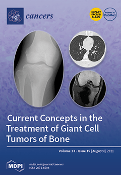
Cover Story (view full-size image): Giant cell tumor of bone is a primary bone tumor of intermediate malignancy. Since the United States Food and Drug Administration approved the use of denosumab for giant cell tumors of bone in 2013, the treatment strategy for giant cell tumors of bone has changed significantly. Denosumab can stop the growth of unresectable lesions and relieve pain. Preoperative administration of denosumab reduces extraosseous lesions, hardens the tumor, and facilitates en bloc resection. On the other hand, administration of denosumab before curettage makes curettage difficult due to osteosclerosis of the lesion. We review the literature on the treatment of giant cell tumors of bone and consider the treatment strategy for giant cell tumors of bone in the denosumab era. View this paper
  • Issues are regarded as officially published after their release is announced to the table of contents alert mailing list.
  • You may sign up for e-mail alerts to receive table of contents of newly released issues.
  • PDF is the official format for papers published in both, html and pdf forms. To view the papers in pdf format, click on the "PDF Full-text" link, and use the free Adobe Reader to open them.
Order results
Result details
Section
Select all
Export citation of selected articles as:
32 pages, 1859 KiB  
Review
Therapeutic Strategies Targeting Tumor Suppressor Genes in Pancreatic Cancer
by Kung-Kai Kuo, Pi-Jung Hsiao, Wen-Tsan Chang, Shih-Chang Chuang, Ya-Han Yang, Kenly Wuputra, Chia-Chen Ku, Jia-Bin Pan, Chia-Pei Li, Kohsuke Kato, Chung-Jung Liu, Deng-Chyang Wu and Kazunari K. Yokoyama
Cancers 2021, 13(15), 3920; https://doi.org/10.3390/cancers13153920 - 03 Aug 2021
Cited by 8 | Viewed by 3688
Abstract
The high mortality of pancreatic cancer is attributed to the insidious progression of this disease, which results in a delayed diagnosis and advanced disease stage at diagnosis. More than 35% of patients with pancreatic cancer are in stage III, whereas 50% are in [...] Read more.
The high mortality of pancreatic cancer is attributed to the insidious progression of this disease, which results in a delayed diagnosis and advanced disease stage at diagnosis. More than 35% of patients with pancreatic cancer are in stage III, whereas 50% are in stage IV at diagnosis. Thus, understanding the aggressive features of pancreatic cancer will contribute to the resolution of problems, such as its early recurrence, metastasis, and resistance to chemotherapy and radiotherapy. Therefore, new therapeutic strategies targeting tumor suppressor gene products may help prevent the progression of pancreatic cancer. In this review, we discuss several recent clinical trials of pancreatic cancer and recent studies reporting safe and effective treatment modalities for patients with advanced pancreatic cancer. Full article
(This article belongs to the Special Issue Tumor Suppressor Genes: Insight into the Cancer Therapy)
Show Figures

Graphical abstract

21 pages, 3071 KiB  
Review
Biomarkers and Lung Cancer Early Detection: State of the Art
by Elisa Dama, Tommaso Colangelo, Emanuela Fina, Marco Cremonesi, Marinos Kallikourdis, Giulia Veronesi and Fabrizio Bianchi
Cancers 2021, 13(15), 3919; https://doi.org/10.3390/cancers13153919 - 03 Aug 2021
Cited by 29 | Viewed by 5372
Abstract
Lung cancer burden is increasing, with 2 million deaths/year worldwide. Current limitations in early detection impede lung cancer diagnosis when the disease is still localized and thus more curable by surgery or multimodality treatment. Liquid biopsy is emerging as an important tool for [...] Read more.
Lung cancer burden is increasing, with 2 million deaths/year worldwide. Current limitations in early detection impede lung cancer diagnosis when the disease is still localized and thus more curable by surgery or multimodality treatment. Liquid biopsy is emerging as an important tool for lung cancer early detection and for monitoring therapy response. Here, we reviewed recent advances in liquid biopsy for early diagnosis of lung cancer. We summarized DNA- or RNA-based biomarkers, proteins, autoantibodies circulating in the blood, as well as circulating tumor cells (CTCs), and compared the most promising studies in terms of biomarkers prediction performance. While we observed an overall good performance for the proposed biomarkers, we noticed some critical aspects which may complicate the successful translation of these biomarkers into the clinical setting. We, therefore, proposed a roadmap for successful development of lung cancer biomarkers during the discovery, prioritization, and clinical validation phase. The integration of innovative minimally invasive biomarkers in screening programs is highly demanded to augment lung cancer early detection. Full article
(This article belongs to the Special Issue Cancer Biomarkers in Body Fluids)
Show Figures

Figure 1

21 pages, 775 KiB  
Review
The Role of Chronic Inflammation in the Development of Breast Cancer
by David N. Danforth
Cancers 2021, 13(15), 3918; https://doi.org/10.3390/cancers13153918 - 03 Aug 2021
Cited by 47 | Viewed by 11399
Abstract
Chronic inflammation contributes to the malignant transformation of several malignancies and is an important component of breast cancer. The role of chronic inflammation in the initiation and development of breast cancer from normal breast tissue, however, is unclear and needs to be clarified. [...] Read more.
Chronic inflammation contributes to the malignant transformation of several malignancies and is an important component of breast cancer. The role of chronic inflammation in the initiation and development of breast cancer from normal breast tissue, however, is unclear and needs to be clarified. A review of the literature was conducted to define the chronic inflammatory processes in normal breast tissue at risk for breast cancer and in breast cancer, including the role of lymphocyte and macrophage infiltrates, chronic active adipocytes and fibroblasts, and processes that may promote chronic inflammation including the microbiome and factors related to genomic abnormalities and cellular injury. The findings indicate that in healthy normal breast tissue there is systemic evidence to suggest inflammatory changes are present and associated with breast cancer risk, and adipocytes and crown-like structures in normal breast tissue may be associated with chronic inflammatory changes. The microbiome, genomic abnormalities, and cellular changes are present in healthy normal breast tissue, with the potential to elicit inflammatory changes, while infiltrating lymphocytes are uncommon in these tissues. Chronic inflammatory changes occur prominently in breast cancer tissues, with important contributions from tumor-infiltrating lymphocytes and tumor-associated macrophages, cancer-associated adipocytes and crown-like structures, and cancer-associated fibroblasts, while the microbiome and DNA damage may serve to promote inflammatory events. Together, these findings suggest that chronic inflammation may play a role in influencing the initiation, development and conduct of breast cancer, although several chronic inflammatory processes in breast tissue may occur later in breast carcinogenesis. Full article
(This article belongs to the Section Cancer Causes, Screening and Diagnosis)
Show Figures

Graphical abstract

22 pages, 42269 KiB  
Article
Adipocytes Promote Breast Cancer Cell Survival and Migration through Autophagy Activation
by Dorine Bellanger, Cléa Dziagwa, Cyrille Guimaraes, Michelle Pinault, Jean-François Dumas and Lucie Brisson
Cancers 2021, 13(15), 3917; https://doi.org/10.3390/cancers13153917 - 03 Aug 2021
Cited by 7 | Viewed by 2762
Abstract
White adipose tissue interacts closely with breast cancers through the secretion of soluble factors such as cytokines, growth factors or fatty acids. However, the molecular mechanisms of these interactions and their roles in cancer progression remain poorly understood. In this study, we investigated [...] Read more.
White adipose tissue interacts closely with breast cancers through the secretion of soluble factors such as cytokines, growth factors or fatty acids. However, the molecular mechanisms of these interactions and their roles in cancer progression remain poorly understood. In this study, we investigated the role of fatty acids in the cooperation between adipocytes and breast cancer cells using a co-culture model. We report that adipocytes increase autophagy in breast cancer cells through the acidification of lysosomes, leading to cancer cell survival in nutrient-deprived conditions and to cancer cell migration. Mechanistically, the disturbance of membrane phospholipid composition with a decrease in arachidonic acid content is responsible for autophagy activation in breast cancer cells induced by adipocytes. Therefore, autophagy might be a central cellular mechanism of white adipose tissue interactions with cancer cells and thus participate in cancer progression. Full article
(This article belongs to the Section Tumor Microenvironment)
Show Figures

Figure 1

11 pages, 540 KiB  
Article
Is Mammographic Breast Density an Endophenotype for Breast Cancer?
by Ellie Darcey, Nina McCarthy, Eric K. Moses, Christobel Saunders, Gemma Cadby and Jennifer Stone
Cancers 2021, 13(15), 3916; https://doi.org/10.3390/cancers13153916 - 03 Aug 2021
Cited by 4 | Viewed by 1762
Abstract
Mammographic breast density (MBD) is a strong and highly heritable predictor of breast cancer risk and a biomarker for the disease. This study systematically assesses MBD as an endophenotype for breast cancer—a quantitative trait that is heritable and genetically correlated with disease risk. [...] Read more.
Mammographic breast density (MBD) is a strong and highly heritable predictor of breast cancer risk and a biomarker for the disease. This study systematically assesses MBD as an endophenotype for breast cancer—a quantitative trait that is heritable and genetically correlated with disease risk. Using data from the family-based kConFab Study and the 1994/1995 cross-sectional Busselton Health Study, participants were divided into three status groups—cases, relatives of cases and controls. Participant’s mammograms were used to measure absolute dense area (DA) and percentage dense area (PDA). To address each endophenotype criterion, linear mixed models and heritability analysis were conducted. Both measures of MBD were significantly associated with breast cancer risk in two independent samples. These measures were also highly heritable. Meta-analyses of both studies showed that MBD measures were higher in cases compared to relatives (β = 0.48, 95% CI = 0.10, 0.86 and β = 0.41, 95% CI = 0.06, 0.78 for DA and PDA, respectively) and in relatives compared to controls (β = 0.16, 95% CI = −0.24, 0.56 and β = 0.16, 95% CI = −0.21, 0.53 for DA and PDA, respectively). This study formally demonstrates, for the first time, that MBD is an endophenotype for breast cancer. Full article
Show Figures

Figure 1

9 pages, 942 KiB  
Article
Effect of Scalp Cooling on the Pharmacokinetics of Paclitaxel
by Leni van Doorn, Mandy M. van Rosmalen, Wendy M. van der Deure, Esther Oomen-de Hoop, Robert Porrazzo, Sophie M. Wijngaard, Ingrid A. Boere, Paola Veenstra, Eman Ibrahim, Peter de Bruijn, Lena E. Friberg, Stijn L. W. Koolen, Ron H. J. Mathijssen and Agnes Jager
Cancers 2021, 13(15), 3915; https://doi.org/10.3390/cancers13153915 - 03 Aug 2021
Cited by 2 | Viewed by 2232
Abstract
Chemotherapy-induced alopecia (CIA), a side effect with high impact, can be prevented by cooling the scalp during the administration of some cytotoxic drugs. However, the effects of this prolonged scalp cooling on the pharmacokinetics of chemotherapy have never been investigated. In this study, [...] Read more.
Chemotherapy-induced alopecia (CIA), a side effect with high impact, can be prevented by cooling the scalp during the administration of some cytotoxic drugs. However, the effects of this prolonged scalp cooling on the pharmacokinetics of chemotherapy have never been investigated. In this study, we compared the pharmacokinetics of the widely used chemotherapeutic agent paclitaxel (weekly dose of 80–100 mg/m2) in female patients with solid tumors using concomitant scalp cooling (n = 14) or not (n = 24). Blood samples were collected in all patients for pharmacokinetic analyses up to 6 h after one course of paclitaxel administration. The primary endpoint was the clearance (L/h) of paclitaxel. Paclitaxel clearance—expressed as relative difference in geometric means—was 6.8% (90% CI: −16.7% to 4.4%) lower when paclitaxel was administered with concomitant scalp cooling versus paclitaxel infusions without scalp cooling. Within the subgroup of patients using scalp cooling, paclitaxel clearance was not statistically significantly different between patients with CIA (alopecia grade 1 or 2) and those without CIA. Hence, scalp cooling did not negatively influence the clearance of paclitaxel treatment. Full article
(This article belongs to the Special Issue Therapeutic Monitoring of Anti-cancer Agents)
Show Figures

Figure 1

17 pages, 2288 KiB  
Article
Consistency of Pituitary Adenoma: Prediction by Pharmacokinetic Dynamic Contrast-Enhanced MRI and Comparison with Histologic Collagen Content
by Kiyohisa Kamimura, Masanori Nakajo, Manisha Bohara, Daigo Nagano, Yoshihiko Fukukura, Shingo Fujio, Tomoko Takajo, Kazuhiro Tabata, Takashi Iwanaga, Hiroshi Imai, Marcel Dominik Nickel and Takashi Yoshiura
Cancers 2021, 13(15), 3914; https://doi.org/10.3390/cancers13153914 - 03 Aug 2021
Cited by 5 | Viewed by 2288
Abstract
Prediction of tumor consistency is valuable for planning transsphenoidal surgery for pituitary adenoma. A prospective study was conducted involving 49 participants with pituitary adenoma to determine whether quantitative pharmacokinetic analysis of dynamic contrast-enhanced magnetic resonance imaging (DCE-MRI) is useful for predicting consistency of [...] Read more.
Prediction of tumor consistency is valuable for planning transsphenoidal surgery for pituitary adenoma. A prospective study was conducted involving 49 participants with pituitary adenoma to determine whether quantitative pharmacokinetic analysis of dynamic contrast-enhanced magnetic resonance imaging (DCE-MRI) is useful for predicting consistency of adenomas. Pharmacokinetic parameters in the adenomas including volume of extravascular extracellular space (EES) per unit volume of tissue (ve), blood plasma volume per unit volume of tissue (vp), volume transfer constant between blood plasma and EES (Ktrans), and rate constant between EES and blood plasma (kep) were obtained. The pharmacokinetic parameters and the histologic percentage of collagen content (PCC) were compared between soft and hard adenomas using Mann–Whitney U test. Pearson’s correlation coefficient was used to correlate pharmacokinetic parameters with PCC. Hard adenomas showed significantly higher PCC (44.08 ± 15.14% vs. 6.62 ± 3.47%, p < 0.01), ve (0.332 ± 0.124% vs. 0.221 ± 0.104%, p < 0.01), and Ktrans (0.775 ± 0.401/min vs. 0.601 ± 0.612/min, p = 0.02) than soft adenomas. Moreover, a significant positive correlation was found between ve and PCC (r = 0.601, p < 0.01). The ve derived using DCE-MRI may have predictive value for consistency of pituitary adenoma. Full article
Show Figures

Figure 1

12 pages, 687 KiB  
Article
Plasma BRAF Mutation Detection for the Diagnostic and Monitoring Trajectory of Patients with LDH-High Stage IV Melanoma
by Sofie H. Tolmeijer, Rutger H. T. Koornstra, Jan Willem B. de Groot, Maartje J. Geerlings, Dirk H. van Rens, Marye J. Boers-Sonderen, Jack A. Schalken, Winald R. Gerritsen, Marjolijn J. L. Ligtenberg and Niven Mehra
Cancers 2021, 13(15), 3913; https://doi.org/10.3390/cancers13153913 - 03 Aug 2021
Cited by 5 | Viewed by 1798
Abstract
For patients with newly diagnosed metastatic melanoma, rapid BRAF mutation (mBRAF) assessment is vital to promptly initiate systemic therapy. Additionally, blood-based biomarkers are desired to monitor and predict treatment response. Circulating tumor DNA (ctDNA) has shown great promise for minimally invasive mBRAF assessment [...] Read more.
For patients with newly diagnosed metastatic melanoma, rapid BRAF mutation (mBRAF) assessment is vital to promptly initiate systemic therapy. Additionally, blood-based biomarkers are desired to monitor and predict treatment response. Circulating tumor DNA (ctDNA) has shown great promise for minimally invasive mBRAF assessment and treatment monitoring, but validation studies are needed. This prospective study utilized longitudinal plasma samples at regular timepoints (0, 6, 12, 18 weeks) to address the clinical validity of ctDNA measurements in stage IV melanoma patients with elevated serum lactate dehydrogenase (LDH > 250U/L) starting first-line systemic treatment. Using droplet digital PCR, the plasma mBRAF abundance was assessed in 53 patients with a BRAFV600 tissue mutation. Plasma mBRAF was detected in 50/51 patients at baseline (98% sensitivity; median fraction abundance of 19.5%) and 0/17 controls (100% specificity). Patients in whom plasma mBRAF became undetectable during the first 12–18 weeks of treatment had a longer progression-free survival (30.2 vs. 4.0 months; p < 0.001) and cancer-specific survival (not reached vs. 10.2 months; p < 0.001) compared to patients with detectable mBRAF. The ctDNA dynamics outperformed LDH and S100 dynamics. These results confirm the clinical validity of ctDNA measurements as a minimally invasive biomarker for the diagnostic and monitoring trajectory of patients with LDH-high stage IV melanoma. Full article
Show Figures

Figure 1

25 pages, 7510 KiB  
Systematic Review
Treatment-Related Adverse Events of Chimeric Antigen Receptor T-Cell (CAR T) in Clinical Trials: A Systematic Review and Meta-Analysis
by Wen Lei, Mixue Xie, Qi Jiang, Nengwen Xu, Ping Li, Aibin Liang, Ken H. Young and Wenbin Qian
Cancers 2021, 13(15), 3912; https://doi.org/10.3390/cancers13153912 - 03 Aug 2021
Cited by 26 | Viewed by 4542
Abstract
Chimeric antigen receptors T (CAR-T) cell therapy of cancer is a rapidly evolving field. It has been shown to be remarkably effective in cases of hematological malignancies, and its approval by the FDA has significantly increased the enthusiasm for wide clinical usage and [...] Read more.
Chimeric antigen receptors T (CAR-T) cell therapy of cancer is a rapidly evolving field. It has been shown to be remarkably effective in cases of hematological malignancies, and its approval by the FDA has significantly increased the enthusiasm for wide clinical usage and development of novel CAR-T therapies. However, it has also challenged physicians and investigators to recognize and deal with treatment-associated toxicities. A total of 2592 patients were included from 84 eligible studies that were systematically searched and reviewed from the databases of PubMed, de, the American Society of Hematology and the Cochrane Library. The meta-analysis and subgroup analysis by a Bayesian logistic regression model were used to evaluate the incidences of therapy-related toxicities such as cytokine release syndrome (CRS) and neurological symptoms (NS), and the differences between different targets and cancer types were analyzed. The pooled all-grade CRS rate and grade ≥ 3 CRS rate was 77% and 29%, respectively, with a significantly higher incidence in the hematologic malignancies (all-grade: 81%; grade ≥ 3: 29%) than in solid tumors (all-grade: 37%; grade ≥ 3: 19%). The pooled estimate NS rate from the individual studies were 40% for all-grade and 28% for grade ≥ 3. It was also higher in the hematologic subgroup than in the solid tumors group. The subgroup analysis by cancer type showed that higher incidences of grade ≥ 3 CRS were observed in anti-CD19 CAR-T therapy for ALL and NHL, anti-BCMA CAR-T for MM, and anti-CEA CAR-T for solid tumors, which were between 24–36%, while higher incidences of grade ≥ 3 NS were mainly observed in CD19-ALL/NHL (23–37%) and BCMA-MM (12%). Importantly, subgroup analysis on anti-CD19 CAR-T studies showed that young patients (vs. adult patients), allologous T cell origin (vs. autologous origin), gamma retrovirus vector, and higher doses of CAR-T cells were associated with high-grade CRS. On the other hand, the patients with NHL (vs ALL), administered with higher dose of CAR-T, and adult patients (vs. young patients) had an increased incidence of grade ≥ 3 NS events. This study offers a comprehensive summary of treatment-related toxicity and will guide future clinical trials and therapeutic designs investigating CAR T cell therapy. Full article
(This article belongs to the Special Issue Advances in Cancer Immunotherapy)
Show Figures

Figure 1

17 pages, 3786 KiB  
Article
Radiation-Associated Angiosarcoma of the Breast and Chest Wall Treated with Thermography-Controlled, Contactless wIRA-Hyperthermia and Hypofractionated Re-Irradiation
by Markus Notter, Emanuel Stutz, Andreas R. Thomsen and Peter Vaupel
Cancers 2021, 13(15), 3911; https://doi.org/10.3390/cancers13153911 - 03 Aug 2021
Cited by 7 | Viewed by 4674
Abstract
Background: Radiation-associated angiosarcoma of the breast (RAASB) is a rare, challenging disease, with surgery being the accepted basic therapeutic approach. In contrast, the role of adjuvant and systemic therapies is a subject of some controversy. Local recurrence rates reported in the literature are [...] Read more.
Background: Radiation-associated angiosarcoma of the breast (RAASB) is a rare, challenging disease, with surgery being the accepted basic therapeutic approach. In contrast, the role of adjuvant and systemic therapies is a subject of some controversy. Local recurrence rates reported in the literature are mostly heterogeneous and are highly dependent on the extent of surgery. In cases of locally recurrent or unresectable RAASB, prognosis is very poor. Methods: We retrospectively report on 10 consecutive RAASB patients, most of them presenting with locally recurrent or unresectable RAASB, which were treated with thermography-controlled water-filtered infrared-A (wIRA) superficial hyperthermia (HT) immediately followed by re-irradiation (re-RT). Patients with RAASB were graded based on their tumor extent before onset of radiotherapy (RT). Results: We recorded a local control (LC) rate dependent on tumor extent ranging from a high LC rate of 100% (two of two patients) in the adjuvant setting with an R0 or R2 resection to a limited LC rate of 33% (one of three patients) in patients with inoperable, macroscopic tumor lesions. Conclusion: Combined HT and re-RT should be considered as an option (a) for adjuvant treatment of RAASB, especially in cases with positive resection margins and after surgery of local recurrence (LR), and (b) for definitive treatment of unresectable RAASB. Full article
(This article belongs to the Special Issue Hyperthermia in Cancer)
Show Figures

Figure 1

20 pages, 1328 KiB  
Review
An Overview on the Histogenesis and Morphogenesis of Salivary Gland Neoplasms and Evolving Diagnostic Approaches
by Janaki Iyer, Arvind Hariharan, Uyen Minh Nha Cao, Crystal To Tam Mai, Athena Wang, Parisa Khayambashi, Bich Hong Nguyen, Lydia Safi and Simon D. Tran
Cancers 2021, 13(15), 3910; https://doi.org/10.3390/cancers13153910 - 03 Aug 2021
Cited by 27 | Viewed by 8354
Abstract
Salivary gland neoplasms (SGN) remain a diagnostic dilemma due to their heterogenic complex behavior. Their diverse histomorphological appearance is attributed to the underlying cellular mechanisms and differentiation into various histopathological subtypes with overlapping fea-tures. Diagnostic tools such as fine needle aspiration biopsy, computerized [...] Read more.
Salivary gland neoplasms (SGN) remain a diagnostic dilemma due to their heterogenic complex behavior. Their diverse histomorphological appearance is attributed to the underlying cellular mechanisms and differentiation into various histopathological subtypes with overlapping fea-tures. Diagnostic tools such as fine needle aspiration biopsy, computerized tomography, magnetic resonance imaging, and positron emission tomography help evaluate the structure and assess the staging of SGN. Advances in molecular pathology have uncovered genetic patterns and oncogenes by immunohistochemistry, fluorescent in situ hybridization, and next–generation sequencing, that may potentially contribute to innovating diagnostic approaches in identifying various SGN. Surgical resection is the principal treatment for most SGN. Other modalities such as radiotherapy, chemotherapy, targeted therapy (agents like tyrosine kinase inhibitors, monoclonal antibodies, and proteasome inhibitors), and potential hormone therapy may be applied, depending on the clinical behaviors, histopathologic grading, tumor stage and location, and the extent of tissue invasion. This review delves into the molecular pathways of salivary gland tumorigenesis, highlighting recent diagnostic protocols that may facilitate the identification and management of SGN. Full article
Show Figures

Graphical abstract

26 pages, 2140 KiB  
Review
MicroRNA and Other Non-Coding RNAs in Epstein–Barr Virus-Associated Cancers
by Kin Israel Notarte, Suranga Senanayake, Imee Macaranas, Pia Marie Albano, Lucia Mundo, Eanna Fennell, Lorenzo Leoncini and Paul Murray
Cancers 2021, 13(15), 3909; https://doi.org/10.3390/cancers13153909 - 03 Aug 2021
Cited by 15 | Viewed by 4634
Abstract
EBV is a direct causative agent in around 1.5% of all cancers. The oncogenic properties of EBV are related to its ability to activate processes needed for cellular proliferation, survival, migration, and immune evasion. The EBV latency program is required for the immortalization [...] Read more.
EBV is a direct causative agent in around 1.5% of all cancers. The oncogenic properties of EBV are related to its ability to activate processes needed for cellular proliferation, survival, migration, and immune evasion. The EBV latency program is required for the immortalization of infected B cells and involves the expression of non-coding RNAs (ncRNAs), including viral microRNAs. These ncRNAs have different functions that contribute to virus persistence in the asymptomatic host and to the development of EBV-associated cancers. In this review, we discuss the function and potential clinical utility of EBV microRNAs and other ncRNAs in EBV-associated malignancies. This review is not intended to be comprehensive, but rather to provide examples of the importance of ncRNAs. Full article
(This article belongs to the Special Issue Epstein-Barr Virus Infection in Cancer)
Show Figures

Figure 1

18 pages, 2337 KiB  
Article
Application of Community Detection Algorithm to Investigate the Correlation between Imaging Biomarkers of Tumor Metabolism, Hypoxia, Cellularity, and Perfusion for Precision Radiotherapy in Head and Neck Squamous Cell Carcinomas
by Ramesh Paudyal, Milan Grkovski, Jung Hun Oh, Heiko Schöder, David Aramburu Nunez, Vaios Hatzoglou, Joseph O. Deasy, John L. Humm, Nancy Y. Lee and Amita Shukla-Dave
Cancers 2021, 13(15), 3908; https://doi.org/10.3390/cancers13153908 - 03 Aug 2021
Cited by 3 | Viewed by 2183
Abstract
The present study aimed to investigate the correlation at pre-treatment (TX) between quantitative metrics derived from multimodality imaging (MMI), including 18F-FDG-PET/CT, 18F-FMISO-PET/CT, DW- and DCE-MRI, using a community detection algorithm (CDA) in head and neck squamous cell carcinoma (HNSCC) patients. Twenty-three [...] Read more.
The present study aimed to investigate the correlation at pre-treatment (TX) between quantitative metrics derived from multimodality imaging (MMI), including 18F-FDG-PET/CT, 18F-FMISO-PET/CT, DW- and DCE-MRI, using a community detection algorithm (CDA) in head and neck squamous cell carcinoma (HNSCC) patients. Twenty-three HNSCC patients with 27 metastatic lymph nodes underwent a total of 69 MMI exams at pre-TX. Correlations among quantitative metrics derived from FDG-PET/CT (SUL), FMSIO-PET/CT (K1, k3, TBR, and DV), DW-MRI (ADC, IVIM [D, D*, and f]), and FXR DCE-MRI [Ktrans, ve, and τi]) were investigated using the CDA based on a “spin-glass model” coupled with the Spearman’s rank, ρ, analysis. Mean MRI T2 weighted tumor volumes and SULmean values were moderately positively correlated (ρ = 0.48, p = 0.01). ADC and D exhibited a moderate negative correlation with SULmean (ρ ≤ −0.42, p < 0.03 for both). K1 and Ktrans were positively correlated (ρ = 0.48, p = 0.01). In contrast, Ktrans and k3max were negatively correlated (ρ = −0.41, p = 0.03). CDA revealed four communities for 16 metrics interconnected with 33 edges in the network. DV, Ktrans, and K1 had 8, 7, and 6 edges in the network, respectively. After validation in a larger population, the CDA approach may aid in identifying useful biomarkers for developing individual patient care in HNSCC. Full article
(This article belongs to the Special Issue Human Papillomavirus and Head and Neck Cancer)
Show Figures

Graphical abstract

18 pages, 5358 KiB  
Article
Distinct Molecular Mechanisms of Altered HLA Class II Expression in Malignant Melanoma
by Stefanie Meyer, Diana Handke, Anja Mueller, Katharina Biehl, Markus Kreuz, Jürgen Bukur, Ulrike Koehl, Maria-Filothei Lazaridou, Mark Berneburg, André Steven, Chiara Massa and Barbara Seliger
Cancers 2021, 13(15), 3907; https://doi.org/10.3390/cancers13153907 - 03 Aug 2021
Cited by 5 | Viewed by 2239
Abstract
Background: The human leukocyte antigen (HLA) class II molecules are constitutively expressed in some melanoma, but the underlying molecular mechanisms have not yet been characterized. Methods: The expression of HLA class II antigen processing machinery (APM) components was determined in melanoma samples by [...] Read more.
Background: The human leukocyte antigen (HLA) class II molecules are constitutively expressed in some melanoma, but the underlying molecular mechanisms have not yet been characterized. Methods: The expression of HLA class II antigen processing machinery (APM) components was determined in melanoma samples by qPCR, Western blot, flow cytometry and immunohistochemistry. Immunohistochemical and TCGA datasets were used for correlation of HLA class II expression to tumor grading, T-cell infiltration and patients’ survival. Results: The heterogeneous HLA class II expression in melanoma samples allowed us to characterize four distinct phenotypes. Phenotype I totally lacks constitutive HLA class II surface expression, which is inducible by interferon-gamma (IFN-γ); phenotype II expresses low basal surface HLA class II that is further upregulated by IFN-γ; phenotype III lacks constitutive and IFN-γ controlled HLA class II expression, but could be induced by epigenetic drugs; and in phenotype IV, lack of HLA class II expression is not recovered by any drug tested. High levels of HLA class II APM component expression were associated with an increased intra-tumoral CD4+ T-cell density and increased patients’ survival. Conclusions: The heterogeneous basal expression of HLA class II antigens and/or APM components in melanoma cells is caused by distinct molecular mechanisms and has clinical relevance. Full article
Show Figures

Figure 1

21 pages, 5335 KiB  
Article
Inhibitors Targeting CDK9 Show High Efficacy against Osimertinib and AMG510 Resistant Lung Adenocarcinoma Cells
by Jaya Padmanabhan, Biswarup Saha, Chase Powell, Qianxing Mo, Bradford A. Perez and Srikumar Chellappan
Cancers 2021, 13(15), 3906; https://doi.org/10.3390/cancers13153906 - 03 Aug 2021
Cited by 9 | Viewed by 3943
Abstract
Non-small cell lung cancer has a 5-year survival rate of less than 12–15%, calling for the development of additional therapeutic strategies to combat this disease. Here we tested the efficacy of inhibiting cyclin-dependent kinase 9 (CDK9) on lung cancer cell lines with K-Ras [...] Read more.
Non-small cell lung cancer has a 5-year survival rate of less than 12–15%, calling for the development of additional therapeutic strategies to combat this disease. Here we tested the efficacy of inhibiting cyclin-dependent kinase 9 (CDK9) on lung cancer cell lines with K-Ras and EGFR mutations and on lung cancer organoids. Three different CDK9 inhibitors reduced the viability and anchorage-independent growth of lung cancer cell lines at very low nanomolar to micromolar concentrations. CDK9 inhibition suppressed the expression of the anti-apoptotic protein, Mcl1, as well as the embryonic stem cell transcription factors, Sox2 and Sox9, which are pro-tumorigenic. In contrast, treatment with CDK9 inhibitors increased the levels of WT p53 and its downstream target p21 in K-Ras mutant cell lines. Furthermore, the CDK9 inhibitors could markedly reduce the viability of Osimertinib-resistant PC9 and AMG510-resistant H23 and H358 cells with comparable efficacy as the parental cells. CDK9 inhibitors could also significantly reduce the growth and viability of lung cancer organoids with high potency. Taken together, the data presented here strongly suggest that CDK9 inhibitors would be efficacious against K-Ras mutant and EGFR mutant NSCLCs, including those that develop resistance to targeted therapies. Full article
(This article belongs to the Special Issue Non-small Cell Lung Cancer--Tumor Biology)
Show Figures

Figure 1

15 pages, 1790 KiB  
Article
Evaluation of Colon-Specific Plasma Nanovesicles as New Markers of Colorectal Cancer
by Inga Nazarova, Maria Slyusarenko, Elena Sidina, Nadezhda Nikiforova, Vladislav Semiglazov, Tatiana Semiglazova, Achim Aigner, Evgeny Rybakov and Anastasia Malek
Cancers 2021, 13(15), 3905; https://doi.org/10.3390/cancers13153905 - 03 Aug 2021
Cited by 5 | Viewed by 2797
Abstract
Purpose: Developing new and efficient approaches for the early diagnosis of colorectal cancer (CRC) is an important issue. Circulating extracellular nanovesicles (ENVs) present a promising class of cancer markers. Cells of well-differentiated adenocarcinomas retain the molecular characteristics of colon epithelial cells, and the [...] Read more.
Purpose: Developing new and efficient approaches for the early diagnosis of colorectal cancer (CRC) is an important issue. Circulating extracellular nanovesicles (ENVs) present a promising class of cancer markers. Cells of well-differentiated adenocarcinomas retain the molecular characteristics of colon epithelial cells, and the ENVs secreted by these cells may have colon-specific surface markers. We hypothesize that an increase in the number of ENVs carrying colon-specific markers could serve as a diagnostic criterion for colorectal cancer. Experimental design: Potential colon-specific markers were selected based on tissue-specific expression profile and cell surface membrane localization data. Plasma was collected from CRC patients (n = 48) and healthy donors (n = 50). The total population of ENVs was isolated with a two-phase polymer system. ENVs derived from colon epithelium cells were isolated using immune-beads with antibodies to colon-specific markers prior to labelling with antibodies against exosomal tetraspanins (CD63 and CD9) and quantification by flow cytometry. Results: The number of ENVs positive for single colon cancer markers was found to be significantly higher in the plasma of CRC patients compared with healthy donors. The efficacy of detection depends on the method of ENV labelling. The diagnostic efficacy was estimated by ROC analysis (the AUC varied between 0.71 and 0.79). The multiplexed isolation of colon-derived ENVs using immune-beads decorated with antibodies against five markers allowed for a further increase in the diagnostic potency of the method (AUC = 0.82). Conclusions: ENVs derived from colon epithelium may serve as markers of differentiated CRC (adenocarcinomas). The composition of ligands used for capturing colon-derived ENVs and their method of labelling are critical for the efficacy of this proposed diagnostic approach. Full article
(This article belongs to the Special Issue Molecular Biomarkers in Colorectal Cancer)
Show Figures

Figure 1

29 pages, 1058 KiB  
Review
PSMA Theranostics: Science and Practice
by Kgomotso Mokoala, Ismaheel Lawal, Thabo Lengana, Mankgopo Kgatle, Frederik L. Giesel, Mariza Vorster and Mike Sathekge
Cancers 2021, 13(15), 3904; https://doi.org/10.3390/cancers13153904 - 02 Aug 2021
Cited by 22 | Viewed by 6136
Abstract
Prostate cancer (PCa) causes significant morbidity and mortality in men globally. While localized PCa may be managed with curative intent by surgery and/or radiation therapy, the management of advanced hormone resistant metastatic disease (mCRPC) is more challenging. Theranostics is a principle based on [...] Read more.
Prostate cancer (PCa) causes significant morbidity and mortality in men globally. While localized PCa may be managed with curative intent by surgery and/or radiation therapy, the management of advanced hormone resistant metastatic disease (mCRPC) is more challenging. Theranostics is a principle based on the ability to use an organ specific ligand and label it to both a diagnostic and a therapeutic agent. The overexpression of prostate specific membrane antigen (PSMA) on prostate cancer cells creates a unique opportunity for development of targeted radionuclide therapy. The use of both beta and alpha emitting particles has shown great success. Several clinical trials have been initiated assessing the efficacy and safety profile of these radionuclide agents. The results are encouraging with PSMA directed radioligand therapy performing well in patients who have exhausted all other standard treatment options. Future studies need to assess the timing of introduction of these radionuclide therapies in the management schema of mCRPC. Drugs or therapies are not without side effects and targeted radionuclide therapies presents a new set of toxicities including xerostomia and myelosuppression. New therapeutic strategies are being explored to improve outcomes while keeping toxicities to a minimum. This review aims to look at the various PSMA labelled tracers that form part of the theragnostic approach and subsequently delve into the progress made in the area of radionuclide therapy. Full article
(This article belongs to the Special Issue Molecular Imaging and Radio-Nuclide Therapy in Cancers)
Show Figures

Figure 1

16 pages, 3154 KiB  
Article
Identification of CNGB1 as a Predictor of Response to Neoadjuvant Chemotherapy in Muscle-Invasive Bladder Cancer
by Anastasia C. Hepburn, Nicola Lazzarini, Rajan Veeratterapillay, Laura Wilson, Jaume Bacardit and Rakesh Heer
Cancers 2021, 13(15), 3903; https://doi.org/10.3390/cancers13153903 - 02 Aug 2021
Cited by 5 | Viewed by 2652
Abstract
Cisplatin-based neoadjuvant chemotherapy (NAC) is recommended prior to radical cystectomy for muscle-invasive bladder cancer (MIBC) patients. Despite a 5–10% survival benefit, some patients do not respond and experience substantial toxicity and delay in surgery. To date, there are no clinically approved biomarkers predictive [...] Read more.
Cisplatin-based neoadjuvant chemotherapy (NAC) is recommended prior to radical cystectomy for muscle-invasive bladder cancer (MIBC) patients. Despite a 5–10% survival benefit, some patients do not respond and experience substantial toxicity and delay in surgery. To date, there are no clinically approved biomarkers predictive of response to NAC and their identification is urgently required for more precise delivery of care. To address this issue, a multi-methods analysis approach of machine learning and differential gene expression analysis was undertaken on a cohort of 30 MIBC cases highly selected for an exquisitely strong response to NAC or marked resistance and/or progression (discovery cohort). RGIFE (ranked guided iterative feature elimination) machine learning algorithm, previously demonstrated to have the ability to select biomarkers with high predictive power, identified a 9-gene signature (CNGB1, GGH, HIST1H4F, IDO1, KIF5A, MRPL4, NCDN, PRRT3, SLC35B3) able to select responders from non-responders with 100% predictive accuracy. This novel signature correlated with overall survival in meta-analysis performed using published NAC treated-MIBC microarray data (validation cohort 1, n = 26, Log rank test, p = 0.02). Corroboration with differential gene expression analysis revealed cyclic nucleotide-gated channel, CNGB1, as the top ranked upregulated gene in non-responders to NAC. A higher CNGB1 immunostaining score was seen in non-responders in tissue microarray analysis of the discovery cohort (n = 30, p = 0.02). Kaplan-Meier analysis of a further cohort of MIBC patients (validation cohort 2, n = 99) demonstrated that a high level of CNGB1 expression associated with shorter cancer specific survival (p < 0.001). Finally, in vitro studies showed siRNA-mediated CNGB1 knockdown enhanced cisplatin sensitivity of MIBC cell lines, J82 and 253JB-V. Overall, these data reveal a novel signature gene set and CNGB1 as a simpler proxy as a promising biomarker to predict chemoresponsiveness of MIBC patients. Full article
(This article belongs to the Special Issue Bladder Cancers)
Show Figures

Figure 1

17 pages, 1901 KiB  
Study Protocol
Irreversible Electroporation and Nivolumab Combined with Intratumoral Administration of a Toll-Like Receptor Ligand, as a Means of In Vivo Vaccination for Metastatic Pancreatic Ductal Adenocarcinoma (PANFIRE-III). A Phase-I Study Protocol
by Bart Geboers, Florentine E. F. Timmer, Alette H. Ruarus, Johanna E. E. Pouw, Evelien A. C. Schouten, Joyce Bakker, Robbert S. Puijk, Sanne Nieuwenhuizen, Madelon Dijkstra, M. Petrousjka van den Tol, Jan J. J. de Vries, Daniela E. Oprea-Lager, C. Willemien Menke-van der Houven van Oordt, Hans J. van der Vliet, Johanna W. Wilmink, Hester J. Scheffer, Tanja D. de Gruijl, Martijn R. Meijerink and on behalf of the Dutch Pancreatic Cancer Group
Cancers 2021, 13(15), 3902; https://doi.org/10.3390/cancers13153902 - 02 Aug 2021
Cited by 16 | Viewed by 3849
Abstract
Irreversible electroporation (IRE) is a novel image-guided tumor ablation technique with the ability to generate a window for the establishment of systemic antitumor immunity. IRE transiently alters the tumor’s immunosuppressive microenvironment while simultaneously generating antigen release, thereby instigating an adaptive immune response. Combining [...] Read more.
Irreversible electroporation (IRE) is a novel image-guided tumor ablation technique with the ability to generate a window for the establishment of systemic antitumor immunity. IRE transiently alters the tumor’s immunosuppressive microenvironment while simultaneously generating antigen release, thereby instigating an adaptive immune response. Combining IRE with immunotherapeutic drugs, i.e., electroimmunotherapy, has synergistic potential and might induce a durable antitumor response. The primary objective of this study is to assess the safety of the combination of IRE with IMO-2125 (a toll-like receptor 9 ligand) and/or nivolumab in patients with metastatic pancreatic ductal adenocarcinoma (mPDAC). In this randomized controlled phase I clinical trial, 18 patients with mPDAC pretreated with chemotherapy will be enrolled in one of three study arms: A (control): nivolumab monotherapy; B: percutaneous IRE of the primary tumor followed by nivolumab; or C: intratumoral injection of IMO-2125 followed by percutaneous IRE of the primary tumor and nivolumab. Assessments include contrast enhanced computed tomography (ceCT), 18F-FDG and 18F-BMS-986192 (PD-L1) positron emission tomography (PET)-CT, biopsies of the primary tumor and metastases, peripheral blood samples, and quality of life and pain questionnaires. There is no curative treatment option for patients with mPDAC, and palliative chemotherapy regimens only moderately improve survival. Consequently, there is an urgent need for innovative and radically different treatment approaches. Should electroimmunotherapy establish an effective and durable anti-tumor response, it may ultimately improve PDAC’s dismal prognosis. Full article
(This article belongs to the Special Issue Recent Advances in Pancreatic Ductal Adenocarcinoma)
Show Figures

Figure 1

13 pages, 2433 KiB  
Article
Effect of Neoadjuvant Therapies on Soft Tissue Sarcomas with Tail-like Lesions: A Multicenter Retrospective Study
by Hisaki Aiba, Kunihiro Ikuta, Kunihiro Asanuma, Katsuhisa Kawanami, Satoshi Tsukushi, Akihiko Matsumine, Daisuke Ishimura, Akihito Nagano, Yoji Shido, Eiji Kozawa, Kenji Yamada, Junji Wasa, Hiroaki Kimura, Takao Sakai, Hideki Murakami, Tomohisa Sakai, Tomoki Nakamura and Yoshihiro Nishida
Cancers 2021, 13(15), 3901; https://doi.org/10.3390/cancers13153901 - 02 Aug 2021
Cited by 1 | Viewed by 1944
Abstract
Several types of soft tissue sarcomas have peripheral infiltrative growth characteristics called tail-like lesions. The efficacy of neoadjuvant therapy for tumors with tail-like lesions has not been elucidated. From 2012 to 2019, we analyzed 36 patients with soft tissue sarcoma with tail-like lesions [...] Read more.
Several types of soft tissue sarcomas have peripheral infiltrative growth characteristics called tail-like lesions. The efficacy of neoadjuvant therapy for tumors with tail-like lesions has not been elucidated. From 2012 to 2019, we analyzed 36 patients with soft tissue sarcoma with tail-like lesions treated with neoadjuvant therapy, including chemotherapy, radiotherapy, or both. The effect of neoadjuvant therapy on the tail sign was investigated by analyzing the change in tail-like lesions during neoadjuvant therapy and histological responses. The median length of the tail-like lesion reduced from 29.5 mm at initiation to 19.5 mm after neoadjuvant therapy. The extent of shrinkage in tail-like lesions was related to the histopathological responses in the main part of the tumor. Complete disappearance of the tail-like lesion was observed in 12 patients; however, it was not related to achieving a microscopically negative margin. The oncologic outcomes did not significantly differ between cases with and without the complete disappearance of tail-like lesions. This study indicated that the shrinkage of tail-like lesions did not have a significant effect on complete resection or improvements of clinical outcomes. A more comprehensive evaluation is needed to elaborate on the surgical strategy. Full article
(This article belongs to the Special Issue Diagnosis and Treatment for Bone Tumor and Sarcoma)
Show Figures

Figure 1

11 pages, 1215 KiB  
Article
Clinicobiological Characteristics and Outcomes of Patients with T-Cell Large Granular Lymphocytic Leukemia and Chronic Lymphoproliferative Disorder of Natural Killer Cells from a Single Institution
by Andrea Rivero, Pablo Mozas, Laura Jiménez, Mónica López-Guerra, Dolors Colomer, Alex Bataller, Juan Correa, Alfredo Rivas-Delgado, Gabriela Bastidas, Tycho Baumann, Alejandra Martínez-Trillos, Julio Delgado, Eva Giné, Elías Campo, Armando López-Guillermo, Neus Villamor, Laura Magnano and Estella Matutes
Cancers 2021, 13(15), 3900; https://doi.org/10.3390/cancers13153900 - 02 Aug 2021
Cited by 11 | Viewed by 2312
Abstract
T-cell large granular lymphocytic leukemia (T-LGLL) and chronic lymphoproliferative disorder of natural killer (NK) cells are two infrequent diseases characterized by clonal expansions of cytotoxic T lymphocytes and NK cells, respectively. Somatic mutations of STAT3 are involved in the pathogenesis of these entities. [...] Read more.
T-cell large granular lymphocytic leukemia (T-LGLL) and chronic lymphoproliferative disorder of natural killer (NK) cells are two infrequent diseases characterized by clonal expansions of cytotoxic T lymphocytes and NK cells, respectively. Somatic mutations of STAT3 are involved in the pathogenesis of these entities. We describe the clinicobiological features, mutational status of STAT3/STAT5B, treatment and outcome of 131 patients. Neutropenia was the most frequent finding at diagnosis, followed by anemia. Concurrent hematological disorders were diagnosed in 37% of patients and autoimmune conditions and solid tumors in 17% and 15%, respectively. All patients who needed treatment belonged to the CD8+CD57+ group. Remarkably, patients included in the CD4+ group had a higher association with solid tumors (p = 0.037). STAT3 mutations were found in 17% of patients, mainly Y640F and D661Y mutations. Patients carrying STAT3 mutations more frequently presented with anemia, neutropenia, high LDH, high large granular lymphocyte counts and need for treatment (p = 0.0037). Methotrexate was the most frequently used agent (72% of cases). The overall response rate to all treatments was 50%. The 10-year overall survival of this series was 78%, with no differences according to the mutational status of STAT3. We compared the survival of these patients with the general Spanish population and no differences were found, confirming the indolent nature of these hematological malignancies. Our study further extends findings documented by others on the clinical behavior of the disease and the impact of STAT3, and for the first time analyzes survival compared to a matched general Spanish population. Full article
(This article belongs to the Special Issue Large Granular Lymphocytic Leukemia: Genomics and Immunome)
Show Figures

Figure 1

15 pages, 1185 KiB  
Review
APLN/APLNR Signaling Controls Key Pathological Parameters of Glioblastoma
by Roland E. Kälin and Rainer Glass
Cancers 2021, 13(15), 3899; https://doi.org/10.3390/cancers13153899 - 02 Aug 2021
Cited by 6 | Viewed by 2784
Abstract
Glioblastoma (GBM) is the most common and aggressive primary brain tumor in adults. GBM-expansion depends on a dense vascular network and, coherently, GBMs are highly angiogenic. However, new intratumoral blood vessels are often aberrant with consequences for blood-flow and vascular barrier function. Hence, [...] Read more.
Glioblastoma (GBM) is the most common and aggressive primary brain tumor in adults. GBM-expansion depends on a dense vascular network and, coherently, GBMs are highly angiogenic. However, new intratumoral blood vessels are often aberrant with consequences for blood-flow and vascular barrier function. Hence, the delivery of chemotherapeutics into GBM can be compromised. Furthermore, leaky vessels support edema-formation, which can result in severe neurological deficits. The secreted signaling peptide Apelin (APLN) plays an important role in the formation of GBM blood vessels. Both APLN and the Apelin receptor (APLNR) are upregulated in GBM cells and control tumor cell invasiveness. Here we summarize the current evidence on the role of APLN/APLNR signaling during brain tumor pathology. We show that targeting APLN/APLNR can induce anti-angiogenic effects in GBM and simultaneously blunt GBM cell infiltration. In addition, we discuss how manipulation of APLN/APLNR signaling in GBM leads to the normalization of tumor vessels and thereby supports chemotherapy, reduces edema, and improves anti-tumorigenic immune reactions. Hence, therapeutic targeting of APLN/APLNR signaling offers an interesting option to address different pathological hallmarks of GBM. Full article
(This article belongs to the Special Issue Targeted Therapies for the Treatment of Glioblastoma)
Show Figures

Figure 1

13 pages, 1365 KiB  
Article
Prognostic Discrimination of Alternative Lymph Node Classification Systems for Patients with Radically Resected Non-Metastatic Colorectal Cancer: A Cohort Study from a Single Tertiary Referral Center
by Dimitrios Prassas, Pablo Emilio Verde, Carlo Pavljak, Alexander Rehders, Sarah Krieg, Tom Luedde, Wolfram Trudo Knoefel and Andreas Krieg
Cancers 2021, 13(15), 3898; https://doi.org/10.3390/cancers13153898 - 02 Aug 2021
Cited by 7 | Viewed by 2117
Abstract
Background: Lymph node ratio (LNR) and the Log odds of positive lymph nodes (LODDS) have been proposed as a new prognostic indicator in surgical oncology. Various studies have shown a superior discriminating power of LODDS over LNR and lymph node category (N) in [...] Read more.
Background: Lymph node ratio (LNR) and the Log odds of positive lymph nodes (LODDS) have been proposed as a new prognostic indicator in surgical oncology. Various studies have shown a superior discriminating power of LODDS over LNR and lymph node category (N) in diverse cancer entities, when examined as a continuous variable. However, for each of the classification systems various cut-off values have been defined, with the question of the most appropriate for patients with CRC still remaining open. The present study aimed to compare the predictive impact of different lymph node classification systems and to define the best cut-off values regarding accurate evaluation of overall survival in patients with resectable, non-metastatic colorectal cancer (CRC). Methods: CRC patients who underwent surgical resection from 1996 to 2018 were extracted from our medical data base. Cox proportional hazards regression models and C-statistics were performed to assess the discriminative power of 25 LNR and 26 LODDS classifications. Regression models were adjusted for age, sex, extent of the tumor, differentiation, tumor size and localization. Results: Our study group consisted of 654 consecutive patients with non-metastatic CRC. C-statistic revealed 2 LNR and 5 LODDS classifications that demonstrated superior prognostic performance in patients with UICC III CRC, compared to the N category. No clear advantage of one classification over another could be demonstrated in any other patient subgroup. Conclusions: Distinct LNR and LODDS classifications demonstrate a prognostic superiority over the N category only in patients with Stage III radically resected CRC. Full article
Show Figures

Figure 1

17 pages, 8206 KiB  
Article
The Most Common VHL Point Mutation R167Q in Hereditary VHL Disease Interferes with Cell Plasticity Regulation
by Stéphanie Buart, Stéphane Terry, M’boyba Khadija Diop, Philippe Dessen, Sophie Couvé, Abdérémane Abdou, Julien Adam, Jérôme Thiery, Pierre Savagner and Salem Chouaib
Cancers 2021, 13(15), 3897; https://doi.org/10.3390/cancers13153897 - 02 Aug 2021
Cited by 3 | Viewed by 2585
Abstract
Von Hippel–Lindau disease (VHL) is a rare hereditary syndrome due to mutations of the VHL tumor suppressor gene. Patients harboring the R167Q mutation of the VHL gene have a high risk of developing ccRCCs. We asked whether the R167Q mutation with [...] Read more.
Von Hippel–Lindau disease (VHL) is a rare hereditary syndrome due to mutations of the VHL tumor suppressor gene. Patients harboring the R167Q mutation of the VHL gene have a high risk of developing ccRCCs. We asked whether the R167Q mutation with critical aspects of pseudo-hypoxia interferes with tumor plasticity. For this purpose, we used wild-type VHL (WT-VHL) and VHL-R167Q reconstituted cells. We showed that WT-VHL and VHL-R167Q expression had a similar effect on cell morphology and colony formation. However, cells transfected with VHL-R167Q display an intermediate, HIF2-dependent, epithelial–mesenchymal phenotype. Using RNA sequencing, we showed that this mutation upregulates the expression of genes involved in the hypoxia pathway, indicating that such mutation is conferring an enhanced pseudo-hypoxic state. Importantly, this hypoxic state correlates with the induction of genes belonging to epithelial–mesenchymal transition (EMT) and stemness pathways, as revealed by GSEA TCGA analysis. Moreover, among these deregulated genes, we identified nine genes specifically associated with a poor patient survival in the TCGA KIRC dataset. Together, these observations support the hypothesis that a discrete VHL point mutation interferes with tumor plasticity and may impact cell behavior by exacerbating phenotypic switching. A better understanding of the role of this mutation might guide the search for more effective treatments to combat ccRCCs. Full article
Show Figures

Figure 1

11 pages, 1085 KiB  
Communication
Soluble SIGLEC5: A New Prognosis Marker in Colorectal Cancer Patients
by Karla Montalbán-Hernández, Ramón Cantero-Cid, Roberto Lozano-Rodríguez, Alejandro Pascual-Iglesias, José Avendaño-Ortiz, José Carlos Casalvilla-Dueñas, Gloria Cristina Bonel Pérez, Jenny Guevara, Cristóbal Marcano, Cristina Barragán, Jaime Valentín, Carlos del Fresno, Luis Augusto Aguirre and Eduardo López Collazo
Cancers 2021, 13(15), 3896; https://doi.org/10.3390/cancers13153896 - 02 Aug 2021
Cited by 7 | Viewed by 2664
Abstract
Colorectal cancer (CRC) is the second most deadly and third most commonly diagnosed cancer worldwide. There is significant heterogeneity among patients with CRC, which hinders the search for a standard approach for the detection of this disease. Therefore, the identification of robust prognostic [...] Read more.
Colorectal cancer (CRC) is the second most deadly and third most commonly diagnosed cancer worldwide. There is significant heterogeneity among patients with CRC, which hinders the search for a standard approach for the detection of this disease. Therefore, the identification of robust prognostic markers for patients with CRC represents an urgent clinical need. In search of such biomarkers, a total of 114 patients with colorectal cancer and 67 healthy participants were studied. Soluble SIGLEC5 (sSIGLEC5) levels were higher in plasma from patients with CRC compared with healthy volunteers. Additionally, sSIGLEC5 levels were higher in exitus than in survivors, and the receiver operating characteristic curve analysis revealed sSIGLEC5 to be an exitus predictor (area under the curve 0.853; cut-off > 412.6 ng/mL) in these patients. A Kaplan–Meier analysis showed that patients with high levels of sSIGLEC5 had significantly shorter overall survival (hazard ratio 15.68; 95% CI 4.571–53.81; p ≤ 0.0001) than those with lower sSIGLEC5 levels. Our study suggests that sSIGLEC5 is a soluble prognosis marker and exitus predictor in CRC. Full article
Show Figures

Figure 1

13 pages, 653 KiB  
Review
Present and Future Research on Anal Squamous Cell Carcinoma
by Laurie Spehner, Jihane Boustani, Luc Cabel, Jérôme Doyen, Angélique Vienot, Christophe Borg and Stefano Kim
Cancers 2021, 13(15), 3895; https://doi.org/10.3390/cancers13153895 - 02 Aug 2021
Cited by 11 | Viewed by 2320
Abstract
Squamous cell carcinoma of the anus is an orphan disease, and after more than three decades of no substantial advances in disease knowledge and treatment, it is finally gaining momentum with the arrival of a taxane-based chemotherapy and immunotherapy. Currently, about 20 combination [...] Read more.
Squamous cell carcinoma of the anus is an orphan disease, and after more than three decades of no substantial advances in disease knowledge and treatment, it is finally gaining momentum with the arrival of a taxane-based chemotherapy and immunotherapy. Currently, about 20 combination clinical trials with an anti-PD1/L1 are ongoing in localized and advanced stages, in association with radiotherapy, chemotherapy, tumor vaccines, anti-CTLA4, anti-EGFR, or antiangiogenic molecules. Moreover, a new biomarker with high sensitivity and specificity such as HPV circulating tumor DNA (HPV ctDNA) by liquid biopsy, is improving not only the prognostic measurement but also the treatment strategy guidance for this disease. Finally, better understanding of potential targets is reshaping the present and future clinical research in this unique, HPV genotype-16-related disease in the great majority of patients. Full article
(This article belongs to the Special Issue Research on Anal Squamous Cell Carcinoma)
Show Figures

Figure 1

13 pages, 1093 KiB  
Article
Comprehensive Immunohistochemical Study of the SWI/SNF Complex Expression Status in Gastric Cancer Reveals an Adverse Prognosis of SWI/SNF Deficiency in Genomically Stable Gastric Carcinomas
by Marie-Isabelle Glückstein, Sebastian Dintner, Tim Tobias Arndt, Dmytro Vlasenko, Gerhard Schenkirsch, Abbas Agaimy, Gernot Müller, Bruno Märkl and Bianca Grosser
Cancers 2021, 13(15), 3894; https://doi.org/10.3390/cancers13153894 - 02 Aug 2021
Cited by 9 | Viewed by 2261
Abstract
The SWI/SNF complex has important functions in the mobilization of nucleosomes and consequently influences gene expression. Numerous studies have demonstrated that mutations or deficiency of one or more subunits can have an oncogenic effect and influence the development, progression, and eventual therapy resistance [...] Read more.
The SWI/SNF complex has important functions in the mobilization of nucleosomes and consequently influences gene expression. Numerous studies have demonstrated that mutations or deficiency of one or more subunits can have an oncogenic effect and influence the development, progression, and eventual therapy resistance of tumor diseases. Genes encoding subunits of the SWI/SNF complex are mutated in approximately 20% of all human tumors. This study aimed to investigate the frequency, association with clinicopathological characteristics, and prognosis of immunohistochemical expression of proteins of the SWI/SNF complexes, SMARCA2, SMARCA4 SMARCB1, ARID1A, ARID1B, and PBRM1 in 477 adenocarcinomas of the stomach and gastroesophageal junction. Additionally, the tumors were classified immunohistochemically in analogy to The Cancer Genome Atlas (TCGA) classification. Overall, 32% of cases demonstrated aberrant expression of the SWI/SNF complex. Complete loss of SMARCA4 was detected in three cases (0.6%) and was associated with adverse clinical characteristics. SWI/SNF aberration emerged as an independent negative prognostic factor for overall survival in genomically stable patients in analogy to TCGA. In conclusion, determination of SWI/SNF status could be suggested in routine diagnostics in genomically stable tumors to identify patients who might benefit from new therapeutic options. Full article
(This article belongs to the Special Issue New Molecular Insights for GC Characterization and Treatment)
Show Figures

Figure 1

14 pages, 401 KiB  
Article
‘I Didn’t Even Recognise Myself’: Survivors’ Experiences of Altered Appearance and Body Image Distress during and after Treatment for Head and Neck Cancer
by Chandrika Gibson, Moira O’Connor, Rohen White, Melanie Jackson, Siddhartha Baxi and Georgia K. B. Halkett
Cancers 2021, 13(15), 3893; https://doi.org/10.3390/cancers13153893 - 02 Aug 2021
Cited by 10 | Viewed by 3025
Abstract
Purpose: Preparation for head and neck cancer treatment is focused on practicalities of treatment. Little or no time is spent prior to treatment discussing aesthetic results of treatment or the psychosocial impact of living with an altered appearance after treatment. The objective of [...] Read more.
Purpose: Preparation for head and neck cancer treatment is focused on practicalities of treatment. Little or no time is spent prior to treatment discussing aesthetic results of treatment or the psychosocial impact of living with an altered appearance after treatment. The objective of this study was to explore the experiences of survivors of head and neck cancers, with a focus on the psychosocial impact of altered appearance. Methods: A qualitative research approach based on social constructionist theory was used. Twenty-one semi-structured interviews were conducted with survivors of head and neck cancer who had been diagnosed in the previous six years. Thematic analysis was used to identify themes. Results: People diagnosed with HNC reported feeling rushed into treatment, with adequate procedural preparation but little or no preparation related to appearance. The main themes included: Preparation (sub-themes: Decision-making; and Preparation for Altered Appearance); Altered Appearance (sub-themes: Weight Loss; Face, Skin and Hair Changes; and Reconstructive Surgery); and Consequences (sub-themes Reactions from Others; Adapting to Altered Appearance). Conclusions: Body image distress related to altered appearance, contributed to psychosocial issues for many people diagnosed with head and neck cancer. Current practice provides information pre-treatment about many aspects of coping; however, the subject of appearance is not routinely addressed. Communication skills training for health professionals that improves their comfort and sensitivity in discussing and conveying compassion around issues of altered appearance, body image, and trauma, is needed to decrease suffering for survivors, support healthy adaptation to living with altered appearance, and increase their satisfaction with health care. Full article
Show Figures

Figure 1

16 pages, 1327 KiB  
Article
Lead Time and Prognostic Role of Serum CEA, CA19-9, IL-6, CRP, and YKL-40 after Adjuvant Chemotherapy in Colorectal Cancer
by Kaisa Lehtomäki, Harri Mustonen, Pirkko-Liisa Kellokumpu-Lehtinen, Heikki Joensuu, Kethe Hermunen, Leena-Maija Soveri, Mogens Karsbøl Boisen, Christian Dehlendorff, Julia Sidenius Johansen, Caj Haglund and Pia Osterlund
Cancers 2021, 13(15), 3892; https://doi.org/10.3390/cancers13153892 - 02 Aug 2021
Cited by 10 | Viewed by 2353
Abstract
In colorectal cancer (CRC), 20–50% of patients relapse after curative-intent surgery with or without adjuvant therapy. We investigated the lead times and prognostic value of post-adjuvant (8 months from randomisation to adjuvant treatment) serum CEA, CA19-9, IL-6, CRP, and YKL-40. We included 147 [...] Read more.
In colorectal cancer (CRC), 20–50% of patients relapse after curative-intent surgery with or without adjuvant therapy. We investigated the lead times and prognostic value of post-adjuvant (8 months from randomisation to adjuvant treatment) serum CEA, CA19-9, IL-6, CRP, and YKL-40. We included 147 radically resected stage II–IV CRC treated with 24 weeks of adjuvant 5-fluorouracil-based chemotherapy in the phase III LIPSYT-study (ISRCTN98405441). All 147 were included in lead time analysis, but 12 relapsing during adjuvant therapy were excluded from post-adjuvant analysis. Elevated post-adjuvant CEA, IL-6, and CRP were associated with impaired disease-free survival (DFS) with hazard ratio (HR) 5.21 (95% confidence interval 2.32–11.69); 3.72 (1.99–6.95); 2.58 (1.18–5.61), respectively, and elevated IL-6 and CRP with impaired overall survival (OS) HR 3.06 (1.64–5.73); 3.41 (1.55–7.49), respectively. Elevated post-adjuvant IL-6 in CEA-normal patients identified a subgroup with impaired DFS. HR 3.12 (1.38–7.04) and OS, HR 3.20 (1.39–7.37). The lead times between the elevated biomarker and radiological relapse were 7.8 months for CEA and 10.0–53.1 months for CA19-9, IL-6, CRP, and YKL-40, and the lead time for the five combined was 27.3 months. Elevated post-adjuvant CEA, IL-6, and CRP were associated with impaired DFS. The lead time was shortest for CEA. Full article
(This article belongs to the Special Issue Molecular Biomarkers in Colorectal Cancer)
Show Figures

Graphical abstract

13 pages, 8739 KiB  
Article
Deep Learning Fast Screening Approach on Cytological Whole Slides for Thyroid Cancer Diagnosis
by Yi-Jia Lin, Tai-Kuang Chao, Muhammad-Adil Khalil, Yu-Ching Lee, Ding-Zhi Hong, Jia-Jhen Wu and Ching-Wei Wang
Cancers 2021, 13(15), 3891; https://doi.org/10.3390/cancers13153891 - 02 Aug 2021
Cited by 27 | Viewed by 3365
Abstract
Thyroid cancer is the most common cancer in the endocrine system, and papillary thyroid carcinoma (PTC) is the most prevalent type of thyroid cancer, accounting for 70 to 80% of all thyroid cancer cases. In clinical practice, visual inspection of cytopathological slides is [...] Read more.
Thyroid cancer is the most common cancer in the endocrine system, and papillary thyroid carcinoma (PTC) is the most prevalent type of thyroid cancer, accounting for 70 to 80% of all thyroid cancer cases. In clinical practice, visual inspection of cytopathological slides is an essential initial method used by the pathologist to diagnose PTC. Manual visual assessment of the whole slide images is difficult, time consuming, and subjective, with a high inter-observer variability, which can sometimes lead to suboptimal patient management due to false-positive and false-negative. In this study, we present a fully automatic, efficient, and fast deep learning framework for fast screening of papanicolaou-stained thyroid fine needle aspiration (FNA) and ThinPrep (TP) cytological slides. To the authors’ best of knowledge, this work is the first study to build an automated deep learning framework for identification of PTC from both FNA and TP slides. The proposed deep learning framework is evaluated on a dataset of 131 WSIs, and the results show that the proposed method achieves an accuracy of 99%, precision of 85%, recall of 94% and F1-score of 87% in segmentation of PTC in FNA slides and an accuracy of 99%, precision of 97%, recall of 98%, F1-score of 98%, and Jaccard-Index of 96% in TP slides. In addition, the proposed method significantly outperforms the two state-of-the-art deep learning methods, i.e., U-Net and SegNet, in terms of accuracy, recall, F1-score, and Jaccard-Index (p<0.001). Furthermore, for run-time analysis, the proposed fast screening method takes 0.4 min to process a WSI and is 7.8 times faster than U-Net and 9.1 times faster than SegNet, respectively. Full article
(This article belongs to the Section Methods and Technologies Development)
Show Figures

Figure 1

Previous Issue
Next Issue
Back to TopTop