Next Issue
Volume 9, July
Previous Issue
Volume 9, May


Vet. Sci., Volume 9, Issue 6 (June 2022) – 56 articles

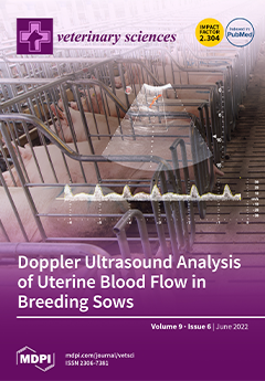
Cover Story (view full-size image): Matching reproductive physiology with new diagnostic methodologies may increase the efficiency of assisted reproductive techniques in pigs. A non-invasive method, the transabdominal Doppler ultrasound, was used in this study to investigate the uterine blood flow indices during the estrus cycle. When an artery is selected for evaluation (top part of the image), red color indicates blood flow towards the transducer and blue blood flow away from the transducer. The bottom part of the image shows a spectral waveform representing the uterine arterial pulse in each cardiac cycle, in contrast with a vein where the flow would not have a pulse. This study has demonstrated how some uterine blood flow indices changed during the estrus cycle (estrus vs. early diestrus). View this paper
  • Issues are regarded as officially published after their release is announced to the table of contents alert mailing list.
  • You may sign up for e-mail alerts to receive table of contents of newly released issues.
  • PDF is the official format for papers published in both, html and pdf forms. To view the papers in pdf format, click on the "PDF Full-text" link, and use the free Adobe Reader to open them.
Order results
Result details
Section
Select all
Export citation of selected articles as:
11 pages, 945 KiB  
Article
Canine Cytokines Profile in an Endemic Region of L. infantum: Related Factors
by Pablo Jesús Marín-García and Lola Llobat
Vet. Sci. 2022, 9(6), 305; https://doi.org/10.3390/vetsci9060305 - 20 Jun 2022
Cited by 5 | Viewed by 2322
Abstract
Canine leishmaniosis is caused by infection with parasite Leishmania infantum, which are transmitted by sandflies Phlebotomus. Canine leishmaniosis is an endemic disease in the Mediterranean region. The immune response could vary between hosts and determines the severity of the disease and clinical [...] Read more.
Canine leishmaniosis is caused by infection with parasite Leishmania infantum, which are transmitted by sandflies Phlebotomus. Canine leishmaniosis is an endemic disease in the Mediterranean region. The immune response could vary between hosts and determines the severity of the disease and clinical features. The aim of this study was to analyze the serum levels of cytokines TNF-α, IFN-γ, IL-2, IL-6, and IL-8, which are related to the activation of Th1 or Th2 immune responses in dogs living in the L. infantum endemic region. Moreover, we intend to relate and correlate these levels with different factors, such as sex, age, diet, lifestyle, and breed. Epidemiological data and serum were recovered for seventy-eight dogs, and serum levels of cytokines described previously were analyzed by using the ELISA method. The results showed differences in serum levels of IFN-γ, IL-2, and IL-8 between breeds. The lifestyle also affected serum levels of IL-2. The main conclusion of this study is that Ibizan hounds and crossbred dogs have a serological profile of cytokines that seems to indicate certain protections against infection by L. infantum compared to boxer and purebred breeds. Full article
Show Figures

Figure 1

13 pages, 2127 KiB  
Article
Urinary Cytology: Potential Role in Canine Urinary Tract Infections
by Ilaria Lippi, Verena Habermaass, Eleonora Gori, Valentina Virginia Ebani, Alessio Pierini and Veronica Marchetti
Vet. Sci. 2022, 9(6), 304; https://doi.org/10.3390/vetsci9060304 - 20 Jun 2022
Cited by 1 | Viewed by 8081
Abstract
The diagnosis of urinary tract infections (UTIs) requires a concomitant evaluation of clinical signs and urine culture, which is of fundamental to start an appropriate antibiotic treatment. Several factors, such as subclinical bacteriuria or pre-analytical errors, may make the interpretation of urine culture [...] Read more.
The diagnosis of urinary tract infections (UTIs) requires a concomitant evaluation of clinical signs and urine culture, which is of fundamental to start an appropriate antibiotic treatment. Several factors, such as subclinical bacteriuria or pre-analytical errors, may make the interpretation of urine culture difficult. The aim of the study was to evaluate the association between the finding of neutrophils and bacteria in unstained and stained canine urine sediment and the presence of clinical signs and positive urine culture. Urine samples from 35 dogs with clinical signs of UTI and 55 asymptomatic dogs with risk factors for UTI were prospectively collected by cystocentesis, divided into three aliquots, and submitted for: (1) physical and chemical Dipstick analysis and unstained urinary sediment (casts, crystals, bacteria, leucocytes, cells, parasites); (2) stained urinary sediment (extra/intracellular bacteria, degenerated and non-degenerated neutrophils); (3) qualitative and quantitative urine culture and antimicrobial sensitivity-test. The association between unstained and stained findings of urinary sediment and urine culture was tested. Sensibility, specificity, and positive/negative predictive values in diagnosing positive urine cultures of bacteria at unstained and stained evaluation were compared. Both wet-mount bacteriuria and the cytological presence of intracellular and extracellular bacteria, neutrophils, and degenerated neutrophils were successively associated with positive urine culture (p < 0.001). The presence of intracellular bacteria was the only independent predictor of positive urine culture. Total bacterial count did not differ significantly between symptomatic and asymptomatic dogs. Detection of extracellular and intracellular bacteriuria at stained urinary sediment significantly improved the sensibility of predicting positive urine culture. Cytologic evaluation of urinary sediment may be helpful in detecting signs of active inflammation, thus enhancing the clinical relevance of a positive urine culture. Full article
(This article belongs to the Special Issue Bacterial Infectious Diseases of Companion Animals)
Show Figures

Figure 1

10 pages, 1701 KiB  
Article
Evaluation of the Fluorescence Polarization Assay for the Diagnosis of Brucellosis in Goat Milk
by Dianelys Sotolongo-Rodríguez, Ricardo Gomez-Flores, Magda Celina Navarro-Soto, Beatriz Arellano-Reynoso, Patricia Tamez-Guerra and Carlos Ramírez-Pfeiffer
Vet. Sci. 2022, 9(6), 303; https://doi.org/10.3390/vetsci9060303 - 20 Jun 2022
Cited by 2 | Viewed by 2196
Abstract
The milk ring test is a detection assay for antibodies against Brucella in bovine milk. It has good sensitivity but tends to give false positive results. In this study, we standardized the application of the fluorescence polarization assay (FPA) for the detection of [...] Read more.
The milk ring test is a detection assay for antibodies against Brucella in bovine milk. It has good sensitivity but tends to give false positive results. In this study, we standardized the application of the fluorescence polarization assay (FPA) for the detection of antibodies against B.melitensis in goat milk. We obtained negative serum and milk samples from healthy goat flocks in the northern zone of Nuevo León. Positive milk and negative, weak, and strong controls were obtained by mixing volumes of positive control serum with negative control milk. Milk samples were treated with citric acid, after which an FPA was performed. Results were then compared with the Rose Bengal test and the FPA in serum. Milk treatment allowed the quantification of antibodies in samples. Significant differences were found between the 2%, 4%, and 6% groups, compared with the control group (F3, 67 = 17.45, p < 0.0001) but not between the 2% and 4% groups (p = 0.0718). The cut-off value was 74.1 mP, with a sensitivity (Se) of 95% and a specificity (Sp) of 100%. Se and Sp values in field milk samples were 84% and 74.55%, respectively. Despite the FPA test on milk samples showed lower Se and Sp than the FPA test on serum samples, its cutoff may be adjusted. It may be recommended as a screening test in goat milk and become useful for the control and eradication of the disease. Full article
Show Figures

Figure 1

13 pages, 633 KiB  
Article
Effects of Backfat Thickness on Oxidative Stress and Inflammation of Placenta in Large White Pigs
by Jian Hu and Peishi Yan
Vet. Sci. 2022, 9(6), 302; https://doi.org/10.3390/vetsci9060302 - 19 Jun 2022
Cited by 5 | Viewed by 2470
Abstract
The purpose of this study was to evaluate the impact of the backfat thickness of sows on reproductive performance and on lipid metabolism, oxidative stress, and inflammation. At farrowing, 60 sows were assigned to three groups: the low-backfat-thickness group (LBF, n = 20): [...] Read more.
The purpose of this study was to evaluate the impact of the backfat thickness of sows on reproductive performance and on lipid metabolism, oxidative stress, and inflammation. At farrowing, 60 sows were assigned to three groups: the low-backfat-thickness group (LBF, n = 20): sows’ backfat thickness was between 9 and 12 mm; the medium-backfat-thickness group (MBF, n = 20): sows’ backfat thickness was between 13 and 20 mm; and the high-backfat-thickness group (HBF, n = 20): sows’ backfat thickness was between 21 and 25 mm. Maternal and fetal blood and placental samples were collected. Compared with the LBF and HBF groups, the MBF group delivered a significantly greater number of live piglets than the LBF or HBF groups. The different backfat thicknesses of sows had different effects on the lipid-related hormones and adipokines of maternal and fetal serum and placenta. Sows with poor or excessive backfat displayed higher levels of oxidative stress and higher levels of pro-inflammatory cytokines. According to these data, the thickness of a sow’s backfat affects the characteristics of farrowing piglets and their lipid metabolism, as well as placental inflammation, maternal inflammation, and oxidative stress. A moderate backfat thickness (between 13 and 20 mm) was associated with greater reproductive performance in sows. Full article
Show Figures

Figure 1

12 pages, 2505 KiB  
Article
Spreading of Pasteurella multocida Infection in a Pet Rabbit Breeding and Possible Implications on Healed Bunnies
by Francesco D’Amico, Gaia Casalino, Giancarlo Bozzo, Antonio Camarda, Roberto Lombardi, Michela Maria Dimuccio and Elena Circella
Vet. Sci. 2022, 9(6), 301; https://doi.org/10.3390/vetsci9060301 - 18 Jun 2022
Cited by 8 | Viewed by 4290
Abstract
The number of pets such as dogs, cats, rabbits, and parrots has increased in European families. Social benefits to owners such as decreasing feelings of loneliness and anxiety are provided by pets which are also used in Animal-assisted Therapy (AAT). Nevertheless, human-animal interactions [...] Read more.
The number of pets such as dogs, cats, rabbits, and parrots has increased in European families. Social benefits to owners such as decreasing feelings of loneliness and anxiety are provided by pets which are also used in Animal-assisted Therapy (AAT). Nevertheless, human-animal interactions are also associated with health problems including allergies, asthma, and zoonosis. Rabbits may carry potential pathogens for humans. One of the most common bacteria that colonizes the oro-pharynx and the upper respiratory tract of rabbits is Pasteurella (P.) multocida. Transmission of the infection to humans results from scratches, licks, and bites but it also can occur from the inhalation of air particles containing the microorganism. Immunocompromised people or persons with pulmonary disorders are particularly susceptible to the infection. Infected rabbits may carry P. multocida with or without clinical signs. In this paper, the sensitivity to antibiotics and the invasiveness ability of P. multocida identified in a farm of pet rabbits affected by severe pasteurellosis were investigated. The strain was P. multocida belonging to capsular type A which is the type most often detected in humans. The identified strain was susceptible to the tested antibiotics, but it appeared equipped with several virulence genes which are responsible for fimbriae production, adhesion processes to host cells, enzyme production, and are involved in iron acquisition processes. These findings are of particular interest because rabbits recovered from pasteurellosis very often become carriers of the bacteria. Therefore, we suggest considering P. multocida screening in the routine medical checks of rabbits, especially if they are meant to be companion animals for children and elder people, given that the transmission of the pathogen cannot be excluded. Full article
(This article belongs to the Special Issue Bacterial Infectious Diseases of Companion Animals)
Show Figures

Figure 1

10 pages, 623 KiB  
Review
Flies as Vectors and Potential Sentinels for Bacterial Pathogens and Antimicrobial Resistance: A Review
by Ji-Hang Yin, Patrick John Kelly and Chengming Wang
Vet. Sci. 2022, 9(6), 300; https://doi.org/10.3390/vetsci9060300 - 16 Jun 2022
Cited by 12 | Viewed by 4316
Abstract
The unique biology of flies and their omnipresence in the environment of people and animals makes them ideal candidates to be important vectors of antimicrobial resistance genes. Consequently, there has been increasing research on the bacteria and antimicrobial resistance genes that are carried [...] Read more.
The unique biology of flies and their omnipresence in the environment of people and animals makes them ideal candidates to be important vectors of antimicrobial resistance genes. Consequently, there has been increasing research on the bacteria and antimicrobial resistance genes that are carried by flies and their role in the spread of resistance. In this review, we describe the current knowledge on the transmission of bacterial pathogens and antimicrobial resistance genes by flies, and the roles flies might play in the maintenance, transmission, and surveillance of antimicrobial resistance. Full article
Show Figures

Figure 1

14 pages, 324 KiB  
Article
Antibacterial Activity of Romanian Propolis against Staphylococcus aureus Isolated from Dogs with Superficial Pyoderma: In Vitro Test
by János Dégi, Viorel Herman, Violeta Igna, Diana Maria Dégi, Anca Hulea, Florin Muselin and Romeo Teodor Cristina
Vet. Sci. 2022, 9(6), 299; https://doi.org/10.3390/vetsci9060299 - 16 Jun 2022
Cited by 8 | Viewed by 2761
Abstract
Staphylococcal infection treatment in dogs is frequently associated with adverse side effects, high costs, prolonged treatment, and resistant strain selection. Staphylococcus aureus is the most frequently isolated staphylococci in cases of canine superficial pyoderma. The number of Staphylococcus strains to exhibit primary resistance [...] Read more.
Staphylococcal infection treatment in dogs is frequently associated with adverse side effects, high costs, prolonged treatment, and resistant strain selection. Staphylococcus aureus is the most frequently isolated staphylococci in cases of canine superficial pyoderma. The number of Staphylococcus strains to exhibit primary resistance to various drugs in vitro is increasing. Propolis has a diverse chemical composition and well-known therapeutic properties against bacterial infections. The current investigation evaluated in vitro the antimicrobial activity of the commercial allopathic antimicrobials, Romanian propolis ethanolic extracts, against clinical Staphylococcus aureus strains isolated from superficial dermatitis clinical samples in dogs and two reference strains: Staphylococcus aureus ATCC 25923 and Staphylococcus aureus ATCC 43300, as the MSSA and MRSA positive controls, respectively, in western Romania. We used the microdilution broth technique to evaluate the susceptibility profile of the bacteria. The minimum inhibitory concentration (MIC) of the Romanian propolis ethanolic extract ranged from 6 to 10 μg/mL for all isolates, determined by the broth microdilution method. The MICs of ethanolic Romanian propolis extracts had a pronounced antibacterial activity. These results indicate that propolis can potentially be used and recommended for in vivo experiments as a promising therapeutic agent against Staphylococcus aureus infections in superficial dermatitis of dogs. Full article
(This article belongs to the Special Issue Antimicrobial Use and Resistance in Animals)
10 pages, 3433 KiB  
Communication
The Haptomonad Stage of Crithidia acanthocephali in Apis mellifera Hindgut
by María Buendía-Abad, Pilar García-Palencia, Luis Miguel de Pablos, Raquel Martín-Hernández and Mariano Higes
Vet. Sci. 2022, 9(6), 298; https://doi.org/10.3390/vetsci9060298 - 16 Jun 2022
Cited by 4 | Viewed by 2091
Abstract
Crithidia acanthocephali is a trypanosomatid species that was initially described in the digestive tract of Hemiptera. However, this parasite was recently detected in honey bee colonies in Spain, raising the question as to whether bees can act as true hosts for this species. [...] Read more.
Crithidia acanthocephali is a trypanosomatid species that was initially described in the digestive tract of Hemiptera. However, this parasite was recently detected in honey bee colonies in Spain, raising the question as to whether bees can act as true hosts for this species. To address this issue, worker bees were experimentally infected with choanomastigotes from the early stationary growth phase and after 12 days, their hindgut was extracted for analysis by light microscopy and TEM. Although no cellular lesions were observed in the honey bee’s tissue, trypanosomatids had differentiated and adopted a haptomonad morphology, transforming their flagella into an attachment pad. This structure allows the protozoa to remain attached to the gut walls via hemidesmosomes-such as junctions. The impact of this species on honey bee health, as well as the pathogenic mechanisms involved, remains unknown. Nevertheless, these results suggest that insect trypanosomatids may have a broader range of hosts than initially thought. Full article
(This article belongs to the Special Issue Challenges and Advances in Bee Health and Diseases)
Show Figures

Figure 1

11 pages, 2490 KiB  
Article
Characterization of Equine Chronic Tendon Lesions in Low- and High-Field Magnetic Resonance Imaging
by Carla Ulrike Doll, Kerstin von Pueckler, Julia Offhaus, Dagmar Berner and Janina Burk
Vet. Sci. 2022, 9(6), 297; https://doi.org/10.3390/vetsci9060297 - 15 Jun 2022
Cited by 1 | Viewed by 2093
Abstract
In equine medicine, experience regarding MRI of chronic tendon lesions is limited, and evidence on the suitability of different sequences in 3 T high-field MRI is scarce. Therefore, macroscopically healthy and altered tendons were examined by histology and in 0.27 T low- and [...] Read more.
In equine medicine, experience regarding MRI of chronic tendon lesions is limited, and evidence on the suitability of different sequences in 3 T high-field MRI is scarce. Therefore, macroscopically healthy and altered tendons were examined by histology and in 0.27 T low- and 3 T high-field MRI, focusing on T1-weighted (T1w) sequences to visualize chronic lesions. In high-field MRI, tendons were positioned parallel (horizontal) and perpendicular (vertical) to the magnetic field, acknowledging the possible impact of the magic angle effect. The images were evaluated qualitatively and signal intensities were measured for quantitative analysis. Qualitative evaluation was consistent with the quantitative results, yet there were differences in lesion detection between the sequences. The low-field T1w GRE sequence and high-field T1w FLASH sequence with vertically positioned tendons displayed all tendon lesions. However, the horizontally scanned high-field T1w SE sequence failed to detect chronic tendon lesions. The agreement regarding tendon signal intensities was higher between high-field sequences scanned in the same orientation (horizontal or vertical) than between the same types of sequence (SE or FLASH), demonstrating the impact of tendon positioning. Vertical scanning was superior for diagnosis of the tendon lesions, suggesting that the magic angle effect plays a major role in detecting chronic tendon disease. Full article
Show Figures

Figure 1

16 pages, 8718 KiB  
Article
Analysis of the Expression of Neurotrophins and Their Receptors in Adult Zebrafish Kidney
by Pietro Cacialli and Carla Lucini
Vet. Sci. 2022, 9(6), 296; https://doi.org/10.3390/vetsci9060296 - 15 Jun 2022
Cited by 3 | Viewed by 2439
Abstract
Neurotrophins and their receptors are involved in the development and maintenance of neuronal populations. Different reports have shown that all neurotrophin/receptor pathways can also play a role in several non-neuronal tissues in vertebrates, including the kidney. These signaling pathways are involved in different [...] Read more.
Neurotrophins and their receptors are involved in the development and maintenance of neuronal populations. Different reports have shown that all neurotrophin/receptor pathways can also play a role in several non-neuronal tissues in vertebrates, including the kidney. These signaling pathways are involved in different events to ensure the correct functioning of the kidney, such as growth, differentiation, and regulation of renal tubule transport. Previous studies in some fish species have identified the neurotrophins and receptors in the kidney. In this study, for the first time, we compare the expression profiles (mRNA and protein) of all neurotrophin/receptor pathways in the kidney of the adult zebrafish. We quantify the levels of mRNA by using qPCR and identify the expression pattern of each neurotrophin/receptor pathway by in situ hybridization. Next, we detect the proteins using Western blotting and immunohistochemistry. Our results show that among all neurotrophins analyzed, NT-3/TrkC is the most expressed in the glomerule and tubule and in the hematopoietic cells, similar to what has been reported in the mammalian kidney. Full article
(This article belongs to the Special Issue Neuropeptides: Role and Function in Species of Veterinary Interest)
Show Figures

Figure 1

9 pages, 1139 KiB  
Article
IL-1R8 as Pathoimmunological Marker for Severity of Canine Chronic Enteropathy
by Federica Riva, Laura Bianchessi, Camilla Recordati, Alessia Inglesi, Vittoria Castiglioni and Lauretta Turin
Vet. Sci. 2022, 9(6), 295; https://doi.org/10.3390/vetsci9060295 - 14 Jun 2022
Cited by 1 | Viewed by 2204
Abstract
Chronic enteropathy (CE) is a severe multifactorial gastrointestinal disease that affects dogs and is driven by poorly characterized inflammatory pathways. Imbalance of pro-inflammatory response regulators, including IL-1R8, may be due to different factors, among which the infection with Helicobacteraceae is known to lead [...] Read more.
Chronic enteropathy (CE) is a severe multifactorial gastrointestinal disease that affects dogs and is driven by poorly characterized inflammatory pathways. Imbalance of pro-inflammatory response regulators, including IL-1R8, may be due to different factors, among which the infection with Helicobacteraceae is known to lead to a vicious circle in which excessive pro-inflammatory signaling and gastrointestinal injury reinforce each other and boost the disease. We investigated the expression of IL-1R8 in large intestine biopsies of dogs with or without clinical signs of CE and with previously assessed enterohepatic Helicobacter spp. colonization status by mean of quantitative real-time PCR. Our study revealed that IL-1R8 is downregulated in both acutely (p = 0.0074) and chronically (p = 0.0159) CE affected dogs compared to healthy controls. The data also showed that IL-1R8 expression tends to decrease with colonization by Helicobacter spp. Interestingly, a negative correlation was detected between the level of expression of IL-1R8 and the severity of macroscopic lesions identified by endoscopy and the crypt hyperplasia score. We further compared the expression levels between males and females and found no statistically significant difference between the two groups. No significant difference was observed in IL-1R8 expression profiles with the age of the animals either. Interestingly, an association was uncovered between IL-1R8 expression level and dog breed. Together, our data advance knowledge on gastrointestinal pathoimmunology in dogs and highlight the potential utilization of IL-1R8 as a diagnostic, prognostic and therapeutic biomarker for canine chronic enteropathy. Full article
(This article belongs to the Section Veterinary Microbiology, Parasitology and Immunology)
Show Figures

Figure 1

15 pages, 688 KiB  
Article
Single Nucleotide Polymorphisms, Gene Expression and Economic Evaluation of Parameters Associated with Mastitis Susceptibility in European Cattle Breeds
by Ahmed I. Ateya, Samer S. Ibrahim and Mona M. Al-Sharif
Vet. Sci. 2022, 9(6), 294; https://doi.org/10.3390/vetsci9060294 - 14 Jun 2022
Cited by 8 | Viewed by 2359
Abstract
The objective of this study was to explore single nucleotide polymorphisms (SNPs), gene expression and economic evaluation of parameters associated with mastitis susceptibility in Holstein and Brown Swiss dairy cows. Two hundred and forty Holstein and Brown Swiss dairy cows (120 cows of [...] Read more.
The objective of this study was to explore single nucleotide polymorphisms (SNPs), gene expression and economic evaluation of parameters associated with mastitis susceptibility in Holstein and Brown Swiss dairy cows. Two hundred and forty Holstein and Brown Swiss dairy cows (120 cows of each breed) were used in this study. The investigated dairy cows in each breed were allocated into two equal-sized groups (60 cows each); mastitis tolerant and affected groups. PCR-DNA sequencing of SELL, ABCG2, SLC11A1, FEZL, SOD1, CAT, GPX1, and AhpC/TSA revealed nucleotide sequence variations in the form of SNPs associated with mastitis tolerance/susceptibility in investigated Holstein and Brown Swiss dairy cows. Levels of SELL, SLC11A1 and FEZL gene expression were significantly up-regulated in mastitic Holstein and Brown Swiss dairy cows than in tolerant ones. Meanwhile, ABCG2, SOD1, CAT, GPX1, and AhpC/TSA genes were significantly downregulated. Regarding the economic parameters, significant differences were recorded for net returns and a reduction in the percentage of net profit, as the higher values of net returns were recorded for tolerant dairy cows than mastitic ones in both breeds; moreover, the net profit was reduced by 39% and 27% in mastitic Holstein and Brown Swiss dairy cows, respectively, when compared to tolerant ones. The results herein confirmed the potential significance of investigated genes as candidates for mastitis tolerance/susceptibility in Holstein and Brown Swiss dairy cows. Mastitis also has detrimental impacts on economic efficiency in dairy farms. Full article
Show Figures

Figure 1

7 pages, 1157 KiB  
Communication
Prevalence of Reticulocytosis in the Absence of Anemia in Dogs with Cardiogenic Pulmonary Edema Due to Myxomatous Mitral Valve Disease: A Retrospective Study
by Sol-Ji Choi, Won-Kyoung Yoon, Hyerin Ahn, Woo-Jin Song and Ul-Soo Choi
Vet. Sci. 2022, 9(6), 293; https://doi.org/10.3390/vetsci9060293 - 14 Jun 2022
Cited by 4 | Viewed by 3214
Abstract
Myxomatous mitral valve disease (MMVD) is the most common heart disease in small breed dogs. Dogs with MMVD commonly show clinical signs of dyspnea due to cardiogenic pulmonary edema (CPE). Reticulocytosis in the absence of anemia (RAA) is a hematological finding in hypoxic [...] Read more.
Myxomatous mitral valve disease (MMVD) is the most common heart disease in small breed dogs. Dogs with MMVD commonly show clinical signs of dyspnea due to cardiogenic pulmonary edema (CPE). Reticulocytosis in the absence of anemia (RAA) is a hematological finding in hypoxic conditions. We aimed to assess the prevalence of RAA in dogs with CPE due to MMVD, and evaluate whether RAA is reversible with amelioration of dyspnea. Twenty-nine client-owned dogs with CPE due to MMVD were included. Dogs who died within 6 weeks of the onset of CPE were included in the non-survival group, while the others comprised the survival group. Of the 21 dogs, RAA was observed in 17 dogs (80.9%). In the RAA group, the absolute reticulocyte count significantly decreased as CPE resolved (p < 0.001). The mean absolute reticulocyte count in the RAA group was 163.90 ± 50.77 on the first measurement and 78.84 ± 25.64 after resolution of CPE. In the RAA group, no significant differences in mean absolute reticulocyte count were observed between the survival and non-survival groups at either the first or second measurement. Our results indicate that RAA occurs in dogs with MMVD-related CPE and can resolve after resolution of CPE. Full article
(This article belongs to the Section Veterinary Internal Medicine)
Show Figures

Figure 1

11 pages, 865 KiB  
Article
Dynamic Models of Within-Herd Transmission and Recommendation for Vaccination Coverage Requirement in the Case of African Swine Fever in Vietnam
by Thi Ngan Mai, Satoshi Sekiguchi, Thi My Le Huynh, Thi Bich Phuong Cao, Van Phan Le, Van Hieu Dong, Viet Anh Vu and Anuwat Wiratsudakul
Vet. Sci. 2022, 9(6), 292; https://doi.org/10.3390/vetsci9060292 - 14 Jun 2022
Cited by 10 | Viewed by 3759
Abstract
African swine fever (ASF) is a highly contagious disease that is caused by the ASF virus (ASFV) with a high fatality rate in domestic pigs resulting in a high socio-economic impact. The pig business in Vietnam was recently affected by ASF for the [...] Read more.
African swine fever (ASF) is a highly contagious disease that is caused by the ASF virus (ASFV) with a high fatality rate in domestic pigs resulting in a high socio-economic impact. The pig business in Vietnam was recently affected by ASF for the first time. This study thus aimed to develop a disease dynamic model to explain how ASFV spreads in Vietnamese pig populations and suggest a protective vaccine coverage level required to prevent future outbreaks. The outbreak data were collected from ten private small-scale farms within the first wave of ASF outbreaks in Vietnam. Three methods were used to estimate the basic reproduction number (R0), including the exponential growth method, maximum likelihood method, and attack rate method. The average R0 values were estimated at 1.49 (95%CI: 1.05–2.21), 1.58 (95%CI: 0.92–2.56), and 1.46 (95%CI: 1.38–1.57), respectively. Based on the worst-case scenario, all pigs in a herd would be infected and removed within 50 days. We suggest vaccinating at least 80% of pigs on each farm once a commercially approved ASF vaccine is available. However, an improvement in biosecurity levels in small-scale farms is still greatly encouraged to prevent the introduction of the virus. Full article
(This article belongs to the Special Issue Emerging and Re-emerging Swine Viral Diseases)
Show Figures

Figure 1

16 pages, 3026 KiB  
Article
Feline Gastrointestinal Eosinophilic Sclerosing Fibroplasia—Extracellular Matrix Proteins and TGF-β1 Immunoexpression
by Néstor Porras, Agustín Rebollada-Merino, Fernando Rodríguez-Franco, Andrés Calvo-Ibbitson and Antonio Rodríguez-Bertos
Vet. Sci. 2022, 9(6), 291; https://doi.org/10.3390/vetsci9060291 - 13 Jun 2022
Cited by 5 | Viewed by 5117
Abstract
Feline gastrointestinal eosinophilic sclerosing fibroplasia (FGESF) has been described as an inflammatory disorder with an eosinophilic component with etiopathogenesis that is still unknown. Sixteen intestinal samples from two veterinary diagnostic services (2014–2017) were included in the study. A histopathological criterion classified the cases [...] Read more.
Feline gastrointestinal eosinophilic sclerosing fibroplasia (FGESF) has been described as an inflammatory disorder with an eosinophilic component with etiopathogenesis that is still unknown. Sixteen intestinal samples from two veterinary diagnostic services (2014–2017) were included in the study. A histopathological criterion classified the cases into three grades (mild, moderate, and severe) according to the distribution of the lesions and the course. An immunohistochemical study of collagen I, collagen III, fibronectin, and transforming growth factor β1 (TGF-β1) was performed in each case. An immunohistochemical study of mild grades shows greater collagen III immunoexpression, compared to collagen I and fibronectin, which suggests an “early” stage of fibrosis. In more intense grades, an increased immunoexpression of collagen I, compared to collagen III, suggests a “late” stage of fibrosis. Otherwise, the highest expression of TGF-β1 was observed in the moderate phase, due to the high proliferation of reactive fibroblast and intense inflammation. The results suggest that the inflammatory infiltrate is the trigger for the elevation in TGF-β1, altering the collagen type III:I ratio. In conclusion, immunohistochemical studies can be a very useful method in diagnosing cases of FGESF of mild grades and could help to apply a differential diagnosis regarding feline eosinophilic chronic enteritis (CEE) in the context of inflammatory bowel disease (IBD). Full article
Show Figures

Figure 1

14 pages, 287 KiB  
Article
Innovative Use of Olive, Winery and Cheese Waste By-Products as Functional Ingredients in Broiler Nutrition
by Eleftherios Bonos, Ioannis Skoufos, Konstantinos Petrotos, Ioannis Giavasis, Chrysanthi Mitsagga, Konstantina Fotou, Konstantina Vasilopoulou, Ilias Giannenas, Evangelia Gouva, Anastasios Tsinas, Angela Gabriella D’Alessandro, Angela Cardinali and Athina Tzora
Vet. Sci. 2022, 9(6), 290; https://doi.org/10.3390/vetsci9060290 - 12 Jun 2022
Cited by 11 | Viewed by 2451
Abstract
The purpose of this study was to evaluate the dietary use of novel silage that was created by combining three agro-industrial wastes produced in bulk, i.e., olive mill wastewater, grape pomace, and deproteinized feta cheese whey, in the diets of broiler chickens. A [...] Read more.
The purpose of this study was to evaluate the dietary use of novel silage that was created by combining three agro-industrial wastes produced in bulk, i.e., olive mill wastewater, grape pomace, and deproteinized feta cheese whey, in the diets of broiler chickens. A total of 216 one-day-old male Ross-308 chicks were randomly allocated to three treatment groups with six replications (12 chicks per pen). Three isocaloric and isonitrogenous diets were formulated to include the examined silage at 0%, 5%, or 10%. Commercial breeding and management procedures were employed throughout the trial. At the end of the trial (day 35), tissue samples were collected for analysis. Feeding 10% silage resulted in increased (p ≤ 0.001) final body weight (p ≤ 0.001) and feed intake. Jejunum and cecum microflora, as well as breast and thigh meat microflora, were modified (p ≤ 0.05) by the dietary inclusion. Thigh meat oxidative stability was improved (p < 0.01) by the silage supplementation. In addition, breast and thigh meat fatty acid profiles were different, respectively, (p < 0.05) in the supplemented treatments compared to the control. The examined silage was successfully tested in broiler diets with potential benefits for their performance and meat quality. Full article
(This article belongs to the Special Issue Agricultural By-Products in Animal Nutrition)
15 pages, 753 KiB  
Article
Repeatability of Health and Welfare Traits and Correlation with Performance Traits in Dairy Goats Reared under Low-Input Farming Systems
by Sotiria Vouraki, Athanasios I. Gelasakis, Vasileia Fotiadou, Georgios Banos and Georgios Arsenos
Vet. Sci. 2022, 9(6), 289; https://doi.org/10.3390/vetsci9060289 - 11 Jun 2022
Cited by 3 | Viewed by 2467
Abstract
The objectives of the study were to estimate the repeatability of health and welfare traits and investigate their association with performance in three breeds of dairy goats reared under low-input farming systems in Greece. A total of 1210 goats of Eghoria (n [...] Read more.
The objectives of the study were to estimate the repeatability of health and welfare traits and investigate their association with performance in three breeds of dairy goats reared under low-input farming systems in Greece. A total of 1210 goats of Eghoria (n = 418), Skopelos (n = 429), and Damascus (n = 363) breeds were assessed. Udder health, parasitic resistance, welfare, milk yield and quality, and body condition score were recorded monthly for two milking periods. Udder health records included somatic cell count (SCC) and total viable count (TVC). Based on combinations of SCC and TVC and thresholds set at >106 cells/mL and >2 × 10⁴ cfu/mL, respectively, additional udder health phenotypes were defined. Parasitism included myiasis, tick infestation, gastrointestinal nematode (GIN) and cestode faecal egg count (FEC), and lungworm faecal larval count (FLC). Infection with each of the endoparasites was defined based on FEC/FLC. Welfare assessment parameters included the presence of ear and horn injuries, ocular and nasal discharge, body and udder abscesses, injury and lesions on the skin of different regions, diarrhoea, hernias, overgrown hooves, arthritis, lameness, and udder asymmetry. Trait repeatability and animal correlations were estimated. Significant (p < 0.05) repeatability was reported for all udder health and most welfare traits in all breeds, GIN and cestode FEC, and GIN and lungworm infection in Eghoria, and myiasis in Skopelos. Correlations of health and most of welfare traits with performance were non-significant or favourable. Overall, results demonstrate potential to improve health and welfare of the studied breeds without compromising performance. Full article
(This article belongs to the Section Veterinary Biomedical Sciences)
Show Figures

Figure 1

17 pages, 1796 KiB  
Review
Describing and Characterizing the Literature Regarding Umbilical Health in Intensively Raised Cattle: A Scoping Review
by Matthew B. Van Camp, David L. Renaud, Todd F. Duffield, Diego E. Gomez, William J. McFarlane, Joanne Marshall and Charlotte B. Winder
Vet. Sci. 2022, 9(6), 288; https://doi.org/10.3390/vetsci9060288 - 11 Jun 2022
Cited by 2 | Viewed by 2394
Abstract
The objective of this scoping review was to describe and characterize the existing literature regarding umbilical health and identify gaps in knowledge. Six databases were searched for studies examining umbilical health in an intensively raised cattle population. There were 4249 articles initially identified; [...] Read more.
The objective of this scoping review was to describe and characterize the existing literature regarding umbilical health and identify gaps in knowledge. Six databases were searched for studies examining umbilical health in an intensively raised cattle population. There were 4249 articles initially identified; from these, 723 full text articles were then screened, with 150 articles included in the review. Studies were conducted in the USA (n = 41), Brazil (n = 24), Canada (n = 13), UK (n = 10), and 37 additional countries. Seventeen were classified as descriptive, 24 were clinical trials, and 109 were analytical observational studies. Umbilical outcomes evaluated in descriptive studies were infection (n = 11), parasitic infection (n = 5), and hernias (n = 2). Of the clinical trials, only one examined treatment of navel infections; the remainder evaluated preventative management factors for navel health outcomes (including infections (n = 17), myiasis (n = 3), measurements (n = 5), hernias (n = 1), and edema (n = 1)). Analytical observational studies examined risk factors for umbilical health (n = 60) and umbilical health as a risk factor (n = 60). Studies examining risk factors for umbilical health included navel health outcomes of infections (n = 28; 11 of which were not further defined), hernias (n = 8), scoring the navel sheath/flap size (n = 16), myiasis (n = 2), and measurements (n = 6). Studies examining umbilical health as a risk factor defined these risk factors as infection (n = 39; of which 13 were not further defined), hernias (n = 8; of which 4 were not further defined), navel dipping (n = 12), navel/sheath scores as part of conformation classification for breeding (n = 2), measurements (n = 3), and umbilical cord drying times (n = 2). This review highlights the areas in need of future umbilical health research such as clinical trials evaluating the efficacy of different treatments for umbilical infection. It also emphasizes the importance for future studies to clearly define umbilical health outcomes of interest, and consider standardization of these measures, including time at risk. Full article
Show Figures

Figure 1

18 pages, 5128 KiB  
Article
Flow Cytometric Analysis of Leukocyte Populations in the Lung Tissue of Dromedary Camels
by Jamal Hussen, Turke Shawaf, Naser Abdallah Al Humam, Sameer M. Alhojaily, Mohammed Ali Al-Sukruwah, Faisal Almathen and Francesco Grandoni
Vet. Sci. 2022, 9(6), 287; https://doi.org/10.3390/vetsci9060287 - 10 Jun 2022
Cited by 2 | Viewed by 2653
Abstract
Respiratory tract infections are among the most common infections in dromedary camels, with a high impact on animal health, production, and welfare. Tissue-specific distribution of immune cells is one of the important factors that influence the nature and outcome of the immune response [...] Read more.
Respiratory tract infections are among the most common infections in dromedary camels, with a high impact on animal health, production, and welfare. Tissue-specific distribution of immune cells is one of the important factors that influence the nature and outcome of the immune response to pathogens. Several protocols have recently been described for the flow cytometric analysis of immune cells in the lung tissue of several species. However, no such protocol currently exists for dromedary camels. The aim of the present study was, therefore, to establish a flow cytometric protocol for the identification of immune cell populations in the camel lung tissue and the evaluation of some of their phenotypic and functional properties. Combined staining of camel lung leukocytes with monoclonal antibodies to the pan-leukocyte marker CD45 and the myeloid cell marker CD172a allowed the identification of myeloid cells (CD45+CD172a+) and lymphoid cells (CD45+CD172a) in the lung of healthy camels. The cell adhesion molecules CD11a and CD18 were found in a higher abundance on myeloid cells compared to lymphoid cells. Based on their differential expression of the LPS receptor CD14, macrophages (CD172a+CD14high cells) were identified as the most abundant immune cell population in the camel lung tissue. In contrast to their dominance in camel peripheral blood, granulocytes (CD172a+CD14low) presented only a minor population in the lung tissue. The higher frequency of γδ T cells in the lung tissue than in peripheral blood suggests a role for these cells in the pulmonary immune system. Flow cytometric analysis of bacterial phagocytosis and ROS production upon bacterial stimulation revealed high antimicrobial activity of camel lung phagocytes, which was comparable with the antimicrobial activity of blood granulocytes. Comparative analysis of immune cell distribution between the cranial and caudal lobes of the camel lung revealed a higher frequency of granulocytes and a lower frequency of macrophages in the cranial compared to the caudal lung lobe. In addition, the higher frequency of cells expressing the M2 macrophage marker CD163 in the caudal lung tissue, with a slightly higher fraction of MHCII-positive cells (M1 phenotype) in the cranial lung tissue, may suggest the distribution of different macrophage subtypes in the different lobes of the camel lung. Such differences between lung lobes could influence the effectiveness of the immune response to infection or vaccination with respiratory pathogens. Collectively, the present study identified some similarities and differences between camels and other farm animals regarding the distribution of the main immune cell populations in their lungs. Further studies are required for comprehensive immunophenotyping of the cellular pulmonary immune system in camels. Full article
(This article belongs to the Special Issue Immune Biomarkers in Veterinary Research: Challenges and Promises)
Show Figures

Figure 1

12 pages, 1309 KiB  
Article
Experimental Infection of Pigs with a ST 245 Brachyspira hyodysenteriae Isolated from an Asymptomatic Pig in a Herd with No History of Swine Dysentery
by José Paulo H. Sato, Amanda G. S. Daniel, Carlos E. R. Pereira, Mariana R. Andrade, Ricardo P. Laub, Michelle P. Gabardo, Luisa V. A. Otoni, Nubia R. Macedo, Javier A. Barrera-Zarate and Roberto M. C. Guedes
Vet. Sci. 2022, 9(6), 286; https://doi.org/10.3390/vetsci9060286 - 10 Jun 2022
Cited by 2 | Viewed by 2866
Abstract
Swine dysentery (SD) is characterized by a severe mucohemorrhagic colitis caused by infection with Brachyspira species. In infected herds the disease causes considerable financial loss due to mortality, slow growth rates, poor feed conversion, and costs of treatment. B. hyodysenteriae is the most [...] Read more.
Swine dysentery (SD) is characterized by a severe mucohemorrhagic colitis caused by infection with Brachyspira species. In infected herds the disease causes considerable financial loss due to mortality, slow growth rates, poor feed conversion, and costs of treatment. B. hyodysenteriae is the most common etiological agent of SD and infection is usually associated with disease. However, isolated reports have described low pathogenic strains of B. hyodysenteriae. The aim of this study was to describe an experimental infection trial using a subclinical B. hyodysenteriae isolated from an animal without clinical signs and from a disease-free herd, to evaluate the pathogenicity and clinical pathological characteristics compared to a highly clinical isolate. Forty-eight 5-week-old pigs were divided into three groups: control, clinical and the subclinical isolates. The first detection/isolation of B. hyodysenteriae in samples of the animals challenged with a known clinical B. hyodysenteriae strain (clinical group) occurred 5th day post inoculation. Considering the whole period of the study, 11/16 animals from this group were qPCR positive in fecal samples, and diarrhea was observed in 10/16 pigs. In the subclinical isolate group, one animal had diarrhea. There were SD large intestine lesions in 3 animals at necropsy and positive B. hyodysenteriae isolation in 7/15 samples of the subclinical group. In the control group, no diarrhea, gross/microscopic lesions, or qPCR positivity were observed. Clinical signs, bacterial isolation, macroscopic and histologic lesions were significantly difference among groups, demonstrating low pathogenicity of the subclinical isolate in susceptible pigs. Full article
(This article belongs to the Special Issue New Findings on Long-Known Pathogens in Pigs)
Show Figures

Figure 1

10 pages, 1894 KiB  
Case Report
Tumor-Homing of Mesenchymal Stem Cells Infected with Oncolytic Virus in a Canine Patient
by Pablo Delgado-Bonet, Beatriz Davinia Tomeo-Martín, Gustavo Ortiz-Díez and Ana Judith Perisé-Barrios
Vet. Sci. 2022, 9(6), 285; https://doi.org/10.3390/vetsci9060285 - 9 Jun 2022
Cited by 3 | Viewed by 2897
Abstract
Intravenous administration of oncolytic adenovirus (OAds) can be challenging, although various vehicles for the delivery of the virus to the tumor have been described. The efficacy of mesenchymal stem cells (MSCs) as a virus vehicle has been reported in mouse models and canine [...] Read more.
Intravenous administration of oncolytic adenovirus (OAds) can be challenging, although various vehicles for the delivery of the virus to the tumor have been described. The efficacy of mesenchymal stem cells (MSCs) as a virus vehicle has been reported in mouse models and canine and human patients, but the actual action mechanism has never been described in patients. It is of importance to determine whether MSCs infected with OAds can reach the tumor and release the virus in a clinical setting. For this purpose, GFP-labeled MSCs were infected with an OAd and inoculated into a companion dog diagnosed with spontaneous lung carcinoma. Forty-eight hours later, the tumor was excised and analyzed microscopically by flow cytometry for GFP fluorescence detection, and a cellular culture was established. Peripheral blood samples were taken to quantify the oncolytic adenovirus by qRT-PCR. Green fluorescence cells detected in the cellular culture by microscopy and flow cytometry revealed 0.69% GFP-positive cells in the tumor. OAd in peripheral blood was confirmed by qRT-PCR during follow-up. For the first time, the tumoral-homing capacity of OAds infected-MSC has been confirmed in a clinical setting, helping to explain the clinical response mechanism, whose efficacy was previously reported in canine and human patients. Full article
(This article belongs to the Special Issue Animal and Disease Models in Biomedical Research)
Show Figures

Graphical abstract

13 pages, 1062 KiB  
Article
Antimicrobial Resistance of Clinical and Commensal Escherichia coli Canine Isolates: Profile Characterization and Comparison of Antimicrobial Susceptibility Results According to Different Guidelines
by Vera Fernandes, Eva Cunha, Telmo Nunes, Elisabete Silva, Luís Tavares, Luísa Mateus and Manuela Oliveira
Vet. Sci. 2022, 9(6), 284; https://doi.org/10.3390/vetsci9060284 - 9 Jun 2022
Cited by 7 | Viewed by 3086
Abstract
Background: Pyometra is a diestrual chronic disease frequently associated with Escherichia coli. Initial pyometra treatment involves empiric antimicrobial therapy whose suitability should be confirmed by antimicrobial susceptibility testing. Antimicrobial resistance is a major health issue for veterinary medicine, rendering surveillance studies essential. [...] Read more.
Background: Pyometra is a diestrual chronic disease frequently associated with Escherichia coli. Initial pyometra treatment involves empiric antimicrobial therapy whose suitability should be confirmed by antimicrobial susceptibility testing. Antimicrobial resistance is a major health issue for veterinary medicine, rendering surveillance studies essential. Our goal was to determine the susceptibility profile of E. coli isolates obtained from healthy and pyometra-presenting dogs and to compare the application of different antimicrobial susceptibility guidelines. Methods: The antimicrobial susceptibility profile (ASP) of 74 E. coli isolates was determined by disk diffusion, using six antimicrobials commonly used in veterinary medicine. Profiles were assessed by CLSI VET01S, CLSI M100 and EUCAST guidelines. β-lactamases-encoding genes blaTEM, blaSHV and blaOXA were detected by multiplex PCR. Biofilm production ability was evaluated by pellicle formation assays in Luria–Bertani medium. Results: Variations in the resistance frequency were observed for amoxicillin/clavulanic acid, cephalexin and cefotaxime (29.7–54.1%, 10.8–16.2% and 1.4–4.1%, respectively). Results varied slightly between clinical and commensal isolates, as well as their biofilm-forming ability. Genes blaTEM, blaSHV and blaOXA were detected in 25.5%, 11.8% and 9.8% of isolates, respectively. Conclusions: Results show the importance of ASP determination in veterinary isolates and the need for using standardized and validated testing methods and harmonized interpretive criteria. Full article
(This article belongs to the Special Issue Antimicrobial Use and Resistance in Animals)
Show Figures

Figure 1

8 pages, 511 KiB  
Article
Prevalence and Predictors of Radiographically Apparent Upper Urinary Tract Urolithiasis in Eight Dog Breeds Predisposed to Calcium Oxalate Urolithiasis and Mixed Breed Dogs
by Alexis M. Hoelmer, Jody P. Lulich, Aaron K. Rendahl and Eva Furrow
Vet. Sci. 2022, 9(6), 283; https://doi.org/10.3390/vetsci9060283 - 9 Jun 2022
Cited by 2 | Viewed by 2787
Abstract
Data on upper urinary tract (UUT) uroliths in dogs are important to understanding their etiology. The aim of this retrospective case-control study was to determine the prevalence and identify predictors of radiographically apparent UUT uroliths in dog breeds at increased risk for calcium [...] Read more.
Data on upper urinary tract (UUT) uroliths in dogs are important to understanding their etiology. The aim of this retrospective case-control study was to determine the prevalence and identify predictors of radiographically apparent UUT uroliths in dog breeds at increased risk for calcium oxalate uroliths (CaOx risk breeds) and mixed breed dogs. Radiologist reports of three-view abdominal radiographs were reviewed from 251 purebred dogs of 8 CaOx risk breeds and 68 mixed breed dogs. UUT uroliths were more common in CaOx risk breeds than mixed breed dogs (23% versus 6%, respectively; OR = 4.8, 95% confidence interval [CI] 1.7–18.9, p < 0.001). UUT uroliths were more common in dogs with lower urinary tract (LUT) uroliths (predominantly calcium-containing) than those without (41% versus 5%, respectively; OR = 13.6, 95% CI 6.3–33.1, p < 0.001), and LUT uroliths predicted the presence of UUT uroliths in the multivariable regression (OR = 6.5, 95% CI 2.8–16.7, p < 0.001). Increasing age (p < 0.001) and lower body weight (p = 0.0016) were also predictors of UUT urolith presence in the multivariable regression. The high prevalence of UUT uroliths in dogs with LUT uroliths supports a shared mechanism for their formation. Full article
(This article belongs to the Special Issue Diseases of the Kidneys and Urinary Tract in Small Animals)
Show Figures

Figure 1

10 pages, 1804 KiB  
Article
Phenotypic and Genotypic Screening of Colistin Resistance Associated with Emerging Pathogenic Escherichia coli Isolated from Poultry
by Heba Badr, Abdelhafez Samir, Essam Ismail El-Tokhi, Momtaz A. Shahein, Flourage M. Rady, Ashraf S. Hakim, Ehab Ali Fouad, Engy Farahat El-Sady and Samah F. Ali
Vet. Sci. 2022, 9(6), 282; https://doi.org/10.3390/vetsci9060282 - 9 Jun 2022
Cited by 9 | Viewed by 3115
Abstract
Chickens continue to be an important reservoir of zoonotic multidrug-resistant illnesses. Antimicrobial resistance correlated with colistin has emerged as a critical concern worldwide in the veterinary field and the public health sector. The current study investigated the prevalence of multidrug-resistant avian pathogenic Escherichia [...] Read more.
Chickens continue to be an important reservoir of zoonotic multidrug-resistant illnesses. Antimicrobial resistance correlated with colistin has emerged as a critical concern worldwide in the veterinary field and the public health sector. The current study investigated the prevalence of multidrug-resistant avian pathogenic Escherichia coli among chicken farms in three Egyptian governorates, focusing on colistin resistance assessment. A total of 56 Escherichia coli isolates were recovered out of 120 pooled samples obtained from diseased chicken broilers (46.7%). The E. coli isolates were serotyped to nine different serotypes; the highest incidence was for O125 (n = 18). The E. coli isolates demonstrated multidrug-resistant patterns against 10 antibiotics, especially clindamycin, tetracycline, streptomycin and ampicillin, by 100, 100, 96.4 and 92.9%, respectively. On the other hand, colistin resistance was 41.1% using AST. All E. coli isolates displayed positive colistin resistance growth on chromogenic medium, but only 25% represented this positivity via MIC estimation and Sensititre kit. PCR results revealed that all isolates harbored mcr-1, but no isolates harbored the other 2–5 mcr genes. In conclusion, the study demonstrated the emergence of multidrug-resistant, especially colistin-resistant, E. coli among chicken broiler flocks, and mcr-1 is the master gene of the colistin resistance feature. Full article
(This article belongs to the Topic Animal Diseases in Agricultural Production Systems)
Show Figures

Figure 1

11 pages, 2570 KiB  
Case Report
Symblepharon, Ankyloblepharon, and Salt Gland Dysfunction in a Loggerhead Sea Turtle (Caretta caretta)
by Andrea Affuso, Cristina Di Palma, Leonardo Meomartino, Antonino Pace, Serena Montagnaro, Valeria Russo, Giuseppina Mennonna, Fabiana Micieli, Fulvio Maffucci, Sandra Hochscheid, Francesco Lamagna, Ilaria D’Aquino and Barbara Lamagna
Vet. Sci. 2022, 9(6), 281; https://doi.org/10.3390/vetsci9060281 - 8 Jun 2022
Cited by 1 | Viewed by 2643
Abstract
Adhesions involving the bulbar and the palpebral conjunctiva (Symblepharon) may interfere with tear drainage, cause chronic conjunctivitis, and reduce ocular motility. This condition may be associated with adhesion of the edges of the upper and lower eyelids (ankyloblepharon). The present case describes bilateral [...] Read more.
Adhesions involving the bulbar and the palpebral conjunctiva (Symblepharon) may interfere with tear drainage, cause chronic conjunctivitis, and reduce ocular motility. This condition may be associated with adhesion of the edges of the upper and lower eyelids (ankyloblepharon). The present case describes bilateral symblepharon, ankyloblepharon and salt gland dysfunction in a juvenile Caretta caretta. The loggerhead presented both eyelids swollen, ulcerated, and not separable when rescued. Eye examination was not possible, but ultrasonography showed right bulbar integrity, while the left eye was smaller, with a thicker cornea that had lost its normal doubled lined structure. Surgical dissection of the fibrous adhesions between the palpebral and bulbar conjunctiva, cornea, and third eyelid was performed, and large dacryoliths were removed. The microscopic findings were consistent with chronic keratoconjunctivitis. Ultrastructurally, no virus-like particles were observed. In addition, tissue samples were negative for herpesvirus by qualitative PCR. The eyelids of both eyes and the corneal epithelium of the right eye healed; moreover, the vision was restored in the right eye. There were no recurrences after 12 months of follow-up, and the turtle was released 16 months after the end of treatments on the southern Tyrrhenian coast in the western Mediterranean Sea. To the authors’ knowledge, this is the first report of symblepharon with ankyloblepharon and salt gland dysfunction in Caretta caretta turtle. Ocular ultrasonography was helpful in the preliminary diagnostic work-up. Full article
(This article belongs to the Special Issue Advances in Veterinary and Comparative Ophthalmology)
Show Figures

Figure 1

14 pages, 4013 KiB  
Review
Inguinal Hernia in Nonhuman Primates: From Asymptomatic to Life-Threatening Events
by Melissa A. de la Garza, Sara R. Hegge and Jaco Bakker
Vet. Sci. 2022, 9(6), 280; https://doi.org/10.3390/vetsci9060280 - 8 Jun 2022
Cited by 2 | Viewed by 6142
Abstract
In this study, a review of available data and literature on the epidemiology and anamnesis of inguinal hernias in nonhuman primates, as well as on their clinical evaluation and surgical management, was conducted. Inguinal hernias are assumed to be relatively common in male [...] Read more.
In this study, a review of available data and literature on the epidemiology and anamnesis of inguinal hernias in nonhuman primates, as well as on their clinical evaluation and surgical management, was conducted. Inguinal hernias are assumed to be relatively common in male nonhuman primates. Clinical signs are usually limited to a visible or palpable mass in the groin region without pain or systemic illness. Most hernias contain omentum. Careful monitoring is an acceptable treatment option for those animals. Size, the danger of incarceration, and the presence of strangulation are important factors when considering surgical repair. A strangulated inguinal hernia is an emergency, requiring prompt surgery to avoid tissue necrosis and death. Imaging techniques, as well as computed tomography (CT), ultrasonography, and magnetic resonance imaging (MRI), provide information about the anatomical characteristics of the suspected region, allowing for a diagnosis and treatment. An inguinal hernia repair can be performed with either open surgery or laparoscopic surgery. The hernia repair can be achieved by mesh or suture. Decisions regarding which repair technique to use depend on the surgeon′s skill level and preference. Complication and recurrence rates are generally low. The most common postsurgical complication is a recurrence of the hernia. Contraceptive measures are not indicated in breeders, as there is no known hereditary component, and the presence of hernia does not appear to affect fertility, nor does it predispose to occurrence, recurrence, or incarceration. Full article
(This article belongs to the Special Issue Trends in Nonhuman Primate Medicine & Care)
Show Figures

Figure 1

12 pages, 2066 KiB  
Article
Small Ruminant Farming in Tribal Areas of Dera Ghazi Khan, Punjab, Pakistan
by Muhammad Ameen Jamal, Samiullah Khan, Yanhua Su, Chang Yang, Heng Zhao, Kaixiang Xu, Deling Jiao, Wenmin Cheng, Abdul Rauf, Mahboob Ali, Sohail Ahmad, Yubo Qing and Hong-Jiang Wei
Vet. Sci. 2022, 9(6), 279; https://doi.org/10.3390/vetsci9060279 - 7 Jun 2022
Cited by 3 | Viewed by 3485
Abstract
Provincially Administered Tribal Areas (PATA) of Punjab-Pakistan are comprised of hilly mountains with small ruminants as a sole source of income. In this study, farming practices, productivity, health and the economic value of sheep were evaluated in PATA through a survey of farmers [...] Read more.
Provincially Administered Tribal Areas (PATA) of Punjab-Pakistan are comprised of hilly mountains with small ruminants as a sole source of income. In this study, farming practices, productivity, health and the economic value of sheep were evaluated in PATA through a survey of farmers (n = 138) holding 11,558 heads of sheep. Out of a total population, 87% were non-descriptive flocks, and 9% and 4% were purebred flocks belonging to the Kajli and Thali populations, respectively. Sheep flocks were mainly (86%) reared under the traditional production system and had a delayed onset of puberty. There was low influence of season on the reproduction, and the majority of flocks (78%) were bred throughout the year. The lack of proper vaccination and poor management exposed the flocks to bacterial, viral and parasitic infections, which lead to high mortality in lambs (~22%) and adults (~32%). The share of sheep in farmers livelihood was 42%, and only 20% of producers’ living standard was improved with sheep farming, but the rise in rearing more sheep was quite low (20%). Although the livestock department arranged farmers’ training, the majority of farmers (83%) never participated in training and had no knowledge of modern technologies. Collectively, the traditional sheep production systems, poor management, lack of vaccination, marketing channels and farmers training hampered the sheep rearing and producers’ livelihood in the PATA of Punjab-Pakistan. However, developing model livestock farms, conducting farmer training, establishing a viable market for dairy products, and introducing subsidy policy interventions can improve the sheep farming in these areas. Full article
(This article belongs to the Special Issue Assisted Reproductive Technologies (ARTs) in Domestic Mammals)
Show Figures

Figure 1

7 pages, 4483 KiB  
Article
Impact of Obesity on Lung Function in Cats with Bronchoconstriction
by Alicia Caro-Vadillo, J. Alberto Montoya-Alonso and Laín García-Guasch
Vet. Sci. 2022, 9(6), 278; https://doi.org/10.3390/vetsci9060278 - 7 Jun 2022
Cited by 4 | Viewed by 3403
Abstract
Obesity is a nutritional disorder commonly diagnosed in adult cats that has been associated with an increased risk of different chronic diseases including respiratory diseases. The main objective of this study is to define if there is a relation between lung function measured [...] Read more.
Obesity is a nutritional disorder commonly diagnosed in adult cats that has been associated with an increased risk of different chronic diseases including respiratory diseases. The main objective of this study is to define if there is a relation between lung function measured by barometric whole-body plethysmography and obesity in cats with bronchoconstriction. Fifty-three cats were included in the study. All animals presented a bronchoconstriction status diagnosed with an Enhanced Pause (Penh) value higher than the reference range. Based on a standardized 9-point body condition scale, 36 cats were normal-weight cats (with BCS < 6), and 17 cats were considered overweight or obese cats (with BCS ≥ 6). Overweight cats were mainly male cats and older, and presented lower tidal volume values, lower minute volume values, and lower peak inspiratory and expiratory flows than normal-weight cats. According to the results of the present study, overweight cats showed a more compromised lung function parameters related to restrictive pattern compared with normal-weight cats. However, overweight cats did not show a higher bronchoconstriction level compared with normal-weight cats. Full article
(This article belongs to the Section Nutritional and Metabolic Diseases in Veterinary Medicine)
Show Figures

Figure 1

16 pages, 3102 KiB  
Article
Evaluation of Tumor Grade and Proliferation Indices before and after Short-Course Anti-Inflammatory Prednisone Therapy in Canine Cutaneous Mast Cell Tumors: A Pilot Study
by Shawna Klahn, Nikolaos Dervisis, Kevin Lahmers and Marian Benitez
Vet. Sci. 2022, 9(6), 277; https://doi.org/10.3390/vetsci9060277 - 7 Jun 2022
Cited by 4 | Viewed by 3241
Abstract
Glucocorticoid administration is a common clinical practice that attempts to decrease the inflammation associated with and improve the resectability of canine mast cell tumors (MCTs). However, the impact of neoadjuvant glucocorticoids on the histological features and proliferation indices of canine MCTs is unknown. [...] Read more.
Glucocorticoid administration is a common clinical practice that attempts to decrease the inflammation associated with and improve the resectability of canine mast cell tumors (MCTs). However, the impact of neoadjuvant glucocorticoids on the histological features and proliferation indices of canine MCTs is unknown. The objective of this study was to evaluate changes in tumor grade, mitotic count, Ki67, AgNOR, and AgNORxKi67 scores following short-course anti-inflammatory neoadjuvant prednisone in canine patients with MCTs. This was a prospective single-arm pilot study. Client-owned dogs with treatment-naïve cytologically confirmed MCTs were enrolled. Patients underwent an initial incisional biopsy followed by a 10–14-day course of anti-inflammatory prednisone and surgical resection. All histological samples were randomized, masked, and evaluated by a single pathologist. Unstained paired pre- and post-treatment samples were submitted to a commercial laboratory for Ki67 and AgNOR immunohistochemical analysis. There were 11 dogs enrolled with 11 tumors. There were no statistical differences between the pre- and post-treatment histological parameters of mitotic index, Ki67, AgNOR, or Ki67xAgNOR. There were no clinically significant alterations between pre-treatment and post-treatment in the assignment of tumor grades. A short course of anti-inflammatory prednisone does not appear to alter the histological parameters that affect grade determination or significantly alter the proliferation indices in canine MCTs. Full article
(This article belongs to the Section Veterinary Biomedical Sciences)
Show Figures

Figure 1

11 pages, 635 KiB  
Article
Changes in the Oxidative Stress Status of Dogs Affected by Acute Enteropathies
by Alessia Candellone, Flavia Girolami, Paola Badino, Watanya Jarriyawattanachaikul and Rosangela Odore
Vet. Sci. 2022, 9(6), 276; https://doi.org/10.3390/vetsci9060276 - 6 Jun 2022
Cited by 8 | Viewed by 2229
Abstract
Canine acute enteropathies (AE) are common morbidities primarily managed with supportive therapy. However, in some cases, unnecessary courses of antibiotics are empirically prescribed. Recent studies in humans have hypothesized the use of antioxidants as a possible alternative and/or support to antimicrobial drugs in [...] Read more.
Canine acute enteropathies (AE) are common morbidities primarily managed with supportive therapy. However, in some cases, unnecessary courses of antibiotics are empirically prescribed. Recent studies in humans have hypothesized the use of antioxidants as a possible alternative and/or support to antimicrobial drugs in uncomplicated cases. Considering the global need to reduce the antibiotic use, the aim of the study was to compare the oxidative burden of the diarrhetic population to that of healthy dogs. Forty-five patients suffering from uncomplicated acute diarrhea (AD) and 30 controls were screened for clinical and biochemical parameters, and serum redox indices (reactive oxygen metabolites, dROMs; serum antioxidant capacity, SAC; oxidative stress index, OSi). The average levels of dROMs in AD dogs were significantly higher (p < 0.001) than in healthy dogs, while SAC did not significantly differ between the two groups. However, the OSi values (ratio between dROMs and SAC) significantly increased (p < 0.001) in AD dogs compared to controls. The study demonstrates that canine AD could induce redox imbalance. Although its role in the etiopathogenesis and evolution of the disease should be further investigated, our results suggest that the improvement of the patient oxidative status, possibly through the dietary administration of antioxidants, could support the management of canine AE, reducing the use of antibiotics. Full article
Show Figures

Figure 1

Previous Issue
Next Issue
Back to TopTop