Abstract
Crithidia acanthocephali is a trypanosomatid species that was initially described in the digestive tract of Hemiptera. However, this parasite was recently detected in honey bee colonies in Spain, raising the question as to whether bees can act as true hosts for this species. To address this issue, worker bees were experimentally infected with choanomastigotes from the early stationary growth phase and after 12 days, their hindgut was extracted for analysis by light microscopy and TEM. Although no cellular lesions were observed in the honey bee’s tissue, trypanosomatids had differentiated and adopted a haptomonad morphology, transforming their flagella into an attachment pad. This structure allows the protozoa to remain attached to the gut walls via hemidesmosomes-such as junctions. The impact of this species on honey bee health, as well as the pathogenic mechanisms involved, remains unknown. Nevertheless, these results suggest that insect trypanosomatids may have a broader range of hosts than initially thought.
1. Introduction
Trypanosomatids are a large group of parasitic protozoa that can infect a wide range of organisms, including plants, insects, and vertebrates. Indeed, some of these species can cause important medical and veterinary diseases, such as Leishmania and Trypanosoma, and consequently, they have been widely studied [1]. While some of these trypanosomatid species can be transmitted by insect vectors (dixenous), the lifecycle of others may be restricted to only one type of organism, known as monoxenous. Despite representing most diversity of the Trypanosomatidae family, these latter species have received less attention [2,3]. Most monoxenous species infect insects, and while the majority do not appear to harm the host’s health, there are some exceptions that include species that infect honey bees and bumble bees [4]. Moreover, it is significant that insect trypanosomatids have also been found to colonize other host groups, including plants, rats, dogs, bats, or even humans [2,5].
The comparative lack of information about insect trypanosomatids has had a strong impact on the establishment of phylogenetic relationships within the family, which in turn has driven constant modifications in their taxonomy and systematics [6]. This is indeed the case of Crithidia acanthocephali and Crithidia flexonema: The former was described and isolated from the hindgut of the hemipteran Acanthocephala femorata in 1961 [7], while C. flexonema was described in 1960 in water strider Aquarius remiges (formerly known as Gerris remiges) [8]. Following the “one host-one parasite” paradigm that governed the classification of trypanosomatids for many years, these were both considered as different species. However, a recent taxonomic revision based on DNA barcoding [9] showed the sequences of both these species to be identical, leading to the proposal to unify the nomenclature under the name of the first chronological described species, C. flexonema. However, to the best of our knowledge there has been no consensus on this proposal and most of the bibliography consulted refers to C. acanthocephali. Indeed, the cultures obtained from ATCC are named after this species; thus, from now on this name will be used here to refer to both these species.
Crithidia acanthocephali is quite widespread geographically [9], and as previous works have proved, it can proliferate inside insects from different orders, such as Diptera, Coleoptera, or Orthoptera, increasing their mortality [10,11]. Although to date there is little information available regarding this trypanosomatid in honey bees, a recent study detected C. acanthocephali for the first time in honey bee colonies in the center of Spain via Ion PGM sequencing, along with other species commonly found in honey bee colonies (e.g., Lotmaria passim and Crithidia mellificae) [12]. Other trypanosomatid species also detected in this study (such as Crithidia bombi or Crithidia expoeki) are commonly detected in other hymenopteran hosts such as bumble bees, but they are not usually found in honey bee colonies.
Both L. passim and C. mellificae have been recently found to modify their promastigote and choanomastigote morphology into an haptomonad form, remodeling their flagella into an attachment pad that allows them to remain attached to the gut walls of their host and cover the epithelial cells [13]. This haptomonad stage and its morphogenesis have been described in other trypanosomatid species, such as Leishmania, and it is regarded as an influential factor for parasite survival and transmission [14,15,16]. Those are key features for monoxenous parasites, but acquisition is also crucial for considering an insect as a true host. In this regard, the presence of C. acanthocephali in honey bees implies that honey bees acquire this parasite naturally, but how this happens, as well as what occurs inside this host, is still unknown. Here, we report that under experimental conditions, C. acanthocephali can establish, thrive, and differentiate into the haptomonad morphotype inside a honey bee’s gut.
2. Materials and Methods
The C. acanthocephali reference strain (ATCC 30251, American Type Culture Collection) was cultured in vitro to generate the inoculum, establishing serial cultures to infect honey bees with trypanosomatids at the same developmental stage on consecutive days. Starting from an initial concentration of 105 cells/mL, the cells were cultured as described previously for C. mellificae and L. passim [13], maintaining them at 27 °C in 25 cm2 flasks (Corning, New York, NY, USA) in Brain Heart Infusion broth (BHI; Sigma-Aldrich, Merck KGaA, Darmstadt, Germany) supplemented with 10% heat-inactivated fetal bovine serum (HIFBS, Gibco, Thermo-Fisher Scientific, Waltham, MA, USA) and 1% penicillin/streptomycin (Lonza, Basel, Switzerland) [13]. After 96–168 h, the cultures had reached the early stationary phase, and the choanomastigote forms of C. acanthocephali were obtained to be used as an inoculum [13]. The cells were counted in a Neubauer chamber and the inoculum concentration was adjusted with Phosphate Buffered Saline (PBS) to 5 × 10⁴ cells/µL.
Brood frames from experimental and control honey bee colonies were kept in the laboratory at 34 ± 1 °C to randomly cage the workers upon their emergence in two experimental groups, infected and non-infected control bees, each with 3 cages of 10 workers per cage (N = 30 workers/group). The bees were maintained for two days at 27 °C in separate incubators (Memmert® IPP500, 0.1 °C, Memmert GmH + Co.KG, Schwabach, Germany) to avoid cross-contamination, and they were fed with 50% sucrose syrup + 2% Promotor L (Laboratorios Calier SA, Barcelona, Spain), which was renewed daily as described elsewhere [13].
To stimulate their appetite, two-day-old bees were starved for two hours. Each bee was then manually inoculated orally with 2 µL of either PBS or the inoculum [17], the latter resulting in a final dose of 105 cells per bee. Worker bees were inoculated twice daily for 12 consecutive days (daily dose/bee: 2 × 10⁵ cells), each dose separated by 6 h, to ensure obtaining images of the trypanosomatids inside the bee’s hindgut (ileum and rectum) [13]. After the second dose each day, they were fed ad libitum as indicated above.
After the 12th day of infection, the bees were sedated with CO2 to extract their digestive tract by pulling from the last abdominal segment [17]. Their gut was washed in PBS and placed on 45 µm cellulose nitrate filters (Sartorius, Gotinga, Germany) to keep them stretched during the fixation process [13]. Half of the bees from each cage and group were fixed for 24 h in buffered formalin (10%: Merck KGaA, Darmstadt, Germany) for light microscopy and then they were embedded in paraffin. The microtome sections obtained (4 µm: Leica® 2155, Leica Biosystems, Wetzlar, Germany) were stained with Haematoxylin-Eosin (H&E) [13]. The remaining guts were processed for TEM analysis: first fixing them with Karnovsky fixative at 4 °C to be later stained with 1% osmium tetroxide (Sigma-Aldrich) and dehydrated in a graded acetone series (Panreac Química S.L.U., © ITW Reagents Division, Castellar del Vallès, Spain) and finally embedding them in a graded Spurr resin-acetone series (Sigma-Aldrich). The ileum and rectum were separated and placed in different resin blocks, obtaining semi-thin sections to locate the areas of interest (0.5 µm: Reichert-Jung Ultracut E microtome, Leica microsystems, Wetzlar, Germany®), which were then trimmed to obtain ultra-thin sections (60 nm). After performing dual-contrast with 2% uranyl acetate (Thermo-Fisher Scientific) in water and lead citrate Reynolds solution (Merck), the sections were analyzed and photographed (Jeol 1010 and Jeol JEM-1400 Electron Microscope, Tokyo, Japan). Further details of the fixation process can be found elsewhere [13].
3. Results
3.1. Light Microscopy Analysis
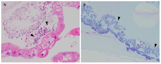
Crithidia acanthocephali had colonized the hindguts of infected bees 12 days after infection, whereas no trypanosomatid forms were observed in the control bees. These trypanosomatids were observed to cover the surface of the digestive tract in all the infected bees analyzed, and images were obtained from both the ileum and rectum (Figure 1). The trypanosomatid cells were observed to form clusters and to also organize as monolayers. While the former clusters were more often observed in the ileum, in the rectum, the monolayer arrangement seemed to predominate. No histological changes were observed in the host’s epithelial cells, which suggests that the trypanosomatids cause no evident damage to the intestinal cells.

Figure 1.
Light microscopy images of the ileum (A) and rectum (B) of workers infected with C. acanthocephali: (A) longitudinal section (40×) of the ileum stained with hematoxylin and eosin (H&E), in which trypanosomatid clusters could be observed (arrowheads); (B) methylene blue-stained longitudinal semi-thin section (20×) of the rectum, covered by a layer of trypanosomatid cells (arrowheads).
3.2. Transmission Electron Microscopy
Although samples from both the ileum and rectum were processed for TEM, we could only obtain images of C. acanthocephali from the latter (Figure 2 and Figure 3). Trypanosomatid cells were found lining the digestive tract surface, and their morphology differed from the choanomastigotes observed in cultures. Instead, they adopted a haptomonad-like form, with their flagella remodeled into an attachment pad. This modification allowed trypanosomatids to attach to the epithelial cells and remain inside the host’s hindgut. Moreover, in accordance with the light microscopy images, these protozoa did not appear to damage host cells.

Figure 2.
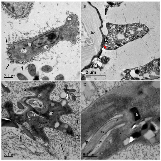
Transmission electron microscopy (TEM) images obtained from the rectum of C. acanthocephali infected bees: (A) longitudinal section of an adherent haptomonad form, with the elongated nucleus (n) visible on the central part of the cell body, with condensed heterochromatin both centrally and around the nuclear membrane, as well as some organelles as acidocalcisomes (ac) and glycosomes (g). A network of fibers of unknown nature can be observed around the trypanosomatid, especially in the posterior part of the cell (black arrows). (B) Haptomonad cell attached to the host cell (hc). Some flagellapodia (fd) that remain attached could also be observed, even though their cell bodies seem to be lost or are not in the same section. (C) Cross-section of a haptomonad cell undergoing division, in which two flagella axonemes (ax) can be observed inside both flagellar pockets. (D) Detailed longitudinal section of a haptomonad cell. The flagellar body (f), with the axoneme visible (ax), is located within half of the length of the cell body, right before the kinetoplast (k). Some vesicles (v) are observed inside the flagellar pocket, while other organelles, such as the mitochondria (m), are also observed in the cell body.

Figure 3.
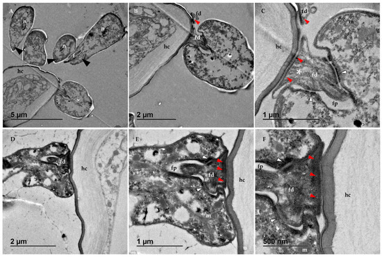
Transmission electron microscopy (TEM) images obtained from the rectum of C. acanthocephali infected bees: (A) longitudinal section that shows different phases of the attachment process, including haptomonads adhered to the host cell (hc) and non-attached trypanosomatids with their flagella (f) oriented to the host surface (black arrowheads). (B,C) Details of the haptomonad cells and its flagellopodium (fd), which is surrounded by the flagellar pocket (fp) and is held together with the cell body through the type A desmosomes (white arrowheads). Hemidesmosome-like complexes are observed between the flagellopodium and the host surface (electron-dense material underneath the attachment pad: red arrowheads), while an array of filaments reinforce the entire complex (asterisk). Some organelles could be observed inside the trypanosomatids, such as the nucleus (n) and the mitochondria (m). (D) Haptomonad attached to the cuticular layer of the honey bee epithelial cells (hc), with the axoneme (ax) visible at the base of the flagellopodium (fd). (E,F) Details of the flagellopodium (fd), where type A desmosomes (white arrowheads) and the hemidesmosome-like complexes (red arrowheads) could be observed.
The magnification of the images allowed us to observe the ultrastructure of the trypanosomatid cells in the magnified images. The nucleus elongated and is centrally located in some cells (Figure 2A) and it moved toward the posterior part of the cell as it approaches the host’s surface (Figure 3A), in which heterochromatin accumulated visibly beneath the membrane (Figure 2A). A prominent disc-shaped kinetoplast could be observed immediately posterior to the start of the flagellum (Figure 2D). A single, large mitochondria can be observed at the peripheral zones of the cells (Figure 2A) and the flagellar pocket seemed to insert up to approximately half the length of the cell body (Figure 2D and Figure 3B,D). This structure (also referred to as the reservoir by some authors [18,19,20]) surrounded the flagellum from its start and throughout its trajectory inside the cell body until it finally emerged at the anterior part of the cell as a modified structure that formed an attachment pad: the flagellopodium (Figure 2A,B and Figure 3B,C,E,F). Doublets of microtubules, not seen in the flagellar pocket, ran along the entire length of the modified flagellum, adopting the typical (9 × 2) + 2 axonemal conformation (Figure 2C,D).
The distal part of this modified flagellum was the point of contact between the trypanosomatid cell and the host’s intestinal surface, forming hemidesmosome-like junction complexes evident as electron-dense areas in the images immediately beneath the flagellopodium membrane (Figure 3B–F). Other junction complexes, such as type A desmosomes, strengthened the entire trypanosomatid cell structure. These desmosomes are responsible for holding the cell body and the flagellopodium together, and they can be seen in the images as dark zones between the membrane of the latter and that of the flagellar pocket (Figure 3C,E,F). To reinforce the entire cell complex, an array of filaments can be seen and they are connected to the axoneme with both types of junctions: hemidesmosome-like complexes and type A desmosomes (Figure 3C,E,F).
Other organelles and cell structures can be observed in the cytoplasm (Figure 2D), including typical trypanosomatid structures such as glycosomes and acidocalcisomes, with different electron densities (Figure 2A). Some vesicles were observed inside the flagellar pocket (Figure 2D) that serve to expand the surface of the reservoir and adapt it to the modifications of the flagellum. A single layer of subpellicular microtubules was found beneath the cell membrane (not shown in the images). In addition, the cells seem to be surrounded by a fiber network of electron-dense particles of unknown nature that seemed to be secreted by the cells themselves (Figure 2 and Figure 3). This fiber network appeared to be more intense at the posterior part of the cell (black arrows). In some cells two axonemes could be observed in two different flagellar pockets (Figure 2C), a clear indicator that events that are part of the multiplication cycle of these haptomonad forms of the organism were underway inside the bee’s rectum.
4. Discussion
For the first time, this study describes the presence of the haptomonad morphotype of the C. acanthocephali trypanosomatid in the hindgut of A. mellifera attached to the intestinal surface through the transformation of the flagellum into an attachment pad. Haptomonad morphology has been observed in the intestinal tract of several insect hosts, such as Anopheles gambiae [21], Phlebotomus papatasi [22], or Lutzomyia longipalpis [23]. This stage was also observed in vitro in culture, since haptomonad cells can attach to synthetic materials [24,25]. Furthermore, a recent study found that Paratrypanosoma confusum, a species that infects mosquitoes and that is phylogenetically located between the parasitic trypanosomatids branch and the bodonids (free-living kinetoplastids), had a similar sedentary stage [26]. Thus, the haptomonad morphotype can be considered a common feature of the Trypanosomatidae family.
In terms of honey bees, previous research described both “spheroid” and “flagellated” trypanosomatid forms colonizing the hindgut of worker bees experimentally infected with the species C. mellificae or L. passim [17,27,28]. A detailed description of the haptomonad form of these two species recently appeared in the honey bee [13], apparently with the aforementioned spheroid morphotype. Crithidia acanthocephali adopts a similar morphology, and it was found to colonize both the ileum and rectum. Although trypanosomatids were observed here at both these sites, the latter seems to be the preferred location since it was where the haptomonad forms were observed by TEM. One hypothesis is that the union between the trypanosomatids and the epithelial cells could be thicker in this region than in the ileum such that they might better resist the fixation process when they are in this part of the gut. However, it could also be due to the rectum containing more parasites than the ileum or the nutritional requirements of this trypanosomatid. Sugars and amino acids are thought to be absorbed by rectal cells [29] in what would be an ideal environment for trypanosomatids to grow. The minimal nutritional requirements of these species have been investigated previously by the omission of individual components [30]. It was discovered that this species can use D-ribose as a carbon source, free or as adenosine, although many other carbohydrates enhanced its growth, especially glucose, fructose, and sucrose. These are precisely the major components of the bees’ diet; therefore, they are commonly found in the honey bee’s gut [31]. Another interesting factor is that purine was found to be vital for this species to grow. According to the metabolomics analysis of different honey bee gut regions [31], the highest concentrations of purines can be found in the rectum, followed by the ileum, whereas in the midgut they are barely found at all. Moreover, the cuticle layer that covers the ileum and the rectum seems to play an important role in trypanosomatid adhesion and the formation of junctions [21,28].
Despite the valuable information about the development of this trypanosomatid within the honey bee that is gained by detecting the haptomonad stage, its pathogenic implications remain unclear, as this is also the case with C. mellificae, L. passim and other insect trypanosomatids [4]. With the information obtained here, we can only hypothesize about both the pathogenicity of this morphotype and its possible mechanisms of virulence. Based on the lack of histological changes in the gut epithelial cells, it seems most probable that these protozoa are active in the lumen. Indeed, their disposition covering the host surface could hinder nutrient absorption [21,32] and the presence of the uncharacterized secreted particles observed could be implicated in this effect. Thus, future research on the relevance of establishing haptomonads, the biochemical mechanisms implicated, and studies into mortality would be of great interest to determine the pathogenic mechanisms induced by this trypanosomatid species. Nevertheless, remaining attached to the host’s intestinal walls allows trypanosomatids to maintain infection of the host for a long time. Although not much is known about the mechanisms of transmission of monoxenous trypanosomatids, some species of Blastocrithidia and Leptomonas, among others, form resistant cells or “cysts” that allow their survival under adverse conditions [33,34]. As far as we know, C. acanthocephali does not form these resistant forms, so lengthening their stay inside the host could increase the chances of transmission to another individual [35]. However, the establishment and attachment of this trypanosomatid will probably be influenced by other factors. For example, the microbiota present in the honey bee’s gut has been proposed as likely to have a protective role against microorganisms [36].
Based on the “one parasite-one host” paradigm, new trypanosomatid species have traditionally been named according to the host in which they were first described [3]. The molecular characterization of their associations and the specificity of several trypanosomatid species in different heteropteran hosts has proved that these interactions may not be that stringent [35], suggesting more promiscuous host–parasite relationships than were initially thought for monoxenous trypanosomatids. Several authors have used C. acanthocephali experimental infection (rectal or haematocele injections) to test this host-parasite specificity in what a priori were considered to be foreign hosts [10,11]. In all cases, dense populations of trypanosomatids were observed to colonize the gut and hemolymph of the host insect, increasing host mortality. Trypanosomatid infection provoked a phagocytic response and the formation of nodules, in which motile flagellates could be observed, sometimes even in stages of division [11], which provides evidence that C. acanthocephali can multiply in foreign hosts. Here, events characteristic of trypanosomatid division were detected in the rectum of the honey bees, such as the presence of two flagella on the same cell, suggesting that this trypanosomatid species can truly infect this insect host.
Crithidia acanthocephali was recently found for the first time in honey bee colonies in Spain [12] and in bumble bees [37]. It was detected in honey bee colonies throughout the year, at all seasons, and no differences were found between interior and forager bees, indicating it is a common organism in bee colonies. Together with the apparent absence of haplotype differentiation in L. passim, C. mellificae, or C. bombi between their hosts [38], these data suggest that insect trypanosomatids may infect a wider range of species than was previously thought.
5. Conclusions
By obtaining the first microscopy images of C. acanthocephali in the honey bee hindgut, this trypanosomatid species can apparently adopt the haptomonad stage in this host in order to remain attached to honey bee hindgut cells. The impact of the presence of trypanosomatids and more specifically of C. acanthocephali on honey bee health remains unclear, making this an interesting area for further research. Nevertheless, the data presented here suggest that insect trypanosomatids have the potential to infect and multiply in several insect species, which is evidence that these organisms have less strict parasite–host specificity than previously thought.
Author Contributions
Conceptualization, M.B.-A. and M.H.; methodology, M.B.-A., P.G.-P. and M.H.; validation, M.B.-A., P.G.-P., L.M.d.P., R.M.-H. and M.H.; formal analysis, M.B.-A., P.G.-P., L.M.d.P. and M.H.; investigation, M.B.-A., P.G.-P. and M.H.; resources, P.G.-P., R.M.-H. and M.H.; data curation, M.B.-A., P.G.-P. and M.H.; writing—original draft preparation, M.B.-A.; writing—review and editing, M.B.-A., P.G.-P., L.M.d.P., R.M.-H. and M.H.; visualization, M.B.-A., P.G.-P. and M.H.; supervision, M.H.; project administration, P.G.-P. and M.H.; funding acquisition, L.M.d.P., R.M.-H. and M.H. All authors have read and agreed to the published version of the manuscript.
Funding
This research was funded by the Instituto Nacional de Investigación y Tecnología Agraria y Alimentaria (INIA, Spain: grant number ERTA2014-00003), by the Spanish Program for Knowledge Generation and Scientific and Technological Strengthening of the R + D + I System, Generación del Conocimiento (grant number PGC2018-098929-A-100), and by the Eva Crane Trust (grant number ECTA_20210308).
Institutional Review Board Statement
Ethical review and approval were waived for this study, in accordance with Directive 2010/63/EU of the European Parliament and of the Council of 22 September 2010 on the protection of animals used for scientific purposes, which among invertebrates is only considered to be applicable to cephalopods. Therefore, no permits were required for experimentation on bees, although all bees were treated according to animal welfare standards to avoid any animal suffering when possible.
Informed Consent Statement
Not applicable.
Acknowledgments
The authors want to thank J. Almagro, J. García, V. Albendea, C. Uceta, M. Gajero, T. Corrales, C. Botías, M. Benito, C. Jabal-Uriel and D. Aguado (at the Bee Pathology Lab of the Centro de Investigación Apícola y Agroambiental -CIAPA, Instituto Regional de Investigación y Desarrollo Agroalimentario y Forestal de Castilla-La Mancha, Spain), and P. Aranda (at the Departamento de Medicina y Cirugía Animal, Facultad de Veterinaria, Universidad Complutense de Madrid, Spain) for their technical support and help. We also thank Marisa García and Miriam González at the Centro Nacional de Microscopía Nacional de Microscopía Electrónica (CNME, UCM, Spain) for their assistance in the preparation, processing, and visualization of the TEM samples. In addition, we want to thank the IRIAF (JCCM) for additional economic support for carrying out this research.
Conflicts of Interest
The authors declare no conflict of interest. The funders had no role in the design of the study; in the collection, analyses, or interpretation of data; in the writing of the manuscript; or in the decision to publish the results.
References
- Lopes, A.H.; Souto-padrón, T.; Dias, F.A.; Gomes, M.T.; Rodrigues, G.C.; Zimmermann, L.T.; Alves, T.L.; Vermelho, A.B. Trypanosomatids: Odd Organisms, Devastating Diseases. Open Parasitol. J. 2010, 4, 30–59. [Google Scholar] [CrossRef]
- Kaufer, A.; Ellis, J.; Stark, D.; Barratt, J. The evolution of trypanosomatid taxonomy. Parasites Vectors 2017, 10, 287. [Google Scholar] [CrossRef] [PubMed]
- Lukeš, J.; Butenko, A.; Hashimi, H.; Maslov, D.A.; Votýpka, J.; Yurchenko, V. Trypanosomatids Are Much More than Just Trypanosomes: Clues from the Expanded Family Tree. Trends Parasitol. 2018, 34, 466–480. [Google Scholar] [CrossRef] [PubMed]
- Schaub, G.A. Pathogenicity of trypanosomatids on insects. Parasitol. Today 1994, 10, 463–468. [Google Scholar] [CrossRef]
- Ghobakhloo, N.; Motazedian, M.H.; Naderi, S.; Ebrahimi, S. Isolation of Crithidia spp. from lesions of immunocompetent patients with suspected cutaneous leishmaniasis in Iran. Trop. Med. Int. Healthy 2019, 24, 116–126. [Google Scholar] [CrossRef]
- Votýpka, J.; d’Avila-Levy, C.M.; Grellier, P.; Maslov, D.A.; Lukeš, J.; Yurchenko, V. New Approaches to Systematics of Trypanosomatidae: Criteria for Taxonomic (Re)description. Trends Parasitol. 2015, 31, 460–469. [Google Scholar] [CrossRef]
- Hanson, W.L.; McGhee, R.B. The Biology and Morphology of Crithidia acanthocephali n. sp., Leptomonas leptoglossi n. sp., and Blastocrithidia euschisti n. sp. J. Protozool. 1961, 8, 200–204. [Google Scholar] [CrossRef]
- Wallace, F.G.; Clark, T.B.; Dyer, M.I.; Collins, T. Two New Species of Flagellates Cultivated from Insects of the Genus Gerris. J. Protozool. 1960, 7, 390–392. [Google Scholar] [CrossRef]
- Boucinha, C.; Caetano, A.R.; Santos, H.L.C.; Helaers, R.; Vikkula, M.; Branquinha, M.H.; Luis, A.; Grellier, P.; Morelli, K.A.; Avila-levy, C.M. Analysing ambiguities in trypanosomatids taxonomy by barcoding. Cad. Saude Publica 2020, 115, 1–14. [Google Scholar] [CrossRef]
- Hanson, W.L.; McGhee, R.B. Experimental Infection of the Hemipteron Oncopeltus fasciatus with Trypanosomatidae Isolated from other Hosts. J. Protozool. 1963, 10, 233–238. [Google Scholar] [CrossRef]
- Schmittner, S.M.; McGhee, R.B. Host Specificity of Various Species of Crithidia Leger. J. Parasitol. 1970, 56, 684. [Google Scholar] [CrossRef]
- Bartolomé, C.; Buendía-Abad, M.; Benito, M.; Sobrino, B.; Amigo, J.; Carracedo, A.; Martín-Hernández, R.; Higes, M.; Maside, X. Longitudinal analysis on parasite diversity in honeybee colonies: New taxa, high frequency of mixed infections and seasonal patterns of variation. Sci. Rep. 2020, 10, 10454. [Google Scholar] [CrossRef] [PubMed]
- Buendía-Abad, M.; García-Palencia, P.; de Pablos, L.M.; Alunda, J.M.; Osuna, A.; Martín-Hernández, R.; Higes, M. First description of Lotmaria passim and Crithidia mellificae haptomonad stages in the honeybee hindgut. Int. J. Parasitol. 2021, 52, 65–75. [Google Scholar] [CrossRef] [PubMed]
- Vickerman, K. The Mode of Attachment of Trypanosoma vivax in the Proboscis of the Tsetse Fly Glossina fuscipes: An Ultrastructural Study of the Epimastigote Stage of the Trypanosome. J. Protozool. 1973, 20, 394–404. [Google Scholar] [CrossRef] [PubMed]
- Bonaldo, M.C.; Souto-Padron, T.; De Souza, W.; Goldenberg, S. Cell-substrate adhesion during Trypanosoma cruzi differentiation. J. Cell Biol. 1988, 106, 1349–1358. [Google Scholar] [CrossRef]
- Hendry, K.A.K.; Vickerman, K. The requirement for epimastigote attachment during division and metacyclogenesis in Trypanosoma congolense. Parasitol. Res. 1988, 74, 403–408. [Google Scholar] [CrossRef]
- Gómez-Moracho, T.; Buendía-Abad, M.; Benito, M.; García-Palencia, P.; Barrios, L.; Bartolomé, C.; Maside, X.; Meana, A.; Jiménez-Antón, M.D.; Olías-Molero, A.I.; et al. Experimental evidence of harmful effects of Crithidia mellificae and Lotmaria passim on honey bees. Int. J. Parasitol. 2020, 50, 1117–1124. [Google Scholar] [CrossRef]
- Molyneux, D.H.; Killick-Kendrick, R.; Ashford, R.W. Leishmania in phlebotomid sandflies III. The ultrastructure of Leishmania mexicana amazonensis in the midgut and pharynx of Lutzomyia longipalpis. Proc. R. Soc. London. Ser. B. Biol. Sci. 1975, 190, 341–357. [Google Scholar] [CrossRef]
- Brooker, B.E. Desmosomes and hemidesmosomes in the flagellate Crithidia fasciculata. Z. Zellforsch. Mikrosk. Anat. 1970, 105, 155–166. [Google Scholar] [CrossRef]
- Wallace, F.G. The trypanosomatid parasites of insects and arachnids. Exp. Parasitol. 1966, 18, 124–193. [Google Scholar] [CrossRef]
- Brooker, B.E. Flagellar attachment and detachment of Crithidia fasciculata to the gut wall of Anopheles gambiae. Protoplasma 1971, 73, 191–202. [Google Scholar] [CrossRef] [PubMed]
- Warburg, A.; Hamada, G.S.; Schlein, Y.; Shire, D. Scanning electron microscopy of Leishmania major in Phlebotomus papatasi. Z. Parasitenkd. 1986, 72, 423–431. [Google Scholar] [CrossRef] [PubMed]
- Killick-Kendrick, R.; Molyneux, D.H.; Ashford, R.W. Leishmania in phlebotomid sandflies I. Modifications of the flagellum associated with attachment to the mid gut and oesophageal valve of the sandfly. Proc. R. Soc. London. Ser. B. Biol. Sci. 1974, 187, 409–419. [Google Scholar] [CrossRef]
- Brooker, B.E. Flagellar adhesion of Crithidia fasciculata to Millipore filters. Protoplasma 1971, 72, 19–25. [Google Scholar] [CrossRef]
- Wakid, M.H.; Bates, P.A. Flagellar attachment of Leishmania promastigotes to plastic film In Vitro. Exp. Parasitol. 2004, 106, 173–178. [Google Scholar] [CrossRef]
- Skalický, T.; Dobáková, E.; Wheeler, R.J.; Tesařová, M.; Flegontov, P.; Jirsová, D.; Votýpka, J.; Yurchenko, V.; Ayala, F.J.; Lukeš, J. Extensive flagellar remodeling during the complex life cycle of Paratrypanosoma, an early-branching trypanosomatid. Proc. Natl. Acad. Sci. USA 2017, 114, 11757–11762. [Google Scholar] [CrossRef]
- Langridge, D.F.; McGhee, R.B. Crithidia mellificae n. sp. an Acidophilic Trypanosomatid of the Honey Bee Apis mellifera. J. Protozool. 1967, 14, 485–487. [Google Scholar] [CrossRef]
- Schwarz, R.S.; Bauchan, G.R.; Murphy, C.A.; Ravoet, J.; De Graaf, D.C.; Evans, J.D. Characterization of two species of trypanosomatidae from the Honey Bee Apis mellifera: Crithidia mellificae Langridge and McGhee, and Lotmaria passim n. gen., n. sp. J. Eukaryot. Microbiol. 2015, 62, 567–583. [Google Scholar] [CrossRef]
- Ramsay, B.Y.J.A. Excretion by the Malpighian Tubules of the Stick Insect, Dixippus morosus (Orthoptera, Phasmidae): Amino Acids, Sugars and Urea. J. Exp. Biol. 1958, 35, 871–891. [Google Scholar] [CrossRef]
- Roitman, I.; Angluster, J.; Fiorini, J.E. Nutrition of the Trypanosomatids Crithidia acanthocephali and C. harmosa. Ribose and Adenosine: Substrates for C. acanthocephali. J. Protozool. 1985, 32, 490–492. [Google Scholar] [CrossRef]
- Zheng, H.; Powell, J.E.; Steele, M.I.; Dietrich, C.; Moran, N.A. Honeybee gut microbiota promotes host weight gain via bacterial metabolism and hormonal signaling. Proc. Natl. Acad. Sci. USA 2017, 114, 4775–4780. [Google Scholar] [CrossRef] [PubMed]
- Molyneux, D.H.; Ashford, R.W. Observations on a trypanosomatid flagellate in a flea, Peromyscopsylla silvatica spectabilis. Ann. Parasitol. Hum. Comp. 1975, 50, 265–274. [Google Scholar] [CrossRef] [PubMed]
- McGhee, R.B.; Cosgrove, W.B. Biology and physiology of the lower trypanosomatidae. Microbiol. Rev. 1980, 44, 140–173. [Google Scholar] [CrossRef] [PubMed]
- Frolov, A.O.; Malysheva, M.N.; Ganyukova, A.I.; Yurchenko, V.; Kostygov, A.Y. Life cycle of Blastocrithidia papi sp. n. (Kinetoplastea, Trypanosomatidae) in Pyrrhocoris apterus (Hemiptera, Pyrrhocoridae). Eur. J. Protistol. 2017, 57, 85–98. [Google Scholar] [CrossRef] [PubMed]
- Kozminsky, E.; Kraeva, N.; Ishemgulova, A.; Dobáková, E.; Lukeš, J.; Kment, P.; Yurchenko, V.; Votýpka, J.; Maslov, D.A. Host-specificity of Monoxenous Trypanosomatids: Statistical Analysis of the Distribution and Transmission Patterns of the Parasites from Neotropical Heteroptera. Protist 2015, 166, 551–568. [Google Scholar] [CrossRef] [PubMed]
- Iorizzo, M.; Letizia, F.; Ganassi, S.; Testa, B.; Petrarca, S.; Albanese, G.; Di Criscio, D.; De Cristofaro, A. Functional Properties and Antimicrobial Activity from Lactic Acid Bacteria as Resources to Improve the Health and Welfare of Honey Bees. Insects 2022, 13, 308. [Google Scholar] [CrossRef]
- Bartolomé, C.; Jabal-Uriel, C.; Buendía-Abad, M.; Benito, M.; Ornosa, C.; De la Rúa, P.; Martín-Hernández, R.; Higes, M.; Maside, X. Wide diversity of parasites in Bombus terrestris (Linnaeus, 1758) revealed by a high-throughput sequencing approach. Environ. Microbiol. 2021, 23, 478–483. [Google Scholar] [CrossRef]
- Bartolomé, C.; Buendía-Abad, M.; Ornosa, C.; De la Rúa, P.; Martín-Hernández, R.; Higes, M.; Maside, X. Bee Trypanosomatids: First Steps in the Analysis of the Genetic Variation and Population Structure of Lotmaria passim, Crithidia bombi and Crithidia mellificae. Microb. Ecol. 2021, 1–12. [Google Scholar] [CrossRef]
Publisher’s Note: MDPI stays neutral with regard to jurisdictional claims in published maps and institutional affiliations. |
© 2022 by the authors. Licensee MDPI, Basel, Switzerland. This article is an open access article distributed under the terms and conditions of the Creative Commons Attribution (CC BY) license (https://creativecommons.org/licenses/by/4.0/).