Article

Canine Cytokines Profile in an Endemic Region of L. infantum: Related Factors

by
Pablo Jesús Marín-García
and
Lola Llobat
*
Department of Animal Production and Health, Veterinary Public Health and Food Science and Technology (PASAPTA), Facultad de Veterinaria, Universidad Cardenal Herrera-CEU, CEU Universities, 46115 Valencia, Spain
*
Author to whom correspondence should be addressed.
Vet. Sci. 2022, 9(6), 305; https://doi.org/10.3390/vetsci9060305
Submission received: 25 May 2022 / Revised: 14 June 2022 / Accepted: 17 June 2022 / Published: 20 June 2022

Abstract

:
Canine leishmaniosis is caused by infection with parasite Leishmania infantum, which are transmitted by sandflies Phlebotomus. Canine leishmaniosis is an endemic disease in the Mediterranean region. The immune response could vary between hosts and determines the severity of the disease and clinical features. The aim of this study was to analyze the serum levels of cytokines TNF-α, IFN-γ, IL-2, IL-6, and IL-8, which are related to the activation of Th1 or Th2 immune responses in dogs living in the L. infantum endemic region. Moreover, we intend to relate and correlate these levels with different factors, such as sex, age, diet, lifestyle, and breed. Epidemiological data and serum were recovered for seventy-eight dogs, and serum levels of cytokines described previously were analyzed by using the ELISA method. The results showed differences in serum levels of IFN-γ, IL-2, and IL-8 between breeds. The lifestyle also affected serum levels of IL-2. The main conclusion of this study is that Ibizan hounds and crossbred dogs have a serological profile of cytokines that seems to indicate certain protections against infection by L. infantum compared to boxer and purebred breeds.

1. Introduction

Leishmaniosis is a zoonotic disease caused by infection with the obligate intracellular protozoan parasite Leishmania spp. that are transmitted by the phlebotomine sandflies from the Psychodidae family, and the genus Phlebotomus is its main vector in the Mediterranean region [1,2,3]. This parasitosis could be presented in mucocutaneous or visceral form, and the latter is the most pathogenic and is caused by the species Leishmania infantum, which is predominant in the Mediterranean area [4,5,6]. Although this parasite can infect different types of mammals, including wild animals and cats [7,8], the domestic dog (Canis lupus familiaris) is considered the principal reservoir of leishmaniosis due to L. infantum in the Mediterranean basin, Middle East, and South America [5,9]. In Spain, the high prevalence of seroprevalence of L. infantum infection in dogs has been demonstrated in the Mediterranean zone, mainly the Balearic Islands, and Valencian Region [10]. However, not all infected dogs develop the disease [3,11,12].
The immune response produced after infection by L. Infantum could vary between hosts and determines the severity of the disease [13,14,15]. Once the parasite has been inoculated by the sandfly’s bite, the host macrophages present the L. infantum antigen to undifferentiated Th0 lymphocyte [4,16]. L. infantum infection actives adaptative immune responses, with a balance between Th1 and Th2 response. The activation of Th1 lymphocytes triggers a cellular response with the activation of macrophages, the synthesis of free oxygen radicals, and the destruction of the parasite, and this response is related with controlling infections [17,18]. The cytokines involved in this pathway are interleukin 2 (IL-2), tumor necrosis factor alpha (TNF-α), and interferon gamma (IFN-γ) [4,15,16]. This response makes it possible to control the infection, and it is known as a protective response, since the host does not show clinical signs of the disease [19]. In fact, elevated serum levels of IFN-γ are related with low parasite counts and few or no clinical signs compared to sick dogs [20], and these levels increase with anti-Leishmania treatment together with clinical improvement and low blood parasitemia [21,22]. Infected dogs exhibit increases in serum levels of IFN-γ, IL-6, and IL-18 and decreases in TNF-α, IL-2, and IL-8 compared to non-infected dogs [23].
Different factors could affect the immune response of dogs. Several studies indicate that nutritional supplements could modify Th1 inflammatory responses [24,25]. In humans, it has been demonstrated that aging affects mainly adaptative immune response, changing cytokines levels and inflammatory responses [26,27,28]. Although studies in this regard in dogs are not as abundant, some studies indicate that aging dogs experience changes like human in inflammation and immune function [29,30]. However, one of the most relevant factors that seems to affect the immune response in Leishmania infection is the canine breed. In fact, the prevalence of infection differs between canine breeds in endemic regions of L. infantum [31], which could indicate immunological protection of some canine breeds compared to others against infection by the parasite and, mainly, the development of leishmaniosis. Several studies show that the activation of Th1 immune response depends on the canine breed such that some breeds present different serum levels of cytokines [22,32,33]. In fact, different studies suggest a certain resistance and/or susceptibility to clinical diseases in dogs related to breed. In this sense, our research group recently published a study where a clear difference was observed in the prevalence of leishmaniosis according to the canine breed, presenting a higher prevalence in breeds such as boxer or Doberman (evolutionarily related) in which the prevalence of clinical signs reaches up to around 50% [31]. This difference in the prevalence of leishmaniosis could be indicating an underlying cause of genetic origin that could be related to different immune responses, or with the activation of different immunity pathways. Solano-Gallego et al. (2000) demonstrated that Ibizan hound dogs have a higher cellular immune response than other breeds and suggested that the Ibizan hound is a special population regarding immune responses against Leishmania infection [34]. Sanchez-Robert et al. (2008) conducted a study with nineteen canine breeds, including Ibizan hounds and boxer breeds, where they analyzed the presence of polymorphisms in the Slc11a gene, which is linked to susceptibility toward canine visceral leishmaniosis [35].
The aim of this study is to relate the serum levels of different cytokines in dogs living in endemic L. infantum regions with different factors that could affect these levels, including canine breeds.

2. Materials and Methods

2.1. Ethics Approval

The experiments involving animals were conducted according to the guidelines of the Declaration of Helsinki and approved by the Animal Experimentation Ethics Committee of the Universidad Cardenal Herrera CEU, with code 2020/VSC/PEA/0216.

2.2. Animals and Data Collection

Seventy-eight dogs living in the Valencia Community region (East of Spain, Mediterranean region) were used for the study, and the serum samples and epidemiological data were recovered from October 2021 to May 2022. For all animals, the following epidemiological data were recovered: sex (two categories: male or female); age (four categories: puppies—less than one year old; young—between one and five years old; adult—between five and ten years old; elder—more than ten years old); breed (four categories: purebred, Ibizan hound, boxer, or crossbred); with official vaccination in order or not; with or without leishmaniosis vaccination; with or without the use of external deworming and type; lived with other animals or not; type of feeding; indoor or outdoor animals; with L. infantum infection detected and, if yes, symptomatic or asymptomatic animals.

2.3. Samples Collection and Cytokines Levels

Ten milliliters of blood samples were collected from cephalic vein with Vacutainer tubes without anticoagulants to recovery serum samples. Serum was obtained and preserved at −20 °C before analysis. Serological testing for L. infantum detected specific antibodies using the indirect immunofluorescent antibody test (IFAT), which was conducted by an external laboratory, and IFAT for anti-Leishmania-specific immunoglobulin G (IgG) antibodies (MegaFLUO LEISH, Megacor Diagnostik GmbH, Hörbranz, Austria). Dog serum was considered seropositive with IFAT titer ≥ 1:80, following the manufacturer’s instructions [36,37].
To recover serum, all samples recovered without anticoagulants were maintained at room temperature and were centrifugated to obtain serum aliquots. Serum levels of TNF-α, IFN-γ, IL-2, IL-6, and IL-8 were measured by a commercial kit of indirect ELISA assay (Canine TNF-α ELISA kit, Canine IFN-γ, Canine IL-2 ELISA kit, Canine IL-6 ELISA kit, and Canine IL-8 ELISA kit; Invitrogen; and Canine IL-18 ELISA kit, MyBiosource, respectively) following the manufacturer recommendations. Briefly, a 50–100 µL of serum was added to the microplate wells. Then, a biotinylated detection antibody specific for different cytokine and Avidin-Horseradish peroxidase (HRP) conjugates were added successively to each microplate well and incubated. Free components were washed away. The substrate solution was added to each well. The enzyme-substrate reaction was determined by using optical density (OD), and it was measured spectrophotometrically at a wavelength of 450 nm. The serum concentration of different cytokines was calculated by comparing the OD of the samples to the standard curve.

2.4. Statistical Analysis

Epidemiological data were recovered, and serum levels of cytokines were analyzed using the general linear model procedure (PROC GLM) of the statistical package SAS (North Carolina State University, USA), after normality and homoscedasticity were tested by Shapiro–Wilks and Levene tests, respectively. The model was carried out with sex, age, type of feed, vaccinated against Leishmania or not, lived with other animals or not, and breed as fixed effects. Pearson’s correlations between cytokine levels were carried out. The statistical significance was set at p-value < 0.05.

3. Results

The epidemiological data recovered are showed in Table 1. Briefly, all animals were outdoor, and most of the animals were owner dogs (91.03%): 51.28% of animals used were males, and 10.26% were puppies (less than one year old), whereas 33.33% were elder (more than ten years old). Most dogs lived with other animals (88.46%), and these animals included other dogs and some tested dogs lived in the same household and were fed commercial diets (88.46%). A percentage of 91.03% with respect to dogs used external deworming, and most dogs have the correct official vaccination, whereas only 10.26% were vaccinated against Leishmania (Table 1).
Only 14.10% of dogs were positive for L. infantum infection by ELISA antibody detection, and 81.82% had at least one clinical sign. All animals with clinical signs presented parasitic burden above 1:100. The clinical signs found, and their prevalence are shown in Table 2.
Table 3 shows the data on the main cytokines controlled in this work. By using a very diverse population (Table 2), the data set used in this study showed a wide range. No expression of TNF-α was found.
Serum levels of cytokines have only been related to breed and living or not with other animals. Animals that do not live with other animals presented higher serum levels of IL-2 than animals that live with other animals (+51%; p = 0.0073) (Figure 1).
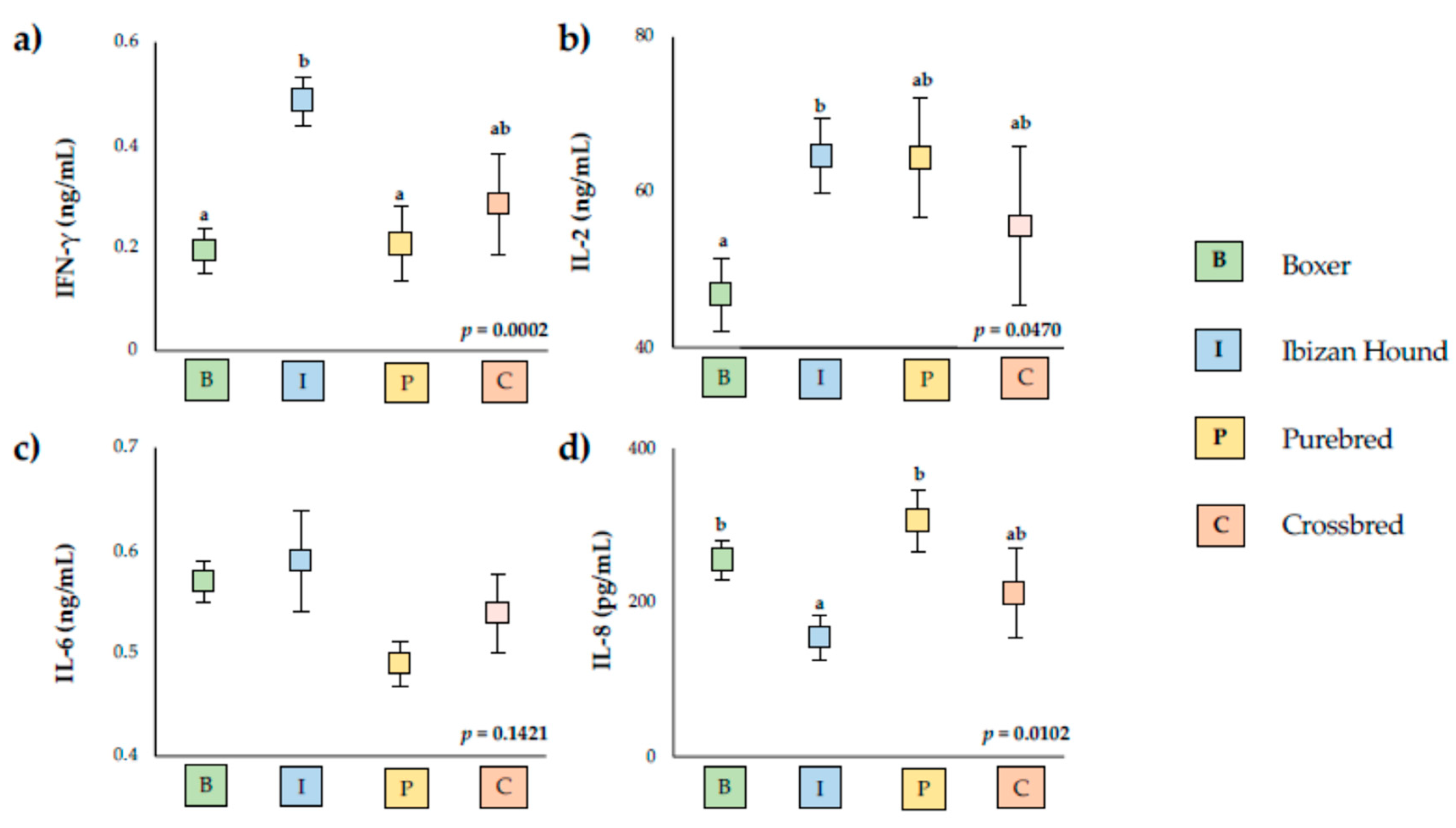
Significant differences were also observed according to boxer breed, Ibizan hound, and other purebreds, and crossbreds presented statistical differences in IFN-γ, IL-2, and IL-8 serum levels (Figure 2).
None of the Ibizan hounds were infected. The serum levels of IFN-γ were similar in boxer, elder animals, and infected animals, and they were lower than Ibizan hounds (p < 0.0001). No differences between infected animals and other groups were found in the serum levels of analyzed cytokines.

4. Discussion

This study analyzed the canine serum levels of cytokines TFN-α, IFN-γ, IL-2, IL-6, and IL-8 in endemic L. infantum regions with different factors such as age, sex, diet, lifestyle, and breed in seventy-eight dogs.
Out of the animals examined, 14.10% were positive to L. infantum infection, in accordance with data observed in other reported studies on Mediterranean areas, where prevalence was estimated between 5 and 57.1% [38]. Previous studies in indoor animals estimated this prevalence in 4.74% in other Mediterranean areas [31], and these higher levels were found probably because the animals used in this study were all outdoor. Of the positive animals, 81.82% presented values above 1:100 of parasitic burden and had at least one clinical feature. Other studies in experimental infected dogs indicated a clinical state in 66% [23], whereas a recent meta-analysis found that only 43% of all animals presented cutaneous and clinical changes [39]. Differences between the prevalence could be due to the parasitic burden quantification technique. Although the Office International des Epizooties recommend the detection for serological methods and the use of parasitological or molecular methods for confirmation in the case of lower titers [24,40], several studies indicate that molecular methods are more sensitive and, concretely, real-time PCR for diagnosis of leishmaniasis is the method chosen in humans (see review [41]). In fact, several studies realized in Brazil and Venezuela recommend the confirmation of leishmaniosis in dogs using molecular methods [42,43]. Related to serological methods, ELISA seems to present higher sensitivity and specificity than IFAT [44]. However, IFAT continues to be the method of choice in many studies. Quantitative serological methods such as ELISA or IFAT determine antibody levels and can confirm the infection of compatible clinical signs, but low antibody levels require confirmation by molecular techniques, according to the recommendation of LeishVet guidelines [12]. However, the most common clinical practice is the detection only using serological methods, mainly the IFAT method.
In agreement with our results, studies realized in dogs naturally infected in Brazil showed that around 90% of dogs infected presented clinical signs [45]. The most common clinical features were lymphadenomegaly (50%) and conjunctivitis (30%), and they are firstly related to high parasitic burden. These results agree with results obtained previously in other studies in Spain [22,46] and with results in experimental infected dogs, where the authors found 50% of lymphadenomegaly and 25% of conjunctivitis [23]. In natural infected animals, the results are similar, with 55.9% of lymphadenopathy found by Freitas et al. (2012) in Brazil. In agree with our results, these authors found hyperglobulinemia. However, they indicated a reduction in red blood cells and hematocrit, unlike the results in this study where the most common biochemical alterations were proteinuria and hyperglobulinemia (30%) [45].
Related to serum cytokines levels, TNF-α is not expressed in the analyzed animals. On the other hand, different authors showed high levels of TFN-α and its expression in skin, lymph node cells, and spleen and liver extracts in animals with visceral leishmaniosis, and these were related to parasitic burden [47,48,49,50]; only Do Nascimento et al. (2013) related the serum levels of TNF-α with parasitic burden in the spleen, indicating that the levels of TNF-α increases with spleen parasitism [51], whereas De lima et al. (2007) did not observe statistical differences in the serum levels of TNF-α in infected dogs with or without visceral symptoms [52]. In our study, the number of parasites in the spleen has not been analyzed; therefore, it is possible that the parasite burden in our animals was so low that serum TNF-α levels were undetectable by the ELISA method. Therefore, the quantification of parasitic burden in spleen and detection of TNF-α by other methods that are more sensitive than PCR would be interesting.
No differences were found in serum cytokines levels related to sex, age, and diet. In humans, several studies were carried out to analyze the sex differences of immune responses. The results were that male monocytes produce higher levels of IL-12, IL-1, and TNF-α and lower levels of IL-2 to lymphocytes than females, which suggests a relative suppression of the cellular immune response of the specific immune system in men [53]. However, according to our results, no differences were found in serum levels of IFN-γ, IL-2, IL-6, and IL-8 that were related to sex in dogs [54,55]. In humans, some studies indicated that the elevated age results in decreased activities in the immune system [28] and in the response to vaccination [56], which is also shown in dogs [57]. However, human studies have shown that this difference is due to the age-associated accumulation of adipose tissue, which is the cause of differences in inflammatory cytokine levels [58,59]; thus, leptin induces the production of TNF-α, IL-6, and IL-12 in obese human and mouse [60,61]. Given that none of our animals were obese or overweight, it is to be expected that there would be no differences in the levels of cytokines analyzed, even though they were of different ages. Although some studies indicate the relationship between immune response and diet [56], only two studies linked certain nutritional supplements with a different immune response in dogs [25,62]. Concretely, Cortese et al. (2015) showed that nutraceuticals supplementation diet could regulate the immune response to L. infantum infection in dogs [25].
Serum levels of IL-2 were higher in dogs living with other animals than those not living with other animals. IL-2 has a key role in the regulation of immune response and is essential for the maturation of Treg [63,64], and it could be a regulator for Th1/Th2 balance [65]. In fact, this cytokine regulates T cells, and it is fundamental to starting immune responses [66]. In serum dogs immunized with Lutzomyia longipalpis salivary protein LJM17, high levels of IL-2 have been found in dogs vaccinated against Leishmania [67], and IL-2 is the most abundant in the serum of cured and asymptomatic dogs than uninfected dogs [68]. The serum levels of IL-2 found in animals that live with other animals could indicate an exposure of these animals to the parasite or even a very early infection, which is still undetectable by IFAT.
The high levels of IL-2 have also been found in Ibizan hound dogs and low levels in Boxer breeds, whereas the groups of purebred and crossbred dogs presented intermediate values. Abbehusen et al. (2018) show that high levels of IL-2 and IL-6 are present in immunized dogs [67]. However, no differences in serum levels of IL-6 were found between breed groups. Similar results were observed by Pinelli et al. (1994) in a study examining infected and non-infected dogs [19], whereas other authors indicate high levels of IL-6 in infected animals, suggesting a mixture of inflammatory and anti-inflammatory responses [23]. The levels of IL-6 have been related to disease progression in visceral leishmaniasis [69,70]. Therefore, no differences in the serum levels of IL-6 found in animals studied could be related to the low number of animals with clinical signs included in our study and with low levels of disease progression observed. Similarly to IL-2, serum levels of IFN-γ were higher in Ibizan hound dogs than boxer and purebred, whereas the crossbred presented intermediate levels. Some studies have demonstrated that high IFN-γ could act as a protective mechanism against infections [32], which could indicate more protection against L. infantum infection in Ibizan hounds and crossbred than boxer and purebred. Recently, it has been demonstrated in L. infantum-infected dogs that purebred dogs present more frequent clinical features than infected crossbred dogs [71]. On the contrary, IL-8 was lower in Ibizan hounds than boxer and purebred dogs, presenting crossbreed intermediate serum levels. The levels of IL-8 have been found to be lower in uninfected than natural and experimentally infected dogs of L. infantum [23], and neutrophils from healthy dogs produced higher levels of IL-8 than neutrophils from Leishmania-infected dogs [72].
These results could indicate an immunological protection mainly in the Ibizan hound, and to a lesser extent in crossbred dogs. Our results show that different immune responses occur depending on the dog’s breed, mainly in Ibizan hound and boxer, according to several studies [31,34,35].

5. Conclusions

The prevalence of L. infantum infection was 14.10%. Of the infected animals, 81.82% presented values above 1:100 of parasitic burden and at least one clinical feature. The most common clinical signs were lymphadenomegaly (50%) and conjunctivitis (30%), whereas the most common biochemical alterations were proteinuria and hyperglobulinemia (30%). The serum levels of cytokines were not affected by the sex, age, and diet of the dog. However, animals that lived with other animals presented higher serum levels of IL-2 than animals that have not lived with other animals. The most relevant factor that affected serum cytokine levels was canine breed. The Ibizan hound showed higher levels of IL-2 and IFN-γ and lower levels of IL-8 than boxer and other purebred animals, suggesting differentiated immune responses in these two canine breeds. Crossbred presented intermediate values of IFN-γ, IL-2, and IL-8. These results could indicate a different immune response depending on canine breed. Cytokine mRNA expression studies in whole blood samples and genetic studies from different canine breeds could be interesting for verifying whether serum cytokine values correspond to differences in gene expression. More studies including other cytokines and other canine breeds are necessary to elucidate the immune response pathway in different genetic animals.

Author Contributions

Conceptualization, L.L.; methodology and formal analysis, P.J.M.-G. and L.L.; data curation, L.L.; writing—original draft preparation, P.J.M.-G. and L.L.; writing—review and editing, L.L.; supervision, L.L.; project administration, L.L.; funding acquisition, L.L. All authors have read and agreed to the published version of the manuscript.

Funding

This work was funded by the grants for emerging research groups of Generalitat Valenciana (GV/2021/104) and IDOC project of University CEU Cardenal Herrera (IDOC21-03). The funding agencies were not involved in the design of study, data collection, analysis of data, or manuscript writing.

Institutional Review Board Statement

The study was conducted according to the guidelines of the Declaration of Helsinki and approved by the Animal Experimentation Ethics Committee of the Universidad Cardenal Herrera CEU (2020/VSC/PEA/0216).

Informed Consent Statement

Informed consent was obtained from all subjects involved in the study.

Data Availability Statement

Not applicable.

Acknowledgments

We are grateful to the Ibizan Hound Association, Boxer Club of Valencia, and Veterinary Hospital of Universidad Cardenal Herrera CEU, especially to Esperanza Montaner.

Conflicts of Interest

The authors declare no conflict of interest.

References

  1. Gramiccia, M. Recent Advances in Leishmaniosis in Pet Animals: Epidemiology, Diagnostics and Anti-Vectorial Prophylaxis. Vet. Parasitol. 2011, 181, 23–30. [Google Scholar] [CrossRef] [PubMed]
  2. Akhoundi, M.; Kuhls, K.; Cannet, A.; Votýpka, J.; Marty, P.; Delaunay, P.; Sereno, D. A Historical Overview of the Classification, Evolution, and Dispersion of Leishmania Parasites and Sandflies. PLoS Negl. Trop. Dis. 2016, 10, e0004349. [Google Scholar] [CrossRef] [PubMed]
  3. Baneth, G.; Koutinas, A.F.; Solano-Gallego, L.; Bourdeau, P.; Ferrer, L. Canine Leishmaniosis—New Concepts and Insights on an Expanding Zoonosis: Part One. Trends Parasitol. 2008, 24, 324–330. [Google Scholar] [CrossRef] [PubMed]
  4. Barbiéri, C.L. Immunology of Canine Leishmaniasis. Parasite Immunol. 2006, 28, 329–337. [Google Scholar] [CrossRef]
  5. Ready, P.D. Epidemiology of Visceral Leishmaniasis. Clin. Epidemiol. 2014, 6, 147–154. [Google Scholar] [CrossRef] [Green Version]
  6. Trájer, A.J.; Sebestyén, V. The Changing Distribution of Leishmania Infantum Nicolle, 1908 and Its Mediterranean Sandfly Vectors in the Last 140 Kys. Sci. Rep. 2019, 9, 11820. [Google Scholar] [CrossRef] [Green Version]
  7. Abbate, J.M.; Arfuso, F.; Napoli, E.; Gaglio, G.; Giannetto, S.; Latrofa, M.S.; Otranto, D.; Brianti, E. Leishmania Infantum in Wild Animals in Endemic Areas of Southern Italy. Comp. Immunol. Microbiol. Infect. Dis. 2019, 67, 101374. [Google Scholar] [CrossRef]
  8. Ahuir-Baraja, A.E.; Ruiz, M.P.; Garijo, M.M.; Llobat, L. Feline Leishmaniosis: An Emerging Public Health Problem. Vet. Sci. 2021, 8, 173. [Google Scholar] [CrossRef]
  9. Mancianti, F.; Gramiccia, M.; Gradoni, L.; Pieri, S. Studies on Canine Leishmaniasis Control. 1. Evolution of Infection of Different Clinical Forms of Canine Leishmaniasis Following Antimonial Treatment. Trans. R. Soc. Trop. Med. Hyg. 1988, 82, 566–567. [Google Scholar] [CrossRef]
  10. Gálvez, R.; Montoya, A.; Cruz, I.; Fernández, C.; Martín, O.; Checa, R.; Chicharro, C.; Migueláñez, S.; Marino, V.; Miró, G. Latest Trends in Leishmania Infantum Infection in Dogs in Spain, Part I: Mapped Seroprevalence and Sand Fly Distributions. Parasit Vectors 2020, 13, 204. [Google Scholar] [CrossRef] [Green Version]
  11. Miró, G.; Cardoso, L.; Pennisi, M.G.; Oliva, G.; Baneth, G. Canine Leishmaniosis--New Concepts and Insights on an Expanding Zoonosis: Part Two. Trends Parasitol. 2008, 24, 371–377. [Google Scholar] [CrossRef] [PubMed]
  12. Solano-Gallego, L.; Miró, G.; Koutinas, A.; Cardoso, L.; Pennisi, M.G.; Ferrer, L.; Bourdeau, P.; Oliva, G.; Baneth, G. The LeishVet Group, LeishVet Guidelines for the Practical Management of Canine Leishmaniosis. Parasit Vectors 2011, 4, 86. [Google Scholar] [CrossRef] [PubMed] [Green Version]
  13. Rossi, M.; Fasel, N. How to Master the Host Immune System? Leishmania Parasites Have the Solutions! Int. Immunol. 2018, 30, 103–111. [Google Scholar] [CrossRef] [PubMed] [Green Version]
  14. Vanloubbeeck, Y.; Jones, D.E. The Immunology of Leishmania Infection and the Implications for Vaccine Development. Ann. N. Y. Acad. Sci. 2004, 1026, 267–272. [Google Scholar] [CrossRef]
  15. Solano-Gallego, L.; Koutinas, A.; Miró, G.; Cardoso, L.; Pennisi, M.G.; Ferrer, L.; Bourdeau, P.; Oliva, G.; Baneth, G. Directions for the Diagnosis, Clinical Staging, Treatment and Prevention of Canine Leishmaniosis. Vet. Parasitol. 2009, 165, 1–18. [Google Scholar] [CrossRef]
  16. Hosein, S.; Blake, D.P.; Solano-Gallego, L. Insights on Adaptive and Innate Immunity in Canine Leishmaniosis. Parasitology 2017, 144, 95–115. [Google Scholar] [CrossRef] [Green Version]
  17. Coffman, R.L.; Seymour, B.W.; Lebman, D.A.; Hiraki, D.D.; Christiansen, J.A.; Shrader, B.; Cherwinski, H.M.; Savelkoul, H.F.; Finkelman, F.D.; Bond, M.W. The Role of Helper T Cell Products in Mouse B Cell Differentiation and Isotype Regulation. Immunol. Rev. 1988, 102, 5–28. [Google Scholar] [CrossRef]
  18. Mosmann, T.R.; Cherwinski, H.; Bond, M.W.; Giedlin, M.A.; Coffman, R.L. Two Types of Murine Helper T Cell Clone. I. Definition According to Profiles of Lymphokine Activities and Secreted Proteins. J. Immunol. 1986, 136, 2348–2357. [Google Scholar]
  19. Pinelli, E.; Killick-Kendrick, R.; Wagenaar, J.; Bernadina, W.; del Real, G.; Ruitenberg, J. Cellular and Humoral Immune Responses in Dogs Experimentally and Naturally Infected with Leishmania Infantum. Infect. Immun. 1994, 62, 229–235. [Google Scholar] [CrossRef] [Green Version]
  20. Ordeix, L.; Montserrat-Sangrà, S.; Martínez-Orellana, P.; Solano-Gallego, L. Toll-Like Receptors 2, 4, and 7, Interferon-Gamma, Interleukin 10, and Programmed Death Ligand 1 Transcripts in Leishmanin Skin Test-Positive Reactions of Ibizan Hound Dogs. J. Immunol. Res. 2020, 2020, 9602576. [Google Scholar] [CrossRef]
  21. Priolo, V.; Martínez-Orellana, P.; Pennisi, M.G.; Masucci, M.; Prandi, D.; Ippolito, D.; Bruno, F.; Castelli, G.; Solano-Gallego, L. Leishmania Infantum-Specific IFN-γ Production in Stimulated Blood from Cats Living in Areas Where Canine Leishmaniosis Is Endemic. Parasit Vectors 2019, 12, 133. [Google Scholar] [CrossRef] [PubMed]
  22. Solano-Gallego, L.; Montserrrat-Sangrà, S.; Ordeix, L.; Martínez-Orellana, P. Leishmania infantum-Specific Production of IFN-γ and IL-10 in Stimulated Blood from Dogs with Clinical Leishmaniosis. Parasit Vectors 2016, 9, 317. [Google Scholar] [CrossRef] [PubMed] [Green Version]
  23. Abbehusen, M.M.C.; Almeida, V.D.A.; da S Solcà, M.; da Silva Pereira, L.; Costa, D.J.; Gil-Santana, L.; Bozza, P.T.; Fraga, D.B.M.; Veras, P.S.T.; Dos-Santos, W.L.C.; et al. Clinical and Immunopathological Findings during Long Term Follow-up in Leishmania Infantum Experimentally Infected Dogs. Sci. Rep. 2017, 7, 15914. [Google Scholar] [CrossRef] [PubMed] [Green Version]
  24. Cortese, L.; Annunziatella, M.; Palatucci, A.T.; Rubino, V.; Piantedosi, D.; Di Loria, A.; Ruggiero, G.; Ciaramella, P.; Terrazzano, G. Regulatory T Cells, Cytotoxic T Lymphocytes and a T(H)1 Cytokine Profile in Dogs Naturally Infected by Leishmania Infantum. Res. Vet. Sci. 2013, 95, 942–949. [Google Scholar] [CrossRef]
  25. Lombardi, P.; Palatucci, A.T.; Giovazzino, A.; Mastellone, V.; Ruggiero, G.; Rubino, V.; Musco, N.; Crupi, R.; Cutrignelli, M.I.; Britti, D.; et al. Clinical and Immunological Response in Dogs Naturally Infected by L. infantum Treated with a Nutritional Supplement. Animals 2019, 9, 501. [Google Scholar] [CrossRef] [Green Version]
  26. Castelo-Branco, C.; Soveral, I. The Immune System and Aging: A Review. Gynecol. Endocrinol. 2014, 30, 16–22. [Google Scholar] [CrossRef]
  27. Pinti, M.; Appay, V.; Campisi, J.; Frasca, D.; Fülöp, T.; Sauce, D.; Larbi, A.; Weinberger, B.; Cossarizza, A. Aging of the Immune System: Focus on Inflammation and Vaccination. Eur. J. Immunol. 2016, 46, 2286–2301. [Google Scholar] [CrossRef]
  28. Sadighi Akha, A.A. Aging and the Immune System: An Overview. J. Immunol. Methods 2018, 463, 21–26. [Google Scholar] [CrossRef]
  29. Alexander, J.E.; Colyer, A.; Haydock, R.M.; Hayek, M.G.; Park, J. Understanding How Dogs Age: Longitudinal Analysis of Markers of Inflammation, Immune Function, and Oxidative Stress. J. Gerontol. A Biol. Sci. Med. Sci. 2018, 73, 720–728. [Google Scholar] [CrossRef] [Green Version]
  30. HogenEsch, H.; Thompson, S.; Dunham, A.; Ceddia, M.; Hayek, M. Effect of Age on Immune Parameters and the Immune Response of Dogs to Vaccines: A Cross-Sectional Study. Vet. Immunol. Immunopathol. 2004, 97, 77–85. [Google Scholar] [CrossRef]
  31. Edo, M.; Marín-García, P.J.; Llobat, L. Is the Prevalence of Leishmania infantum Linked to Breeds in Dogs? Characterization of Seropositive Dogs in Ibiza. Animals 2021, 11, 2579. [Google Scholar] [CrossRef] [PubMed]
  32. Martínez-Orellana, P.; Marí-Martorell, D.; Montserrat-Sangrà, S.; Ordeix, L.; Baneth, G.; Solano-Gallego, L. Leishmania infantum-Specific IFN-γ Production in Stimulated Blood from Dogs with Clinical Leishmaniosis at Diagnosis and during Treatment. Vet. Parasitol. 2017, 248, 39–47. [Google Scholar] [CrossRef] [PubMed]
  33. Sanchez-Robert, E.; Altet, L.; Sanchez, A.; Francino, O. Polymorphism of Slc11a1 (Nramp1) Gene and Canine Leishmaniasis in a Case-Control Study. J. Hered. 2005, 96, 755–758. [Google Scholar] [CrossRef] [PubMed]
  34. Solano-Gallego, L.; Llull, J.; Ramos, G.; Riera, C.; Arboix, M.; Alberola, J.; Ferrer, L. The Ibizian Hound Presents a Predominantly Cellular Immune Response against Natural Leishmania Infection. Vet. Parasitol. 2000, 90, 37–45. [Google Scholar] [CrossRef]
  35. Sanchez-Robert, E.; Altet, L.; Utzet-Sadurni, M.; Giger, U.; Sanchez, A.; Francino, O. Slc11a1 (Formerly Nramp1) and Susceptibility to Canine Visceral Leishmaniasis. Vet. Res. 2008, 39, 36. [Google Scholar] [CrossRef] [Green Version]
  36. Olías-Molero, A.I.; Corral, M.J.; Jiménez-Antón, M.D.; Alunda, J.M. Early Antibody Response and Clinical Outcome in Experimental Canine Leishmaniasis. Sci. Rep. 2019, 9, 18606. [Google Scholar] [CrossRef]
  37. Miró, G.; Checa, R.; Montoya, A.; Hernández, L.; Dado, D.; Gálvez, R. Current Situation of Leishmania infantum Infection in Shelter Dogs in Northern Spain. Parasit Vectors 2012, 5, 60. [Google Scholar] [CrossRef] [Green Version]
  38. Morillas, F.; Sanchez Rabasco, F.; Ocaña, J.; Martin-Sanchez, J.; Ocaña-Wihelmi, J.; Acedo, C.; Sanchiz-Marin, M.C. Leishmaniosis in the Focus of the Axarquía Region, Malaga Province, Southern Spain: A Survey of the Human, Dog, and Vector. Parasitol. Res. 1996, 82, 569–570. [Google Scholar] [CrossRef]
  39. Oliveira, C.S.; Ratzlaff, F.R.; Pötter, L.; Romão, P.R.T.; de Avila Botton, S.; Vogel, F.S.F.; Sangioni, L.A. Clinical and Pathological Aspects of Canine Cutaneous Leishmaniasis: A Meta-Analysis. Acta Parasitol. 2019, 64, 916–922. [Google Scholar] [CrossRef]
  40. Maroli, M.; Feliciangeli, M.D.; Bichaud, L.; Charrel, R.N.; Gradoni, L. Phlebotomine Sandflies and the Spreading of Leishmaniases and Other Diseases of Public Health Concern. Med. Vet. Entomol. 2013, 27, 123–147. [Google Scholar] [CrossRef]
  41. Galluzzi, L.; Ceccarelli, M.; Diotallevi, A.; Menotta, M.; Magnani, M. Real-Time PCR Applications for Diagnosis of Leishmaniasis. Parasit Vectors 2018, 11, 273. [Google Scholar] [CrossRef] [PubMed]
  42. Paz, G.F.; Rugani, J.M.N.; Marcelino, A.P.; Gontijo, C.M.F. Implications of the Use of Serological and Molecular Methods to Detect Infection by Leishmania spp. in Urban Pet Dogs. Acta Trop. 2018, 182, 198–201. [Google Scholar] [CrossRef] [PubMed]
  43. Rivas, A.K.; Alcover, M.M.; Martínez-Orellana, P.; Montserrat-Sangrà, S.; Nachum-Biala, Y.; Fisa, R.; Riera, C.; Baneth, G.; Solano-Gallego, L. Serological and Molecular Survey of Leishmania Infection in Dogs from Venezuela. Vet. Parasitol. Reg. Stud. Rep. 2020, 21, 100420. [Google Scholar] [CrossRef]
  44. de Arruda, M.M.; Figueiredo, F.B.; Marcelino, A.P.; Barbosa, J.R.; Werneck, G.L.; Noronha, E.F.; Romero, G.A.S. Sensitivity and Specificity of Parallel or Serial Serological Testing for Detection of Canine Leishmania Infection. Mem. Inst. Oswaldo Cruz. 2016, 111, 168–173. [Google Scholar] [CrossRef] [PubMed]
  45. de Freitas, J.C.C.; Nunes-Pinheiro, D.C.S.; Neto, B.E.L.; Santos, G.J.L.; de Abreu, C.R.A.; Braga, R.R.; de Morais Campos, R.; de Oliveira, L.F. Clinical and Laboratory Alterations in Dogs Naturally Infected by Leishmania chagasi. Rev. Soc. Bras. Med. Trop. 2012, 45, 24–29. [Google Scholar] [CrossRef] [PubMed] [Green Version]
  46. de Ybáñez, R.R.; del Río, L.; Martínez-Carrasco, C.; Segovia, M.; Cox, J.; Davies, C.; Berriatua, E. Questionnaire Survey on Canine Leishmaniosis in Southeastern Spain. Vet. Parasitol. 2009, 164, 124–133. [Google Scholar] [CrossRef] [PubMed]
  47. Alves, C.F.; de Amorim, I.F.G.; Moura, E.P.; Ribeiro, R.R.; Alves, C.F.; Michalick, M.S.; Kalapothakis, E.; Bruna-Romero, O.; Tafuri, W.L.; Teixeira, M.M.; et al. Expression of IFN-Gamma, TNF-Alpha, IL-10 and TGF-Beta in Lymph Nodes Associates with Parasite Load and Clinical Form of Disease in Dogs Naturally Infected with Leishmania (Leishmania) chagasi. Vet. Immunol. Immunopathol. 2009, 128, 349–358. [Google Scholar] [CrossRef]
  48. DE F Michelin, A.; Perri, S.H.V.; De Lima, V.M.F. Evaluation of TNF-α, IL-4, and IL-10 and Parasite Density in Spleen and Liver of L. (L.) chagasi Naturally Infected Dogs. Ann. Trop. Med. Parasitol. 2011, 105, 373–383. [Google Scholar] [CrossRef] [Green Version]
  49. Pereira-Fonseca, D.C.M.; Oliveira-Rovai, F.M.; Rodas, L.A.C.; Beloti, C.A.C.; Torrecilha, R.B.P.; Ito, P.K.R.K.; Avanço, S.V.; Cipriano, R.S.; Utsunomiya, Y.T.; Hiramoto, R.M.; et al. Dog Skin Parasite Load, TLR-2, IL-10 and TNF-α Expression and Infectiousness. Parasite Immunol. 2017, 39. [Google Scholar] [CrossRef]
  50. Venturin, G.L.; Chiku, V.M.; Silva, K.L.O.; de Almeida, B.F.M.; de Lima, V.M.F. M1 Polarization and the Effect of PGE2 on TNF-α Production by Lymph Node Cells from Dogs with Visceral Leishmaniasis. Parasite Immunol. 2016, 38, 698–704. [Google Scholar] [CrossRef] [Green Version]
  51. do Nascimento, P.R.P.; Martins, D.R.A.; Monteiro, G.R.G.; Queiroz, P.V.; Freire-Neto, F.P.; Queiroz, J.W.; Morais Lima, A.L.; Jeronimo, S.M.B. Association of Pro-Inflammatory Cytokines and Iron Regulatory Protein 2 (IRP2) with Leishmania Burden in Canine Visceral Leishmaniasis. PLoS ONE 2013, 8, e73873. [Google Scholar] [CrossRef] [PubMed] [Green Version]
  52. de Lima, V.M.F.; Peiro, J.R.; de Oliveira Vasconcelos, R. IL-6 and TNF-Alpha Production during Active Canine Visceral Leishmaniasis. Vet. Immunol. Immunopathol. 2007, 115, 189–193. [Google Scholar] [CrossRef] [PubMed]
  53. Bouman, A.; Schipper, M.; Heineman, M.J.; Faas, M.M. Gender Difference in the Non-Specific and Specific Immune Response in Humans. Am. J. Reprod. Immunol. 2004, 52, 19–26. [Google Scholar] [CrossRef] [PubMed]
  54. Allison, L.N.; Jaffey, J.A.; Bradley-Siemens, N.; Tao, Z.; Thompson, M.; Backus, R.C. Immune Function and Serum Vitamin D in Shelter Dogs: A Case-Control Study. Vet. J. 2020, 261, 105477. [Google Scholar] [CrossRef] [PubMed]
  55. Calvalido, J.; Wood, G.A.; Mutsaers, A.J.; Wood, D.; Sears, W.; Woods, J.P. Comparison of Serum Cytokine Levels between Dogs with Multicentric Lymphoma and Healthy Dogs. Vet. Immunol. Immunopathol. 2016, 182, 106–114. [Google Scholar] [CrossRef] [PubMed]
  56. Zimmermann, P.; Curtis, N. Factors That Influence the Immune Response to Vaccination. Clin. Microbiol. Rev. 2019, 32, e00084-18. [Google Scholar] [CrossRef] [PubMed] [Green Version]
  57. Schultz, R.D.; Thiel, B.; Mukhtar, E.; Sharp, P.; Larson, L.J. Age and Long-Term Protective Immunity in Dogs and Cats. J. Comp. Pathol. 2010, 142 Suppl 1, S102–S108. [Google Scholar] [CrossRef]
  58. Park, H.S.; Park, J.Y.; Yu, R. Relationship of Obesity and Visceral Adiposity with Serum Concentrations of CRP, TNF-Alpha and IL-6. Diabetes Res. Clin. Pract. 2005, 69, 29–35. [Google Scholar] [CrossRef]
  59. Sewter, C.P.; Digby, J.E.; Blows, F.; Prins, J.; O’Rahilly, S. Regulation of Tumour Necrosis Factor-Alpha Release from Human Adipose Tissue in Vitro. J. Endocrinol. 1999, 163, 33–38. [Google Scholar] [CrossRef]
  60. La Cava, A.; Matarese, G. The Weight of Leptin in Immunity. Nat. Rev. Immunol. 2004, 4, 371–379. [Google Scholar] [CrossRef]
  61. Sarraf, P.; Frederich, R.C.; Turner, E.M.; Ma, G.; Jaskowiak, N.T.; Rivet, D.J.; Flier, J.S.; Lowell, B.B.; Fraker, D.L.; Alexander, H.R. Multiple Cytokines and Acute Inflammation Raise Mouse Leptin Levels: Potential Role in Inflammatory Anorexia. J. Exp. Med. 1997, 185, 171–175. [Google Scholar] [CrossRef] [PubMed] [Green Version]
  62. Cortese, L.; Annunziatella, M.; Palatucci, A.T.; Lanzilli, S.; Rubino, V.; Di Cerbo, A.; Centenaro, S.; Guidetti, G.; Canello, S.; Terrazzano, G. An Immune-Modulating Diet Increases the Regulatory T Cells and Reduces T Helper 1 Inflammatory Response in Leishmaniosis Affected Dogs Treated with Standard Therapy. BMC Vet. Res. 2015, 11, 295. [Google Scholar] [CrossRef] [PubMed] [Green Version]
  63. Bendickova, K.; Fric, J. Roles of IL-2 in Bridging Adaptive and Innate Immunity, and as a Tool for Cellular Immunotherapy. J. Leukoc. Biol. 2020, 108, 427–437. [Google Scholar] [CrossRef] [PubMed]
  64. Cheng, G.; Yu, A.; Dee, M.J.; Malek, T.R. IL-2R Signaling Is Essential for Functional Maturation of Regulatory T Cells during Thymic Development. J. Immunol. 2013, 190, 1567–1575. [Google Scholar] [CrossRef] [Green Version]
  65. Liu, R.; Zhou, Q.; La Cava, A.; Campagnolo, D.I.; Van Kaer, L.; Shi, F.-D. Expansion of Regulatory T Cells via IL-2/Anti-IL-2 MAb Complexes Suppresses Experimental Myasthenia. Eur. J. Immunol. 2010, 40, 1577–1589. [Google Scholar] [CrossRef] [Green Version]
  66. Kalia, V.; Sarkar, S. Regulation of Effector and Memory CD8 T Cell Differentiation by IL-2-A Balancing Act. Front. Immunol. 2018, 9, 2987. [Google Scholar] [CrossRef] [Green Version]
  67. Abbehusen, M.M.C.; Cunha, J.; Suarez, M.S.; Teixeira, C.; Almeida, V.D.A.; da Silva Pereira, L.; Bordoni, M.; Gil-Santana, L.; da Silva Solcà, M.; Fraga, D.B.M.; et al. Immunization of Experimental Dogs With Salivary Proteins From Lutzomyia longipalpis, Using DNA and Recombinant Canarypox Virus Induces Immune Responses Consistent With Protection Against Leishmania infantum. Front. Immunol. 2018, 9, 2558. [Google Scholar] [CrossRef] [Green Version]
  68. Costa, A.S.A.; Costa, G.C.; de Aquino, D.M.C.; de Mendonça, V.R.R.; Barral, A.; Barral-Netto, M.; Caldas, A. de J.M. Cytokines and Visceral Leishmaniasis: A Comparison of Plasma Cytokine Profiles between the Clinical Forms of Visceral Leishmaniasis. Mem. Inst. Oswaldo Cruz. 2012, 107, 735–739. [Google Scholar] [CrossRef]
  69. Samant, M.; Sahu, U.; Pandey, S.C.; Khare, P. Role of Cytokines in Experimental and Human Visceral Leishmaniasis. Front. Cell Infect. Microbiol. 2021, 11, 624009. [Google Scholar] [CrossRef]
  70. Lima, M.L.; Abengózar, M.A.; Nácher-Vázquez, M.; Martínez-Alcázar, M.P.; Barbas, C.; Tempone, A.G.; López-Gonzálvez, Á.; Rivas, L. Molecular Basis of the Leishmanicidal Activity of the Antidepressant Sertraline as a Drug Repurposing Candidate. Antimicrob. Agents Chemother. 2018, 62, e01928-18. [Google Scholar] [CrossRef] [Green Version]
  71. Cabré, M.; Planellas, M.; Ordeix, L.; Solano-Gallego, L. Is Signalment Associated with Clinicopathological Findings in Dogs with Leishmaniosis? Vet. Rec. 2021, 189, e451. [Google Scholar] [CrossRef] [PubMed]
  72. Wardini, A.B.; Pinto-da-Silva, L.H.; Nadaes, N.R.; Nascimento, M.T.; Roatt, B.M.; Reis, A.B.; Viana, K.F.; Giunchetti, R.C.; Saraiva, E.M. Neutrophil Properties in Healthy and Leishmania infantum-Naturally Infected Dogs. Sci. Rep. 2019, 9, 6247. [Google Scholar] [CrossRef] [PubMed] [Green Version]
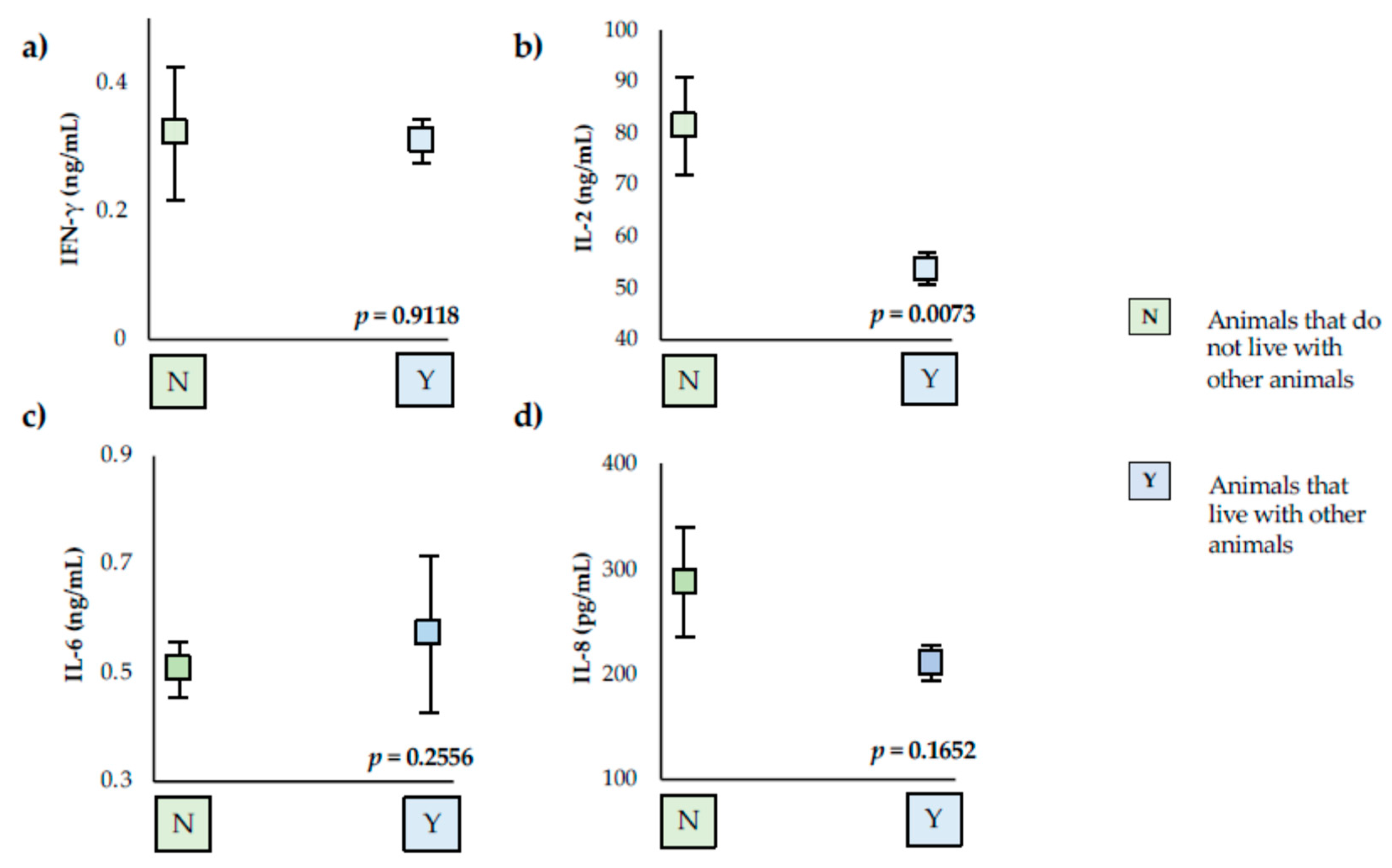
Figure 1. Serum levels of the cytokines studies in animals that do not live with other animals (N) and that live with other animals (Y). (a) Interferon gamma (IFN-γ), (b) Interleukin 2 (IL-2), (c) Interleukin 6 (IL-6), and (d) Interleukin 8 (IL-8). Squares represent mean values for each group analyzed, and vertical lines represent standard deviation. The values for IL-8 are pg/mL, and for IFN-γ, IL-2 and IL-6, they are ng/mL. Each cytokine value possessing different letters differs significantly (p < 0.05).
Vetsci 09 00305 g001
Figure 2. Serum levels of the cytokines studies in boxer (B) and Ibizan hound (I) and another purebred (p) and crossbred (C). (a) Interferon gamma (IFN-γ), (b) Interleukin 2 (IL-2), (c) Interleukin 6 (IL-6), and (d) Interleukin 8 (IL-8). Squares represent mean values for each group analyzed, and vertical lines represent standard deviation. The values for IL-8 are pg/mL, and for IFN-γ, IL-2, and IL-6, they are ng/mL. Each cytokine value possessing different letters differs significantly (p < 0.05).
Vetsci 09 00305 g002
Table 1. Epidemiological data recovered of the animals studied.
VariableCategoriesNo. of Dogs (%)
Dog populationHouseholds71 (91.03)
Animal shelters7 (8.91)
GenderMale40 (51.28)
Female38 (48.72)
AgePuppy (<1 year)8 (10.26)
Young (1 to 5 years)21 (26.92)
Adult (5 to 10 years)23 (29.49)
Elder (>10 years)26 (33.33)
BreedIbizan hound28 (35.90)
boxer31 (39.74)
Purebred (no Ibizan hound and boxer)28 (35.90)
Crossbred7 (8.97)
DietCommercial69 (88.46)
Home prepared/raw food consumption9 (11.54)
Lived with other animalsYes69 (88.46)
No9 (11.54)
Obligatory vaccinationYes69 (88.46)
No9 (11.54)
Anti-Leishmania vaccinationYes8 (10.26)
No70 (89.74)
External dewormingYes71 (91.03)
No7 (8.91)
Overall 78 (100.00)
Table 2. Clinical features and anti-Leishmania antibodies titer observed in positive animals and its prevalence.
Clinical FeatureNo. of Dogs (%)Anti-Leishmania Antibodies Titer
Lymphadenomegaly5 (50)1:640 to 1:1280
Weight loss2 (20)1:640
Lethargy1 (10)1:1280
Pale mucous membranes2 (20)1:640
Splenomegaly1 (10)1:100
Exfoliative dermatitis2 (20)1:640
Onychogryphosis1 (10)1:100
Blepharitis1 (10)1:640
Conjunctivitis3 (30)1:200
Thrombocytopenic anemia1 (10)1:100
Leukocytosis/leukopenia1 (10)1:200
Thrombocytopenia2 (20)1:200
Hyperglobulinemia3 (30)1:200 to 1:1280
Hypoalbuminemia2 (20)1:200
Reduced ALB/GLOB ratio2 (20)1:200
Proteinuria3 (30)1:200
Renal azotemia1 (10)1:200
Hepatic transaminase elevation2 (20)1:640
Overall10 (100)1:100 to 1:1280
Table 3. Ranges of different cytokines analyzed. The table shows the number of data analyzed (n), range, mean ± standard deviation (SD), and coefficient of variation (CV).
Cytokine 1nRange 2Mean ± SD 2CV (%)
IFN-γ720.1–1.290.31 ± 0.0388
IL-27510.37–148.0056.71 ± 3.0146
IL-6700.57–0.010.37 ± 0.9520
IL-8770–624.00223.00 ± 17.1567
1 IFN-γ: interferon gamma; IL: interleukin. 2 The values for IL-8 are expressed pg/mL, and for IFN-γ, IL-2, and IL-6, they are expressed ng/mL.
Publisher’s Note: MDPI stays neutral with regard to jurisdictional claims in published maps and institutional affiliations.