Next Issue
Volume 11, October
Previous Issue
Volume 11, August


Vet. Sci., Volume 11, Issue 9 (September 2024) – 63 articles

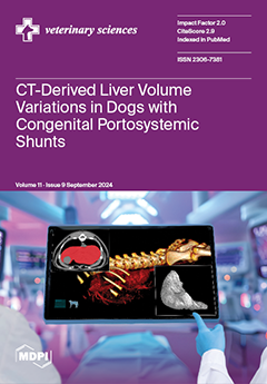
Cover Story (view full-size image): Congenital portosystemic shunts (PSSs) cause blood to bypass the liver, leading to microhepatia. The type of PSS may influence shunt fraction and the severity of microhepatia. Using computed tomographic hepatic volumetry, we investigated liver size differences in dogs with various congenital PSSs, including intrahepatic and different extrahepatic types. Findings showed that the severity of microhepatia varied based on the PSS type and body size, with smaller dogs being more affected. Although normalized liver volumes were consistent across shunt types, microhepatia was significantly more severe in dogs with extrahepatic portocaval and portoazygos shunts. This study offers valuable insights into the clinical management of PSSs in dogs. View this paper
  • Issues are regarded as officially published after their release is announced to the table of contents alert mailing list.
  • You may sign up for e-mail alerts to receive table of contents of newly released issues.
  • PDF is the official format for papers published in both, html and pdf forms. To view the papers in pdf format, click on the "PDF Full-text" link, and use the free Adobe Reader to open them.
Order results
Result details
Section
Select all
Export citation of selected articles as:
15 pages, 4537 KiB  
Article
Analytical Validation of Two Point-of-Care Assays for Hematological Analysis in the Miranda Donkey
by Céline Costa, Ana Patrícia Sousa, Grasiene Silva, Felisbina Queiroga, Ângela Martins, Daniela Andrade and Ana C. Silvestre-Ferreira
Vet. Sci. 2024, 11(9), 450; https://doi.org/10.3390/vetsci11090450 - 22 Sep 2024
Abstract
Miranda donkeys are an endangered autochthonous breed of Portugal. The HemoCue WBC DIFF and HemoCue Hb 201 portable analyzers, developed as a simplified alternative method for total and differential WBC count and hemoglobin measurement, respectively, may be relevant tools in veterinary practice. This [...] Read more.
Miranda donkeys are an endangered autochthonous breed of Portugal. The HemoCue WBC DIFF and HemoCue Hb 201 portable analyzers, developed as a simplified alternative method for total and differential WBC count and hemoglobin measurement, respectively, may be relevant tools in veterinary practice. This study aimed to validate these instruments using Miranda donkey blood samples. For the HemoCue WBC DIFF, most parameters showed acceptable intra- and inter-assay precision with coefficients of variation (CV) below 5%, except for monocytes and eosinophils with higher CVs. The HemoCue Hb 201 showed CVs of 1.98% and 4.07%. Linearity correlation coefficients (r) ranged from 0.53 to 0.99 for HemoCue WBC DIFF and 0.99 for HemoCue Hb 201. Significant levels for neutrophils, lymphocytes, monocytes, eosinophils, and Hb measurements varied. Comparisons with ProCyte Dx showed an excellent correlation for WBC (rs = 0.96), neutrophils (rs = 0.91), lymphocytes (rs = 0.94), and eosinophils (rs = 0.90) but a poor correlation for monocytes and basophils. The HemoCue Hb 201 showed a correlation of rs = 0.96 with ProCyte Dx. In conclusion, both analyzers provided reliable results and are suitable for use in Miranda’s donkey breed for WBC counts and Hb measurements. Full article
Show Figures

Figure 1

7 pages, 225 KiB  
Article
Dog Blood Type DEA 1 in Two Municipalities of Luanda Province of Angola (Sub-Saharan Africa)
by Ana C. Silvestre-Ferreira, Hugo Vilhena, Ana C. Oliveira, José R. Mendoza, Maria Garcia Aura and Josep Pastor
Vet. Sci. 2024, 11(9), 449; https://doi.org/10.3390/vetsci11090449 - 22 Sep 2024
Abstract
In dogs, the risk of an acute hemolytic transfusion reaction at the first transfusion is negligible; however, mismatched transfusions may produce alloimmunization. To avoid fatal acute hemolytic reactions in subsequent blood transfusions, it is important to recognize blood groups and to blood type [...] Read more.
In dogs, the risk of an acute hemolytic transfusion reaction at the first transfusion is negligible; however, mismatched transfusions may produce alloimmunization. To avoid fatal acute hemolytic reactions in subsequent blood transfusions, it is important to recognize blood groups and to blood type both the donor and the recipient. Prevalence of dog blood groups varies geographically and between breeds. Our aim was to determine DEA 1 prevalence in a canine population in Luanda (Angola) and to assess alloimmunization risk after a mismatched blood transfusion. Blood samples were typed using an immunochromatographic strip technique. Of the 112 dogs tested (59 males; 53 females), 52.68% were DEA 1 positive and 47.32% DEA 1 negative. Females tended to be DEA 1 positive, and males DEA 1 negative (p = 0.0085). In a first-time mismatched blood transfusion, the calculated probability of a dog becoming sensitized was 24.9% and the probability of an acute hemolytic reaction following a second incompatible blood transfusion was 6.21%. DEA 1 prevalence obtained was similar to that reported worldwide, but differs from other African countries. The risk of alloimmunization and acute hemolytic transfusion reactions in mismatched blood transfusions is higher than that in other African regions. Blood typing is recommended prior to transfusion. Full article
12 pages, 1379 KiB  
Article
A Multiplex PCR Assay for Simultaneous Detection of Giardia duodenalis, Cryptosporidium parvum, Blastocystis spp. and Enterocytozoon bieneusi in Goats
by Xingang Yu, Hui Xu, Xuanru Mu, Kaijian Yuan, Yilong Li, Nuo Xu, Qiaoyu Li, Wenjing Zeng, Shengfeng Chen and Yang Hong
Vet. Sci. 2024, 11(9), 448; https://doi.org/10.3390/vetsci11090448 - 22 Sep 2024
Abstract
Giardia duodenalis, Cryptosporidium parvum, Blastocystis spp. and Enterocytozoon bieneusi are four common zoonotic parasites associated with severe diarrhea and enteric diseases. In this study, we developed a multiplex PCR assay for the simultaneous detection of these four zoonotic protozoans in goat [...] Read more.
Giardia duodenalis, Cryptosporidium parvum, Blastocystis spp. and Enterocytozoon bieneusi are four common zoonotic parasites associated with severe diarrhea and enteric diseases. In this study, we developed a multiplex PCR assay for the simultaneous detection of these four zoonotic protozoans in goat stool samples and assessed its detection efficiency. Specific primers were designed from conserved gene sequences retrieved from GenBank, and the PCR conditions were optimized. Genomic DNA from 130 samples was subjected to both single-target PCR and multiplex PCR. The multiplex PCR assay successfully amplified specific gene fragments (G. duodenalis, 1400 bp; C. parvum, 755 bp; Blastocystis spp., 573 bp; E. bieneusi, 314 bp). The assay sensitivity was ≥102 copies of pathogenic DNA clones with high specificity confirmed by negative results for other intestinal parasites. The detection rates were 23.08% (30/130) for G. duodenalis, 24.62% (32/130) for C. parvum, 41.54% (54/130) for Blastocystis spp., and 12.31% (16/130) for E. bieneusi, matching the single-target PCR results. The sensitivity and predictive values were 100.00%. This multiplex PCR provided a rapid, sensitive, specific, and cost-effective approach for detecting these four parasites. It also provided essential technical support for the rapid detection and epidemiological investigation of G. duodenalis, C. parvum, Blastocystis spp., and E. bieneusi infections in goat fecal samples. Full article
Show Figures

Figure 1

13 pages, 2348 KiB  
Article
Characterization Studies on the sugC Gene of Streptococcus suis Serotype 2 in Adhesion, Invasion, and Virulence in Mice
by Zhimin Dong, Cheng Li, Xiangxue Tian, Xiaoran Guo, Xiuli Li, Weike Ren, Jingjing Chi, Li Zhang, Fuqiang Li, Yao Zhu, Wanjiang Zhang and Minghua Yan
Vet. Sci. 2024, 11(9), 447; https://doi.org/10.3390/vetsci11090447 - 21 Sep 2024
Abstract
The sugC gene of Streptococcus suis (S. suis) is a coding gene for the ATP-binding transporter-associated protein with strong pathogenicity. In order to reveal the effect of the sugC gene on the virulence of S. suis serotype 2, a wild-type strain [...] Read more.
The sugC gene of Streptococcus suis (S. suis) is a coding gene for the ATP-binding transporter-associated protein with strong pathogenicity. In order to reveal the effect of the sugC gene on the virulence of S. suis serotype 2, a wild-type strain of TJS75, isolated from fattening pigs’ brain tissue samples, was used as a parent strain, and a knockout sugC gene (ΔsugC) and complementary strain (CΔsugC) were successfully constructed via homologous recombination technology. The biological characteristics of TJS75, ΔsugC and CΔsugC were compared and analyzed through growth curves, biochemical characteristics, hemolysis characteristics, cell infection tests and pathogenicity tests on BALB/c mice. The results of the growth characteristic experiments in vitro showed that the plateau stage growth period of ΔsugC was delayed compared to the TJS75 strain, but there was no difference in the total number of bacteria. The biochemical characteristics and hemolysis ability of ΔsugC in sheep blood had no difference compared with TJS75, but its adhesion and invasion abilities in PK-15 cells were decreased. Knockout of the sugC gene had no impact on the expression levels of adhesion-related genes in TJS75 in real-time PCR analysis. In addition, the LD50 of ΔsugC in BALB/c mice was 1.47 × 10⁸ CFU, seven times higher than that of TJS75 (LD50 = 2.15 × 10⁷ CFU). These results illustrate that the deletion of sugC reduced the virulence of TJS75 to BALB/c mice, but its role in the adhesion and invasion of PK-15 cells in this strain needs to be further explored. In summary, this study provides evidence that the sugC gene is a virulence-related gene in the S. suis serotype 2 strain and plays a crucial role in the adhesion and invasion of S. suis. This study lays a foundation for the further exploration of the potential virulence factors and pathogenesis of S. suis. Full article
Show Figures

Figure 1

12 pages, 277 KiB  
Article
Saliva as a Potential Source of Biomarkers in Cows with Metritis: A Pilot Study
by Pedro J. Vallejo-Mateo, María D. Contreras-Aguilar, Alberto Muñoz-Prieto, María Botia, Asta Tvarijonaviciute, Camila Peres Rubio, Rasa Zelvyte, José J. Cerón and Lorena Franco-Martínez
Vet. Sci. 2024, 11(9), 446; https://doi.org/10.3390/vetsci11090446 - 21 Sep 2024
Abstract
Metritis affects 5–20% of cows after parturition, negatively impacting animal welfare and the profitability of dairy farms, increasing culling rates and costs, and decreasing productivity and reproduction rates. This study compared the results of a comprehensive biochemical panel consisting of 25 salivary and [...] Read more.
Metritis affects 5–20% of cows after parturition, negatively impacting animal welfare and the profitability of dairy farms, increasing culling rates and costs, and decreasing productivity and reproduction rates. This study compared the results of a comprehensive biochemical panel consisting of 25 salivary and 31 serum analytes between healthy cows (n = 16) and cows with metritis (n = 12). Descriptive parameters such as depression, rectal temperature, body condition score (BCS), heart rate, respiratory rate, mucous color, ruminal motility, vaginal discharge, milk production, and complete hematology analyses were also assessed for comparative purposes. The biochemistry analytes comprised five analytes related to stress, five to inflammation, five to oxidative status, and nineteen to general metabolism. The two-way ANOVA analysis revealed that, in saliva, eight biomarkers (lipase, adenosine deaminase (ADA), haptoglobin (Hp), total proteins, g-glutamyl transferase (gGT), aspartate aminotransferase (AST), alkaline phosphatase (ALP), and creatine kinase (CK)) were significant higher in cows with metritis. In serum, eight biomarkers (ADA, Hp, serum amyloid A (SAA), fibrinogen, ferritin, AOPPs/albumin ratio, non-esterified fatty acids (NEFAs), and bilirubin) were significantly higher in cows with metritis, whereas six (total esterase (TEA), albumin, urea, lactate, phosphorus, and calcium) were lower. Of the total number of 23 biomarkers that were measured in both saliva and serum, significant positive correlations between the two biofluids were found for six of them (Hp, FRAP, CUPRAC, AOPPs, urea, and phosphorus). Urea showed an R = 0.7, and the correlations of the other analytes were weak (R < 0.4). In conclusion, cows with metritis exhibited differences in biomarkers of stress, inflammation, cellular immune system, and general metabolism in both salivary and serum biochemistry profiles. These changes were of different magnitudes in the two biofluids. In addition, with the exception of ADA and Hp, the analytes that showed changes in the saliva and serum profiles of cows affected by metritis were different. Overall, this report opens a new window for the use of saliva as potential source of biomarkers in cows with metritis. Full article
(This article belongs to the Section Veterinary Reproduction and Obstetrics)
15 pages, 3751 KiB  
Article
Transcriptomic Analysis of the Hepatopancreas in the Sex-Related Size Differences of Macrobrachium nipponense
by Yabing Wang, Guangde Qiao, Yanfeng Yue, Shiming Peng and Hongtuo Fu
Vet. Sci. 2024, 11(9), 445; https://doi.org/10.3390/vetsci11090445 - 21 Sep 2024
Abstract
Macrobrachium nipponense, a commercially popular crustacean species within the Chinese context, is recognized for its exceptional nutritional composition and palatability. There are significant differences in growth between male and female M. nipponense. Herein, transcriptomics was used to determine the hepatopancreas transcriptome [...] Read more.
Macrobrachium nipponense, a commercially popular crustacean species within the Chinese context, is recognized for its exceptional nutritional composition and palatability. There are significant differences in growth between male and female M. nipponense. Herein, transcriptomics was used to determine the hepatopancreas transcriptome differences between sex-related size differences in M. nipponense. We identified 974 differentially expressed genes (DEGs) between the SHE (female) and BHE (male) groups, which were validated using RT-qPCR. The genes encoding matrix metalloproteinase-9 (MM9), Ribosome-binding protein 1 (RBP1), Aly/REF export factor 2, and hematological and neurological expressed 1 (HN1) may play a role in modulating the sex-related size differences observed in M. nipponense. Clusters of orthologous groups and gene ontology functional analysis demonstrated that the DEGs for sex-related size in M.nipponense were associated with various biological functions. The Kyoto Encyclopedia of Genes and Genomes pathways analysis demonstrated that upregulated DEGs were mainly enriched in lysine biosynthesis, tryptophan metabolism, and lysine degradation pathways, whereas the downregulated DEGs were mainly enriched in ascorbate and aldarate metabolism, retinol metabolism, and drug metabolism-cytochrome P450 pathways. The results indicated the molecular mechanism underlying the sex-related size differences and identified key genes. This data will be invaluable to support explanations of individual differences between male and female prawns. Full article
(This article belongs to the Section Veterinary Physiology, Pharmacology, and Toxicology)
Show Figures

Figure 1

11 pages, 1187 KiB  
Article
Assessing the Relationship between proAKAP4 Level and Longevity of Sexed Sperm Quality after Thawing
by İlktan Bastan, Fırat Korkmaz, Derya Şahin, Seher Şimşek and Ufuk Kaya
Vet. Sci. 2024, 11(9), 444; https://doi.org/10.3390/vetsci11090444 - 21 Sep 2024
Abstract
ProAKAP4 is a sperm structural protein that regulates motility through the PKA-dependent cAMP signaling pathway, which is synthesized as an X chromosome-linked member of the gene family. This study aims to determine the optimal level of proAKAP4 for evaluating sexed semen through investigating [...] Read more.
ProAKAP4 is a sperm structural protein that regulates motility through the PKA-dependent cAMP signaling pathway, which is synthesized as an X chromosome-linked member of the gene family. This study aims to determine the optimal level of proAKAP4 for evaluating sexed semen through investigating its relationship with the longevity of sperm quality in sexed Holstein bull sperm. A total of 30 sexed sperm samples (bearing X chromosomes) from 30 distinct Holstein bulls (n = 30) were analyzed. The frozen bull sperm samples were assessed for their proAKAP4 levels, mitochondrial membrane potential, plasma membrane and acrosome integrity (PMAI), and spermatozoa movement parameters at hours 0 and 3 after thawing. The proAKAP4 levels in the sexed sperm samples ranged from 16.35 to 72.10 ng/10 M spz, with an average of 37.18 ± 15.1 ng/10 M spz. A strong positive correlation was observed between proAKAP4 levels and total motility, progressive motility, PMAI, high mitochondrial membrane potential, VAP, and VCL values after 3 h of incubation, when compared to post-thaw analyses. The results also reveal that spermatozoa with proAKAP4 levels of ≥40 ng/10 M spz exhibit higher quality. In conclusion, the level of proAKAP4 in sexed sperm aligns with previous studies and shows potential as a biomarker for assessing the longevity of sexed sperm quality. Full article
(This article belongs to the Special Issue Sperm Biotechnology in Animals Reproduction)
Show Figures

Figure 1

13 pages, 4202 KiB  
Case Report
Intraluminal Contrast-Enhanced Ultrasonography Application in Dogs and Cats
by Saran Chhoey, Soyeon Kim, Eunjee Kim, Dongjae Lee, Kroesna Kang, Sath Keo, Jezie Alix Acorda, Junghee Yoon and Jihye Choi
Vet. Sci. 2024, 11(9), 443; https://doi.org/10.3390/vetsci11090443 - 20 Sep 2024
Abstract
Administering intraluminal fluid can improve the acoustic window for the visualization of the lumen and wall layers in the cavitary organs. Microbubbles in ultrasound contrast agents can also be used for intracavitary applications to enhance visualization of the lesion in human patients. However, [...] Read more.
Administering intraluminal fluid can improve the acoustic window for the visualization of the lumen and wall layers in the cavitary organs. Microbubbles in ultrasound contrast agents can also be used for intracavitary applications to enhance visualization of the lesion in human patients. However, there was no literature extending the clinical application of intraluminal contrast-enhanced ultrasonography (CEUS) to patients with naturally occurring diseases in veterinary medicine. This case series aims to describe the detailed application and diagnostic value of intraluminal CEUS in six clinical cases with naturally occurring gastrointestinal (GI) and urinary tract diseases. Full article
Show Figures

Figure 1

12 pages, 1026 KiB  
Article
The Diagnostic Value of qPCR Quantification of Paenibacillus larvae in Hive Debris and Adult Bees for Predicting the Onset of American Foulbrood
by Bojan Papić, Lucija Žvokelj, Metka Pislak Ocepek, Barbara Hočevar, Monika Kozar, Rene Rus, Urška Zajc and Darja Kušar
Vet. Sci. 2024, 11(9), 442; https://doi.org/10.3390/vetsci11090442 - 20 Sep 2024
Abstract
American foulbrood (AFB) is a serious infectious disease of honeybees (Apis mellifera) caused by Paenibacillus larvae. Increased P. larvae count in hive-related material is associated with an increased risk of AFB. Here, we quantified P. larvae cells in 106 adult [...] Read more.
American foulbrood (AFB) is a serious infectious disease of honeybees (Apis mellifera) caused by Paenibacillus larvae. Increased P. larvae count in hive-related material is associated with an increased risk of AFB. Here, we quantified P. larvae cells in 106 adult bee and 97 hive debris samples using quantitative PCR (qPCR); 66/106 adult bee and 66/97 hive debris samples were collected simultaneously from the same bee colony (paired-sample design). The corresponding bee colonies were also examined for the presence of AFB clinical signs. A binary logistic regression model to distinguish between AFB-affected and unaffected honeybee colonies showed a strong diagnostic accuracy of both sample types for predicting the onset of AFB based on P. larvae counts determined by qPCR. The colonies with a P. larvae count greater than 4.5 log cells/adult bee or 7.3 log cells/mL hive debris had a 50% probability of being clinically affected and were categorized as high-risk. The AFB-unaffected colonies had significantly lower P. larvae counts than the AFB-affected colonies, but the latter did not differ significantly in P. larvae counts in relation to the severity of clinical signs. Both bee-related sample types had a high diagnostic value for predicting disease outcome based on P. larvae counts. These results improve the understanding of the relationship between P. larvae counts and AFB occurrence, which is essential for early detection of high-risk colonies. Full article
(This article belongs to the Section Veterinary Microbiology, Parasitology and Immunology)
Show Figures

Graphical abstract

18 pages, 6852 KiB  
Article
Predictive Neuromarker Patterns for Calcification Metaplasia in Early Tendon Healing
by Melisa Faydaver, Valeria Festinese, Oriana Di Giacinto, Mohammad El Khatib, Marcello Raspa, Ferdinando Scavizzi, Fabrizio Bonaventura, Valentina Mastrorilli, Paolo Berardinelli, Barbara Barboni and Valentina Russo
Vet. Sci. 2024, 11(9), 441; https://doi.org/10.3390/vetsci11090441 - 19 Sep 2024
Abstract
Unsuccessful tendon healing leads to fibrosis and occasionally calcification. In these metaplastic drifts, the mouse AT preclinical injury model represents a robust experimental setting for studying tendon calcifications. Previously, calcium deposits were found in about 30% of tendons after 28 days post-injury. Although [...] Read more.
Unsuccessful tendon healing leads to fibrosis and occasionally calcification. In these metaplastic drifts, the mouse AT preclinical injury model represents a robust experimental setting for studying tendon calcifications. Previously, calcium deposits were found in about 30% of tendons after 28 days post-injury. Although a neuromediated healing process has previously been documented, the expression patterns of NF200, NGF, NPY, GAL, and CGRP in mouse AT and their roles in metaplastic calcific repair remain to be explored. This study included a spatiotemporal analysis of these neuromarkers during the inflammatory phase (7 days p.i.) and the proliferative/early-remodelling phase (28 days p.i.). While the inflammatory phase is characterised by NF200 and CGRP upregulation, in the 28 days p.i., the non-calcified tendons (n = 16/24) showed overall NGF, NPY, GAL, and CGRP upregulation (compared to 7 days post-injury) and a return of NF200 expression to values similar to pre-injury. Presenting a different picture, in calcified tendons (n = 8), NF200 persisted at high levels, while NGF and NPY significantly increased, resulting in a higher NPY/CGRP ratio. Therefore, high levels of NF200 and imbalance between vasoconstrictive (NPY) and vasodilatory (CGRP) neuromarkers may be indicative of calcification. Tendon cells contributed to the synthesis of neuromarkers, suggesting that their neuro-autocrine/paracrine role is exerted by coordinating growth factors, cytokines, and neuropeptides. These findings offer insights into the neurobiological mechanisms of early tendon healing and identify new neuromarker profiles predictive of tendon healing outcomes. Full article
Show Figures

Figure 1

13 pages, 6272 KiB  
Article
Punicalagin Inhibits African Swine Fever Virus Replication by Targeting Early Viral Stages and Modulating Inflammatory Pathways
by Renhao Geng, Dan Yin, Yingnan Liu, Hui Lv, Xiaoyu Zhou, Chunhui Bao, Lang Gong, Hongxia Shao, Kun Qian, Hongjun Chen and Aijian Qin
Vet. Sci. 2024, 11(9), 440; https://doi.org/10.3390/vetsci11090440 - 19 Sep 2024
Abstract
African swine fever (ASF), caused by the African swine fever virus (ASFV), has resulted in significant losses in the global pig industry. Considering the absence of effective vaccines, developing drugs against ASFV may be a crucial strategy for its prevention and control in [...] Read more.
African swine fever (ASF), caused by the African swine fever virus (ASFV), has resulted in significant losses in the global pig industry. Considering the absence of effective vaccines, developing drugs against ASFV may be a crucial strategy for its prevention and control in the future. In this study, punicalagin, a polyphenolic substance extracted from pomegranate peel, was found to significantly inhibit ASFV replication in MA-104, PK-15, WSL, and 3D4/21 cells by screening an antiviral compound library containing 536 compounds. Time-of-addition studies demonstrated that punicalagin acted on early viral replication stages, impinging on viral attachment and internalization. Meanwhile, punicalagin could directly inactivate the virus according to virucidal assay. RT-qPCR and Western blot results indicated that punicalagin modulated the NF-κB/STAT3/NLRP3 inflammasome signaling pathway and reduced the levels of inflammatory mediators induced by ASFV. In conclusion, this study reveals the anti-ASFV activity of punicalagin and the mechanism of action, which may have great potential for developing effective drugs against ASFV. Full article
Show Figures

Figure 1

26 pages, 5003 KiB  
Article
Oral Fecal Microbiota Transplantation in Dogs with Tylosin-Responsive Enteropathy—A Proof-of-Concept Study
by Mohsen Hanifeh, Elisa Scarsella, Connie A. Rojas, Holly H. Ganz, Mirja Huhtinen, Tarmo Laine and Thomas Spillmann
Vet. Sci. 2024, 11(9), 439; https://doi.org/10.3390/vetsci11090439 - 18 Sep 2024
Abstract
A clinical trial was conducted to evaluate the effect of fecal microbiota transplantation (FMT) on the canine chronic enteropathy clinical activity index (CCECAI), fecal consistency, and microbiome of dogs with tylosin-responsive enteropathy (TRE). The trial consisted of four phases: (1) screening with discontinuation [...] Read more.
A clinical trial was conducted to evaluate the effect of fecal microbiota transplantation (FMT) on the canine chronic enteropathy clinical activity index (CCECAI), fecal consistency, and microbiome of dogs with tylosin-responsive enteropathy (TRE). The trial consisted of four phases: (1) screening with discontinuation of tylosin for 4 weeks, (2) inclusion with re-introduction of tylosin for 3–7 days, (3) treatment with FMT/placebo for 4 weeks, and (4) post-treatment with follow-up for 4 weeks after treatment cessation. The study found that the treatment efficacy of FMT (71.4%) was slightly higher than that of placebo (50%), but this difference was not statistically significant due to underpowering. The most abundant bacterial species detected in the fecal microbiomes of dogs with TRE before FMT or placebo treatment were Blautia hansenii, Ruminococcus gnavus, Escherichia coli, Clostridium dakarense, Clostridium perfringens, Bacteroides vulgatus, and Faecalimonas umbilicata. After FMT, the microbiomes exhibited increases in Clostridium dakarense, Clostridium paraputrificum, and Butyricicoccus pullicaecorum. The microbiome alpha diversity of TRE dogs was lower when on tylosin treatment compared to healthy dogs, but it increased after treatment in both the FMT and placebo groups. Comparisons with the stool donor showed that, on average, 30.4% of donor strains were engrafted in FMT recipients, with the most common strains being several Blautia sp., Ruminococcus gnavus, unclassified Lachnoclostridium, Collinsella intestinalis, and Fournierella massiliensis. Full article
(This article belongs to the Special Issue Small Animal Gastrointestinal Diseases: Challenges and Advances)
Show Figures

Figure 1

14 pages, 1554 KiB  
Article
Homemade Diet as a Paramount for Dogs’ Health: A Descriptive Analysis
by Giulia Pignataro, Paolo Emidio Crisi, Elena Landolfi, Benedetta Belà, Isa Fusaro, Luana Clerico and Alessandro Gramenzi
Vet. Sci. 2024, 11(9), 438; https://doi.org/10.3390/vetsci11090438 - 17 Sep 2024
Abstract
Pet nutrition awareness has risen enormously, with an increasing interest in homemade diets aimed at controlling food composition. The literature in this field is scarce, especially regarding the homemade diet’s long-term effects on pathological conditions. This prospective study encompassed 167 healthy and sick [...] Read more.
Pet nutrition awareness has risen enormously, with an increasing interest in homemade diets aimed at controlling food composition. The literature in this field is scarce, especially regarding the homemade diet’s long-term effects on pathological conditions. This prospective study encompassed 167 healthy and sick dogs subjected to a customized dietary plan based on homemade food. After an initial visit, dog owners provided questionnaires with follow-up data on their perceptions of physical sign changes or symptom modifications. A total of 104 (62%) subjects maintained the homemade diet, while 63 reverted to their previous diet. The median follow-up was 14 months. Out of 31 healthy dogs that continued the nutritional plan, 70% exhibited improvements in their coat condition and 47% a decrease in evacuation frequency. Regarding weight loss goals, 67% of dogs achieved their target. The 67 pathological dogs that completed the follow-up were primarily affected by gastrointestinal and dermatological disorders. Dogs with chronic enteropathy improved their symptoms in 95% of the cases, subjects with dermatological pathologies in 83%, and patients presenting both disorders in 100%. These clinical results are promising. Personalized and well-balanced homemade diets noticeably enhanced the overall pet’s health, with an almost complete remission of symptoms in pathological dogs. Full article
(This article belongs to the Special Issue Effects of Diet on Small Animal Health—2nd Edition)
Show Figures

Figure 1

10 pages, 1426 KiB  
Article
Evaluation of Totarol for Promoting Open Wound Healing in Dogs
by Somphong Hoisang, Supranee Jitpean, Suvaluk Seesupa, Phanthit Kamlangchai, Tossawarn Makpunpol, Pimsiri Ngowwatana, Saikam Chaimongkol, Duangdaow Khunbutsri, Jeerasak Khlongkhlaeo and Naruepon Kampa
Vet. Sci. 2024, 11(9), 437; https://doi.org/10.3390/vetsci11090437 - 16 Sep 2024
Abstract
This study investigates the susceptibility of common pathogens to totarol and assesses its clinical effectiveness in promoting wound healing in client-owned dogs with open wounds. Twenty-three client-owned dogs with open wounds were divided into two groups: (1) the treatment group (T-group) and (2) [...] Read more.
This study investigates the susceptibility of common pathogens to totarol and assesses its clinical effectiveness in promoting wound healing in client-owned dogs with open wounds. Twenty-three client-owned dogs with open wounds were divided into two groups: (1) the treatment group (T-group) and (2) the control group (C-group). Clinical samples were collected from the wounds for the bacterial identification and determination of the minimum inhibitory concentrations (MICs) of totarol. In the T-group, wounds were treated with standard wound care together with the application at a dosage of 0.3 mL (two sprays) of commercial totarol product per 25 cm2 of the wound area. The C-group received only standard wound care. This in vitro study found that totarol exhibited antimicrobial activity against both standard pathogens and clinical wound pathogens. The MIC values of totarol dissolved in absolute ethyl alcohol were 4 µg/mL for Gram-positive pathogens and ranged from 256 to 512 µg/mL for Gram-negative pathogens. However, the MIC values of the commercial totarol product ranged from 512 to 1024 for both Gram-positive and Gram-negative pathogens. Clinically, the use of a commercial totarol product as an adjunctive therapy significantly improved wound healing, as indicated by a greater percentage of wound area reduction (p < 0.05). From day 2 to day 7 of the treatment, the percentage of wound area reduction differed significantly between the T-group and the C-group. At the end of the study, the average percentage of wound area reduction was 69.18% ± 18.12 and 41.50% ± 20.23 in the T-group and C-group, respectively. The finding of this study illustrates the antimicrobial properties of totarol and its product against prevalent wound pathogens. These results suggest the potential of totarol as an adjunctive option for canine wound care. Full article
(This article belongs to the Section Veterinary Surgery)
Show Figures

Figure 1

13 pages, 3131 KiB  
Article
Isolation, Genomic Characterization and Evolution of Six Porcine Rotavirus A Strains in a Pig Farming Group
by Zhendong Zhang, Chengyue Wu, Yue Chen, Yubo Li, Duo Li, Wenqiang Wang, Wei Wen, Zhenbang Zhu and Xiangdong Li
Vet. Sci. 2024, 11(9), 436; https://doi.org/10.3390/vetsci11090436 - 14 Sep 2024
Abstract
Porcine rotavirus (PoRV) is a significant enteric pathogen causing gastroenteritis in piglets, which causes huge economic loss to the Chinese pig industry. In this study, six porcine rotavirus A strains were isolated from three adjacent sow farms belonging to the same company within [...] Read more.
Porcine rotavirus (PoRV) is a significant enteric pathogen causing gastroenteritis in piglets, which causes huge economic loss to the Chinese pig industry. In this study, six porcine rotavirus A strains were isolated from three adjacent sow farms belonging to the same company within one year, which suffered severe diarrhea outbreaks. AHBZ2303 (G11P[7]) and AHBZ2305 (G9P[23]), AHBZ2304 (G9P[23]) and AHBZ2312 (G4P[6]), AHBZ2310 (G9P[23]) and AHBZ2402 (G5P[23]) were isolated from Farm A, B and C, respectively. All six isolates were related to human rotavirus through complete genome analysis, suggesting the potential cross–species infection between humans and pigs. Evolutionary analysis revealed that AHBZ2303 and AHBZ2304 likely emerged simultaneously in Farm A and B, and then AHBZ2304 was introduced to Farm A and C, leading to the emergence of AHBZ2305 and AHBZ2310. Recombination and large variation were identified for AHBZ2312 and AHBZ2402. These findings provided insights into the transmission and evolution of PoRV among farms and underscored the need for enhanced monitoring to mitigate the risk of outbreaks from novel variants. Full article
(This article belongs to the Special Issue Emerging and Re-emerging Swine Viruses)
Show Figures

Figure 1

13 pages, 979 KiB  
Article
Exploring the Epidemiology of Melanocytic Tumors in Canine and Feline Populations: A Comprehensive Analysis of Diagnostic Records from a Single Pathology Institution in Italy
by Adriana Lo Giudice, Ilaria Porcellato, Giuseppe Giglia, Monica Sforna, Elvio Lepri, Maria Teresa Mandara, Leonardo Leonardi, Luca Mechelli and Chiara Brachelente
Vet. Sci. 2024, 11(9), 435; https://doi.org/10.3390/vetsci11090435 - 14 Sep 2024
Abstract
MTs are prevalent in dogs, representing the most frequent oral malignancy, compared to cats, in which ocular melanomas predominate. This study investigates the canine and feline MT epidemiology (2005–2024) of cases submitted to the Veterinary Pathology Service (University of Perugia). Among the canine [...] Read more.
MTs are prevalent in dogs, representing the most frequent oral malignancy, compared to cats, in which ocular melanomas predominate. This study investigates the canine and feline MT epidemiology (2005–2024) of cases submitted to the Veterinary Pathology Service (University of Perugia). Among the canine neoplasms, 845 (4%) were melanocytic: 329 (39%) melanocytomas; 512 (61%) melanomas. Of these, 485 (57%) were cutaneous (4% of canine cutaneous neoplasms), 193 (23%) were oral (50% of oral canine neoplasms), and 104 (12%) were mucocutaneous. The average age of affected dogs was 10 years. Older dogs were more likely to have melanomas compared to melanocytomas (p < 0.001). There were 60 (1%) feline MTs: 6 (10%) melanocytomas; 53 (88%) melanomas. Of these, 29 (48%) were cutaneous (1% of feline cutaneous tumors), 18 (30%) were ocular, and 9 (15%) were oral (22% of feline oral tumors). The average age of affected cats was 11 years. In dogs, mucocutaneous melanomas were more common compared to cutaneous ones (p < 0.05); oral melanomas were more common compared to all other sites (p < 0.001). In cats, ocular melanomas were more common compared to cutaneous ones (p < 0.05). Our study provides the MT prevalence in a selected canine and feline population, revealing MT epidemiological patterns, highlighting species-specific differences in the tumor prevalence, localization, and age distribution. Full article
(This article belongs to the Special Issue Focus on Tumours in Pet Animals)
Show Figures

Figure 1

6 pages, 856 KiB  
Brief Report
Accurate Preoperative Localization of Thoracolumbar Disc Extrusion in Dogs: A Prospective Controlled Study
by William McCartney, Ciprian Ober and Christos Yiapanis
Vet. Sci. 2024, 11(9), 434; https://doi.org/10.3390/vetsci11090434 - 13 Sep 2024
Abstract
Intervertebral disc disease (IVDD) is a very common cause of spinal cord compression in dogs. The errors of human surgeons operating on the wrong level or wrong side is a recognized problem and has been largely analyzed. To date, little information is available [...] Read more.
Intervertebral disc disease (IVDD) is a very common cause of spinal cord compression in dogs. The errors of human surgeons operating on the wrong level or wrong side is a recognized problem and has been largely analyzed. To date, little information is available regarding these errors in dogs. The objective of this study was to assess the accuracy of disc localization prior to possible surgery for IVDD to ensure the surgeon begins their incision directly over the extruded disc. Eighty-five chondrodystrophic or brachycephalic dogs that presented with IVDD confirmed by CT or MRI scan were included in the study. Two small Backhaus clamps were placed cranial and caudal to the lesion site and a control clamp placed at the wing of the ilium. The main interest was whether the surgeon was able to preoperatively pinpoint the correct site for the surgery. Dorsoventral radiographs were taken to verify by another person if the clamps had been placed in the correct position. If the result was incorrect, the surgeon was asked to try again without knowing that the clamps were incorrect, and another radiograph was taken. This was repeated until the position was correct. The results were recorded as correct or incorrect and the number of attempts were registered. The results suggested no significant trend over time for any of the outcomes examined. There were some slight improvements over time, but none of the results was close to statistical significance. The findings of the study showed that in the thoracolumbar region the surgeon has a higher chance of incorrectly marking the exact site for surgery. Full article
(This article belongs to the Section Veterinary Surgery)
Show Figures

Figure 1

25 pages, 4349 KiB  
Review
Assisting the Learning of Clinical Reasoning by Veterinary Medical Learners with a Case Example
by Gustavo Ferlini Agne, Amanda (Mandi) Nicole Carr, Roy Neville Kirkwood and Kiro Risto Petrovski
Vet. Sci. 2024, 11(9), 433; https://doi.org/10.3390/vetsci11090433 - 13 Sep 2024
Abstract
Effective clinical reasoning is essential for veterinary medical education, particularly in managing complex cases. This review explores strategies for learning clinical reasoning by veterinary medical learners, using a case example of mastitis to illustrate key concepts. Clinical reasoning encompasses cognitive, metacognitive, social, and [...] Read more.
Effective clinical reasoning is essential for veterinary medical education, particularly in managing complex cases. This review explores strategies for learning clinical reasoning by veterinary medical learners, using a case example of mastitis to illustrate key concepts. Clinical reasoning encompasses cognitive, metacognitive, social, and situational activities, yet the literature on practical applications in veterinary education remains limited. The review discusses various stages of clinical reasoning, including data collection, problem representation, differential diagnosis, and management planning. It emphasizes the importance of integrating client-centered care and iterative evaluation into the clinical decision-making process. Key learning strategies include facilitation in using the domains of clinical reasoning—concepts, data collection, and analysis, taking action, and reflection on encounters. This review highlights best practices such as forward and backward reasoning, reflective practice, and the use of practical examples to enhance learners’ diagnostic accuracy and patient outcomes. The insights provided aim to enhance the training of veterinary learners, ensuring they can navigate day 1 as well as complex cases with improved diagnostic accuracy and patient outcomes. Full article
Show Figures

Figure 1

9 pages, 412 KiB  
Review
Reflections on Small-Class Teaching in Veterinary Medicine Undergraduate Programs in China
by Pengpeng Xia, Ziyue Chen, Yi Luo, Xiangyu Li, Xin Ma and Siqi Lian
Vet. Sci. 2024, 11(9), 432; https://doi.org/10.3390/vetsci11090432 - 13 Sep 2024
Abstract
As a cornerstone of higher education in China, the quality of undergraduate teaching is crucial for nurturing high-caliber innovative talents that meet the needs of national and social development. Small-class teaching has emerged as a focal point in the reform of undergraduate education [...] Read more.
As a cornerstone of higher education in China, the quality of undergraduate teaching is crucial for nurturing high-caliber innovative talents that meet the needs of national and social development. Small-class teaching has emerged as a focal point in the reform of undergraduate education and represents an important approach to cultivating top-notch innovative talents. Veterinary medicine is a scientific discipline that encompasses the prevention, control, diagnosis, and treatment of animal diseases. It also involves efforts to prevent the transmission of animal diseases to humans. The training of professionals in this field should emphasize the integration of theoretical knowledge with practical skills. Therefore, small-class teaching facilitates open communication between educators and students, which is instrumental in fostering a new generation of veterinarians equipped with robust professional knowledge, excellent practical abilities, and strong scientific literacy. This paper provides a preliminary exploration into the strategy and direction of small-class teaching within China’s veterinary medicine undergraduate programs by summarizing its characteristics, advantages, and existing challenges. The unique traits of veterinary medicine are also taken into consideration during this analysis. Full article
Show Figures

Figure 1

12 pages, 891 KiB  
Article
Effect of Exogenous Melatonin on Performance and Mastitis in Dairy Cows
by Yunmeng Li, Zhiqiang Cheng, Wenting Ma, Yaqi Qiu, Tuo Liu, Bingyu Nan, Mengfei Li, Long Sun, Wentao Liu, Haina Yin, Caidie Wang, Xiaobin Li and Changjiang Zang
Vet. Sci. 2024, 11(9), 431; https://doi.org/10.3390/vetsci11090431 - 13 Sep 2024
Abstract
Mastitis is an important factor affecting the health of cows that leads to elevated somatic cell counts in milk, which can seriously affect milk quality and result in huge economic losses for the livestock industry. Therefore, the aim of this trial was to [...] Read more.
Mastitis is an important factor affecting the health of cows that leads to elevated somatic cell counts in milk, which can seriously affect milk quality and result in huge economic losses for the livestock industry. Therefore, the aim of this trial was to investigate the effect of melatonin on performance and mastitis in dairy cows. Forty-eight Holstein cows with a similar body weight (470 ± 10 kg), parity (2.75 ± 1.23), number of lactation days (143 ± 43 days), BCS (3.0–3.5), milk yield (36.80 ± 4.18 kg), and somatic cell count (300,000–500,000 cells/mL) were selected and randomly divided into four groups: control (CON group), trial Ⅰ (T80 group), trial Ⅱ (T120 group), and trial Ⅲ (T160 group). Twelve cows in trial groups I, II, and III were pre-dispensed 80, 120, and 160 mg of melatonin in edible glutinous rice capsules along with the basal ration, respectively, while the control group was fed an empty glutinous rice capsule along with the ration. The trial period was 37 days, which included a 7-day adaptive phase followed by a 30-day experimental period. At the end of the trial period, feeding was ended and the cows were observed for 7 days. Milk samples were collected on days 0, 7, 14, 21, 28, and 37 to determine the somatic cell number and milk composition. Blood samples were collected on days 0, 15, 30, and 37 of the trial to determine the serum biochemical indicators, antioxidant and immune indicators, and the amount of melatonin in the blood. The results showed that the somatic cell counts of lactating cows in the CON group were lower than those in the T120 group on days 14 (p < 0.05) and 28 (p < 0.01) at 1 week after melatonin cessation. The milk protein percentage and milk fat percentage of cows in the T120 group were higher than those in the CON group (p < 0.01). The total protein and globulin content in the T120 group were higher than those in the CON group (p < 0.01). In terms of antioxidant capacity and immunity, the cows 1 week after melatonin cessation showed higher superoxide dismutase activity and interleukin-10 contents (p < 0.01) compared with the CON group and lower malondialdehyde and tumor necrosis factor-alpha contents (p < 0.01) compared with the T120 group. The melatonin content in the T120 group was increased relative to that in the other groups. In conclusion, exogenous melatonin can increase the content of milk components, reduce the somatic cell count, and improve the antioxidant capacity and immune responses to a certain extent. Under the experimental conditions, 120 mg/day melatonin is recommended for mid- to late-lactation cows. Full article
(This article belongs to the Special Issue Effects of Nutrition on Ruminants Production Performance and Health)
Show Figures

Figure 1

16 pages, 4618 KiB  
Article
Impact of Toceranib Phosphate and Carprofen on Survival and Quality of Life in Dogs with Inflammatory Mammary Carcinomas
by Miguel Garcia-de la Virgen, Isabel Del Portillo Miguel, Elisa Maiques, Ignacio Pérez Roger, Enric Poch and Juan Borrego
Vet. Sci. 2024, 11(9), 430; https://doi.org/10.3390/vetsci11090430 - 13 Sep 2024
Abstract
Canine inflammatory mammary carcinoma (IMC) is an aggressive and rare type of mammary gland cancer in dogs where vascular endothelial growth factor and cyclooxigenase-2 overexpression usually occur, which contribute to its invasive and angiogenic nature. This study aimed to evaluate the efficacy and [...] Read more.
Canine inflammatory mammary carcinoma (IMC) is an aggressive and rare type of mammary gland cancer in dogs where vascular endothelial growth factor and cyclooxigenase-2 overexpression usually occur, which contribute to its invasive and angiogenic nature. This study aimed to evaluate the efficacy and safety of a combined treatment regimen of toceranib phosphate and carprofen in dogs with measurable IMC. Fifteen female dogs with histopathologically confirmed IMC were included, undergoing a regimen of toceranib (2.4–2.75 mg/kg PO, three times weekly) and carprofen (4.4 mg/kg/24 h PO). Initial evaluations included physical exams, tumor measurements, complete blood count, biochemistry, urinalysis, three view thoracic radiographs, and abdominal ultrasound. Follow-up assessments of physical condition and quality of life (QOL) were conducted bi-weekly, with tumor response evaluations monthly, using RECIST v1.0 criteria. While no complete or partial responses were observed, 60% of the dogs maintained stable disease, with a median progression-free survival of 76 days and an overall survival of 90 days. Notably, 60% of the dogs showed clinical benefit through improved QOL and disease stabilization. The treatment was well-tolerated, with only grade I/II toxicities reported. Despite limited biological activity against the cancer, this protocol may enhance QOL in dogs with IMC, offering a valuable palliative option. Full article
(This article belongs to the Special Issue Focus on Tumours in Pet Animals)
Show Figures

Figure 1

14 pages, 6502 KiB  
Article
A Morphometric Study on the Dimensions of the Vertebral Canal and Intervertebral Discs from Th1 to S1 in Cats and Their Relevance for Spinal Diseases
by Jessica Richter, Christoph K. W. Mülling and Nicole Röhrmann
Vet. Sci. 2024, 11(9), 429; https://doi.org/10.3390/vetsci11090429 - 13 Sep 2024
Abstract
As part of the spine, the vertebral canal represents a central structure protecting the spinal cord running within it. Since alterations to the spinal canal and adjacent structures can have a significant impact on the spinal cord, knowledge of the physiological vertebral canal [...] Read more.
As part of the spine, the vertebral canal represents a central structure protecting the spinal cord running within it. Since alterations to the spinal canal and adjacent structures can have a significant impact on the spinal cord, knowledge of the physiological vertebral canal dimensions is essential. Compression of spinal nerves at the lumbosacral junction is the primary cause of cauda equina syndrome (CES). Although CES is common in dogs, it is rarely documented in cats. Given the lack of information on normal vertebral canal dimensions in cats, it is necessary to collect data and verify currently used measurements, to determine if and to what extent comparisons with dogs are valid. In 50 cats, interpedicular (ID) and midsagittal (SD) diameters were examined from the first thoracic (Th1) to the first sacral vertebra (S1). In 28 of these animals, the intervertebral disc width (IVDW) was measured. All data were gathered through gross anatomical dissection of the cats. Significant lumen reduction was evident in all cats from L6 to S1 with the narrowest point at S1. Narrowings were also found in the thoracic spine. The widest points coincide with the spinal cord enlargements. IVDW shows relatively constant values up to Th10–Th11 and peaks at L7–S1 in 95.65% of cats. While distinct similarities to dogs were observed, differences exist. The findings allow conclusions as to whether relations between the parameters and resulting predispositions to pathological changes can be derived. This could help the understanding of the pathogenesis of feline spinal diseases, particularly compressive myelopathies. Further studies are necessary to investigate the impact of age, sex and breed. Full article
(This article belongs to the Section Anatomy, Histology and Pathology)
Show Figures

Figure 1

20 pages, 14568 KiB  
Article
Combined Genome-Wide Association Study and Haplotype Analysis Identifies Candidate Genes Affecting Growth Traits of Inner Mongolian Cashmere Goats
by Xiaofang Ao, Youjun Rong, Mingxuan Han, Xinle Wang, Qincheng Xia, Fangzheng Shang, Yan Liu, Qi Lv, Zhiying Wang, Rui Su, Yanjun Zhang and Ruijun Wang
Vet. Sci. 2024, 11(9), 428; https://doi.org/10.3390/vetsci11090428 - 12 Sep 2024
Abstract
In this study, genome-wide association analysis was performed on the growth traits (body height, body length, chest circumference, chest depth, chest width, tube circumference, and body weight) of Inner Mongolian cashmere goats (Erlangshan type) based on resequencing data. The population genetic parameters were [...] Read more.
In this study, genome-wide association analysis was performed on the growth traits (body height, body length, chest circumference, chest depth, chest width, tube circumference, and body weight) of Inner Mongolian cashmere goats (Erlangshan type) based on resequencing data. The population genetic parameters were estimated, haplotypes were constructed for the significant sites, and association analysis was conducted between the haplotypes and phenotypes. A total of two hundred and eighty-four SNPs and eight candidate genes were identified by genome-wide association analysis, gene annotation, and enrichment analysis. The phenotypes of 16 haplotype combinations were significantly different by haplotype analysis. Combined with the above results, the TGFB2, BAG3, ZEB2, KCNJ12, MIF, MAP2K3, HACD3, and MEGF11 functional candidate genes and the haplotype combinations A2A2, C2C2, E2E2, F2F2, I2I2, J2J2, K2K2, N2N2, O2O2, P2P2, R1R1, T1T1, W1W1, X1X1, Y1Y1, and Z1Z1 affected the growth traits of the cashmere goats and could be used as molecular markers to improve the accuracy of early selection and the economic benefits of breeding. Full article
Show Figures

Figure 1

14 pages, 860 KiB  
Article
Extracted Plasma Cell-Free DNA Concentrations Are Elevated in Colic Patients with Systemic Inflammation
by Rosemary L. Bayless, Bethanie L. Cooper and M. Katie Sheats
Vet. Sci. 2024, 11(9), 427; https://doi.org/10.3390/vetsci11090427 - 12 Sep 2024
Abstract
Colic is a common and potentially life-threatening condition in horses; in many cases, it remains challenging for clinicians to determine the cause, appropriate treatment, and prognosis. One approach that could improve patient care and outcomes is identification of novel diagnostic and prognostic biomarkers. [...] Read more.
Colic is a common and potentially life-threatening condition in horses; in many cases, it remains challenging for clinicians to determine the cause, appropriate treatment, and prognosis. One approach that could improve patient care and outcomes is identification of novel diagnostic and prognostic biomarkers. Plasma cell-free DNA (cfDNA) is a biomarker that shows promise for characterizing disease severity and predicting survival in humans with acute abdominal pain or requiring emergency abdominal surgery. In horses, we recently determined that extracted plasma cfDNA concentrations are elevated in colic patients compared to healthy controls. For this current study, we hypothesized that extracted plasma cfDNA concentrations would be significantly higher in horses with strangulating or inflammatory colic lesions, in colic patients with systemic inflammatory response syndrome (SIRS), and in non-survivors. Cell-free DNA concentrations were measured in extracted plasma samples using a compact, portable Qubit fluorometer. Colic patients that met published criteria for equine SIRS had significantly higher median extracted plasma cfDNA compared to non-SIRS colic patients. There were no significant differences in extracted plasma cfDNA concentrations between other groups of interest. Our data offer early evidence that extracted plasma cfDNA concentration may provide information about systemic inflammation in colic patients, and additional research is warranted to expand on these findings. Full article
(This article belongs to the Section Veterinary Microbiology, Parasitology and Immunology)
Show Figures

Figure 1

17 pages, 311 KiB  
Review
The Role of Plant Extracts in Enhancing Nutrition and Health for Dogs and Cats: Safety, Benefits, and Applications
by Xinzi Guo, Yifei Wang, Zhaoxuan Zhu and Lian Li
Vet. Sci. 2024, 11(9), 426; https://doi.org/10.3390/vetsci11090426 - 12 Sep 2024
Abstract
Plant extracts, derived from various natural sources, encompass primary and secondary metabolites, which include plant polysaccharides, polyphenols, alkaloids, flavonoids, glycosides, terpenes, and volatile oils. These compounds exhibit a range of biological activities such as antioxidant, anti-inflammatory, and antimicrobial functions. Currently, polyphenols and other [...] Read more.
Plant extracts, derived from various natural sources, encompass primary and secondary metabolites, which include plant polysaccharides, polyphenols, alkaloids, flavonoids, glycosides, terpenes, and volatile oils. These compounds exhibit a range of biological activities such as antioxidant, anti-inflammatory, and antimicrobial functions. Currently, polyphenols and other bioactive compounds are being incorporated into the diets of farm animals, fish, and pets to promote health benefits. Despite this, the application and potential of plant extracts in canine and feline nutrition have not been comprehensively explored. Many aspects of the mechanisms underlying the action of these plant metabolites remain to be analyzed and elucidated. Furthermore, leveraging natural plant extracts for the treatment of clinical conditions in dogs and cats is a crucial component of clinical nutrition. Consequently, this review aims to highlight the impact of plant extracts on overall health, gastrointestinal health, immune health, cardiovascular health, redox balance, and pathology in dogs and cats. Full article
(This article belongs to the Topic Research on Companion Animal Nutrition)
14 pages, 401 KiB  
Review
Review of the Current Status on Ruminant Abortigenic Pathogen Surveillance in Africa and Asia
by George Peter Semango and Joram Buza
Vet. Sci. 2024, 11(9), 425; https://doi.org/10.3390/vetsci11090425 - 12 Sep 2024
Abstract
Ruminant abortion events cause economic losses. Despite the importance of livestock production for food security and the livelihoods of millions of people in the world’s poorest communities, very little is known about the scale, magnitude, or causes of these abortions in Africa and [...] Read more.
Ruminant abortion events cause economic losses. Despite the importance of livestock production for food security and the livelihoods of millions of people in the world’s poorest communities, very little is known about the scale, magnitude, or causes of these abortions in Africa and Asia. The aim of this review was to determine the current status of surveillance measures adopted for ruminant abortigenic pathogens in Africa and Asia and to explore feasible surveillance technologies. A systematic literature search was conducted using PRISMA guidelines for studies published between 1 January 1990 and 1 May 2024 that reported epidemiological surveys of abortigenic pathogens Africa and Asia. A meta-analysis was used to estimate the species-specific sero-prevalence of the abortigenic agents and the regions where they were detected. In the systematic literature search, 39 full-text manuscripts were included. The most prevalent abortigenic pathogens with sero-prevalence greater than 10% were BHV-1, Brucella, Chlamydia abortus, Neospora caninum, RVFV, and Waddlia chondrophila in cattle, BVDV in sheep, and RVFV and Toxoplasma gondii in goats in Africa. In Asia, Anaplasma, BHV-1, Bluetongue virus, Brucella, and BVDV were prevalent in cattle, whereas Mycoplasma was important in goats and sheep. Full article
Show Figures

Figure 1

14 pages, 4660 KiB  
Article
β-Tocotrienol and δ-Tocotrienol as Additional Inhibitors of the Main Protease of Feline Infectious Peritonitis Virus: An In Silico Analysis
by Manos C. Vlasiou, Georgios Nikolaou, Kyriakos Spanoudes and Daphne E. Mavrides
Vet. Sci. 2024, 11(9), 424; https://doi.org/10.3390/vetsci11090424 - 11 Sep 2024
Abstract
Feline infectious peritonitis (FIP) is a severe and invariably fatal disease affecting both domestic and wild felines with limited effective therapeutic options available. By considering the significant immunomodulatory effects of vitamin E observed in both animal and human models under physiological and pathological [...] Read more.
Feline infectious peritonitis (FIP) is a severe and invariably fatal disease affecting both domestic and wild felines with limited effective therapeutic options available. By considering the significant immunomodulatory effects of vitamin E observed in both animal and human models under physiological and pathological conditions, we have provided a full in silico investigation of vitamin E and related compounds and their effect on the crystal structure of feline infectious peritonitis virus 3C-like protease (FIPV-3CLpro). This work revealed the β-tocotrienol and δ-tocotrienol analogs as inhibitor candidates for this protein, suggesting their potential as possible drug compounds against FIP or their supplementary use with current medicines against this disease. Full article
(This article belongs to the Section Veterinary Physiology, Pharmacology, and Toxicology)
Show Figures

Figure 1

22 pages, 1508 KiB  
Article
Longitudinal Baboon (Papio anubis) Neutrophil to Lymphocyte Ratio (NLR), and Correlations with Monthly Sedation Rate and Within-Group Sedation Order
by Sarah J. Neal, Steven J. Schapiro and Elizabeth R. Magden
Vet. Sci. 2024, 11(9), 423; https://doi.org/10.3390/vetsci11090423 - 11 Sep 2024
Cited by 1
Abstract
Neutrophil to lymphocyte ratio (NLR) is a simple marker of stress and inflammation, but there is limited research regarding NLR in nonhuman primates (NHPs), with studies showing associations with longevity, certain medical conditions, and stressful circumstances. Here, we examined baboon NLR longitudinally, and [...] Read more.
Neutrophil to lymphocyte ratio (NLR) is a simple marker of stress and inflammation, but there is limited research regarding NLR in nonhuman primates (NHPs), with studies showing associations with longevity, certain medical conditions, and stressful circumstances. Here, we examined baboon NLR longitudinally, and as a function of health parameters. We also examined whether NLR was affected by sedation rate, as well as the order of sedation within a group, given that sedation events during clinical and research practices can induce stress in NHPs. While older adult and geriatric baboon NLR did not differ longitudinally, juvenile and young adult NLR tended to increase, primarily driven by increases in females. Additionally, baboons sedated later within a group showed significantly higher NLRs than those sedated earlier in the process. However, baboons with higher sedation rates per month showed lower NLRs. These data indicate that NLR may be dysregulated in different ways as a function of different types of stress, with sedation order (i.e., acute stress) causing pathological increases in NLR, and sedation rate over time (i.e., chronic stress) causing decreases. Importantly, we propose that NLR, a routinely obtained veterinary measure, has potential utility as a welfare indicator of stress resulting from clinical and research practices, as well as a measure that can inform behavioral management practices and interventions. Full article
Show Figures

Figure 1

8 pages, 1255 KiB  
Article
IgG Subtype Response against Virulence-Associated Protein A in Foals Naturally Infected with Rhodococcus equi
by Yuya Mizuguchi, Nao Tsuzuki, Marina Dee Ebana, Yasunori Suzuki and Tsutomu Kakuda
Vet. Sci. 2024, 11(9), 422; https://doi.org/10.3390/vetsci11090422 - 9 Sep 2024
Abstract
Rhodococcus equi is an intracellular bacterium that causes suppurative pneumonia in foals. T-helper (Th) 1 cells play an important role in the protective response against R. equi. In mice and humans, the directionality of IgG switching reflects the polarization of Th-cell responses, [...] Read more.
Rhodococcus equi is an intracellular bacterium that causes suppurative pneumonia in foals. T-helper (Th) 1 cells play an important role in the protective response against R. equi. In mice and humans, the directionality of IgG switching reflects the polarization of Th-cell responses, but this has not been fully elucidated in horses. In this 4-year study, we classified R. equi-infected foals into surviving and non-surviving group and investigated differences in IgG subclass response to virulence-associated protein A, the main virulence factor of R. equi, between the groups. IgGa, IgGb, and IgG(T) titers were significantly higher in the non-surviving group compared with the surviving group. The titers of IgGa and IgG(T), IgGb and IgG(T), and IgGa and IgGb, respectively, were positively correlated, and the IgG(T)/IgGb ratio in the non-surviving group was significantly higher than that in the surviving group. The IgG(T) titer tended to increase more than the IgGa and IgGb titers in the non-surviving group compared with the surviving group. Our findings suggest that the IgG(T) bias in IgG subclass responses reflects the immune status, which exacerbates R. equi infection. Full article
(This article belongs to the Section Veterinary Microbiology, Parasitology and Immunology)
Show Figures

Figure 1

23 pages, 1762 KiB  
Review
Review of Liquid Vitamin A and E Formulations in Veterinary and Livestock Production: Applications and Perspectives
by Yauheni Shastak and Wolf Pelletier
Vet. Sci. 2024, 11(9), 421; https://doi.org/10.3390/vetsci11090421 - 9 Sep 2024
Abstract
Vitamins A and E are vital fat-soluble micronutrients with distinct yet intertwined roles in various biological processes. This review delves into their functions, nutritional requirements across different animal species, the consequences of deficiencies, and the impact of liquid formulations on veterinary medicine and [...] Read more.
Vitamins A and E are vital fat-soluble micronutrients with distinct yet intertwined roles in various biological processes. This review delves into their functions, nutritional requirements across different animal species, the consequences of deficiencies, and the impact of liquid formulations on veterinary medicine and livestock production. Vitamin A exists in multiple forms, essential for vision, immunity, and growth, while vitamin E acts primarily as an antioxidant, safeguarding cell membranes from oxidative damage. Hypovitaminosis in these vitamins can lead to severe health consequences, affecting vision, immunity, growth, reproduction, and neurological functions. Hence, supplementation, particularly through innovative liquid formulations, becomes pivotal in addressing deficiencies and enhancing overall animal health and productivity. Injectable forms of vitamins A and E show promise in enhancing reproductive performance, growth, and immune function in livestock. Administering these vitamins through drinking water offers a convenient way to enhance livestock health and productivity, particularly during times of stress or increased nutritional needs. Liquid vitamin A and E drops offer a flexible and effective solution in veterinary practice, allowing precise dosing and easy administration, particularly for companion animals. Future research may aim to optimize formulations and explore targeted therapies and precision feeding via nutrigenomics, promising advancements in veterinary medicine and livestock production. Full article
Show Figures

Figure 1

Previous Issue
Next Issue
Back to TopTop