


Biomedicines, Volume 13, Issue 11 (November 2025) – 268 articles
Cover Story (view full-size image):
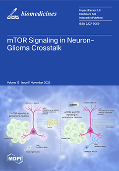
mTOR is a central regulator of cellular growth and neural activity, positioned at the intersection of oncogenic signaling and neural circuit dynamics. In adult diffusely infiltrating glioma, dysregulated mTOR not only facilitates tumor proliferation and metabolism, but it also reshapes communication between tumor cells and surrounding neurons. Molecular, electrophysiological, and imaging studies reveal that mTOR-dependent signaling drives synaptic remodeling, alters excitatory–inhibitory balance, and increases network hyperexcitability. These changes contribute to both glioma-associated seizures and cognitive decline. In this review, we highlight mTOR as a key mediator of neuron–glioma crosstalk and a potential therapeutic target capable of addressing tumor-induced neurological dysfunction in patients with glioma. View this paper
- Issues are regarded as officially published after their release is announced to the table of contents alert mailing list.
- You may sign up for e-mail alerts to receive table of contents of newly released issues.
- PDF is the official format for papers published in both, html and pdf forms. To view the papers in pdf format, click on the "PDF Full-text" link, and use the free Adobe Reader to open them.
Previous Issue
Next Issue