Article

Evaluation of Stereopsis Performance, Gaze Direction and Pupil Diameter in Post-COVID Syndrome Using Machine Learning

1
Department of Ophthalmology, Universitätsklinikum, Friedrich-Alexander-University of Erlangen-Nürnberg, 91054 Erlangen, Germany
2
Department of Internal Medicine 3–Rheumatology and Immunology, Deutsches Zentrum Immuntherapie and Center for Rare Diseases Erlangen, Universitätsklinikum Erlangen, Friedrich-Alexander-Universität Erlangen-Nürnberg, 91054 Erlangen, Germany
3
Talkingeyes & More, Erlangen, 91052 Erlangen, Germany
*
Authors to whom correspondence should be addressed.
These authors contributed equally to this work.
Biomedicines 2025, 13(11), 2828; https://doi.org/10.3390/biomedicines13112828
Submission received: 30 September 2025 / Revised: 7 November 2025 / Accepted: 10 November 2025 / Published: 20 November 2025

Abstract

Background/Objectives: Post-COVID syndrome (PCS) encompasses symptoms that persist for at least 12 weeks after the onset of a COVID-19 infection and cannot be explained by other causes. The most common symptoms are fatigue, cognitive impairments, and physical limitations. The objective diagnosis of PCS is still challenging, as specific biomarkers are lacking. One possibility to measure cognitive impairment is the virtual-reality-oculomotor-test-system (VR-OTS, Talkingeyes & More, Germany). It shows stereoscopic stimuli in a VR-environment to the test person. While working on the visual tasks, many features are recorded. These features can be categorized into three groups: stereopsis performance, gaze direction, and pupil diameter. The aim of this study was to investigate which of these three feature groups is best to distinguish patients with PCS from a healthy control group. Methods: In total, 429 patients with PCS were recruited within the disCOVer 1.0 and disCOVer 2.0 study at the Department of Ophthalmology, Universitätsklinikum (Erlangen, Germany). All patients received VR-OTS measurements. From these measurements, a total of 95 features were extracted, which can be categorized into three groups: gaze direction, pupil diameter, and stereopsis performance. In the first step, support vector machines (SVMs) were trained on these different feature sets and evaluated using the area under receiver operating characteristic (AUROC) as the evaluation metric. In the second step, the same procedure was repeated with each feature independently to investigate which were most the predictive per group. Results: The SVM using the pupil diameter features yielded an AUROC of 0.73, the one using the gaze direction features resulted in an AUROC of 0.68. and the stereopsis performance features produced an AUROC of 0.66. The SVM using all VR-OTS data showed an AUROC of 0.68. For the single features, the index of pupillary activity (IPA) showed the best discrimination. Moreover, all features that were evaluated at different difficulties showed the same pattern—that the more difficult test proved to be more predictive. Conclusions: The study showed that VR-OTS can distinguish between patients with PCS and healthy control probands. Since different features showed a better performance than others, it makes sense for further studies to use a subset of the available features for further analysis.

1. Introduction

In December 2019, the severe acute respiratory syndrome coronavirus 2 (SARS-CoV-2) was observed in Wuhan, Hubei province, China. The virus is transmitted by inhalation or contact with infected droplets. It rapidly spread and caused a global pandemic [1,2]. According to the WHO, more than 778 million cases and over 7 million deaths caused by SARS-CoV-2 had been reported by August 2025 [3]. Even though the infection rate and mortality of SARS-CoV-2 have decreased since the outbreak in 2019, the long disease progression and the limitations in daily activities of patients with Post-COVID syndrome (PCS) makes COVID-19 still a long-lasting societal problem [4,5,6]. PCS is considered a diagnosis by exclusion. Therefore, various diagnostic procedures, including tests like blood samples and pulmonary function tests, are conducted to eliminate other possible medical causes. Since there is no distinct diagnosis for PCS, it is desirable to find objective biomarkers.
Because COVID-19 affects multiple organs, various manifestations can occur, such as pulmonary, renal, hematologic, cardiovascular neuropsychiatric, endocrinal, gastrointestinal, and dermatologic symptoms. [7,8]. A significant number of people suffer from prolonged symptoms after the initial acute phase. [6,9]. If this lasts more than 12 weeks, it is defined as Long COVID(LC)/post-COVID-19-syndrome (PCS)/post-acute sequelae of COVID-19 (PASC) by the WHO [10,11,12]. In most cases, the symptoms are fatigue, aches and pains in the muscles, feeling breathless, headaches, alterations in taste, and cognitive impairment [12].
One method to measure cognitive impairment is the virtual-reality-oculomotor-test-system (VR-OTS, Talkingeyes & More, Germany). The device generates tasks using a virtual 3D environment. During task execution, several of the participant’s biomarkers are measured. These biomarkers can be categorized into three groups: stereopsis performance, pupil diameter, and the gaze direction. Mehringer et al. [13] performed a study using the VR-OTS to distinguish patients with PCS from a healthy cohort. However, they only used 73 features that were provided by the device. All three groups of features (stereopsis performance, gaze direction, and pupil diameter) showed conspicuous values for patients with PCS. However, the disadvantage of the study by Mehringer et al. [13] was the very small patient cohort. Güttes et al. [14] also used the VR-OTS device to measure patients with PCS, and showed significantly prolonged reaction times and worse accuracy for patients with PCS. There are some studies investigating similar processes with a simpler setup, using a stimulus on a 2D-screen in combination with a mobile pupil or gaze tracker. Schmidt et al. [15] documented both a prolonged reaction time and smaller pupils with higher oscillations for patients with PCS. Birirgen et al. [16] demonstrated a lower latency and a shorter duration of the pupil response of COVID-19 patients, while Yurtaser et al. [17] revealed a smaller pupil with lower speed. Garcia et al. [18] showed alterations in the eye movement of patients with PCS [18].
Since the aforementioned studies showed that VR-OTS has great potential for measuring cognitive impairment and distinguishing patients with PCS from healthy controls, pupil and gaze alterations detected by VR-OTS could prove to be valuable biomarkers for PCS. However, VR-OTS records many features. So, the goal of this study was to extract 95 features from the VR-OTS measurements and evaluate which of them performed best. To do so, the different VR-OTS features were used as input to support vector machines (SVMs) and the area under receiver operator characteristic (AUROC) was computed to evaluate their performance. In the first step, the parameters were categorized into three groups (gaze data, pupil diameter, and stereopsis performance). Each of these groups was used as input to the SVM. Moreover, an overall model was trained including all features. In the second step, each parameter was evaluated on its own to see which features are most predictive.

2. Materials and Methods

2.1. Study Population

A total of 429 participants (242 female, 187 male, mean age 39.7 ± 12.7) were recruited at the Department of Ophthalmology, Universitätsklinikum Erlangen, Friedrich-Alexander Universität Erlangen-Nürnberg, Erlangen, Germany, during the course of the disCOVer1.0- and disCOVer2.0-study: 330 patients with PCS (193 female, 137 male, mean age 40.9) and 99 healthy controls (female 49, male 50, mean age 35.7). The demographic data are given in Table 1. For inclusion, patients of the PCS group had to prove the infection by a Polymerase Chain Reaction (PCR) Test and had to have suffered from symptoms for at least 3 months after acute infection according to the German guidelines [10]. Exclusion criteria were an age below 18 years, pre-existing ocular disorders, or systemic disorders with possible ocular involvement and a visual acuity below 0.8 on the weaker eye. Informed written consent was obtained from all participants. The study was reviewed and approved by the ethics committee of the Friedrich-Alexander Universität Erlangen-Nürnberg (295_20_B) and was performed in accordance with the tenets of the Declaration of Helsinki.

2.2. VR-OTS

The virtual-reality-oculomotor-test-system (VR-OTS, Talkingeyes & More, Erlangen, Germany) is a device to analyze ocular function and cognitive impairment. It uses the HTC Vive Pro Eye (HTC Corporation, Taoyuan, Taiwan) to create a virtual 3D-environment. Four balls in a rhomboid arrangement are presented at a virtual distance of 200 cm in front of the patient. The set of the four balls can occur in nine possible positions (central, down, lower right, right, upper right, up, upper left, left, lower left). The arrangement of the balls is displayed at Figure 1. One of the balls is rendered closer to the patient. The difference in the distance between the ball rendered closer and the remaining balls defines the difficulty of the task. In VR-OTS, the patients are presented with tasks of three different difficulty levels (disparities of 250, 550, 1100 arc-seconds (arcsec), where a lower disparity indicates a higher difficulty). The patient must choose the closest of the four balls by pressing the analog key on a keyboard. In each run, all combinations of the three disparities and the nine testing positions are displayed three times in a random order. This makes a total of 81 stereoscopic tasks per run. Each patient performs three trials. On average, the whole procedure takes about 5 min. Only the third run is used for analysis, while the first two serve as practice. In total, 79 features of three data groups were measured, which are explained in more detail in the following.

2.2.1. Stereopsis Performance Features

One data group was about the stereopsis performance of the patient. Reaction time and the number of correct responses were recorded. Beside the accuracy, which is the percentage of correctly given responses, 9 statistical parameters (maximum, minimum, median, mean, standard deviation, variance, skewness, kurtosis) of the reaction time were calculated for each difficulty. For the reaction time, only the correctly identified balls were evaluated. Moreover, the gain, which is defined as the difference between the mean reaction time of two difficulties, was computed for the disparities 275 and 550 and 275 and 1100 [19]. This led to a total of 29 features in the stereopsis performance group.

2.2.2. Pupil Diameter Features

The second data group was about pupil diameter, which was recorded by an eyetracker. The VR-OTS determines the maximum, minimum, range, median, mean, standard deviation, variance, skewness, kurtosis of the pupil diameter for each eye. However, in this study, only the data of the right eye was used, because we assume that there is a correlation between the right and the left eye of the same patient. Moreover, the mean of the index of pupillary activity (IPA) and the mean of the low/high index of pupillary activity (LHIPA) are computed across the entire pupillary signal. IPA [20] and LHIPA [21] are indices to measure cognitive load based on the oscillation and variation in pupil diameter signal. Additionally, values for the signal slopes are calculated. These signal slopes are the linear regression of the pupil diameter for every stimulus. Because the pupil reaction to a stimulus is delayed, every slope is divided into two subsections. The first one is mainly influenced by the prior stimulus. The second one shows the reaction to the current stimulus [22]. IPA, LHIPA, and the slope values were calculated for every disparity. This led to a total of 26 features.

2.2.3. Gaze Behavior

The VR-OTS can also document the distance of gaze direction changes via an eye tracker. From these changes, the angular velocity can be computed using the asymptotic model described by Duchowski et al. [23]. From the gaze direction change and angular velocity, the maximum, median, mean, standard deviation, variance, skewness, kurtosis, and entropy were calculated. Moreover, the number of fixations and fixations per trial were counted. Also, the time every target (background, each ball) got fixated was recorded. This led to a total of 24 values about gaze directions and fixation.

2.2.4. Evaluation

For the evaluation, the available data was split into a 60% training, 20% validation, and 20% test set. In the first step, the features of the three groups (stereopsis performance, gaze direction, and pupil diameter) and all acquired features were used as input to train multiple support vector machines (SVMs) with different architectures on the training set. All of them were subsequently evaluated on the validation set using the area under receiver operating characteristics (AUROC) as the evaluation metric. A 10-fold cross-validation was performed. For each of these four groups, the SVM architecture that achieved the highest mean AUROC on the validation set was chosen for the final evaluation on the test set. In a second step, the same procedure was repeated for each feature individually to evaluate which of them showed the best performance per group.
The programming was done in python (version 3.9) using the scikit-learn library (version 1.3.2).

3. Results

All three feature groups (stereopsis performance, the pupil diameter, and the gaze direction) showed a mean AUROC above 0.66 and are consequently suitable methods to distinguish patients with PCS from healthy controls. The best discrimination was achieved by the pupil diameter, which had a mean AUROC of 0.73, followed by gaze direction and stereopsis performance. The mean AUROC values are shown in detail in Table 2.
The stereopsis feature group showed the lowest AUROC of 0.66. For the single features of this group, the minimum reaction time at the hardest difficulty level (disparity 275) yielded the best AUROC of 0.68. The best mean AUROC values of single features in this group was achieved by the mean (mean 275: 0.68; mean 550: 0.67; mean 1100: 0.67) and median (median 275: 0.68; median 550: 0.68; median 1100: 0.66) of the reaction time, while accuracy was less predictive (accuracy 275: 0.59; accuracy 550: 0.56; accuracy 1100: 0.53). For the mean and median of reaction time and accuracy, mean AUROC values decreased along with difficulty.
The gaze direction feature group achieved an AUROC of 0.68. The best feature in this group was the fixation duration, which had an AUROC of 0.70. The fixation time on the background also showed predictive value (0.67) and exceeded the fixation time on the four balls (right ball: 0.65; top ball: 0.61; left ball: 0.62; bottom ball: 0.64). The maximum of angular velocity and the amplitude of the gaze direction yielded the worst AUROC values of 0.52 and 0.54 in this group.
Pupil diameter was the best performing group of parameters, with an AUROC of 0.73. The values for mean IPA (IPA 275: 0.70; IPA 550: 0.71; IPA 1100: 0.66) and mean LHIPA (LHIPA 275: 0.67; LHIPA 550: 0.67; LHIPA 1100: 0.65) of the individual difficulties performed best. The minimum of the pupil diameter at the hardest difficulty yielded an AUROC of 0.65. The worst mean AUROC values in this group were achieved by the slope features for any difficulty, with mean AUROC-values between 0.48 (Slope 2 550) and 0.58 (Slope 275).
The mean AUROC values and their standard deviations for all single features can be found in Table A1, Table A2 and Table A3 in the Appendix A.

4. Discussion

PCS is defined as symptoms persisting more than 12 weeks after an acute COVID-19 infection. Most patients report neurological symptoms such as significantly impaired thinking or concentration problems [7,12,24].
These neurological symptoms could be explained by neurotropism in COVID-19, which is examined in multiple studies [25]. While Voruz et al. [26] conclude that the central nervous system (CNS) is affected by SARS-CoV-2, Whu et al. [27] describe involvement of cerebrospinal fluid and the brain. This CNS damage can lead to alterations in pupil response [16,17] and eye movement [18,28]. In earlier investigations, stereoscopic visual tasks were used to quantify traumatic brain injuries [29]. We assume that this can also be applied to measure cognitive impairment in patients with PCS.
The VR-OTS is a functional test system that can examine this cognitive impairment. The device shows 4 balls in a 3D-enviroment. The participant must identify one ball, which is projected closer to the patient than the remaining three balls, while measurements are performed. A total of 95 features are extracted from the measurement and can be categorized into three groups: stereopsis performance, gaze direction, and pupil diameter. Since these are many features, the aim of this study was to examine which of them is best to discriminate PCS-patients from healthy controls to simplify diagnosis in future.
Similar approaches have been described in the literature: Cena et al. [18] and Duan et al. [30] analyzed the gaze behavior of patients with PCS fixating on a 2D-screen. Vinela-Navarro et al. extended the experimental setup by incorporating a pupil tracker [31]. In the study of Carbone et al. [32], in addition to observing the gaze behavior on a 2D-screen, a task for measuring the reaction time was added. Maiorana et al. determined the reaction time via a visual detection task on a phone. None of the methods presented measures stereopsis performance, gaze directions, and pupil diameter simultaneously. Moreover, none of them used a 3D stimulus, in contrast to VR-OTS.
In our study, stereopsis performance revealed the worst mean AUROC-value, while in the study of Mehringer et al. [13] this group of features outperformed gaze direction and the pupil diameter. A reason for this might be that Mehringer et al. only included 35 patients (20 patients with PCS and 15 controls) in their study, which may have led to different results compared to our study with 429 patients (330 patients with PCS and 99 controls).
In our study, the minimum reaction time at a disparity of 275 had the best discrimination. Also, the median and mean of the reaction time showed high AUROC values. One observation here was that the AUROC dropped along with the difficulty. This is in line with previous studies. Mehringer et al. [13] showed that the mean and median are increased in patients with PCS. In a further study of Güttes et al. [14], the reaction time of patients with PCS was significantly prolonged, while the p-value decreased inverse to the difficulty [14]. Prolonged reaction times in PCS patients were also found in multiple other studies [15,33,34]. An increase in differences with more complex tasks was also reported by Santoyo-Mora et al. [34]. The reason therefore is presumably cognitive deficiencies. Santoyo-Mora et al. [34] attribute this to a higher cognitive performance load, which causes patients with PCS to require more time. This is consistent with our observation that the AUROC values increase along with the difficulty, particularly regarding the minimum reaction time at our highest difficulty level.
The accuracy values for all difficulties of the stereopsis feature group were among the worst predictive parameters. In the case of Güttes et al. [14], accuracy was significantly reduced in patients with PCS, although the level of significance was notably lower than reaction time. However, this study only investigated whether there is a statistically significant difference between patients with PCS and healthy controls; it did not investigate the performance of the features. Mehringer et al. [13] could also show that the accuracy is less predictive than the reaction time. In our study, the accuracy features at higher difficulties proved to be more predictive. Güttes et al. [14] could also show that the statistical significance decreases with lower difficulty. One possible explanation therefore might be that patients with PCS usually do not report worse visual acuity. Consequently, they should be able to correctly identify the closer ball. However, they might have a prolonged cognitive processing time, leading to a higher reaction time. Moreover, in our study, only patients with a visual acuity larger than 0.8 were included, which can also eliminate further differences in the accuracy.
The AUROC value of the gaze direction feature group performs better in our study than the stereopsis group but worse than the pupil diameter group. From this group, the fixation duration emerges as the most predictive parameter, which was evident in all possible fixation directions. This is consistent with the results of Mehringer et al. [13]. According to the author, the main reason might be that patients with PCS take longer to complete the entire procedure, just like the reaction times in the stereopsis performance feature group. The fixation on the background was the second-best performing feature. We suspect that a possible cause for this might be a prolonged fixation on the background caused by a high cognitive load, as described by Martinez-Cedello et al. [35]. The maximum of the angular velocity and the maximum of the amplitude of gaze direction were the worst parameters of the gaze direction feature group. Mehringer et al. [13] observed the same effect.
The pupil diameter feature group showed the best mean AUROC-values in our study and performed better than in the earlier study by Mehringer et al. [13]. This may be again due to the small sample size used in their study. Multiple other studies also observed differences in the pupil behavior in patients with PCS compared to healthy controls [15,16,17]. The AUROC of the pupil diameter feature achieved a value of 0.73, indicating a moderate to good ability to discriminate between healthy patients and patients with PCS [36]. In our study, IPA and LHIPA showed the highest mean AUROC values at every difficulty and that they correlate with difficulty. Since IPA and LHIPA are direct measures of the cognitive load, this is a strong hint that there is a cognitive impairment in patients with PCS. Mehringer et al. [13] found in their study that only LHIPA at the hardest difficulty could distinguish between patients with PCS and healthy controls. A reason for this might again be the small sample size, which could only detect stronger effects. The minimum diameter also seemed to be a good feature for the differentiation of patients with PCS from healthy controls. A smaller peak diameter for patients with a higher cognitive load could be described by [37,38]. Consequently, patients with PCS could show smaller pupil sizes due to their increased cognitive load.
The worst mean AUROC values in the pupil diameter group were generated by the slope features. Mehringer et al. [13] could also not find a substantial difference in these features between patients with PCS and healthy controls. They attribute this to the flexible processing time of each stimulus. To improve these features, the test system would need to be modified to include a fixed time per stimulus [13].
The study is not without limitations. One limitation of the present study is the lack of differentiation between subgroups of patients. It is assumed that different molecular mechanisms such as persisting viral reservoirs or viral reactivation [39], abnormal blood clotting [40], endothelial dysfunction [41], and immune and autoimmune dysregulation [42] can lead to PCS. Consequently, cognitive impairment might not be present in all patients with PCS, which could influence our results. Thus, investigating stereopsis performance, gaze direction, and pupil diameter in different PCS subgroups might increase its diagnostic value. In addition, further stratification can be done by duration of illness, severity of symptoms, type of pathogenesis, or gender. Furthermore, only a European patient population was included. Moreover, the PCS group contained more patients than the control group, in particular more female patients. Also, only 429 patients were included. Consequently, the results might not be generalizable or transferrable to other ethnicities. Richer or semantically engaging targets might increase the attentional engagement and modulate gaze behavior. Therefore, future studies should compare the current minimalistic stereotarget with more visually engaging environments to evaluate whether additional visual richness improves the signal-to-noise ratio without compromising task specificity.

5. Conclusions

The study showed that VR-OTS is a useful tool for distinguishing between patients with PCS and a healthy control group. Since different features showed a better performance than others, it would make sense for further studies to use a subset of the available features for further analysis. For example, the IPA values were the best performing, indicating that patients with PCS suffer from cognitive impairment. Furthermore, it was shown that tasks with a higher difficulty leaded to more predictive results.

Author Contributions

Conceptualization, B.H., C.Y.M. and J.S.; methodology, J.S. and T.S.K.; software, J.S. and T.S.K.; validation, J.S.; formal analysis, J.S.; investigation, A.S., F.S., H.S., M.G., M.I., M.J., T.S.K. and J.Z.; resources, B.H.; data curation, T.S.K.; writing—original draft preparation, J.S. and T.S.K.; writing—review and editing, A.S., B.H., C.Y.M., F.S., G.M., H.S., J.R., J.S., M.I., M.G., M.J., T.S.K. and J.Z.; visualization, J.S. and T.S.K.; supervision, B.H., C.Y.M. and J.S.; project administration, B.H.; funding acquisition, B.H. All authors have read and agreed to the published version of the manuscript.

Funding

The present study was funded by the (1) Bavarian Health and Food Safety Authority (LGL; 2490-PC-2021-V14: Discover–Establishment and evaluation of clinical algorithms for objective diagnosis of subtypes of Long-COVID as an essential basis for effective patient care; Gesamt-2490-PCS-2023-V6 disCOVer 2.0), the (2) Deutsche Forschungsgemeinschaft (DFG; GRK 2504/1: Novel antiviral approaches from small molecules to immune intervention), and the (3) Federal Ministry of Education and Research (BMBF; iIMMUNE_ACS; 01EO2105: iIMMUNE_ACS–Förderung von Advanced Clinician Scientists im Bereich Immunmedizin). The funders had no role in the design of the study, in the collection, analysis, or interpretation of data, in writing or in the decision to publish results.

Institutional Review Board Statement

The studies involving human participants were reviewed and approved by the ethics committee of the University of Erlangen-Nürnberg (Approval Code: 295_20B Approval Date: 18 August 2020).

Informed Consent Statement

The patients/participants provide their written informed consent to participate in this study.

Data Availability Statement

The dataset analyzed during the current study is not publicly available due to general data privacy regulations but is available from the corresponding author.

Acknowledgments

The present work was performed in fulfillment of requirements for obtaining the degree Dr. Med. for T.K. The Department of ophthalmology is part of the Universität of Erlangen-Nürnberg, Friedrich Alexander-Universität Erlangen-Nürnberg (FAU), Germany.

Conflicts of Interest

G.M. is owner of Talkingeyes and More GmbH, which developed VR-OTS. The remaining authors declare no competing interests.

Abbreviations

The following abbreviations are used in this manuscript:
arcsecarc-sec
AUROCarea under receiver operating characteristic
CNSCentral nervous system
IPAIndex of Pupillary Activity
LClong COVID
LHIPALow/High Index of Pupillary Activity
PASCpost-acute sequelae of COVID-19
PCSPost-COVID-19-syndrome
PCRPolymerase Chain Reaction
SARS-CoV-2severe acute respiratory syndrome coronavirus 2
SVMSupport vector machines
VR-OTSVirtual-Reality-oculomotor-test-system

Appendix A

Table A1. Mean AUROC values and their standard deviation of features of the stereopsis performance.
Parameter DifficultyMean AUROC ± Standard Deviation
Accuracy2750.59 ± 0.10
maximum (reaction time) 2750.63 ± 0.13
minimum (reaction time)2750.68 ± 0.10
median (reaction time)2750.68 ± 0.10
mean (reaction time)2750.68 ± 0.09
standard deviation (reaction time)2750.64 ± 0.10
variance (reaction time)2750.62 ± 0.11
skewness (reaction time)2750.62 ± 0.06
kurtosis (reaction time)2750.63 ± 0.09
accuracy (reaction time)5500.56 ± 0.07
maximum (reaction time)5500.68 ± 0.08
minimum (reaction time)5500.64 ± 0.09
median (reaction time)5500.68 ± 0.08
mean (reaction time)5500.67 ± 0.09
standard deviation (reaction time)5500.66 ± 0.08
variance (reaction time)5500.66 ± 0.07
skewness (reaction time)5500.54 ± 0.09
kurtosis (reaction time)5500.54 ± 0.12
accuracy (reaction time)11000.53 ± 0.07
maximum (reaction time)11000.64 ± 0.10
minimum (reaction time)11000.65 ± 0.08
median (reaction time)11000.66 ± 0.10
mean (reaction time)11000.67 ± 0.10
standard deviation (reaction time)11000.62 ± 0.10
variance (reaction time)11000.62 ± 0.10
skewness (reaction time)11000.59 ± 0.09
kurtosis (reaction time)11000.60 ± 0.09
median gain Disp 275-1100275-11000.60 ± 0.10
median gain Disp 550-1100550-11000.60 ± 0.07
Table A2. Mean AUROC values and their standard deviation of features of the gaze direction.
Parameter Mean AUROC ± Standard Deviation
maximum (amplitude)0.54 ± 0.06
median (amplitude)0.58 ± 0.17
mean (amplitude)0.61 ± 0.11
Standard deviation (amplitude)0.59 ± 0.10
variance (amplitude)0.59 ± 0.10
skewness (amplitude)0.65 ± 0.08
kurtosis (amplitude)0.61 ± 0.11
entropy (amplitude)0.61 ± 0.11
maximum (angular velocity)0.52 ± 0.07
median (angular velocity)0.58 ± 0.17
mean (angular velocity)0.61 ± 0.12
Standard deviation (angular velocity)0.60 ± 0.11
variance (angular velocity)0.60 ± 0.11
skewness (angular velocity)0.62 ± 0.09
kurtosis (angular velocity)0.62 ± 0.10
entropy (angular velocity)0.62 ± 0.09
fixation duration0.70 ± 0.09
fixation duration (background)0.67 ± 0.08
fixation duration (ball up)0.61 ± 0.07
fixation duration (ball down)0.64 ± 0.10
fixation duration (ball left)0.62 ± 0.10
fixation duration (ball right)0.65 ± 0.10
ball transitions0.64 ± 0.10
mean ball transitions per trial0.64 ± 0.10
Table A3. Mean AUROC values and their standard deviation of features of the pupil diameter.
Parameter DifficultyMean AUROC ± Standard Deviation
maximum (diameter) 275-550-11000.60 ± 0.09
minimum (diameter)275-550-11000.65 ± 0.09
median (diameter)275-550-11000.63 ± 0.09
mean (diameter)275-550-11000.63 ± 0.09
standard deviation (diameter)275-550-11000.55 ± 0.09
variance (diameter)275-550-11000.55 ± 0.10
skewness (diameter)275-550-11000.48 ± 0.09
kurtosis (diameter)275-550-11000.51 ± 0.10
range (diameter)275-550-11000.51 ± 0.08
mean IPA275-550-11000.53 ± 0.15
mean LHIPA275-550-11000.55 ± 0.08
mean IPA2750.70 ± 0.09
mean LHIPA2750.67 ± 0.10
mean slope 2750.58 ± 0.07
mean slope 12750.52 ± 0.11
mean slope 22750.52 ± 0.13
mean IPA5500.71 ± 0.14
mean LHIPA5500.67 ± 0.05
mean slope 5500.57 ± 0.08
mean slope 15500.50 ± 0.10
mean slope 25500.48 ± 0.09
mean IPA11000.66 ± 0.09
mean LHIPA11000.65 ± 0.06
mean slope 11000.49 ± 0.09
mean slope 111000.57 ± 0.10
mean slope 211000.52 ± 0.12

References

  1. Singhal, T. A review of Coronavirus Disease-2019 (COVID-19). Indian J. Pediatr. 2020, 87, 281–286. [Google Scholar] [CrossRef]
  2. Wang, C.; Horby, P.W.; Hayden, F.G.; Gao, G.F. A novel coronavirus outbreak of global health concern. Lancet 2020, 395, 470–473. [Google Scholar] [CrossRef]
  3. WHO. WHO COVID-19 Dashboard. 8 October 2025. Available online: https://data.who.int/dashboards/covid19 (accessed on 1 September 2025).
  4. Al-Aly, Z.; Davis, H.; McCorkell, L.; Soares, L.; Wulf-Hanson, S.; Iwasaki, A.; Topol, E.J. Long COVID science, research and policy. Nat. Med. 2024, 30, 2148–2164. [Google Scholar] [CrossRef]
  5. de Sire, A.; Moggio, L.; Marotta, N.; Agostini, F.; Tasselli, A.; Drago Ferrante, V.; Curci, C.; Calafiore, D.; Ferraro, F.; Bernetti, A.; et al. Impact of Rehabilitation on Fatigue in Post-COVID-19 Patients: A Systematic Review and Meta-Analysis. Appl. Sci. 2022, 12, 8593. [Google Scholar] [CrossRef]
  6. Chung, Y.S.; Lam, C.Y.; Tan, P.H.; Tsang, H.F.; Wong, S.C.C. Comprehensive Review of COVID-19: Epidemiology, Pathogenesis, Advancement in Diagnostic and Detection Techniques, and Post-Pandemic Treatment Strategies. Int. J. Mol. Sci. 2024, 25, 8155. [Google Scholar] [CrossRef]
  7. Nalbandian, A.; Sehgal, K.; Gupta, A.; Madhavan, M.V.; McGroder, C.; Stevens, J.S.; Cook, J.R.; Nordvig, A.S.; Shalev, D.; Sehrawat, T.S.; et al. Post-acute COVID-19 syndrome. Nat. Med. 2021, 27, 601–615. [Google Scholar] [CrossRef]
  8. National Institute for Health and Care Excellence. COVID-19 Rapid Guideline: Managing the Long-Term Effects of COVID-19; NICE Guideline; NICE: London, UK, 2024. [Google Scholar]
  9. Davis, H.E.; Assaf, G.S.; McCorkell, L.; Wei, H.; Low, R.J.; Re’em, Y.; Redfield, S.; Austin, J.P.; Akrami, A. Characterizing long COVID in international cohort: 7 months of symptoms and their impact. EClinical Med. 2021, 38, 101019. [Google Scholar] [CrossRef] [PubMed]
  10. Bundesärztekammer. Bekanntmachung “Post-COVID-Syndrom (PCS)”. Dtsch. Ärzteblatt 2022, 1–43. [Google Scholar] [CrossRef]
  11. WHO. Post COVID-19 Condition (Long COVID). 7 December 2022. Available online: https://www.who.int/europe/news-room/fact-sheets/item/post-COVID-19-condition (accessed on 20 June 2025).
  12. WHO. Post COVID-19 Condition (Long COVID). 26 February 2025. Available online: https://www.who.int/news-room/fact-sheets/detail/post-covid-19-condition-(long-covid) (accessed on 20 June 2025).
  13. Mehringer, W.; Stoeve, M.; Krauss, D.; Ring, M.; Steussloff, F.; Güttes, M.; Zott, J.; Hohberger, B.; Michelson, G.; Eskofier, B. Virtual reality for assessing stereopsis performance and eye characteristics in Post-COVID. Sci. Rep. 2023, 13, 13167. [Google Scholar] [CrossRef] [PubMed]
  14. Güttes, M.; Lucio, M.; Skornia, A.; Rühl, E.; Steußloff, F.; Zott, J.; Mardin, C.; Mehringer, W.; Ganslmayer, M.; Michelson, G.; et al. A case-control study of reaction time deficits in a 3D virtual reality in patients with Post-COVID syndrome. Sci. Rep. 2024, 14, 27204. [Google Scholar] [CrossRef]
  15. Schmidt, L.M.; Klingner, C.; Petersen, I.; Volkmer, A.; Schreiber, M.; Schmidt, A.; Reuken, P.; Besteher, B.; Geis, C.; Ullsperger, M.; et al. Cognitive impairment and associated neurobehavioral dysfunction in post-COVID syndrome. Psychiatry Res. 2025, 349, 116522. [Google Scholar] [CrossRef] [PubMed]
  16. Bitirgen, S. Abnormal quantitative pupillary light responses in patients with COVID-19. Int. Ophthalmol. 2022, 42, 2847–2854. [Google Scholar] [CrossRef]
  17. Yurttaser Ocak, S.; Ozturan, S.G.; Bas, E. Pupil responses in patients with COVID-19. Int. Ophthalmol. 2022, 42, 385–391. [Google Scholar] [CrossRef] [PubMed]
  18. García Cena, C.; Costa, M.C.; Saltarén Pazmiño, R.; Santos, C.P.; Gómez-Andrés, D.; Benito-León, J. Eye movement alterations in Post-COVID-19 condition: A proof-of-concept study. Sensors 2022, 22, 1481. [Google Scholar] [CrossRef] [PubMed]
  19. Michelson, G. Europäische Patentanmeldung. Europe Patent EP 3 287 074 A1, 28 February 2018. [Google Scholar]
  20. Duchowski, A.T.; Krejtz, K.; Gehrer, N.A.; Bafna, T.; Bækgaard, P. The low/high index of pupillary activity. In Proceedings of the 2020 CHI Conference on Human Factors in Computing Systems, Honolulu, HI, USA, 25–30 April 2020. [Google Scholar]
  21. Duchowski, A.T.; Krejtz, K.; Krejtz, I.; Biele, C.; Niedzielska, A.; Kiefer, P.; Raubal, M.; Giannopoulos, I. The index of pupillary activity: Measuring cognitive load vis-á-vis task difficulty with pupil oscillation. In Proceedings of the 2018 CHI Conference on Human Factors in Computing Systems, Montreal, QC, Canada, 21–26 April 2018. [Google Scholar]
  22. Baltaci, S.; Gokcay, D. Stress detection in human-computer detection: Fusion of pupillary activity and facial temperature features. Int. J. Hum. Comput. Interact. 2016, 32, 956–966. [Google Scholar] [CrossRef]
  23. Duchowski, A.T.; Medlin, E.; Cournia, N.; Gramopadhye, A.; Melloy, B.; Nair, S. 3D eye movement analysis for VR visual inspection training. In Proceedings of the 2002 Symposium on Eye Tracking Research & Applications, New Orleans, LA, USA, 25–27 March 2002. [Google Scholar]
  24. Ladek, A.M.; Lucio, M.; Weiß, A.; Knauer, T.; Sarmiento, H.; Ilgner, M.; Jakobi, M.; Barteczko, L.; Ganslmayer, M.; Rech, J.; et al. Deoxygenation Trends and Their Multivariate Association with Self-Reported Fatigue in Post-COVID Syndrome. Biomedicines 2025, 13, 1371. [Google Scholar] [CrossRef]
  25. Mendonca Filho, V.C.M.; de Oliveira, A.G.; Maia, I.D.F.V.C.; de Falcone, A.C.M.; Betini, B.G.; Rezende, L.B.; Alves, F.H.M. COVID-19 in the nervous system: Physiopathology and neurological manifestations. Arq. Neuro-Psiquiatr. 2023, 81, 756–763. [Google Scholar] [CrossRef]
  26. Voruz, P.; Allali, G.; Benzakour, L.; Nuber-Champier, A.; Thomasson, M.; Jacot de Alcântara, I.; Pierce, J.; Lalive, P.H.; Lövblad, K.O.; Braillard, O.; et al. Long COVID neuropsychological deficits after severe, moderate, or mild infection. Clin. Transl. Neurosci. 2022, 6, 9. [Google Scholar] [CrossRef]
  27. Wu, Y.; Xu, X.; Chen, Z.; Duan, J.; Hashimoto, K.; Yang, L.; Liu, C.; Yang, C. Nervous system involvement after infection with COVID-19 and other coronaviruses. Brain Behav. Immun. 2020, 97, 18–22. [Google Scholar] [CrossRef] [PubMed]
  28. Wennmo, C.; Hindfelt, B. Eye movements in brainstem lesions. Acta Oto-Laryngol. 1980, 90, 230–236. [Google Scholar] [CrossRef] [PubMed]
  29. Kara, D.D.; Ring, M.; Hennig, F.F.; Michelson, G. Effects of mild traumatic brain injury on stereopsis detected by a virtual reality system: Attempt to develop a screening test. J. Med. Biol. Eng. 2020, 40, 639–647. [Google Scholar] [CrossRef]
  30. Duan, X.; Huang, Z.; Zhang, S.; Zhu, G.; Wang, R.; Wang, Z. Duan. SARS-CoV-2 infection impairs oculomotor functions: A longitudinal eye-tracking study. J. Eye Mov. Res. 2024, 17, 10–16910. [Google Scholar] [CrossRef] [PubMed]
  31. Vinuela-Navarro, V.; Goset, J.; Aldaba, M.; Mestre, C.; Rovira-Gay, C.; Cano, N.; Ariza, M.; Delàs, B.; Garolera, M.; Vilaseca, M. Eye movements in patients with post-COVID condition. Biomed. Opt. Express 2023, 14, 3936–3949. [Google Scholar] [CrossRef] [PubMed]
  32. Carbone, F.; Zamarian, L.; Rass, V.; Bair, S.; Ritter, M.; Beer, R.; Mahlknecht, P.; Heim, B.; Limmert, V.; Peball, M.; et al. Cognitive dysfunction 1 year after COVID-19: Evidence from eye tracking. Ann. Clin. Transl. Neurol. 2022, 9, 1826–1831. [Google Scholar] [CrossRef]
  33. Maiorana, N.V.; Aiello, E.N.; Poletti, B.; Carusi, F.; De Sandi, A.; Guidetti, M.; Prandin, R.; Marceglia, S.; Ticozzi, N.; Silani, V.; et al. Relationship between Reaction Times and Post-COVID-19. Healthcare 2023, 11, 284. [Google Scholar] [CrossRef] [PubMed]
  34. Santoyo-Mora, M.; Villaseñor-Mora, C.; Cardona-Torres, L.M.; Martínez-Nolasco, J.J.; Barranco-Gutiérrez, A.I.; Padilla-Medina, J.A.; Bravo-Sánchez, M.G. COVID-19 Long-Term Effects: Is There an Impact on the Simple Reaction Time and Alternative-Forced Choice on Recovered Patients? Brain Sci. 2022, 12, 1258. [Google Scholar] [CrossRef]
  35. Martinez-Cedillo, A.P.; Gavrila, N.; Mishra, A.; Geangu, E.; Foulsham, T. Cognitive load affects gaze dynamics during real-world tasks. Exp. Brain Res. 2025, 243, 82. [Google Scholar] [CrossRef] [PubMed]
  36. Çorbacıoğlu, Ş.K.; Aksel, G. Receiver operating characteristic curve analysis in diagnostic accuracy studies: A guide to interpreting the area under the curve value. Turk. J. Emerg. Med. 2023, 23, 195–198. [Google Scholar] [CrossRef]
  37. Opwonya, J.; Kim, K.; Lee, K.H.; Kim, J.I.; Kim, J.U. Task-evoked pupillary responses as potential biomarkers of mild cognitive impairment. Alzheimer’s Dement. Diagn. Assess. Dis. Monit. 2024, 16, e70019. [Google Scholar] [CrossRef]
  38. El Haj, M.; Chapelet, G.; Moustafa, A.A.; Boutoleau-Bretonnière, C. Pupil size as an indicator of cognitive activity in mild Alzheimer’s disease. EXCLI J. 2022, 21, 307. [Google Scholar]
  39. Proal, A.D.; VanElzakker, M.B.; Aleman, S.; Bach, K.; Boribong, B.P.; Buggert, M.; Cherry, S.; Chertow, D.S.; Davies, H.E.; Dupont, C.L.; et al. SARS-CoV-2 reservoir in post-acute sequelae of COVID-19 (PASC). Nat. Immunol. 2023, 24, 1616–1627. [Google Scholar] [CrossRef] [PubMed]
  40. Kell, D.B.; Laubscher, G.J.; Pretorius, E. A central role for amyloid fibrin microclots in long COVID/PASC: Origins and therapeutic implications. Biochem. J. 2022, 479, 537–559. [Google Scholar] [CrossRef] [PubMed]
  41. Davis, H.E.; McCorkell, L.; Vogel, J.M.; Topol, E.J. Long COVID: Major findings, mechanisms and recommendations. Nat. Rev. Microbiol. 2023, 21, 133–146. [Google Scholar] [CrossRef] [PubMed]
  42. Wallukat, G.; Hohberger, B.; Wenzel, K.; Fürst, J.; Schulze-Rothe, S.; Wallukat, A.; Hönicke, A.S.; Müller, J. Functional autoantibodies against G-protein coupled receptors in patients with persistent Long-COVID-19 symptoms. J. Transl. Autoimmun. 2021, 4, 100100. [Google Scholar] [CrossRef]
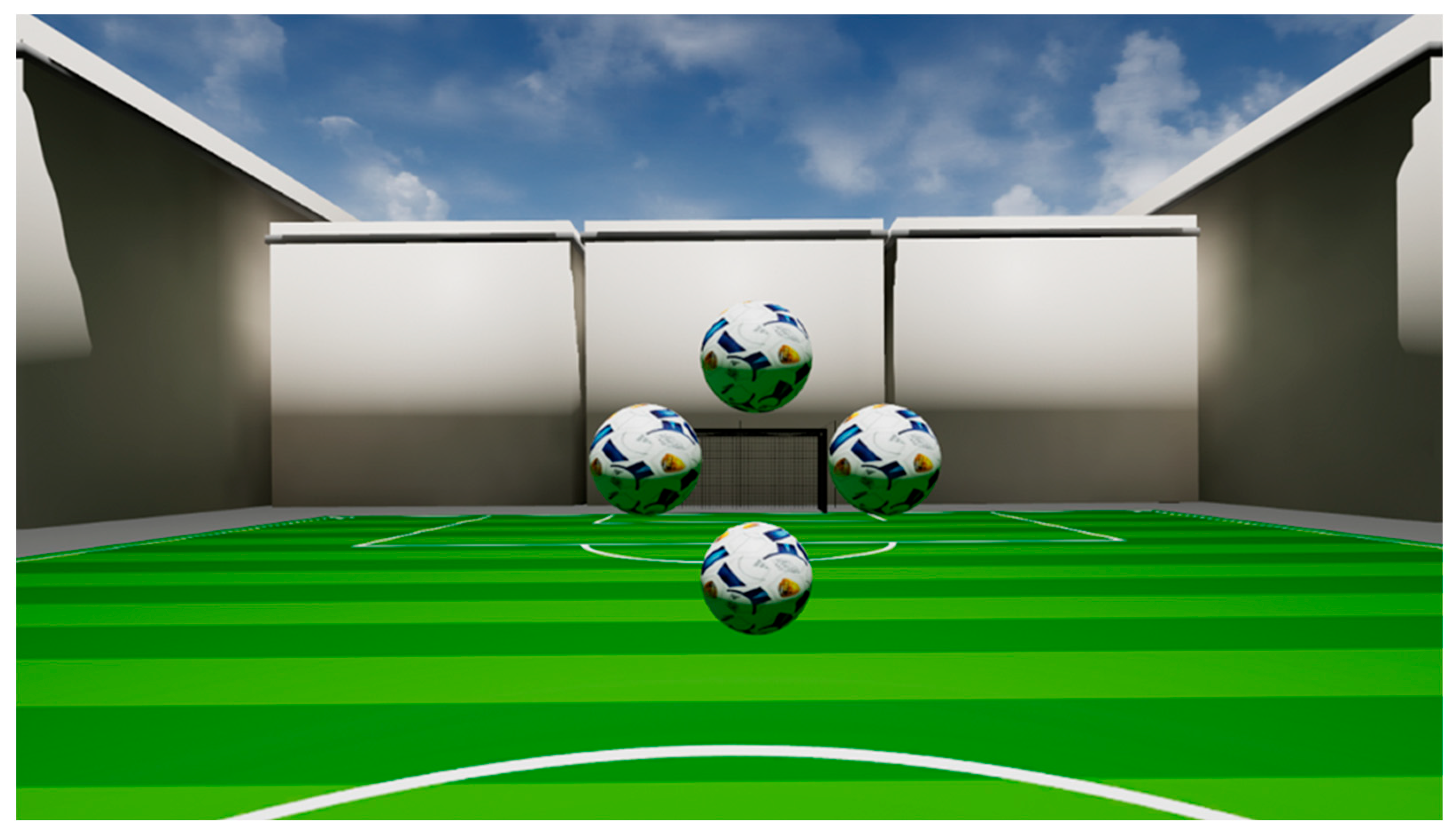
Figure 1. Visual depiction of the visual stimulus presented to a participant: Four balls (central position) are rendered in a rhomboid arrangement. The patient has to detect which of the balls is rendered closer.
Biomedicines 13 02828 g001
Table 1. Demographic data of patients with PCS and healthy controls.
PCS (n = 330)Control (n = 99)
Sex (male/female)137 (41.5%)/193 (58.5%)50 (50.5%)/49 (49.5%)
Age40.9 (±11.9)35.7 (±14.5)
Table 2. Mean AUROC values and their standard deviation of stereopsis performance, pupil diameter, gaze direction and all VR-OTS features.
Parameter GroupMean AUROC ± Standard Deviation
pupil diameter0.73 ± 0.09
gaze direction0.68 ± 0.07
stereopsis performance0.66 ± 0.09
all extracted VR-OTS0.71 ± 0.07
Disclaimer/Publisher’s Note: The statements, opinions and data contained in all publications are solely those of the individual author(s) and contributor(s) and not of MDPI and/or the editor(s). MDPI and/or the editor(s) disclaim responsibility for any injury to people or property resulting from any ideas, methods, instructions or products referred to in the content.