




Next Issue
Volume 16, August
Previous Issue
Volume 16, June


Biomolecules, Volume 16, Issue 7 (July 2026) – 149 articles
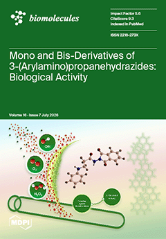
Cover Story (view full-size image): Redox-active small molecules are promising modulators of oxidative processes and cellular redox balance. Since oxidative stress is closely linked to microbial toxicity and antibiotic resistance, antioxidant and antibacterial activities are often interconnected, supporting their biomedical potential. Comparative evaluation of antioxidant and antibacterial properties of the synthesized N’-(4-nitrobenzylidene)propanehydrazides and corresponding bishydrazones, as well as N-(1,3-dioxoisoindolin-2-yl)propanamides and corresponding bis(propanamides), identified several derivatives with pronounced antioxidant activity in multiple in vitro assays, while several compounds also exhibited moderate broad-spectrum antibacterial effects, highlighting the relationship between redox-regulating capacity and antimicrobial activity. View this paper
  • Issues are regarded as officially published after their release is announced to the table of contents alert mailing list.
  • You may sign up for e-mail alerts to receive table of contents of newly released issues.
  • PDF is the official format for papers published in both, html and pdf forms. To view the papers in pdf format, click on the "PDF Full-text" link, and use the free Adobe Reader to open them.
Order results
Result details
Section
Select all
Export citation of selected articles as:
22 pages, 1789 KB  
Article
Increasing Bioactive Compound Production in Lettuce by Application of Trichoderma sp. Strain STP8
by Božidar Benko, Mia Dujmović, Sanja Radman, Jana Šic Žlabur and Snježana Topolovec-Pintarić
Biomolecules 2026, 16(7), 1073; https://doi.org/10.3390/biom16071073 - 22 Jul 2026
Viewed by 878
Abstract
Improving the nutritional quality of food through advanced and sustainable agricultural practices has become a key objective of modern vegetable crop production. Emphasis is placed on increasing the content of health-promoting bioactive compounds, such as vitamins and polyphenols, particularly flavonoids whose accumulation is [...] Read more.
Improving the nutritional quality of food through advanced and sustainable agricultural practices has become a key objective of modern vegetable crop production. Emphasis is placed on increasing the content of health-promoting bioactive compounds, such as vitamins and polyphenols, particularly flavonoids whose accumulation is strongly affected by various biotic and abiotic stress factors. To mitigate stress-induced limitations and enhance plant performance, biostimulants are increasingly applied. Among them, Trichoderma spp. are widely recognized for their ability to promote plant growth and resilience, primarily through enzymatic activity and the production of bioactive metabolites. The aim of this study was to evaluate the potential of the native Trichoderma sp. strain STP8 to enhance the production of bioactive compounds through seed and soil applications at planting and 26 days after planting (DAP), applied individually or in combination. A spore suspension (4 × 10⁶ spores mL−1) was used. The experiment was arranged in a randomized complete block design with five replicates. At harvest (43 DAP), dry matter, ascorbic acid, chlorophyll, and carotenoid contents were determined. Additionally, flavonoids and non-flavonoids, total phenolics, individual phenolic compounds, and antioxidant capacity were analyzed. Achieved results demonstrate that the effects of the native Trichoderma sp. strain STP8 on lettuce secondary metabolism and antioxidant properties are strongly dependent on the developmental stage at which inoculation is performed, providing further insight into the stage-specific interactions between plans and Trichoderma. Practically, a single application at planting proved to be the most effective strategy for enhancing the accumulation of bioactive compounds, indicating that optimized application timing may improve the efficacy of Trichoderma-based biostimulants, while avoiding unnecessary repeated applications. These findings support the potential use of native Trichoderma strains as sustainable tools for improving the nutritional and functional quality of lettuce. Further research integrating physiological, biochemical, and molecular analyses is required to elucidate the mechanisms by which the native Trichoderma sp. strain STP8 regulates the biosynthesis of bioactive compounds in lettuce. Full article
(This article belongs to the Special Issue Plant Secondary Metabolism Engineering and Bioactive Compounds)
Show Figures

Figure 1

33 pages, 879 KB  
Review
Non-Mammalian Models for Mitochondria Research in CNS Disorders
by Dubravka Svob Strac, Vedrana Filic, Ana Filosevic Vujnovic, Ivana Vrhovac Madunic, Josip Madunic, Ana Cipak Gasparovic, Ana Havelka Mestrovic and Rozi Andretic Waldowski
Biomolecules 2026, 16(7), 1072; https://doi.org/10.3390/biom16071072 - 22 Jul 2026
Viewed by 560
Abstract
Mitochondrial dysfunction is increasingly recognized as a major contributor to central nervous system (CNS) disorders, including neurodegenerative and neuropsychiatric diseases. Animal models are essential for elucidating disease mechanisms and supporting the development of new therapeutic strategies. Among these models, non-mammalian organisms offer distinct [...] Read more.
Mitochondrial dysfunction is increasingly recognized as a major contributor to central nervous system (CNS) disorders, including neurodegenerative and neuropsychiatric diseases. Animal models are essential for elucidating disease mechanisms and supporting the development of new therapeutic strategies. Among these models, non-mammalian organisms offer distinct advantages, including low cost, rapid life cycles, genetic tractability, and suitability for large-scale, high-throughput studies. Organisms such as Saccharomyces cerevisiae, Dictyostelium discoideum, Caenorhabditis elegans, Drosophila melanogaster, and Danio rerio have substantially advanced the understanding of mitochondrial processes relevant to CNS pathology. Studies using these models have revealed conserved mechanisms involving mitophagy, mitochondrial quality control, respiratory function, bioenergetic signaling, and neurodegenerative pathways. Their strengths, including scalability, live imaging capacity, and efficient genetic manipulation, have accelerated disease modeling and therapeutic discovery. However, simplified physiology, evolutionary distance from humans, and the incomplete representation of complex CNS organization limit their translational relevance and often require validation in higher-order organisms. Nevertheless, integrating these models into CNS research, particularly alongside emerging technologies, provides a powerful strategy for linking fundamental mitochondrial biology with translational neuroscience. This review summarizes the use of non-mammalian models in neuroscience research, with an emphasis on mitochondrial dysfunction in CNS disorders and their potential to support future therapeutic advances. Full article
(This article belongs to the Special Issue Mitochondria and Central Nervous System Disorders: 3rd Edition)
Show Figures

Figure 1

22 pages, 12480 KB  
Article
Integrative Multi-Omics Reveal Metabolic Reprogramming by Ketogenic Diet in Melanoma Xenografts
by Rohit Dnyansagar, Natalie Bordag, Rodolphe Poupardin, Julia Tevini, Victoria E. Stefan, Sophia Derdak, Martin Bilban, Nikolaus Fortelny, Barbara Kofler, Roland Lang and Daniela D. Weber
Biomolecules 2026, 16(7), 1071; https://doi.org/10.3390/biom16071071 - 22 Jul 2026
Viewed by 761
Abstract
The ketogenic diet (KD) has demonstrated anti-proliferative effects across multiple tumor types, yet the underlying metabolic and transcriptomic mechanisms remain incompletely understood. This study employed integrated multi-omics analysis combining targeted metabolomics and RNA sequencing to elucidate KD-induced metabolic reprogramming in BRAF/NRAS wild-type, BRAF [...] Read more.
The ketogenic diet (KD) has demonstrated anti-proliferative effects across multiple tumor types, yet the underlying metabolic and transcriptomic mechanisms remain incompletely understood. This study employed integrated multi-omics analysis combining targeted metabolomics and RNA sequencing to elucidate KD-induced metabolic reprogramming in BRAF/NRAS wild-type, BRAF mutant, and NRAS mutant melanoma xenografts, which showed delayed tumor growth when treated with the KD. Despite pronounced metabolic and transcriptional heterogeneity across models with minimal overlap in individual KD-responsive genes, pathway-level analysis revealed convergent biological signatures. Using VIP score-based integration and supervised latent variable modeling (mixOmics DIABLO), we identified consistent KD-associated alterations in cancer-related pathways including the PI3K-Akt, MAPK, sphingolipid as well as HIF-1 signaling pathways. The KD enhanced sphingomyelin and ceramide levels and additionally induced transcriptional signatures, indicating increased ceramide synthesis and reduced ceramide breakdown. Moreover, the KD reduced transcript levels of genes encoding critical tumor regulators, including PI3K, AKT, HIF, MEK, and ERK. These findings demonstrate that despite metabolic and transcriptomic heterogeneity, the KD drives coordinated metabolic reprogramming at the pathway level, indicative of shifting lipid metabolism toward pro-apoptotic ceramides and attenuating key oncogenic signaling cascades. Our results provide insights into the KD’s anti-tumor efficacy and identify metabolic nodes amenable to therapeutic intervention in melanoma. Full article
Show Figures

Figure 1

27 pages, 15320 KB  
Review
GDF15: A Hijacked Metabo-Hormone Orchestrating Cachexia and Immunosuppression in Cancer
by Dong-Yang Qi, Yong-Fei Wang and Wei-Lin Jin
Biomolecules 2026, 16(7), 1070; https://doi.org/10.3390/biom16071070 - 22 Jul 2026
Viewed by 982
Abstract
Cancer is responsible for systemic burdens, most notably cachexia and immunosuppression, that extend far beyond local tumor growth and collectively dictate poor outcomes. While often studied separately, these debilitating syndromes are deeply interconnected. On the basis of emerging evidence of growth differentiation factor [...] Read more.
Cancer is responsible for systemic burdens, most notably cachexia and immunosuppression, that extend far beyond local tumor growth and collectively dictate poor outcomes. While often studied separately, these debilitating syndromes are deeply interconnected. On the basis of emerging evidence of growth differentiation factor 15 (GDF15)’s dual actions in immunity and metabolism, we propose that the stress-responsive hormone GDF15 is hijacked by tumors and repurposed as a central metaboceptive hub that integrates diverse oncogenic stress signals to launch a coordinated, dual pathological cascade. Systemically, it disrupts brain–body communication via glial cell line-derived neurotrophic factor family receptor alpha-like (GFRAL) activation in the brainstem, driving anorexia, metabolic rewiring, and progressive wasting of skeletal muscle and adipose tissue that define cachexia. GDF15 acts as a potent immunosuppressor within the local tumor microenvironment, impairing T cell cytotoxicity and increasing the abundance of regulatory T cells. Crucially, these effects are not parallel but interlinked, forming a self-reinforcing detrimental cycle that accelerates host deterioration and therapeutic failure. This positions the GDF15-GFRAL axis as a unique dual-benefit therapeutic target with the potential to simultaneously ameliorate cachexia, improve patient function and quality of life, and revitalize anti-tumor immunity. Reframing cancer through the lens of a hijacked metabolic sensing system provides an integrated perspective that transforms this formidable challenge of concurrent host wasting and immune evasion into a druggable opportunity, charting a course for novel host-directed therapies that restore systemic homeostasis. Full article
(This article belongs to the Special Issue Cancer Research: Molecular Insights and Therapeutic Strategies)
Show Figures

Graphical abstract

21 pages, 2226 KB  
Article
Coffee Pulp and Silverskin Mitigate Fructose-Induced Intestinal Alterations in Rats
by Francisca Silva, Nelson Andrade, Ilda Rodrigues, Cláudia Marques, Juliana A. Barreto-Peixoto, Maria B. P. P. Oliveira, Rita C. Alves and Fátima Martel
Biomolecules 2026, 16(7), 1069; https://doi.org/10.3390/biom16071069 - 22 Jul 2026
Viewed by 1835
Abstract
Excessive fructose consumption is associated with metabolic syndrome (MS). This study evaluated the effect of two coffee by-products, coffee pulp (CP) and coffee silverskin (SK), on fructose-induced intestinal changes. Sprague–Dawley rats were assigned to six groups (n = 6/group) for 10 weeks: [...] Read more.
Excessive fructose consumption is associated with metabolic syndrome (MS). This study evaluated the effect of two coffee by-products, coffee pulp (CP) and coffee silverskin (SK), on fructose-induced intestinal changes. Sprague–Dawley rats were assigned to six groups (n = 6/group) for 10 weeks: Control, Fructose (FRU; 20% fructose in drinking water), CP, CP + FRU, SK, and SK + FRU. CP and SK were administered by oral gavage (250 mg/kg/day) using corn oil as vehicle. Intestinal morphology, gene expression (RT-qPCR), and gut microbiota composition (16S rRNA sequencing) were assessed. Fructose significantly increased jejunal expression of the glucose transporters SGLT1 and GLUT2. CP and SK reversed SGLT1 and GLUT2 overexpression and reduced GLUT5 expression relative to the FRU group. Fructose also markedly increased expression of sweet taste receptors TAS1R2 and TAS1R3 and the transcription factors SREBP-1c and ChREBP. Both CP and SK normalized TAS1R2 and TAS1R3 expression, whereas SK additionally prevented SREBP-1c and ChREBP overexpression. Both by-products restored fructose-induced reductions in microbial richness and alpha diversity. CP also modified beta diversity and increased the abundance of the genus Blautia compared with FRU. In conclusion, CP and SK reversed several fructose-induced intestinal alterations, namely in the jejunal expression of sugar-sensing and absorption-related genes. Additionally, CP showed microbiota-modulating effects, whereas SK modulated the jejunal expression of key transcription factors (SREBP-1c and ChREBP) involved in carbohydrate and lipid metabolism. Overall, these findings suggest that CP and SK may represent promising candidates for mitigating fructose-induced intestinal alterations. Full article
Show Figures

Graphical abstract

24 pages, 3689 KB  
Review
Helicobacter pylori Vacuolating Cytotoxin A: Structure, Biological Functions, Genetic Polymorphisms, and Therapeutic Perspectives
by Xiaona Song, Xiaoqiong Tang, Alfred Tay, Mohammed Benghezal, Barry J. Marshall, Hong Tang and Hong Li
Biomolecules 2026, 16(7), 1068; https://doi.org/10.3390/biom16071068 - 22 Jul 2026
Viewed by 956
Abstract
VacA (vacuolating cytotoxin A) is a key virulence factor in Helicobacter pylori infection, contributing to chronic gastritis and gastric adenocarcinoma. It induces vacuolation, disrupts cellular functions, and modulates immune responses, aiding bacterial survival in the harsh gastric environment. Genetic diversity in the vacA [...] Read more.
VacA (vacuolating cytotoxin A) is a key virulence factor in Helicobacter pylori infection, contributing to chronic gastritis and gastric adenocarcinoma. It induces vacuolation, disrupts cellular functions, and modulates immune responses, aiding bacterial survival in the harsh gastric environment. Genetic diversity in the vacA gene, particularly alleles like s1 and m1, is associated with more severe clinical outcomes. Recent advances in structural biology, especially cryo-electron microscopy, have revealed VacA’s oligomeric structure and its ability to form anion-selective channels in host cell membranes, providing important insights into its cytotoxic mechanisms. Understanding VacA’s structure and function is essential for unraveling its role in immune evasion and cellular damage. These findings also pave the way for targeted therapeutic strategies, such as subunit vaccines designed to neutralize VacA’s immunosuppressive effects, potentially leading to more effective control of H. pylori infections. Full article
(This article belongs to the Section Molecular Biology)
Show Figures

Figure 1

14 pages, 717 KB  
Perspective
The Cytoplasmic Domain of MHC Class I Molecules as a Molecular Switch: A Perspective from Short Linear Motifs and Intrinsically Disordered Regions
by Fernando A. Arosa and Elsa M. Cardoso
Biomolecules 2026, 16(7), 1067; https://doi.org/10.3390/biom16071067 - 22 Jul 2026
Viewed by 588
Abstract
Classical Major Histocompatibility Complex Class I (MHC-I) molecules are traditionally viewed as stable peptide-presenting structures expressed on the surface of all nucleated cells. Their expression by professional antigen-presenting dendritic cells (DCs) enables CD8+ T-cell activation, differentiation, and immune surveillance. However, accumulating evidence indicates [...] Read more.
Classical Major Histocompatibility Complex Class I (MHC-I) molecules are traditionally viewed as stable peptide-presenting structures expressed on the surface of all nucleated cells. Their expression by professional antigen-presenting dendritic cells (DCs) enables CD8+ T-cell activation, differentiation, and immune surveillance. However, accumulating evidence indicates that cell-surface MHC-I molecules exist in three major conformational states: (1) β2m-associated, peptide-loaded conformers that originate in the endoplasmic reticulum and pass through the Golgi apparatus after binding proteasome-generated cytosolic peptides (hereafter referred to as closed conformers); (2) β2m-free, peptide-empty conformers that arise following β2m dissociation from closed conformers either at the plasma membrane or after internalization and recycling (hereafter referred to as open conformers); and (3) β2m-associated, peptide-empty conformers that represent an intermediate state between closed and open conformers. Here, we propose a conceptual framework, supported by computational predictors of intrinsically disordered regions, in which transitions between closed and open MHC-I conformers are coupled to intracellular regulatory processes, including post-translational modifications of conserved motifs, intracellular trafficking, and signaling. Although direct experimental evidence linking these processes remains limited, we integrate independent observations into a working model that may guide future investigations into MHC-I-mediated cell–cell communication in both immune and non-immune contexts, in health and disease. For clarity, in this article we define “open conformers” as structurally competent, β2m-free, and peptide-deficient MHC-I molecules. Full article
Show Figures

Figure 1

16 pages, 1195 KB  
Review
Ferroportin at the Crossroads of Iron Biology: Disease, Regulation and Modulation
by Pramudi Hasanga Rathnayake, Nina E. Ryan, Ryan Atkins, Daniel F. Wallace and V. Nathan Subramaniam
Biomolecules 2026, 16(7), 1066; https://doi.org/10.3390/biom16071066 - 21 Jul 2026
Viewed by 676
Abstract
Iron is an essential element for almost all living beings. Ferroportin is the only known cellular iron exporter and is responsible for maintaining iron homeostasis. The hepcidin-ferroportin axis is central to iron regulation. Dysregulation of ferroportin is thus associated with iron disorders. Understanding [...] Read more.
Iron is an essential element for almost all living beings. Ferroportin is the only known cellular iron exporter and is responsible for maintaining iron homeostasis. The hepcidin-ferroportin axis is central to iron regulation. Dysregulation of ferroportin is thus associated with iron disorders. Understanding how ferroportin is regulated will provide greater insight into iron metabolism and potential therapies for iron-related disease. This review synthesizes current knowledge on ferroportin biology with a particular focus on its regulatory modulators and their therapeutic potential and provides an updated perspective on the molecular pathogenesis and clinical spectrum of ferroportin disease. Elucidating these mechanisms will be essential for the development of targeted interventions to correct iron dysregulation in diverse human diseases. Full article
(This article belongs to the Special Issue Iron Metabolism in Cells)
Show Figures

Figure 1

37 pages, 3581 KB  
Review
Plasticity of Non-Apoptotic Residual Tumor Cells After Neoadjuvant Immunochemotherapy: Epigenetic and Microenvironmental Determinants
by Wenjun Meng, Ruiyue Li, Peiliang Xie, Bangyi Xiang and Qing Li
Biomolecules 2026, 16(7), 1065; https://doi.org/10.3390/biom16071065 - 21 Jul 2026
Viewed by 1005
Abstract
Neoadjuvant immunochemotherapy (NICT), mainly anti-PD-1/PD-L1 therapy combined with cytotoxic chemotherapy, significantly improved perioperative outcomes for resectable solid tumors such as lung cancer and breast cancer. But a large number of patients still had residual lesions and eventually relapsed. Residual tumor cells are not [...] Read more.
Neoadjuvant immunochemotherapy (NICT), mainly anti-PD-1/PD-L1 therapy combined with cytotoxic chemotherapy, significantly improved perioperative outcomes for resectable solid tumors such as lung cancer and breast cancer. But a large number of patients still had residual lesions and eventually relapsed. Residual tumor cells are not simply unremoved cellular debris, but represent a therapy-selected and therapy-amplified subset of a pre-existing heterogeneous and plastic tumor ecosystem. To avoid implying that therapy generates a new form of tumor plasticity de novo, we use the term “plasticity of non-apoptotic residual tumor cells” to describe the plastic behavior of viable malignant cells that survive treatment-induced cytotoxicity rather than entering apoptosis. In this review, we define the plasticity of non-apoptotic residual tumor cells as the capacity of residual malignant cells to preserve, switch, or re-enter phenotypic states such as dormancy, hybrid EMT, stem-like regeneration, and immune evasion under the combined influence of intrinsic tumor heterogeneity, systemic therapy pressure, and microenvironmental protection. Before the NICT-specific discussion, we outline general theoretical frameworks including therapeutic stress response, apoptosis-induced regeneration, genetic and non-genetic heterogeneity, as well as spatial heterogeneity of involved lymph nodes, so as to provide a more robust interpretation of residual lesion biology under NICT. Also, this review proposes that residual disease may be reconceptualized as a treatment-shaped plastic niche, whose biological behavior is jointly shaped by clonal selection, reversible phenotypic transformation, and microenvironmental ecological protection. We summarize several key states of residual tumor cells: persistent-like/resting state, hybrid EMT/invasive plasticity state, stem-like/regenerative state, and immune escape state, and elucidate the underlying epigenetic basis, including DNA methylation, histone modification, chromatin remodeling, and non-coding RNA network reprogramming. Meanwhile, niche factors such as immune stress, CAF/TAM enrichment, fibrotic matrix, hypoxia, and metabolic stress can further stabilize these states and promote the survival of relapse seeds. Based on this, we propose that future postoperative assessments should be upgraded from residual volume to a stratified residual state, and dynamically identified by combining single-cell omics, spatial pathology, and ctDNA/MRD monitoring. Furthermore, treatment strategies should shift from simply shrinking tumors to plasticity-locking therapy, that is, identifying, classifying, and blocking the plasticity escape pathways of residual lesions before they evolve into recurrence. Full article
(This article belongs to the Special Issue Molecular Mechanisms of Cell Reprogramming and Differentiation)
Show Figures

Graphical abstract

17 pages, 8290 KB  
Article
Interaction of Alkannin with CPEB4 Contributes to Its Antitumor Effects in Melanoma
by Parwen Parhat, Min Li, Wenying Li, Jinyan Li, Mubarak Obulkasim and Yinglan Ma
Biomolecules 2026, 16(7), 1064; https://doi.org/10.3390/biom16071064 - 21 Jul 2026
Viewed by 562
Abstract
Melanoma is a highly aggressive malignancy characterized by strong invasive and metastatic potential. CPEB4 has been implicated in melanoma progression and may serve as a potential therapeutic target. Alkannin has previously been reported to exert antitumor activity against melanoma; however, its in vivo [...] Read more.
Melanoma is a highly aggressive malignancy characterized by strong invasive and metastatic potential. CPEB4 has been implicated in melanoma progression and may serve as a potential therapeutic target. Alkannin has previously been reported to exert antitumor activity against melanoma; however, its in vivo efficacy and direct molecular interaction with CPEB4 remain unclear. In this study, a subcutaneous xenograft model using BALB/c nude mice was used to assess the in vivo antitumor effects of alkannin, and CPEB4 expression was analyzed via Western blotting. DARTS, CETSA, and SPR investigations were used to elucidate the interaction between alkannin and CPEB4. In addition, stable CPEB4-knockdown A375 melanoma cells were established to examine the effects of alkannin on cell proliferation, apoptosis, cell cycle progression, migration, invasion, and downstream signaling molecules. Alkannin markedly suppressed tumor growth in the xenograft model and reduced CPEB4 expression in a dose-dependent manner compared with the model group. DARTS and CETSA demonstrated alkannin-induced stabilization of CPEB4, while SPR analysis using purified recombinant CPEB4 showed a direct physical interaction with alkannin, with micromolar affinity. At the molecular level, alkannin downregulated CPEB4 and PRC1 expression (p < 0.05), whereas CPEB4 knockdown markedly suppressed MITF and PRC1 (p < 0.05). Notably, alkannin treatment alone did not significantly alter MITF protein expression under the present experimental conditions. Alkannin exerts antitumor activity against melanoma, while its interaction with CPEB4 and the associated molecular changes may contribute to cellular responses involving proliferation, survival, migration, invasion-related phenotypes, and mitotic regulation. Full article
(This article belongs to the Section Chemical Biology)
Show Figures

Figure 1

19 pages, 2973 KB  
Review
Hesperidin and Hesperetin: Epigenetic-Stemness Crosstalk, Antitumor Mechanisms, Preclinical Data and Translation Barriers
by Mengqi Guo, Linxin Shao, Huiqing Yin, Qianrui Kou, Lele Shang, Haixia Guan and Fang Li
Biomolecules 2026, 16(7), 1063; https://doi.org/10.3390/biom16071063 - 21 Jul 2026
Viewed by 994
Abstract
Hesperidin is a natural flavonoid derived from citrus plants, which can be hydrolyzed into hesperetin in vivo. Both compounds have anti-inflammatory, antioxidant and antitumor activities. At present, there is a lack of reviews focusing on the epigenetic regulation of cancer stem cells (CSCs) [...] Read more.
Hesperidin is a natural flavonoid derived from citrus plants, which can be hydrolyzed into hesperetin in vivo. Both compounds have anti-inflammatory, antioxidant and antitumor activities. At present, there is a lack of reviews focusing on the epigenetic regulation of cancer stem cells (CSCs) mediated by hesperidin and hesperetin. This review summarizes the molecular crosstalk between hesperidin/hesperetin and CSCs mediated via three major epigenetic pathways, including direct regulatory effects, indirect modulatory actions, and mechanistic relationships proposed based on scientific hypotheses. We elaborate their effects on inhibiting the self-renewal, invasion and metastasis of CSCs as well as reversing chemoresistance, and analyze the crosstalk between epigenetic networks and classical signaling pathways of CSCs. Furthermore, we discuss the core bottlenecks restricting the clinical transformation of these two compounds and introduce improvement strategies such as nanodelivery systems. Current research is still confronted with problems including CSC heterogeneity and the potential off-target toxicity of drugs. In conclusion, hesperidin and hesperetin may serve as potential candidate agents for epigenetic regulation targeting CSCs, which can offer novel theoretical basis for comprehensive tumor therapy. Full article
(This article belongs to the Section Natural and Bio-derived Molecules)
Show Figures

Figure 1

18 pages, 803 KB  
Review
Lithium as a Potential Neuroprotective Strategy in Glaucoma: Mechanisms and Therapeutic Perspectives
by Alessio Martucci, Giulia Napoleoni, Annagrazia Adornetto, Francesco Aiello, Raffaele Mancino, Massimo Cesareo, Giacinto Bagetta, Carlo Nucci, Enrico Romano and Rossella Russo
Biomolecules 2026, 16(7), 1062; https://doi.org/10.3390/biom16071062 - 20 Jul 2026
Viewed by 593
Abstract
Glaucoma is a major global health concern, identified as the foremost cause of irreversible blindness, affecting nearly 95 million individuals. It is characterized by the progressive degeneration of retinal ganglion cells (RGCs), leading to significant vision-related disabilities and an extensive socio-economic impact. The [...] Read more.
Glaucoma is a major global health concern, identified as the foremost cause of irreversible blindness, affecting nearly 95 million individuals. It is characterized by the progressive degeneration of retinal ganglion cells (RGCs), leading to significant vision-related disabilities and an extensive socio-economic impact. The concept that glaucoma should be viewed not solely as an ocular condition but also as a neurodegenerative disorder, sharing pathophysiological features with diseases like Alzheimer’s and Parkinson’s, is now widely accepted. This review examines the convergence of molecular mechanisms, including the roles of amyloid precursor proteins and neuroinflammation, that contribute to RGC loss. Notably, lithium, traditionally used as a mood stabilizer, has emerged as a potential neuroprotective agent for the treatment of Alzheimer’s disease. In light of the common neurodegenerative mechanisms linking glaucoma with central neurodegenerative diseases, here, we review the current evidence supporting lithium’s therapeutic potential in glaucoma, emphasizing the need for further clinical studies to determine its effectiveness in preserving optic nerve health and improving patient outcomes. Full article
(This article belongs to the Special Issue New Discoveries in the Field of Neuropharmacology: 2nd Edition)
Show Figures

Graphical abstract

19 pages, 1265 KB  
Review
Narrative Review of the Pathophysiology of Post-Infectious Bronchiolitis Obliterans
by Alessandro Zago, Caterina Cocchi, Massimo Maschio, Laura Badina, Francesca Policastro, Alessandro Amaddeo, Egidio Barbi and Sergio Ghirardo
Biomolecules 2026, 16(7), 1061; https://doi.org/10.3390/biom16071061 - 20 Jul 2026
Cited by 1 | Viewed by 2490
Abstract
Post-Infectious Bronchiolitis Obliterans (PIBO) is a rare chronic obstructive lung disease characterized by irreversible airflow limitation due to inflammation, fibrosis, and obliteration of the small airways following severe lower respiratory tract infection. Adenovirus is the most frequently associated pathogen, followed by other viruses [...] Read more.
Post-Infectious Bronchiolitis Obliterans (PIBO) is a rare chronic obstructive lung disease characterized by irreversible airflow limitation due to inflammation, fibrosis, and obliteration of the small airways following severe lower respiratory tract infection. Adenovirus is the most frequently associated pathogen, followed by other viruses and atypical bacteria. PIBO pathogenesis appears to result from a complex interaction between severe epithelial injury, dysregulated immune response, persistent neutrophilic inflammation, abnormal tissue repair, and genetic susceptibility. Epithelial damage triggers the release of inflammatory cytokines and epithelial-derived alarmins, promoting chronic inflammation and airway remodeling through fibrosis and airway obliteration driven by activation of the TGF-β/CTGF pathway, epithelial–mesenchymal transition, macrophage–fibroblast interactions, and extracellular matrix deposition. Genetic factors affecting mucociliary clearance, innate immunity, and fibrotic pathways may further predispose certain individuals to abnormal repair and fibrosis consequently. Histologically, PIBO progresses from inflammatory bronchiolitis to fibroproliferative remodeling and constrictive bronchiolitis with luminal obliteration. Understanding these mechanisms supports a stage-based therapeutic approach targeting inflammation in early disease and fibrotic remodeling in advanced stages. Full article
(This article belongs to the Special Issue Molecular Insights into Bronchiolitis Obliterans)
Show Figures

Graphical abstract

44 pages, 2663 KB  
Review
GDF15 in Liver Fibrosis: Molecular Mechanisms, Immunoregulatory Functions, and Therapeutic Potential
by Xinyun Gan, Longze Zhang, Ting Yu, Sikan Jin, Yan Wu, Rui Xu, Yaqi Zhang, Jidong Zhang, Lin Xu and Xianyao Wang
Biomolecules 2026, 16(7), 1060; https://doi.org/10.3390/biom16071060 - 20 Jul 2026
Viewed by 998
Abstract
Liver fibrosis is a chronic pathological process driven by the activation of hepatic stellate cell (HSC) and characterized by the excessive deposition of extracellular matrix (ECM) components in response to persistent liver injury. This condition can lead to progressive hepatic dysfunction, cirrhosis, and [...] Read more.
Liver fibrosis is a chronic pathological process driven by the activation of hepatic stellate cell (HSC) and characterized by the excessive deposition of extracellular matrix (ECM) components in response to persistent liver injury. This condition can lead to progressive hepatic dysfunction, cirrhosis, and ultimately liver failure. Growth differentiation factor 15 (GDF15) has emerged as a pivotal regulator in the initiation and progression of liver fibrosis, exhibiting context-dependent profibrotic and antifibrotic effects. GDF15 modulates multiple cellular processes, including HSC activation and macrophage polarization, as well as the functions of T cells, natural killer cells, B cells and mesenchymal stem cells. This review provides a comprehensive overview of the role of GDF15 in regulating HSC activation and immune cell responses and elaborates on its immunomodulatory functions in attenuating liver fibrosis. Furthermore, we discuss the therapeutic potential of targeting GDF15 for the treatment of liver fibrosis. Ultimately, this review aims to provide a theoretical foundation and propose novel intervention strategies for the early diagnosis and targeted therapy of liver fibrosis. Full article
(This article belongs to the Topic Biomarker Development and Application, 2nd Edition)
Show Figures

Figure 1

29 pages, 25534 KB  
Article
Bellidifolin Improves Pulmonary Artery Smooth Muscle Cells Proliferation by Targeting the IGFBP5-Mediated PI3K-AKT-mTOR Pathway and Dilates the Pulmonary Artery
by Qiuqin Hu, Hongmai Wang, Yujie Qiao, Qingqing Xia, Jiacheng Hu, Xiangyun Gai, Yulin Li, Tao Chen and Zhanqiang Li
Biomolecules 2026, 16(7), 1059; https://doi.org/10.3390/biom16071059 - 19 Jul 2026
Viewed by 591
Abstract
Pulmonary hypertension (PH) is a progressive disease that severely compromises right ventricular function, characterized by two major pathological features: pulmonary arterial constriction and pulmonary arterial remodeling. Bellidifolin (BEL), a natural ketone compound, exhibits potent anti-inflammatory and antioxidant effects; however, its role in PH [...] Read more.
Pulmonary hypertension (PH) is a progressive disease that severely compromises right ventricular function, characterized by two major pathological features: pulmonary arterial constriction and pulmonary arterial remodeling. Bellidifolin (BEL), a natural ketone compound, exhibits potent anti-inflammatory and antioxidant effects; however, its role in PH remains unexplored. This study evaluated the impact of BEL on the two pathological processes: pulmonary arterial constriction and pulmonary arterial remodeling. First, the effects of BEL on pulmonary arterial constriction were evaluated using wire myography. The study revealed that BEL (6–96 μmol/L) inhibited the contraction response of intact endothelial and denuded pulmonary arterial rings precontracted with norepinephrine (1 μmol/L) in a concentration-dependent manner. BEL (90 μmol/L) suppressed pulmonary constriction induced by intracellular calcium release and extracellular calcium influx. In cellular experiments, BEL inhibited the proliferation, migration, and phenotypic transformation of pulmonary arterial smooth muscle cells (PASMCs) induced by 10 μmol/L CoCl2 (72 h), as evidenced by upregulation of the contractile phenotype markers α-SMA and SM22α and downregulation of the synthetic phenotype markers OPN, vimentin, and PCNA. Multi-omics analysis identified Aldh1A1, Mgp, Col4a6, and Igfbp5 as significantly enriched candidates. Among these candidates, Cellular Thermal Shift Assay demonstrated that BEL enhanced the thermal stability of Igfbp5 in PASMCs, suggesting that Igfbp5 may be a potential direct target of BEL. Moreover, KEGG pathway analysis revealed significant enrichment of the PI3K-AKT-mTOR pathway, which is known to be involved in cell proliferation and is regulated by Igfbp5. BEL may inhibit the PI3K-AKT-mTOR pathway by suppressing Igfbp5. The results indicate that BEL may regulate Igfbp5 to inhibit the PI3K-AKT-mTOR pathway, thereby exerting anti-proliferative and inhibitory effects on migration and phenotypic transformation. Full article
Show Figures

Graphical abstract

36 pages, 6792 KB  
Review
Targeting Protein Tyrosine Phosphatase 1B: Recent Advances in Natural, Synthetic, and Multitarget Inhibitors for Diabetes Therapy
by Laura Braconi, Lorenzo Mattolini, Maria Novella Romanelli, Elisabetta Teodori and Dina Manetti
Biomolecules 2026, 16(7), 1058; https://doi.org/10.3390/biom16071058 - 19 Jul 2026
Cited by 1 | Viewed by 872
Abstract
Diabetes mellitus, particularly type 2 diabetes mellitus (T2DM), represents a major global health challenge, driven by the increasing prevalence of obesity and sedentary lifestyles. T2DM is characterized by insulin resistance and progressive β-cell dysfunction, leading to chronic hyperglycemia and multiple complications. Among the [...] Read more.
Diabetes mellitus, particularly type 2 diabetes mellitus (T2DM), represents a major global health challenge, driven by the increasing prevalence of obesity and sedentary lifestyles. T2DM is characterized by insulin resistance and progressive β-cell dysfunction, leading to chronic hyperglycemia and multiple complications. Among the molecular targets investigated for therapeutic intervention, protein tyrosine phosphatase 1B (PTP1B) has emerged as a key negative regulator of insulin signaling. By dephosphorylating the insulin receptor and its downstream substrates, PTP1B attenuates insulin action and contributes to metabolic dysfunction. In addition to its role in glucose homeostasis, PTP1B is implicated in obesity, diabetic complications, neurodegenerative disorders, and cancer, highlighting its relevance as a multifunctional therapeutic target. However, the development of PTP1B inhibitors remains challenging due to the highly conserved and polar nature of its catalytic site, which limits selectivity and cell permeability. Recent research has focused on alternative strategies, including allosteric modulation and multi-site inhibition, to overcome these limitations. This review provides a comprehensive overview of PTP1B inhibitors from both synthetic (2019–2025) and natural sources, with particular emphasis on natural products reported from 2022 onwards, while including selected earlier studies to provide historical context and illustrate representative structural classes and inhibition mechanisms. Although PTP1B remains an attractive therapeutic target, its clinical validation for diabetes treatment has yet to be achieved. Continued advances in medicinal chemistry and allosteric modulation may help overcome the current translational barriers. Full article
(This article belongs to the Section Chemical Biology)
Show Figures

Figure 1

23 pages, 1127 KB  
Review
DADA2 as a Model of Monogenic Immune Vasculopathy: From Immunopathogenesis to Precision Therapeutics
by Hao Peng, Chunxia Li, Chune Mo, Bihui Li and Minglin Ou
Biomolecules 2026, 16(7), 1057; https://doi.org/10.3390/biom16071057 - 19 Jul 2026
Viewed by 881
Abstract
Deficiency of adenosine deaminase 2 (DADA2) is a monogenic autoinflammatory disorder caused by biallelic loss-of-function mutations in the ADA2 gene (formerly CECR1). First described in 2014, DADA2 has emerged as a paradigm for monogenic vasculitis, bridging the gap between primary immunodeficiencies and [...] Read more.
Deficiency of adenosine deaminase 2 (DADA2) is a monogenic autoinflammatory disorder caused by biallelic loss-of-function mutations in the ADA2 gene (formerly CECR1). First described in 2014, DADA2 has emerged as a paradigm for monogenic vasculitis, bridging the gap between primary immunodeficiencies and systemic vasculitides. The disease is characterized by a remarkably broad clinical spectrum encompassing early-onset lacunar stroke, systemic vasculitis resembling polyarteritis nodosa (PAN), hematologic abnormalities ranging from pure red cell aplasia to pancytopenia, humoral immunodeficiency, and variable lymphoproliferation. ADA2, predominantly secreted by myeloid cells, serves dual functions as a growth factor for endothelial cells and a modulator of extracellular adenosine metabolism. Its deficiency leads to a proinflammatory state driven by macrophage dysregulation, excessive tumor necrosis factor (TNF) production, neutrophil extracellular trap (NET) formation, and endothelial dysfunction. The genotype–phenotype correlation is complex, with certain mutations predisposing to vasculitic versus hematologic-predominant phenotypes. Emerging evidence further links ADA2 deficiency to cellular senescence and inflammaging pathways, suggesting a connection between monogenic vasculitis and aging-related biological mechanisms. Anti-TNF therapy has revolutionized disease management, achieving sustained remission in the majority of vasculitic manifestations. Hematopoietic stem cell transplantation (HSCT) offers a definitive cure for severe hematologic disease, while gene therapy approaches are under active investigation. This review synthesizes current knowledge on the immunopathogenesis, clinical heterogeneity, genotype–phenotype correlations, multi-omics insights, and evolving precision therapeutic strategies for DADA2, positioning it as an instructive model for understanding monogenic immune vasculopathy. Despite this progress, fundamental questions remain—including the relative contribution of ADA2 enzymatic versus growth factor functions to disease pathogenesis, the mechanisms underlying tissue-specific vulnerability, the basis of differential treatment responsiveness, and the identity of genetic and environmental modifiers that determine phenotypic heterogeneity—that define the frontier of current DADA2 research. This review critically evaluates both established knowledge and persistent uncertainties, positioning DADA2 as an instructive model for the study of monogenic immune vasculopathy. Full article
(This article belongs to the Topic Inflammaging: The Immunology of Aging, 2nd Edition)
Show Figures

Figure 1

28 pages, 5396 KB  
Review
PFAS Exposure and Metabolic Disorders: Mechanistic Insights into Lipid and Glucose Homeostasis
by Xinyi Chen, Weijing Wen, Simeng Gu, Fanjia Guo, Zhe Mo, Zhijian Chen, Sujun Yan and Xiaofeng Wang
Biomolecules 2026, 16(7), 1056; https://doi.org/10.3390/biom16071056 - 19 Jul 2026
Viewed by 1338
Abstract
Per- and polyfluoroalkyl substances (PFAS) are a class of synthetic fluorinated chemicals characterized by high environmental persistence and widespread occurrence, which have attracted increasing attention due to their potential adverse effects on human health. As the global burden of chronic diseases related to [...] Read more.
Per- and polyfluoroalkyl substances (PFAS) are a class of synthetic fluorinated chemicals characterized by high environmental persistence and widespread occurrence, which have attracted increasing attention due to their potential adverse effects on human health. As the global burden of chronic diseases related to lipid and glucose dysregulation rises, identifying environmental contributors is increasingly important. Growing evidence links PFAS exposure to metabolic disorders, particularly those involving lipid and glucose metabolism. This review summarizes current findings on the mechanisms by which PFAS disrupt metabolic balance, with a focus on pathways involved in fatty acid uptake and oxidation, nuclear receptor signaling, oxidative stress, inflammation and insulin signaling. Furthermore, we highlight the convergence of molecular pathways involved in PFAS-induced lipid and glucose metabolic alterations, which may provide a mechanistic basis for understanding the development of metabolic disorders. Finally, we discuss current research limitations and future perspectives, including the potential application of computational approaches and emerging technologies to further elucidate PFAS-related metabolic effects. A comprehensive understanding of these mechanisms may help identify targets for the prevention and treatment of metabolic diseases, including obesity and type 2 diabetes, and provide regulatory policies on PFAS. Full article
Show Figures

Figure 1

20 pages, 4955 KB  
Article
Role of Endogenous Myoglobin in Anthracycline Response in Breast Cancer
by Ilona Rybinska, Andreas Petry, Thomas Hankeln, Thomas A. Gorr and Gaetano Cairo
Biomolecules 2026, 16(7), 1055; https://doi.org/10.3390/biom16071055 - 18 Jul 2026
Viewed by 502
Abstract
Anthracyclines such as doxorubicin (DOX) remain central components of breast cancer (BC) chemotherapy, although their efficacy is frequently limited by drug resistance. Myoglobin (MB), an oxygen-binding heme protein expressed in breast tumors, has been implicated in the detoxification of DOX in cardiomyocytes, but [...] Read more.
Anthracyclines such as doxorubicin (DOX) remain central components of breast cancer (BC) chemotherapy, although their efficacy is frequently limited by drug resistance. Myoglobin (MB), an oxygen-binding heme protein expressed in breast tumors, has been implicated in the detoxification of DOX in cardiomyocytes, but its role in BC remains unclear. Using MB-expressing and MB-knockout (MBKO) MDA-MB-468 BC cells, we demonstrate that MB confers hypoxia-dependent resistance to DOX. Under hypoxia, MB-expressing cells exhibited reduced intracellular DOX-associated fluorescence, enhanced superoxide generation, and decreased sensitivity to DOX, findings consistent with altered redox cycling and oxidative processing of the drug. Re-expression of MB in MBKO cells restored resistance, whereas pharmacological modulation of MB function using carbon monoxide-releasing molecule-3 and tert-butoxycarbonyl-alanine reversed MB-dependent reductions in intracellular DOX accumulation. In contrast, aclarubicin, an anthracycline lacking the hydroquinone moiety required for efficient redox cycling, failed to reproduce MB-dependent effects. Analyses of four independent neoadjuvant BC cohorts further demonstrated that elevated MB expression was consistently associated with reduced probability of achieving pathological complete response following anthracycline-containing chemotherapy. Collectively, these findings identify MB as a previously unrecognized modulator of BC response to redox-active anthracyclines and support its potential utility as both a predictive biomarker and therapeutic target. Full article
(This article belongs to the Special Issue Iron Metabolism in Cells)
Show Figures

Figure 1

25 pages, 1138 KB  
Review
Analytical Methods and Application of Single-Cell and Single-Nucleus Transcriptomics in the Study of Ischemic Stroke
by Changqing Mu, Yuchuan Ding, Alexander Weiss, Sydni Rosenfeld, Fengwu Li and Xiaokun Geng
Biomolecules 2026, 16(7), 1054; https://doi.org/10.3390/biom16071054 - 18 Jul 2026
Cited by 1 | Viewed by 1148
Abstract
Background: Ischemic stroke remains a leading cause of mortality and long-term disability worldwide, with complex and heterogeneous pathophysiological processes. Single-cell and single-nucleus RNA sequencing (sc/snRNA-seq) has been increasingly applied to investigate cellular heterogeneity at high resolutions. Methods: We systematically searched PubMed, Web of [...] Read more.
Background: Ischemic stroke remains a leading cause of mortality and long-term disability worldwide, with complex and heterogeneous pathophysiological processes. Single-cell and single-nucleus RNA sequencing (sc/snRNA-seq) has been increasingly applied to investigate cellular heterogeneity at high resolutions. Methods: We systematically searched PubMed, Web of Science, and Embase to identify studies that applied sc/snRNA-seq in ischemic stroke research. Based on the retrieved literature, we summarized the bioinformatic analytical methods and application strategies reported in these studies, focusing on how sc/snRNA-seq has been utilized across different research contexts. Results: The application of sc/snRNA-seq in ischemic stroke has expanded rapidly across species and sample types. A wide range of downstream bioinformatic analyses have been employed, including clustering, differential expression analysis, trajectory inference, gene regulatory network analysis, and cell–cell communication analysis. These approaches have been applied to investigate diverse biological processes in ischemic stroke. In addition, these analytical strategies have been extended to multiple biological contexts, including extracerebral tissues, stroke-related modifiers, and their associated complications. Furthermore, integrative analytical approaches that combine multiple datasets, bulk transcriptomics, and other omics data have been increasingly utilized. Advances in temporal and spatial resolutions have enabled analyses across different stages and anatomical regions. Conclusions: This review systematically summarizes the analytical methods and application strategies of sc/snRNA-seq in ischemic stroke. These approaches provide a structured perspective for understanding the application of single-cell technologies in this field. Future studies may benefit from standardized designs and coordinated analytical strategies to facilitate more systematic investigations. Full article
Show Figures

Figure 1

23 pages, 2261 KB  
Article
Shrinking of Extracellular Space During Metabolic Stress Accelerates Amyloid-β Aggregation
by Laura F De Oliveira, Kanchana Karunarathne, Dalton Zona, Martin Muschol and Ghanim Ullah
Biomolecules 2026, 16(7), 1053; https://doi.org/10.3390/biom16071053 - 18 Jul 2026
Viewed by 406
Abstract
Pathological states associated with metabolic stress, such as traumatic brain injury (TBI), hypoxia, ischemic stroke, and migraine, are considered elevated risk factors for developing Alzheimer’s disease (AD). However, the mechanism underlying the effect of these conditions on the progression of AD remains largely [...] Read more.
Pathological states associated with metabolic stress, such as traumatic brain injury (TBI), hypoxia, ischemic stroke, and migraine, are considered elevated risk factors for developing Alzheimer’s disease (AD). However, the mechanism underlying the effect of these conditions on the progression of AD remains largely unknown. Here, we determine how metabolic stress associated with spreading depolarization (SD)—a hallmark of stroke, hypoxia, TBI, and migraine—modulates amyloid β (Aβ42) aggregation kinetics through dynamic changes in extracellular space (ECS). To achieve this, we used ThT fluorescence to determine how the formation of different Aβ42 aggregate species depends on Aβ42 concentrations. Based on this input, we build a multiscale computational framework that integrates volume regulation, including its dependence on neuronal ion homeostasis, and Aβ42 aggregation kinetics. Our model predicts that neuronal swelling during SD accelerates aggregation, where the impact of metabolic stress is highly dependent on the timing relative to aggregation progression and the initial monomer concentration. At low monomer concentrations, early SD events promote off-pathway oligomer formation, while at higher concentrations they rapidly drive fibril formation to saturation. In the absence of mature fibrils, recurrent metabolic stress events further amplify oligomer accumulation, whereas pre-existing fibril nuclei suppress oligomer formation at the expense of fibril nucleation and growth. Increasing the intensity of metabolic stress prolongs ECS shrinkage and enhances oligomer formation. These findings reveal a mechanistic link between SD-induced microenvironmental changes and Aβ aggregation dynamics, providing a quantitative framework for understanding how acute brain injury and metabolic stress may contribute to early AD pathogenesis. Full article
(This article belongs to the Section Bioinformatics and Systems Biology)
Show Figures

Figure 1

42 pages, 5230 KB  
Review
From Unmet Medical Need to Drug Candidate: A Translational Therapeutic Development Roadmap Illustrated by Dual-Payload Antibody–Drug Conjugates
by Takeshi Honda and Gui-Dong Zhu
Biomolecules 2026, 16(7), 1052; https://doi.org/10.3390/biom16071052 - 18 Jul 2026
Cited by 1 | Viewed by 1281
Abstract
Transformative therapeutic innovation should not begin with a molecule—or even a molecular target. It should begin with a clearly defined unmet clinical need. Here, we present a seven-step Translational Therapeutic Development Roadmap that systematically connects an unmet medical need to a developable drug [...] Read more.
Transformative therapeutic innovation should not begin with a molecule—or even a molecular target. It should begin with a clearly defined unmet clinical need. Here, we present a seven-step Translational Therapeutic Development Roadmap that systematically connects an unmet medical need to a developable drug candidate through the disciplined sequence of (i) defining the need, (ii) understanding disease and resistance biology, (iii) building a mechanistic hypothesis, (iv) defining a target product profile (TPP), (v) molecular design and experimental validation, (vi) developability and manufacturability assessment, and (vii) clinical translation. A central conclusion emerging from this review is that resistance biology should be viewed not merely as a cause of therapeutic failure, but as a primary design input for next-generation therapeutic innovation. Our analysis identifies continuous alignment among unmet clinical needs, resistance biology, mechanistic hypothesis, molecular design, developability, and clinical translation as the defining characteristic of successful therapeutic development. We use dual-payload antibody–drug conjugates (ADCs) as a contemporary and highly illustrative case study of this resistance-informed therapeutic development approach. Single-payload ADCs such as trastuzumab deruxtecan and sacituzumab govitecan have transformed treatment across multiple solid tumors, yet most patients ultimately relapse through antigen loss, defective intracellular trafficking, drug efflux, payload-target alterations, and tumor heterogeneity, creating an emerging post-ADC treatment gap. Dual-payload ADCs, which deliver two mechanistically distinct warheads from a single antibody, represent a form of molecular combination therapy designed to increase the barrier to resistance and address multiple escape pathways simultaneously, as well as provide a clinically relevant model for resistance-informed therapeutic design. Using dual-payload ADCs as a worked example, we demonstrate how resistance biology directly informs payload pairing, molecular architecture, conjugation strategy, experimental validation, and developability. Our analysis indicates that successful dual-payload ADC design depends not simply on combining two cytotoxic payloads, but on selecting complementary mechanisms with non-overlapping resistance liabilities while satisfying predefined target product profiles and manufacturability requirements. We further summarize resistance-guided payload pairing strategies, including topoisomerase I plus tubulin inhibitors, topoisomerase I plus DNA-damage-response inhibitors, cytotoxic plus immunomodulatory payloads, and cell-permeable plus non-permeable combinations; the conjugation chemistries that enable defined dual-payload products; the preclinical validation, pharmacological optimization, and developability hurdles that separate promising biology from viable therapeutics; and the rapidly expanding clinical landscape, including the first-in-human program KH815 and emerging bispecific dual-payload constructs. Finally, we demonstrate that the same translational roadmap extends beyond ADCs to radiopharmaceutical conjugates, multispecific antibodies, targeted protein degraders, and cell and gene therapies, indicating that it represents a general framework for therapeutic innovation rather than an ADC-specific strategy. Collectively, this review supports the concept that therapeutic innovation is most successful when unmet clinical needs, resistance biology, molecular design, developability, and clinical translation are considered as an integrated continuum rather than as independent stages of drug discovery. This Translational Therapeutic Development Roadmap provides an organizing framework for guiding the rational development of next-generation targeted therapeutics across diverse therapeutic modalities. Full article
Show Figures

Figure 1

30 pages, 5819 KB  
Review
Polysaccharide-Based Approaches for Inflammation Treatment: Anti-Inflammatory Mechanisms, Delivery Approaches and Translational Prospects
by Xiaoyu Ren, Yuan Zhou, Fei Liu, Lili Jiang, Ai Yuan, Zhouchunxiao Du and Dezhi Sui
Biomolecules 2026, 16(7), 1051; https://doi.org/10.3390/biom16071051 - 18 Jul 2026
Viewed by 503
Abstract
Inflammation is a physiological response of the body to infection and tissue damage; dysregulated inflammatory signaling can cause the initiation and aggravation of a variety of chronic diseases. Although anti-inflammatory drugs have been commercialized and showed beneficial effects clinically, their long-term application is [...] Read more.
Inflammation is a physiological response of the body to infection and tissue damage; dysregulated inflammatory signaling can cause the initiation and aggravation of a variety of chronic diseases. Although anti-inflammatory drugs have been commercialized and showed beneficial effects clinically, their long-term application is often limited by loss of effects and adverse side effects. Natural polysaccharides are multifunctional biomacromolecules with strong potentials in inflammation therapy due to their inherent biocompatibility, wide immunomodulation property and drug capacity. In this Review, we offer a holistic summary of recent advances in polysaccharide anti-inflammatory strategies by coupling the characteristics of structure, biological mechanism and polysaccharide-based delivery systems. This review gives a comprehensive overview of the rational design of anti-inflammatory strategies based on polysaccharides, and also points out the current limitations of polysaccharide drugs in applications, translation difficulties and future trends. Full article
(This article belongs to the Section Bio-Engineered Materials)
Show Figures

Graphical abstract

29 pages, 2758 KB  
Review
ENO1 as an Immunoregulatory Hub in Cancer: Mechanisms and Translational Implications
by Giovanni Perconti, Angela Bonura, Patrizia Rubino and Agata Giallongo
Biomolecules 2026, 16(7), 1050; https://doi.org/10.3390/biom16071050 - 18 Jul 2026
Viewed by 752
Abstract
Alpha-enolase (ENO1) is a multifunctional protein frequently overexpressed in solid tumors, where elevated levels are associated with aggressive behavior and poor prognosis. Beyond its canonical glycolytic role, ENO1 participates in immunoregulatory processes through distinct subcellular pools. Intracellular ENO1 shapes tumor-associated metabolic programs, while [...] Read more.
Alpha-enolase (ENO1) is a multifunctional protein frequently overexpressed in solid tumors, where elevated levels are associated with aggressive behavior and poor prognosis. Beyond its canonical glycolytic role, ENO1 participates in immunoregulatory processes through distinct subcellular pools. Intracellular ENO1 shapes tumor-associated metabolic programs, while surface-exposed ENO1 functions as a plasminogen receptor and can engage innate immune signaling pathways. Post-translational modifications—particularly citrullination and phosphorylation—generate structurally altered epitopes that expand ENO1 antigenicity and enable adaptive immune recognition, including coordinated humoral and T-cell responses in cancer patients. These determinants of ENO1 immunogenicity have downstream consequences within the tumor microenvironment: immune-accessible ENO1 modulates myeloid cell recruitment, dendritic cell maturation, and macrophage polarization, while ENO1-dependent metabolic and signaling programs contribute to immune suppression and escape through multiple interconnected axes. Together, these mechanisms position ENO1 at the interface between tumor metabolism and immune regulation. Preclinical evidence demonstrates that ENO1-directed strategies—including antibody-based targeting, DNA vaccination, and vaccines incorporating post-translationally modified ENO1 peptides—can generate productive antitumor immunity and synergize with checkpoint blockade, supporting the rationale for ENO1 as an immunotherapeutic target. This review synthesizes current evidence within an integrated framework linking ENO1 dysregulation to its immunological consequences in cancer and discusses translational implications for ENO1-centered immunotherapy and immunoprevention. Full article
Show Figures

Figure 1

28 pages, 8539 KB  
Article
AUKAT: Conditional VAE-Driven Augmentation and Neural Modeling of Enzyme Turnover Numbers
by Mengmeng Liu, Xialong Ni and Michal Brylinski
Biomolecules 2026, 16(7), 1049; https://doi.org/10.3390/biom16071049 - 18 Jul 2026
Viewed by 543
Abstract
Accurate prediction of enzyme turnover numbers (kcat) is essential for applications in systems biology, metabolic engineering, and drug discovery, yet remains challenging due to the limited availability and uneven distribution of experimental data. Here, we present AUKAT, an [...] Read more.
Accurate prediction of enzyme turnover numbers (kcat) is essential for applications in systems biology, metabolic engineering, and drug discovery, yet remains challenging due to the limited availability and uneven distribution of experimental data. Here, we present AUKAT, an integrated framework that combines conditional generative modeling with deep neural prediction to improve kcat estimation. A conditional variational autoencoder generates synthetic training instances in embedding space, followed by a selection pipeline that retains samples with strong agreement across independent evaluators, thereby ensuring data reliability. A hybrid convolutional neural network and transformer-based architecture is then used to predict kcat from substrate, enzyme functional, and species embeddings. Incorporating synthetic data improved predictive performance for both random forest and neural network models in five-fold cross-validation, with larger gains observed for the neural network architecture. Benchmarking against DLKcat demonstrated comparable predictive accuracy on the standard test set, while evaluation on stricter unseen subsets indicated improved generalization for low-similarity substrates and enzymes. Feature importance analysis further showed that AUKAT leverages substrate, enzyme functional, and species information in a more balanced manner rather than relying predominantly on a single feature source. In addition, AUKAT-human, a specialized model trained using a pre-training and fine-tuning strategy, achieved improved prediction accuracy for human enzyme kinetics. Overall, AUKAT provides a scalable approach for enzyme kinetics prediction and offers a practical solution to data scarcity in biochemical modeling. Full article
Show Figures

Figure 1

28 pages, 21711 KB  
Review
The Role of AP-1 in Cancer: Regulation, Tumor Microenvironment and Therapeutic Targeting
by Maria A. Katsianou, Dimitrios Vrachas and Christos Adamopoulos
Biomolecules 2026, 16(7), 1048; https://doi.org/10.3390/biom16071048 - 17 Jul 2026
Cited by 1 | Viewed by 824
Abstract
The activator protein-1 (AP-1) transcription factor is a regulatory dimeric transcription factor complex, that responds to a wide range of intracellular and extracellular stimuli and controls gene expression involved in tumor initiation and progression. Comprised primarily of members of Jun and Fos protein [...] Read more.
The activator protein-1 (AP-1) transcription factor is a regulatory dimeric transcription factor complex, that responds to a wide range of intracellular and extracellular stimuli and controls gene expression involved in tumor initiation and progression. Comprised primarily of members of Jun and Fos protein subfamilies, AP-1 is activated downstream of major oncogenic signaling pathways such as the mitogen-activated protein kinase (MAPK) pathway and controls cellular processes including differentiation, invasion, proliferation and apoptosis. In various cancer types, AP-1 contributes to tumor growth by promoting tumor-like phenotypes and facilitating metastatic behavior. Furthermore, AP-1 can affect the tumor microenvironment by modulating inflammation and interaction with immune cells. AP-1 deregulation is linked to tumor heterogeneity and resistance to chemotherapy and radiation. Therefore, AP-1 has emerged as a potential therapeutic target. In preclinical models, direct and indirect targeting via upstream pathways of AP-1 components has demonstrated encouraging results. In addition, combinatorial approaches targeting AP-1 and other regulators may improve the effectiveness of treatment and overcome therapy resistance. In this review, we highlight the AP-1’s role as a critical hub in tumorigenesis that links oncogenic signaling to transcriptional regulation. We also focus on its regulation, function in the tumor microenvironment, and therapeutic potential in combating tumors. Full article
Show Figures

Figure 1

17 pages, 22553 KB  
Article
Expression of Rate-Limiting Enzymes of Melatonin Synthesis in Several Extrapineal Organs During Pregnancy in Ewes
by Yanshu Xu, Haozhe Hou, Yang Yang, Dongsheng Gao, Leying Zhang and Ling Yang
Biomolecules 2026, 16(7), 1047; https://doi.org/10.3390/biom16071047 - 17 Jul 2026
Viewed by 369
Abstract
Melatonin is not only produced by the pineal gland but is also synthesized in extrapineal organs. It plays a key role in antioxidant defense and immune regulation during pregnancy. Two key rate-limiting enzymes are involved in the melatonin synthesis, aralkylamine N-acetyltransferase (AANAT), and [...] Read more.
Melatonin is not only produced by the pineal gland but is also synthesized in extrapineal organs. It plays a key role in antioxidant defense and immune regulation during pregnancy. Two key rate-limiting enzymes are involved in the melatonin synthesis, aralkylamine N-acetyltransferase (AANAT), and acetylserotonin O-methyltransferase (ASMT). Nevertheless, it remained unclear whether pregnancy affects the expression of AANAT and ASMT in extrapineal organs. In this study, ovine maternal thymus, spleen, liver, lymph nodes, thyroid, duodenum, and endometrium were collected on day 16 of the estrous cycle (N16), and on days 13, 16, 25, and 70 (G70) of gestation (n = 6 per group). The expression of AANAT and ASMT in these extrapineal organs was analyzed using RT-qPCR, Western blot, and immunohistochemistry. The results revealed that pregnancy upregulated the expression of AANAT in the liver and ASMT in the thymus and duodenum, but downregulated AANAT expression in the lymph nodes and duodenum. Moreover, AANAT expression was elevated in the thymus and spleen but reduced in the thyroid and endometrium at G70 compared with N16. ASMT expression was increased in the lymph nodes yet decreased in the spleen, liver, and thyroid at G70 compared with N16. Notably, endometrial ASMT expression showed a pregnancy-stage-specific manner with no significant difference between N16 and G70. In summary, this paper reports, for the first time in sheep, that pregnancy modulates the expression of AANAT and ASMT in these extrapineal organs in a pregnancy-stage- and tissue-specific manner. Full article
(This article belongs to the Section Molecular Reproduction)
Show Figures

Figure 1

23 pages, 2313 KB  
Article
Effects of Amino Acid Point Mutations on the Local Hydrophobicity, Structural Stability, and Conformational Flexibility of P-Glycoprotein
by Alexandra Ioana Năstasie and Adriana Isvoran
Biomolecules 2026, 16(7), 1046; https://doi.org/10.3390/biom16071046 - 17 Jul 2026
Viewed by 516
Abstract
P-glycoprotein (P-gp, ABCB1) plays a central role in multidrug resistance and drug pharmacokinetics. In this study, an integrated computational analysis of point amino acid substitutions across the P-gp sequence was performed to evaluate their predicted pathogenicity and structural impact. Variant classification using AlphaMissense [...] Read more.
P-glycoprotein (P-gp, ABCB1) plays a central role in multidrug resistance and drug pharmacokinetics. In this study, an integrated computational analysis of point amino acid substitutions across the P-gp sequence was performed to evaluate their predicted pathogenicity and structural impact. Variant classification using AlphaMissense and PolyPhen-2 revealed concordant predictions for a substantial subset of amino acid substitutions, with 21 variants consistently classified as benign and 13 as pathogenic, while discrepancies for other variants reflected methodological differences between the tools. Notably, substitutions located in transmembrane domains were more frequently predicted to be deleterious compared with those in cytoplasmic or extracellular regions, consistent with the structural and functional constraints imposed on membrane-spanning helices. Changes in local hydropathicity, average flexibility, protein stability (ΔΔG), hydrogen-bonding patterns, surface hydrophobicity and electrostatic potential distributions were also evaluated. Stability predictions obtained using I-Mutant2.0 and DynaMut2.0 indicated that many substitutions tend to destabilize P-gp, although differences in ΔΔG values were observed between methods due to distinct algorithmic approaches. Structural superposition analyses demonstrated generally minor backbone deviations, yet local alterations in surface hydrophobicity, electrostatic potential, and hydrogen bond networks were evident. These physicochemical perturbations, even when subtle, may influence conformational dynamics and coupling between nucleotide-binding and transmembrane domains. Overall, these findings suggest that single amino acid substitutions may alter the local structural environment of P-gp and could potentially influence its transport function. Full article
Show Figures

Graphical abstract

26 pages, 10894 KB  
Article
Solvent-Free Mechanochemical Synthesis, Antispasmodic Activity, and Integrated In Silico Mechanistic Analysis of a Dapsone-Derived Phenylaminojuglone
by Ricardo E. Zavaleta-Miñano, Elena Mantilla-Rodríguez, Roberto O. Ybañez-Julca, Daniel Asunción-Alvarez, Cinthya Enriquez-Lara, Justo Huertas-Córdova, Iván M. Quispe-Díaz, Rafael Jara-Aguilar, Edison Vásquez-Corales, Wilfredo O. Gutiérrez-Alvarado, Osvaldo Yañez and Julio Benites
Biomolecules 2026, 16(7), 1045; https://doi.org/10.3390/biom16071045 - 17 Jul 2026
Viewed by 671
Abstract
The structural hybridization of bioactive quinones is a promising strategy for generating pharmacologically active compounds through sustainable synthetic approaches. This study aimed to synthesize and evaluate a dapsone-derived phenylaminojuglone as a potential intestinal smooth muscle relaxant. Juglone (AJ) was functionalized with [...] Read more.
The structural hybridization of bioactive quinones is a promising strategy for generating pharmacologically active compounds through sustainable synthetic approaches. This study aimed to synthesize and evaluate a dapsone-derived phenylaminojuglone as a potential intestinal smooth muscle relaxant. Juglone (AJ) was functionalized with dapsone (D) via a solvent-free mechanochemical aza-Michael reaction using silica gel as a mild acid catalyst. The resulting compound (AJ-D) was characterized and evaluated in isolated rat ileum preparations. Pharmacological studies were complemented by molecular docking, density functional theory (DFT) calculations, and in silico ADMET predictions. AJ-D was obtained with complete regioselectivity at the C-3 position and required shorter reaction times than conventional solution-based methods. The compound exhibited significant spasmolytic and antispasmodic effects under basal conditions and against acetylcholine- and KCl-induced contractions. Its relaxant activity was not significantly affected by muscarinic receptor blockade or K+ channel inhibition, whereas verapamil reduced its potency. Calcium reintroduction experiments suggested the involvement of extracellular Ca2+ influx pathways. Docking studies suggested favorable interactions with the CaV1.2 L-type calcium channel, whereas DFT and ADMET analyses indicated suitable electronic and drug-like properties. AJ-D is a promising juglone-derived scaffold with antispasmodic activity, likely associated with the modulation of extracellular calcium influx pathways involved in intestinal smooth muscle contraction. Full article
(This article belongs to the Section Chemical Biology)
Show Figures

Graphical abstract

20 pages, 5174 KB  
Article
Green Fabrication of Keratin Nanoparticles from Yak Horn by Steam Flash Explosion: Structure–Property Evolution
by Zhisong Qian, Haiyue Feng, Kun Meng, Xiaoyong Chen, Zhen Hong Chang, Gege Yan, Jiayu Cheng, Mohd Shaiful Sajab, Peer Mohamed Abdul, Gongtao Ding and Yanbin Wang
Biomolecules 2026, 16(7), 1044; https://doi.org/10.3390/biom16071044 - 17 Jul 2026
Viewed by 520
Abstract
Yak horn represents a valuable resource for fabricating keratin-derived materials; however, its dense, highly cross-linked protein network resists efficient processing via conventional extraction methods. This study introduces a single-step steam flash explosion (SFE) approach to produce keratin nanoparticles (KNPs) from yak horn. The [...] Read more.
Yak horn represents a valuable resource for fabricating keratin-derived materials; however, its dense, highly cross-linked protein network resists efficient processing via conventional extraction methods. This study introduces a single-step steam flash explosion (SFE) approach to produce keratin nanoparticles (KNPs) from yak horn. The impact of SFE operating pressure (0–1.6 MPa) on KNP morphology, protein secondary/tertiary structures, thermal behavior, and in vitro biocompatibility was systematically investigated. Morphological evaluations revealed that SFE pressure successfully regulates particle dimensions, yielding optimal uniformity with a minimum average particle height of 11.5 nm at 1.45 MPa. Structural characterization indicated that high-pressure treatment induced a shift in disulfide bonds from stable gauche-gauche-gauche (g-g-g) states (515 cm−1) toward higher-energy trans-gauche-trans (t-g-t) arrangements (540 cm−1). This structural deconstruction led to a decrease in thermal degradation temperatures (from 321.9 °C at 0 MPa to <300 °C at 1.6 MPa) but significantly enhanced dehydration efficiency (ΔH = −470.44 J/g at 1.6 MPa). In vitro biocompatibility assessments demonstrated that the prepared KNPs maintain excellent cytocompatibility, supporting L929 and HaCaT cell viabilities above 95%. These findings demonstrated the versatility and effectiveness of SFE as a sustainable strategy for tuning KNP properties, highlighting its great potential in biomedical applications and green material processing. Full article
(This article belongs to the Section Biomacromolecules: Proteins, Nucleic Acids and Carbohydrates)
Show Figures

Figure 1

Previous Issue
Next Issue
Back to TopTop
