_Kwok.png)
Hesperidin and Hesperetin: Epigenetic-Stemness Crosstalk, Antitumor Mechanisms, Preclinical Data and Translation Barriers


Abstract
1. Introduction
1.1. Research Potential of Natural Compounds in Tumor Therapy
1.2. Roles of Epigenetic Regulation in Tumor Oncogenesis and Progression
1.3. Roles of Cancer Stem Cells in Tumor Oncogenesis and Progression
2. Regulatory Effects of Hesperidin and Hesperetin on Tumor Growth, Metastasis and Chemoresistance
3. Key Signaling Pathways and Molecular Targets of Hesperidin and Hesperetin in Regulating Cancer Stem Cells and Epigenetics
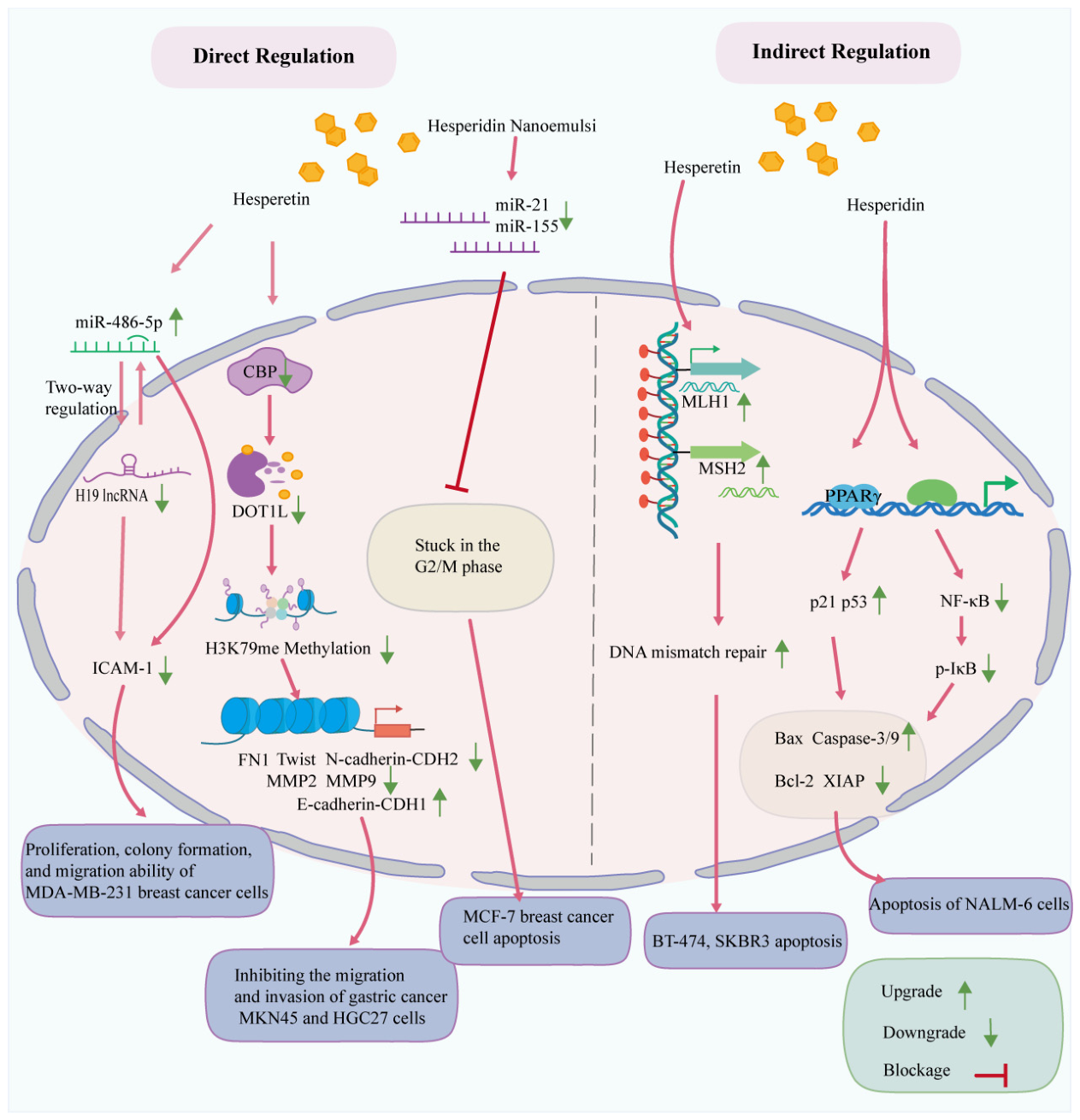
3.1. Molecular Mechanisms of Hesperidin and Hesperetin in Tumor Epigenetic Modification
3.1.1. Direct Regulation
3.1.2. Indirect Regulation
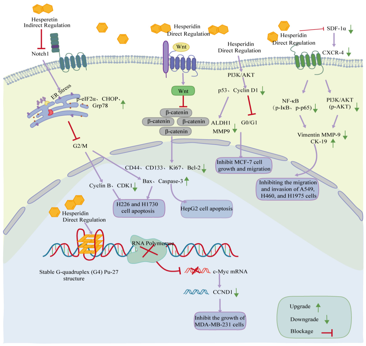
3.2. Molecular Mechanisms of Hesperidin and Hesperetin in Regulating CSCs
3.2.1. Direct Regulation
3.2.2. Indirect Regulation
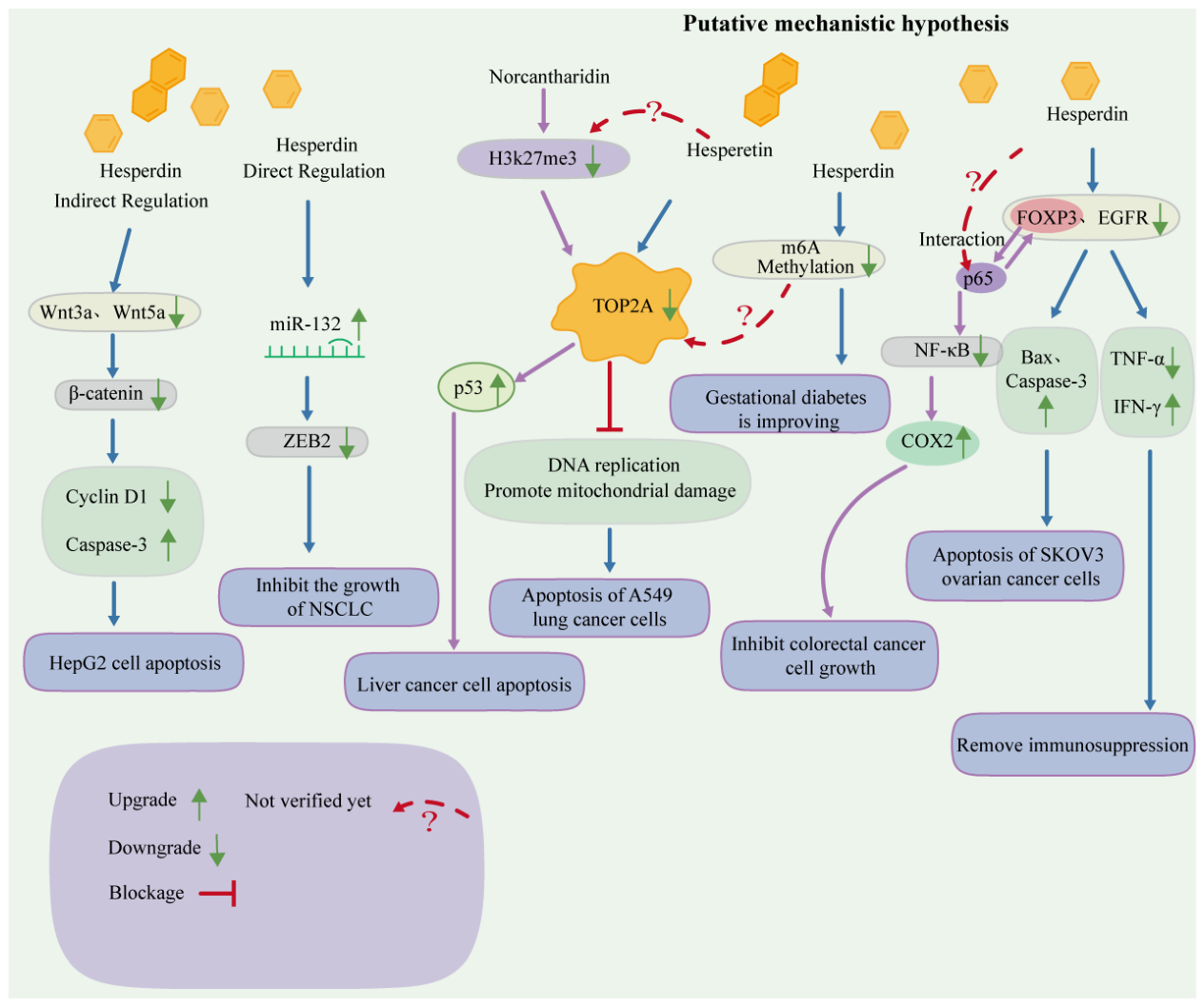
3.3. Crosstalk Between Epigenetic Modification and CSCs: Mechanisms Underlying the Effects of Hesperidin and Hesperetin
3.3.1. Direct Regulation
3.3.2. Indirect Regulation
3.3.3. Putative Mechanistic Hypothesis
4. Preclinical Research on Hesperidin: Challenges and Optimization Strategies
4.1. Optimization of Drug Delivery Systems: Addressing the Bottleneck of Low Bioavailability
4.2. Spatiotemporal Specificity Regulation: Avoiding Damage to Normal Stem Cells
4.3. Management of CSC Heterogeneity: Improving Therapeutic Consistency
4.4. Safety Evaluation and Dose Optimization: Defining the Therapeutic Window
5. Conclusions and Future Perspectives
Author Contributions
Funding
Institutional Review Board Statement
Informed Consent Statement
Data Availability Statement
Acknowledgments
Conflicts of Interest
Abbreviations
| ALDH1 | Aldehyde dehydrogenase 1 |
| BCL-2 | B-cell lymphoma 2 |
| CD | Cluster of differentiation |
| CDH1 | Cadherin 1 (E-cadherin) |
| CDH2 | Cadherin 2 (N-cadherin) |
| CK-19 | Cytokeratin 19 |
| CSCs | Cancer stem cells |
| CXCR-4 | C-X-C chemokine receptor type 4 |
| DNMTs | DNA methyltransferases |
| DOT1L | DOT1-like histone lysine methyltransferase |
| EAC | Ehrlich ascites carcinoma |
| EMT | Epithelial–mesenchymal transition |
| FN1 | Fibronectin 1 |
| FOXP3 | Forkhead box P3 |
| GSH | Glutathione |
| HDACs | Histone deacetylases |
| HIF-1α | Hypoxia-inducible factor-1α |
| HMC | Hesperidin methyl chalcone |
| HP-NEM | Hesperidin nanoemulsions |
| ICAM-1 | Intercellular adhesion molecule 1 |
| IFN-γ | Interferon-gamma |
| IL-1β | Interleukin-1 beta |
| IL-6 | Interleukin-6 |
| IκB | Inhibitor of nuclear factor kappa-B |
| JAK-STAT | Janus kinase-signal transducer and activator of transcription |
| Ki-67 | Marker of proliferation Ki-67 |
| lncRNA | Long non-coding RNA |
| LGR5 | Leucine-rich repeat-containing G-protein coupled receptor 5 |
| MDA | Malondialdehyde |
| MDM2 | Mouse double minute 2 homolog |
| miRNA/miR | MicroRNA |
| MLH1 | MutL homolog 1 |
| MMP | Matrix metalloproteinase |
| MSH2 | MutS homolog 2 |
| MYC/c-Myc | MYC proto-oncogene |
| NANOG | Nanog homeobox |
| NF-κB | Nuclear factor kappa-B |
| Notch | Notch signaling pathway |
| Nrf2 | Nuclear factor erythroid 2-related factor 2 |
| OCT4 | Octamer-binding transcription factor 4 |
| OSCC | Oral squamous cell carcinoma |
| p21 | Cyclin-dependent kinase inhibitor 1A |
| p53 | Tumor protein p53 |
| PI3K/AKT | Phosphatidylinositol 3-kinase/Protein kinase B |
| PLGA | Poly(lactic-co-glycolic acid) |
| PPARγ | Peroxisome proliferator-activated receptor gamma |
| PTX | Paclitaxel |
| ROS | Reactive oxygen species |
| SDF-1α | Stromal cell-derived factor-1 alpha |
| SOX2 | SRY-box transcription factor 2 |
| Survivin | Baculoviral IAP repeat-containing protein 5 |
| TGF-β/SMAD | Transforming growth factor-beta/Smad signaling pathway |
| TNF-α | Tumor necrosis factor-alpha |
| TOP2A | DNA topoisomerase II alpha |
| TRPM2 | Transient receptor potential melastatin 2 |
| VEGF | Vascular endothelial growth factor |
| Vimentin | Vimentin cytoskeletal protein |
| Wnt/β-catenin | Wnt/β-catenin signaling pathway |
| XIAP | X-linked inhibitor of apoptosis protein |
| ZEB2 | Zinc finger E-box binding homeobox 2 |
| ΔΨm | Mitochondrial membrane potential |
| m6A | N6-methyladenosine |
| CpG | Cytosine-phosphate-Guanine |
| 5-FU | 5-fluorouracil |
References
- Bray, F.; Laversanne, M.; Sung, H.; Ferlay, J.; Siegel, R.L.; Soerjomataram, I.; Jemal, A. Global cancer statistics 2022: GLOBOCAN estimates of incidence and mortality worldwide for 36 cancers in 185 countries. CA Cancer J. Clin. 2024, 74, 229–263. [Google Scholar] [CrossRef] [Scilit] [PubMed]
- Bendrihem, K.A.; Mouane, A.; Azzi, M.; Mihoubi, M.A.; Atanassova, M.; Sawicka, B.; Zahnit, W.; Messaoudi, M. The Role of Medicinal Plants in Modulating Epigenetic Mechanisms: Implications for Cancer Prevention and Therapy. Phytother. Res. 2025, 39, 2571–2608. [Google Scholar] [CrossRef] [Scilit] [PubMed]
- Pyrzynska, K. Hesperidin: A Review on Extraction Methods, Stability and Biological Activities. Nutrients 2022, 14, 2387. [Google Scholar] [CrossRef] [Scilit] [PubMed]
- Choi, S.S.; Lee, S.H.; Lee, K.A. A Comparative Study of Hesperetin, Hesperidin and Hesperidin Glucoside: Antioxidant, Anti-Inflammatory, and Antibacterial Activities In Vitro. Antioxidants 2022, 11, 1618. [Google Scholar] [CrossRef] [Scilit] [PubMed]
- Kharwade, R.; Mahajan, N.M.; Telange, D.R.; Yadav, P.N.; More, S.R. Folate receptor-targeted PEGylated PLGA nanoparticles for the site-specific delivery of hesperidin in epithelial ovarian cancer. Artif. Cells Nanomed. Biotechnol. 2026, 54, 180–205. [Google Scholar] [CrossRef] [Scilit] [PubMed]
- Hu, H.; Zhang, C.; Liu, Z. Hesperidin Inhibits Proliferation and Metastasis of Human Breast Cancer Cells by Suppressing MMP-2/9 Activity and Reversing Epithelial-Mesenchymal Transition. Cancer Inform. 2026, 25, 11769351261456238. [Google Scholar] [CrossRef] [Scilit] [PubMed]
- Hosseini, S.S.; Esmailzadeh, E.; Zangooei, M.; Bagheri, V. Hesperetin increases membrane progesterone receptor expression in human myeloid leukemia cells and reduces ROS. Med. Oncol. 2025, 42, 398. [Google Scholar] [CrossRef] [Scilit] [PubMed]
- Jaenisch, R.; Bird, A. Epigenetic regulation of gene expression: How the genome integrates intrinsic and environmental signals. Nat. Genet. 2003, 33, 245–254. [Google Scholar] [CrossRef] [Scilit] [PubMed]
- Moore, L.D.; Le, T.; Fan, G. DNA methylation and its basic function. Neuropsychopharmacology 2013, 38, 23–38. [Google Scholar] [CrossRef] [Scilit] [PubMed]
- Jones, P.A.; Baylin, S.B. The Epigenomics of Cancer. Cell 2007, 128, 683–692. [Google Scholar] [CrossRef] [Scilit] [PubMed]
- Yao, W.; Hu, X.; Wang, X. Crossing epigenetic frontiers: The intersection of novel histone modifications and diseases. Signal Transduct. Target. Ther. 2024, 9, 232. [Google Scholar] [CrossRef] [Scilit] [PubMed]
- Chen, G.; Zhu, X.; Li, J.; Zhang, Y.; Wang, X.; Zhang, R.; Qin, X.; Chen, X.; Wang, J.; Liao, W.; et al. Celastrol inhibits lung cancer growth by triggering histone acetylation and acting synergically with HDAC inhibitors. Pharmacol. Res. 2022, 185, 106487. [Google Scholar] [CrossRef] [Scilit] [PubMed]
- Yeruva, S.L.H.; Zhao, F.; Miller, K.D.; Tevaarwerk, A.J.; Wagner, L.I.; Gray, R.J.; Sparano, J.A.; Connolly, R.M. E2112: Randomized phase iii trial of endocrine therapy plus entinostat/placebo in patients with hormone receptor-positive advanced breast cancer. npj Breast Cancer 2018, 4, 1. [Google Scholar] [CrossRef] [Scilit] [PubMed]
- Li, M.; Ni, Y.; Wu, J.; Zou, X.; Chen, Y.; Qiu, J.; Li, Y.; Cai, H.; Wang, L.; Wang, F.; et al. NEK8 kinase-mediated lactate increase impairs antitumor immunity decreasing radiotherapy sensitivity in colorectal cancer. Nat. Commun. 2026, 17, 4565. [Google Scholar] [CrossRef] [Scilit] [PubMed]
- Giudice, F.S.; Pinto, D.S., Jr.; Nör, J.E.; Squarize, C.H.; Castilho, R.M. Inhibition of histone deacetylase impacts cancer stem cells and induces epithelial-mesenchyme transition of head and neck cancer. PLoS ONE 2013, 8, e58672. [Google Scholar] [CrossRef] [Scilit] [PubMed]
- Slack, F.J.; Chinnaiyan, A.M. The Role of Non-coding RNAs in Oncology. Cell 2019, 179, 1033–1055. [Google Scholar] [CrossRef] [Scilit] [PubMed]
- Batlle, E.; Clevers, H. Cancer stem cells revisited. Nat. Med. 2017, 23, 1124–1134. [Google Scholar] [CrossRef] [Scilit] [PubMed]
- Nassar, D.; Blanpain, C. Cancer Stem Cells: Basic Concepts and Therapeutic Implications. Annu. Rev. Pathol. 2016, 11, 47–76. [Google Scholar] [CrossRef] [Scilit] [PubMed]
- Marin, J.J.G.; Asensio, M.; Álvarez-Fernández, L.; Hortelano-Hernandez, N.; Delgado-Calvo, K.; Marijuan, R.P.; Perez-Silva, L.; Benizri, L.O.; Gallai, D.; Lozano, E.; et al. Impact of CD44 alternative splicing on the response to anticancer drugs. Biochem. Pharmacol. 2026, 251, 118111. [Google Scholar] [CrossRef] [Scilit] [PubMed]
- Xue, Y.; Zheng, Y.; Zhong, Y.; Li, J.; Yang, T.; Hong, S.; Wang, S.; Xie, J.; Xiong, W.; Yang, X.; et al. Modified Fuzheng Yiliu Decoction chemosensitizes colorectal cancer by inhibiting cancer stem cell metabolism and stemness through CAV1/ZNF460/GRP78 signaling. Phytomedicine 2026, 157, 158317. [Google Scholar] [CrossRef] [Scilit] [PubMed]
- Kholodenko, I.V.; Saidova, A.A.; Potashnikova, D.M.; Arzumanian, V.A.; Romashin, D.D.; Tvorogova, A.V.; Poverennaya, E.V.; Yarygin, K.N.; Kim, Y.S. Contrasting Impacts of Targeted Disruption of the Cancer Stem Cell Marker CD133 and Its Epigenetic Regulator TRIM28 in Colorectal Cancer Cells. Int. J. Mol. Sci. 2025, 26, 10862. [Google Scholar] [CrossRef] [Scilit] [PubMed]
- García-Fernández, J.; Díez-Villares, S.; Cascallar, M.; Rivadulla Costa, L.; Martins, A.S.; Groba de Antas, S.; Tarasco, M.C.; Pantano, E.; Taiè, G.; Bertolini, G.; et al. Targeted delivery of therapeutics to metastatic lung cancer cells using aptamer-conjugated nanoemulsions. J. Control. Release 2025, 385, 114031. [Google Scholar] [CrossRef] [Scilit] [PubMed]
- Klose, K.; Packeiser, E.M.; Müller, P.; Granados-Soler, J.L.; Schille, J.T.; Goericke-Pesch, S.; Kietzmann, M.; Murua Escobar, H.; Nolte, I. Metformin and sodium dichloroacetate effects on proliferation, apoptosis, and metabolic activity tested alone and in combination in a canine prostate and a bladder cancer cell line. PLoS ONE 2021, 16, e0257403. [Google Scholar] [CrossRef] [Scilit] [PubMed]
- Maehara, O.; Sato, F.; Natsuizaka, M.; Asano, A.; Kubota, Y.; Itoh, J.; Tsunematsu, S.; Terashita, K.; Tsukuda, Y.; Nakai, M.; et al. A pivotal role of Krüppel-like factor 5 in regulation of cancer stem-like cells in hepatocellular carcinoma. Cancer Biol. Ther. 2015, 16, 1453–1461. [Google Scholar] [CrossRef] [Scilit] [PubMed]
- Zheng, W.; Peng, W.; Qian, F.; Zhang, M.; Duan, B.; Fan, Z.; Xie, Y.; Fu, X. Vitamin D suppresses CD133+/CD44 + cancer stem cell stemness by inhibiting NF-κB signaling and reducing NLRP3 expression in triple-negative breast cancer. Cancer Chemother. Pharmacol. 2024, 94, 67–78. [Google Scholar] [CrossRef] [Scilit] [PubMed]
- Khammanivong, A.; Gopalakrishnan, R.; Dickerson, E.B. SMURF1 silencing diminishes a CD44-high cancer stem cell-like population in head and neck squamous cell carcinoma. Mol. Cancer 2014, 13, 260. [Google Scholar] [CrossRef] [Scilit] [PubMed]
- Ginestier, C.; Hur, M.H.; Charafe-Jauffret, E.; Monville, F.; Dutcher, J.; Brown, M.; Jacquemier, J.; Viens, P.; Kleer, C.G.; Liu, S.; et al. ALDH1 is a marker of normal and malignant human mammary stem cells and a predictor of poor clinical outcome. Cell Stem Cell 2007, 1, 555–567. [Google Scholar] [CrossRef] [Scilit] [PubMed]
- Bhaijee, F.; Pepper, D.J.; Pitman, K.T.; Bell, D. Cancer stem cells in head and neck squamous cell carcinoma: A review of current knowledge and future applications. Head. Neck 2012, 34, 894–899. [Google Scholar] [CrossRef] [Scilit] [PubMed]
- Huang, E.H.; Hynes, M.J.; Zhang, T.; Ginestier, C.; Dontu, G.; Appelman, H.; Fields, J.Z.; Wicha, M.S.; Boman, B.M. Aldehyde dehydrogenase 1 is a marker for normal and malignant human colonic stem cells (SC) and tracks SC overpopulation during colon tumorigenesis. Cancer Res. 2009, 69, 3382–3389. [Google Scholar] [CrossRef] [Scilit] [PubMed]
- Okudela, K.; Woo, T.; Mitsui, H.; Tajiri, M.; Masuda, M.; Ohashi, K. Expression of the potential cancer stem cell markers, CD133, CD44, ALDH1, and β-catenin, in primary lung adenocarcinoma—Their prognostic significance. Pathol. Int. 2012, 62, 792–801. [Google Scholar] [CrossRef] [Scilit] [PubMed]
- Verona, F.; Pantina, V.D.; Modica, C.; Lo Iacono, M.; D’Accardo, C.; Porcelli, G.; Cricchio, D.; Turdo, A.; Gaggianesi, M.; Di Franco, S.; et al. Targeting epigenetic alterations in cancer stem cells. Front. Mol. Med. 2022, 2, 1011882. [Google Scholar] [CrossRef] [Scilit] [PubMed]
- Dakal, T.C.; Bhushan, R.; Xu, C.; Gadi, B.R.; Cameotra, S.S.; Yadav, V.; Maciaczyk, J.; Schmidt-Wolf, I.G.H.; Kumar, A.; Sharma, A. Intricate relationship between cancer stemness, metastasis, and drug resistance. MedComm 2024, 5, e710. [Google Scholar] [CrossRef] [Scilit] [PubMed]
- Chen, D.; Zhang, X.; Wang, X.; Ren, T.; Wang, R. The inhibitory effects of hesperidin and its combination effects with paclitaxel against oral squamous cell car cinoma in nude mice. J. Pract. Stomatol. 2020, 36, 870–875. Available online: https://kns.cnki.net/kcms2/article/abstract?v=wKWn8Yb3X3sWXkJiZuR5pfp1kk0MbjsZMxSHSQzFqaJXu0QP9Ya-ws9xQPN2lDzbFl55aR5pEVPX2_AdahpDwLUze__oeDw6Ci9_6LMg7KKncCJWLBS4zb_sFCMSh1QvpDNM2YLqt7bwkChmC7bM9cs4z8HFGszFK-HR0STUvI0=&uniplatform=NZKPT (accessed on 1 May 2026).
- Wu, D.; Li, J.; Hu, X.; Ma, J.; Dong, W. Hesperetin inhibits Eca-109 cell proliferation and invasion by suppressing the PI3K/AKT signaling pathway and synergistically enhances the anti-tumor effect of 5-fluorouracil on esophageal cancer in vitro and in vivo. RSC Adv. 2018, 8, 24434–24443. [Google Scholar] [CrossRef] [Scilit] [PubMed]
- Saleh, N.; Allam, T.; Korany, R.M.S.; Abdelfattah, A.M.; Omran, A.M.; Attia Abd Eldaim, M.; Borai El-Borai, N. Hesperidin exacerbates the therapeutic potency of cisplatin against hepatocytotoxicity of Ehrlich ascites carcinoma in mice. Sci. Rep. 2025, 15, 20319. [Google Scholar] [CrossRef] [Scilit] [PubMed]
- Karabat, M.U.; Tuncer, M.C.; Özdemir, İ. In Vitro Evaluation of Cytotoxic and Pro-Apoptotic Effects of Hesperidin Alone and in Combination with Cisplatin on Human Malignant Melanoma Cell Line (A431). Pharmaceuticals 2025, 18, 854. [Google Scholar] [CrossRef] [Scilit] [PubMed]
- Çınar, R.; Yıldızhan, K.; Altıner, H.; Yağcı, T. TRPM2 Channel Involvement in the Hesperidin-Mediated Potentiation of Cisplatin’s Antitumor Action in Laryngeal Carcinoma Cells. Int. J. Mol. Sci. 2026, 27, 1141. [Google Scholar] [CrossRef] [Scilit] [PubMed]
- Afşin, Y.; Özdemir, İ.; Toprak, V.; Tuncer, M.C.; Öztürk, Ş. Combined Hesperidin and Gemcitabine Therapy Modulates Apoptosis and Angiogenesis Pathways in ISHIKAWA Human Endometrial Adenocarcinoma Cells. Medicina 2025, 61, 1599. [Google Scholar] [CrossRef] [Scilit] [PubMed]
- Özdemir, İ.; Afşin, Y.; Tuncer, M.C.; Öztürk, Ş. Combined Hesperidin and Doxorubicin Treatment Induces Apoptosis and Modulates Inflammatory Cytokines in HeLa Cervical Cancer Cells. Int. J. Mol. Sci. 2025, 26, 8753. [Google Scholar] [CrossRef] [Scilit] [PubMed]
- Sun, N.; Sun, X. Hesperidin targeting β-catenin inhibits hepatocellular carcinoma stemness and enhances sorafenib sensitivity. Chin. Tradit. Herb. Drugs 2025, 56, 3187–3196. Available online: https://kns.cnki.net/kcms2/article/abstract?v=wKWn8Yb3X3sr4mAvV3wXM-q4lFc2d1Cy_UKtM4MScfVDMYFANTZXWPgbzKCrhtvvGiX-5jbdN5CIJnHkvkaifmXIazFyjPitd1qP2ez2uDmntxCodCzOrkX82-LMrPyzaHhb9YrwNhkA8tO1uioImcOUk2EDX2vplch8thIVFuM=&uniplatform=NZKPT (accessed on 1 May 2026).
- Keyvani-Ghamsari, S.; Khorsandi, K.; Rasul, A.; Zaman, M.K. Current understanding of epigenetics mechanism as a novel target in reducing cancer stem cells resistance. Clin. Epigenet. 2021, 13, 120. [Google Scholar] [CrossRef] [Scilit] [PubMed]
- Wang, S.W.; Sheng, H.; Zheng, F.; Zhang, F. Hesperetin promotes DOT1L degradation and reduces histone H3K79 methylation to inhibit gastric cancer metastasis. Phytomedicine 2021, 84, 153499. [Google Scholar] [CrossRef] [Scilit] [PubMed]
- Jurj, A.; Dragomir, M.P.; Li, Z.; Calin, G.A. MicroRNAs in oncology: A translational perspective in the era of AI. Nat. Rev. Clin. Oncol. 2026, 23, 239–259. [Google Scholar] [CrossRef] [Scilit] [PubMed]
- Degheidy, M.S.; Abou-Elalla, A.A.; Kamel, M.M.; Abdel-Ghany, S.; Arneth, B.; Sabit, H. Regulatory Roles of miR-155-5p, miR-21-5p, miR-93-5p, and miR-140-5p in Breast Cancer Progression. Curr. Issues Mol. Biol. 2025, 47, 377. [Google Scholar] [CrossRef] [Scilit] [PubMed]
- Magura, J.; Hassan, D.; Moodley, R.; Mackraj, I. Hesperidin-loaded nanoemulsions improve cytotoxicity, induce apoptosis, and downregulate miR-21 and miR-155 expression in MCF-7. J. Microencapsul. 2021, 38, 486–495. [Google Scholar] [CrossRef] [Scilit] [PubMed]
- Abdallah, R.M.; Elkhouly, A.M.; Soliman, R.A.; El Mechawy, N.; El Sebaei, A.; Motaal, A.A.; El-Askary, H.; Youness, R.A.; Assal, R.A. Hindering the Synchronization Between miR-486-5p and H19 lncRNA by Hesperetin Halts Breast Cancer Aggressiveness Through Tuning ICAM-1. Anticancer Agents Med. Chem. 2022, 22, 586–595. [Google Scholar] [CrossRef] [Scilit] [PubMed]
- Saleh, N.H.; Al-Khafaji, A.S.K.; Babaei, E. Study of hesperetin effect on modulating transcription levels of MLH1 and MSH2 genes in SKBR3 breast cancer cell line. J. Adv. Pharm. Technol. Res. 2023, 14, 338–344. [Google Scholar] [CrossRef] [Scilit] [PubMed]
- Salman, A.M.; Babaei, E.; Al-Khafaji, A.S.K. Exploring the modulation of MLH1 and MSH2 gene expression in hesperetin-treated breast cancer cells (BT-474). J. Adv. Pharm. Technol. Res. 2024, 15, 43–48. [Google Scholar] [CrossRef] [Scilit] [PubMed]
- Lu, Y.; Wajapeyee, N.; Turker, M.S.; Glazer, P.M. Silencing of the DNA mismatch repair gene MLH1 induced by hypoxic stress in a pathway dependent on the histone demethylase LSD1. Cell Rep. 2014, 8, 501–513. [Google Scholar] [CrossRef] [Scilit] [PubMed]
- Vaňková, B.; Vaněček, T.; Ptáková, N.; Hájková, V.; Dušek, M.; Michal, M.; Švajdler, P.; Daum, O.; Daumová, M.; Michal, M.; et al. Targeted next generation sequencing of MLH1-deficient, MLH1 promoter hypermethylated, and BRAF/RAS-wild-type colorectal adenocarcinomas is effective in detecting tumors with actionable oncogenic gene fusions. Genes Chromosomes Cancer 2020, 59, 562–568. [Google Scholar] [CrossRef] [Scilit] [PubMed]
- Ghorbani, A.; Nazari, M.; Jeddi-Tehrani, M.; Zand, H. The citrus flavonoid hesperidin induces p53 and inhibits NF-κB activation in order to trigger apoptosis in NALM-6 cells: Involvement of PPARγ-dependent mechanism. Eur. J. Nutr. 2012, 51, 39–46. [Google Scholar] [CrossRef] [Scilit] [PubMed]
- Porcuna, J.; Mínguez-Martínez, J.; Ricote, M. The PPARα and PPARγ Epigenetic Landscape in Cancer and Immune and Metabolic Disorders. Int. J. Mol. Sci. 2021, 22, 10573. [Google Scholar] [CrossRef] [Scilit] [PubMed]
- Zhang, Z.; Zhang, Y. Transcriptional regulation of cancer stem cell: Regulatory factors elucidation and cancer treatment strategies. J. Exp. Clin. Cancer Res. 2024, 43, 99. [Google Scholar] [CrossRef] [Scilit] [PubMed]
- Chatterjee, S.; Sil, P.C. Targeting the crosstalks of Wnt pathway with Hedgehog and Notch for cancer therapy. Pharmacol. Res. 2019, 142, 251–261. [Google Scholar] [CrossRef] [Scilit] [PubMed]
- Borlongan, M.C.; Wang, H. Profiling and targeting cancer stem cell signaling pathways for cancer therapeutics. Front. Cell Dev. Biol. 2023, 11, 1125174. [Google Scholar] [CrossRef] [Scilit] [PubMed]
- Caspa Gokulan, R.; Devaraj, H. Stem Cell Markers CXCR-4 and CD133 Predict Aggressive Phenotype and Their Double Positivity Indicates Poor Prognosis of Oral Squamous Cell Carcinoma. Cancers 2021, 13, 5895. [Google Scholar] [CrossRef] [Scilit] [PubMed]
- Choudhury, S.D.; Ghosh, S.; Kumar, P.; Bhardwaj, A.; Singh, K.; Singh, A.; Kumar, A.; Basu, B.; Giri, R.; Choudhury, D. Attenuation of c-Myc expression in breast cancer by hesperidin-mediated stabilization of its promoter proximal G quadruplex region. Int. J. Biol. Macromol. 2025, 309, 143000. [Google Scholar] [CrossRef] [Scilit] [PubMed]
- Xia, R.; Xu, G.; Huang, Y.; Sheng, X.; Xu, X.; Lu, H. Hesperidin suppresses the migration and invasion of non-small cell lung cancer cells by inhibiting the SDF-1/CXCR-4 pathway. Life Sci. 2018, 201, 111–120. [Google Scholar] [CrossRef] [Scilit] [PubMed]
- Katoh, M. Canonical and non-canonical WNT signaling in cancer stem cells and their niches: Cellular heterogeneity, omics reprogramming, targeted therapy and tumor plasticity (Review). Int. J. Oncol. 2017, 51, 1357–1369. [Google Scholar] [CrossRef] [Scilit] [PubMed]
- Verona, F.; Di Bella, S.; Schirano, R.; Manfredi, C.; Angeloro, F.; Bozzari, G.; Todaro, M.; Giannini, G.; Stassi, G.; Veschi, V. Cancer stem cells and tumor-associated macrophages as mates in tumor progression: Mechanisms of crosstalk and advanced bioinformatic tools to dissect their phenotypes and interaction. Front. Immunol. 2025, 16, 1529847. [Google Scholar] [CrossRef] [Scilit] [PubMed]
- Hermawan, A.; Khumaira, A.; Ikawati, M.; Putri, H.; Jenie, R.I.; Angraini, S.M.; Muflikhasari, H.A. Identification of key genes of hesperidin in inhibition of breast cancer stem cells by functional network analysis. Comput. Biol. Chem. 2021, 90, 107427. [Google Scholar] [CrossRef] [Scilit] [PubMed]
- Xie, Q.; He, Z.; Tan, L.; Li, M.; Zhuang, M.; Liu, C.; Chen, S.; Jin, L.; Sui, Y. Hesperetin induces apoptosis in lung squamous carcinoma cells via G(2)/M cycle arrest, inhibition of the Notch1 pathway and activation of endoplasmic reticulum stress. Int. J. Mol. Med. 2025, 55, 77. [Google Scholar] [CrossRef] [Scilit] [PubMed]
- Barat, S.; Chen, X.; Cuong Bui, K.; Bozko, P.; Götze, J.; Christgen, M.; Krech, T.; Malek, N.P.; Plentz, R.R. Gamma-Secretase Inhibitor IX (GSI) Impairs Concomitant Activation of Notch and Wnt-Beta-Catenin Pathways in CD44(+) Gastric Cancer Stem Cells. Stem Cells Transl. Med. 2017, 6, 819–829. [Google Scholar] [CrossRef] [Scilit] [PubMed]
- Galassi, C.; Esteller, M.; Vitale, I.; Galluzzi, L. Epigenetic control of immunoevasion in cancer stem cells. Trends Cancer 2024, 10, 1052–1071. [Google Scholar] [CrossRef] [Scilit] [PubMed]
- Li, S.; Han, Z.; Zhao, N.; Zhu, B.; Zhang, Q.; Yang, X.; Sheng, D.; Hou, J.; Guo, S.; Wei, L.; et al. Inhibition of DNMT suppresses the stemness of colorectal cancer cells through down-regulating Wnt signaling pathway. Cell Signal 2018, 47, 79–87. [Google Scholar] [CrossRef] [Scilit] [PubMed]
- Huang, T.; Song, X.; Xu, D.; Tiek, D.; Goenka, A.; Wu, B.; Sastry, N.; Hu, B.; Cheng, S.Y. Stem cell programs in cancer initiation, progression, and therapy resistance. Theranostics 2020, 10, 8721–8743. [Google Scholar] [CrossRef] [Scilit] [PubMed]
- Joshi, G.; Basu, A. Epigenetic control of cell signalling in cancer stem cells. Int. Rev. Cell Mol. Biol. 2024, 383, 67–88. [Google Scholar] [CrossRef] [Scilit] [PubMed]
- Galassi, C.; Manic, G.; Esteller, M.; Galluzzi, L.; Vitale, I. Epigenetic regulation of cancer stemness. Signal Transduct. Target. Ther. 2025, 10, 243. [Google Scholar] [CrossRef] [Scilit] [PubMed]
- Kyriazi, A.A.; Papiris, E.; Kitsos Kalyvianakis, K.; Sakellaris, G.; Baritaki, S. Dual Effects of Non-Coding RNAs (ncRNAs) in Cancer Stem Cell Biology. Int. J. Mol. Sci. 2020, 21, 6658. [Google Scholar] [CrossRef] [Scilit] [PubMed]
- Barhela, K.; Chaudhary, A.A.; Mishra, R.; Nupur, N.; Magani, S.K.J.; Rudayni, H.A.; Kumar, S.; Kumar, B. Interplay between Epigenetic and Transcription Factors Mediating Drug Resistance via Stem Cells in Breast Cancer. ACS Omega 2025, 10, 61140–61158. [Google Scholar] [CrossRef] [Scilit] [PubMed]
- Tan, S.; Dai, L.; Tan, P.; Liu, W.; Mu, Y.; Wang, J.; Huang, X.; Hou, A. Hesperidin administration suppresses the proliferation of lung cancer cells by promoting apoptosis via targeting the miR-132/ZEB2 signalling pathway. Int. J. Mol. Med. 2020, 46, 2069–2077. [Google Scholar] [CrossRef] [Scilit] [PubMed]
- Zaghloul, R.A.; Elsherbiny, N.M.; Kenawy, H.I.; El-Karef, A.; Eissa, L.A.; El-Shishtawy, M.M. Hepatoprotective effect of hesperidin in hepatocellular carcinoma: Involvement of Wnt signaling pathways. Life Sci. 2017, 185, 114–125. [Google Scholar] [CrossRef] [Scilit] [PubMed]
- Roman-Gomez, J.; Jimenez-Velasco, A.; Cordeu, L.; Vilas-Zornoza, A.; San Jose-Eneriz, E.; Garate, L.; Castillejo, J.A.; Martin, V.; Prosper, F.; Heiniger, A.; et al. WNT5A, a putative tumour suppressor of lymphoid malignancies, is inactivated by aberrant methylation in acute lymphoblastic leukaemia. Eur. J. Cancer 2007, 43, 2736–2746. [Google Scholar] [CrossRef] [Scilit] [PubMed]
- Kaviyaprabha, R.; Miji, T.V.; Apsara, U.; Sindhurani, S.; Sabanayagam, R.; Muthusami, S.; Bharathi, M. Inhibitory Effects of Hesperetin and EGCG in the Lung Cancer Progression involves impairment in TOP2A gene expression regulation. Appl. Biochem. Biotechnol. 2025, 197, 7552–7580. [Google Scholar] [CrossRef] [Scilit] [PubMed]
- Roganowicz, M.; Bär, D.; Bersaglieri, C.; Aprigliano, R.; Santoro, R. BAZ2A-RNA mediated association with TOP2A and KDM1A represses genes implicated in prostate cancer. Life Sci. Alliance 2023, 6, e202301950. [Google Scholar] [CrossRef] [Scilit] [PubMed]
- Kirk, J.S.; Schaarschuch, K.; Dalimov, Z.; Lasorsa, E.; Ku, S.; Ramakrishnan, S.; Hu, Q.; Azabdaftari, G.; Wang, J.; Pili, R.; et al. Top2a identifies and provides epigenetic rationale for novel combination therapeutic strategies for aggressive prostate cancer. Oncotarget 2015, 6, 3136–3146. [Google Scholar] [CrossRef] [Scilit] [PubMed]
- Li, L.; Du, C.; Sun, N.; Xiao, X.; Li, K.; Wu, H.; Gong, J. TOP2A, Stabilized by IGF2BP3 in an m6A-Dependent Manner, Drives Macrophage Recruitment and M2 Polarization in Hepatocellular Carcinoma by YAP1-Mediated CCL2 Activation. Mol. Carcinog. 2026, 65, 123–137. [Google Scholar] [CrossRef] [Scilit] [PubMed]
- Chen, Q.; Hu, K.; Shi, J.; Li, H.; Li, W. Hesperidin inhibits methylation and autophagy in LPS and high glucose-induced human villous trophoblasts. Biochem. Biophys. Res. Commun. 2023, 671, 278–285. [Google Scholar] [CrossRef] [Scilit] [PubMed]
- Wu, R.; Yuan, H.; Wang, Y.; Gou, X.; Hou, W.; Zhou, Z.; Wang, X.; Deng, X.; Wang, C.; Wang, H.; et al. Norcantharidin inhibits TOP2A expression via H3K27me3 mediated epigenetic regulation to alleviate the progression of hepatocellular carcinoma. Front. Pharmacol. 2025, 16, 1541298. [Google Scholar] [CrossRef] [Scilit] [PubMed]
- Li, X.; Liu, Y.; Chen, W.; Fang, Y.; Xu, H.; Zhu, H.H.; Chu, M.; Li, W.; Zhuang, G.; Gao, W.Q. TOP2Ahigh is the phenotype of recurrence and metastasis whereas TOP2Aneg cells represent cancer stem cells in prostate cancer. Oncotarget 2014, 5, 9498–9513. [Google Scholar] [CrossRef] [Scilit] [PubMed]
- Uusküla-Reimand, L.; Wilson, M.D. Untangling the roles of TOP2A and TOP2B in transcription and cancer. Sci. Adv. 2022, 8, eadd4920. [Google Scholar] [CrossRef] [Scilit] [PubMed]
- Güler, A.E.; Tuncer, M.C.; Özdemir, İ. Integrated Experimental and Bioinformatic Analysis Reveals Synergistic Apoptotic, Antioxidant, and Immunomodulatory Effects of Hesperidin and Adriamycin in SKOV3 Ovarian Cancer Cells. Biomedicines 2025, 13, 2798. [Google Scholar] [CrossRef] [Scilit] [PubMed]
- Li, J.; Xu, B.; He, M.; Zong, X.; Cunningham, T.; Sha, C.; Fan, Y.; Cross, R.; Hanna, J.H.; Feng, Y. Control of Foxp3 induction and maintenance by sequential histone acetylation and DNA demethylation. Cell Rep. 2021, 37, 110124. [Google Scholar] [CrossRef] [Scilit] [PubMed]
- Liu, S.; Zhang, C.; Zhang, K.; Gao, Y.; Wang, Z.; Li, X.; Cheng, G.; Wang, S.; Xue, X.; Li, W.; et al. FOXP3 inhibits cancer stem cell self-renewal via transcriptional repression of COX2 in colorectal cancer cells. Oncotarget 2017, 8, 44694–44704. [Google Scholar] [CrossRef] [Scilit] [PubMed]
- Wdowiak, K.; Walkowiak, J.; Pietrzak, R.; Bazan-Woźniak, A.; Cielecka-Piontek, J. Bioavailability of Hesperidin and Its Aglycone Hesperetin-Compounds Found in Citrus Fruits as a Parameter Conditioning the Pro-Health Potential (Neuroprotective and Antidiabetic Activity)—Mini-Review. Nutrients 2022, 14, 2647. [Google Scholar] [CrossRef] [Scilit] [PubMed]
- Manach, C.; Morand, C.; Gil-Izquierdo, A.; Bouteloup-Demange, C.; Rémésy, C. Bioavailability in humans of the flavanones hesperidin and narirutin after the ingestion of two doses of orange juice. Eur. J. Clin. Nutr. 2003, 57, 235–242. [Google Scholar] [CrossRef] [Scilit] [PubMed]
- Bussmann, A.J.C.; Zaninelli, T.H.; Saraiva-Santos, T.; Fattori, V.; Guazelli, C.F.S.; Bertozzi, M.M.; Andrade, K.C.; Ferraz, C.R.; Camilios-Neto, D.; Casella, A.M.B.; et al. The Flavonoid Hesperidin Methyl Chalcone Targets Cytokines and Oxidative Stress to Reduce Diclofenac-Induced Acute Renal Injury: Contribution of the Nrf2 Redox-Sensitive Pathway. Antioxidants 2022, 11, 1261. [Google Scholar] [CrossRef] [Scilit] [PubMed]
- Yaghoubi, N.; Gholamzad, A.; Naji, T.; Gholamzad, M. In vitro evaluation of PLGA loaded hesperidin on colorectal cancer cell lines: An insight into nano delivery system. BMC Biotechnol. 2024, 24, 52. [Google Scholar] [CrossRef] [Scilit] [PubMed]
- Bakhshan, M.A.; Sheikhzadeh, S.; Delirezh, N. Enhanced pro-apoptotic and pro-oxidative effects of hesperidin on LNCaP prostate cancer cell line through nano-emulsification. Mol. Biol. Rep. 2025, 52, 973. [Google Scholar] [CrossRef] [Scilit] [PubMed]
- Deiab, N.S.; Kodous, A.S.; Mahfouz, M.K.; Said, A.M.; Ghobashy, M.M.; Abozaid, O.A.R. Smart Hesperidin/Chitosan Nanogel Mitigates Apoptosis and Endoplasmic Reticulum Stress in Fluoride and Aluminum-Induced Testicular Injury. Biol. Trace Elem. Res. 2024, 202, 4106–4124. [Google Scholar] [CrossRef] [Scilit] [PubMed]
- Al-Zuhairy, S.; Elhabal, S.F.; Mohamed Elrefai, M.F.; Hababeh, S.; Nelson, J.; Fady, M.; Elzohairy, N.A.; Ewedah, T.M.; Mousa, I.S.; Hamdan, A.M.E. Polylactic-Co-Glycolic Acid/Alginate/Neem Oil-Reduced Graphene Oxide as a pH-Sensitive Nanocarrier for Hesperidin Drug Delivery: Antimicrobial and Acute Otitis Media Assessments. Pharmaceuticals 2025, 18, 381. [Google Scholar] [CrossRef] [Scilit] [PubMed]
- Babaloo, H.; Barati, S.; Haghir, H.; Gholami, A.A.; Moharreri, P.; Fallahnezhad, S.; Asl, E.R.; Noorzehi, G.; Tahmasebi, F. The effect of PU/MWCNT nanofiber scaffolds containing hesperidin nanoparticles and mesenchymal stem cells on the microglia and astrocyte phenotype in the spinal cord injury model. Neuroscience 2025, 583, 53–62. [Google Scholar] [CrossRef] [Scilit] [PubMed]
- Huang, F.Y.; Trumpp, A.; Stelmach, P. Resolving leukemic stem cell heterogeneity and plasticity with single-cell multiomics. Semin. Hematol. 2025, 62, 218–225. [Google Scholar] [CrossRef] [Scilit] [PubMed]
- Fetahu, I.S.; Esser-Skala, W.; Dnyansagar, R.; Sindelar, S.; Rifatbegovic, F.; Bileck, A.; Skos, L.; Bozsaky, E.; Lazic, D.; Shaw, L.; et al. Single-cell transcriptomics and epigenomics unravel the role of monocytes in neuroblastoma bone marrow metastasis. Nat. Commun. 2023, 14, 3620. [Google Scholar] [CrossRef] [Scilit] [PubMed]
- Ma, H.; Zhang, V.X.; Tsui, Y.M.; Lee, J.M.; Lee, E.; Lu, J.; Deng, H.; Zeng, F.; Ho, D.W.; Hui, C.; et al. Targeting sterol O-acyltransferase 1 rewires fatty acid metabolism and uncovers immune vulnerability in hepatocellular carcinoma. Hepatology 2025. Publish ahead of print. [Google Scholar] [CrossRef] [Scilit] [PubMed]
- Fang, Y.; Gu, Y.; Xu, T.; Wang, P.; Wu, X.; Shen, H.; Xu, Y.; Xu, Z.; Cao, L.; Li, X.; et al. SENP1 drives glycolysis and cisplatin resistance in gastric cancer via desumoylating ENO1. J. Exp. Clin. Cancer Res. 2025, 44, 285. [Google Scholar] [CrossRef] [Scilit] [PubMed]
- Rahmani, A.H.; Babiker, A.Y.; Anwar, S. Hesperidin, a Bioflavonoid in Cancer Therapy: A Review for a Mechanism of Action through the Modulation of Cell Signaling Pathways. Molecules 2023, 28, 5152. [Google Scholar] [CrossRef] [Scilit] [PubMed]
- Tabeshpour, J.; Hosseinzadeh, H.; Hashemzaei, M.; Karimi, G. A review of the hepatoprotective effects of hesperidin, a flavanon glycoside in citrus fruits, against natural and chemical toxicities. DARU J. Pharm. Sci. 2020, 28, 305–317. [Google Scholar] [CrossRef] [Scilit] [PubMed]
- Zhou, Y.; Dai, Q.; Xu, Y.; Wu, S.; Cheng, M.; Zhao, B. PharmaFormer predicts clinical drug responses through transfer learning guided by patient derived organoid. npj Precis. Oncol. 2025, 9, 282. [Google Scholar] [CrossRef] [Scilit] [PubMed]



| Tumor Type | Combined Chemotherapeutic Drug | Core Biological Effects | Key Molecular Regulatory Mechanisms |
|---|---|---|---|
| Oral squamous cell carcinoma (OSCC) | Paclitaxel (PTX) | Reduce tumor cell viability, increase apoptotic cell proportion, suppress cell migration; achieve high tumor inhibition rate superior to PTX monotherapy | Upregulate Bax and Caspase-3 to trigger apoptosis; downregulate EMT-related MMP-9 and N-cadherin, upregulate E-cadherin to inhibit EMT and cell migration [33] |
| Esophageal carcinoma (Eca-109 cells) | 5-Fluorouracil (5-FU) | Better anti-proliferative and anti-invasive effects than single drug; induce tumor cell apoptosis | 1. Arrest cells at G0/G1 phase, inhibit PI3K/AKT pathway, upregulate p21, downregulate Cyclin D1, MMP-2 and MMP-9; 2. activate mitochondrial apoptotic pathway, reduce Bcl-2/Bax ratio, activate Caspase-3/9 [34] |
| Malignant melanoma (A431 cells); Ehrlich ascites carcinoma (EAC) mouse model | Cisplatin | Synergistically reduce cell viability; enhance proliferation inhibition and apoptosis induction compared with monotherapy | 1. In EAC model: downregulate proliferation marker Ki-67, upregulate Caspase-3; 2. in A431 cells: upregulate pro-apoptotic Bax, Caspase-3/7, downregulate anti-apoptotic Survivin [35,36] |
| Laryngeal squamous cell carcinoma (Hep-2 cells) | Cisplatin | Strengthen cisplatin-mediated cytotoxicity; aggravate oxidative damage and induce massive tumor cell apoptosis | Activate TRPM2 channel to trigger extracellular Ca2+ overload and calcium homeostasis disorder; induce ROS accumulation, GSH depletion and MDA elevation to exacerbate oxidative stress and lipid peroxidation; decrease mitochondrial membrane potential to cause mitochondrial dysfunction; upregulate pro-inflammatory IL-1β and TNF-α [37] |
| Human endometrial adenocarcinoma (ISHIKAWA cells) | Gemcitabine | Synergistically inhibit tumor proliferation; exert dual effects of chemosensitization and reduce chemotherapy-induced oxidative toxicity | 1. Modulate mitochondrial apoptosis: upregulate Bax, downregulate Bcl-2, activate Caspase-3/7; 2. downregulate HIF-1α and VEGF to block tumor angiogenesis and hypoxia adaptation; 3. scavenge excessive ROS to relieve oxidative injury induced by gemcitabine [38] |
| Cervical cancer (HeLa cells) | Doxorubicin | Promote apoptosis of cervical cancer cells; reduce inflammatory response | Upregulate Caspase-3; downregulate Bcl-2, pro-inflammatory cytokines IL-1β, IL-6 and TNF-α [39] |
| Hepatocellular carcinoma (HepG2 cells) | Sorafenib | Improve chemosensitivity; inhibit cancer stem cell stemness; suppress cell proliferation and induce apoptosis | Downregulate CSC markers CD44, CD133 and β-catenin; upregulate Caspase-3 [40] |
Disclaimer/Publisher’s Note: The statements, opinions and data contained in all publications are solely those of the individual author(s) and contributor(s) and not of MDPI and/or the editor(s). MDPI and/or the editor(s) disclaim responsibility for any injury to people or property resulting from any ideas, methods, instructions or products referred to in the content. |
© 2026 by the authors. Licensee MDPI, Basel, Switzerland. This article is an open access article distributed under the terms and conditions of the Creative Commons Attribution (CC BY) license.
Share and Cite
Guo, M.; Shao, L.; Yin, H.; Kou, Q.; Shang, L.; Guan, H.; Li, F. Hesperidin and Hesperetin: Epigenetic-Stemness Crosstalk, Antitumor Mechanisms, Preclinical Data and Translation Barriers. Biomolecules 2026, 16, 1063. https://doi.org/10.3390/biom16071063
Guo M, Shao L, Yin H, Kou Q, Shang L, Guan H, Li F. Hesperidin and Hesperetin: Epigenetic-Stemness Crosstalk, Antitumor Mechanisms, Preclinical Data and Translation Barriers. Biomolecules. 2026; 16(7):1063. https://doi.org/10.3390/biom16071063
Chicago/Turabian StyleGuo, Mengqi, Linxin Shao, Huiqing Yin, Qianrui Kou, Lele Shang, Haixia Guan, and Fang Li. 2026. "Hesperidin and Hesperetin: Epigenetic-Stemness Crosstalk, Antitumor Mechanisms, Preclinical Data and Translation Barriers" Biomolecules 16, no. 7: 1063. https://doi.org/10.3390/biom16071063
APA StyleGuo, M., Shao, L., Yin, H., Kou, Q., Shang, L., Guan, H., & Li, F. (2026). Hesperidin and Hesperetin: Epigenetic-Stemness Crosstalk, Antitumor Mechanisms, Preclinical Data and Translation Barriers. Biomolecules, 16(7), 1063. https://doi.org/10.3390/biom16071063