Review

Plasticity of Non-Apoptotic Residual Tumor Cells After Neoadjuvant Immunochemotherapy: Epigenetic and Microenvironmental Determinants


1
Department of Pain Management, West China Hospital, Sichuan University, Chengdu 610041, China
2
Outpatient Department, West China Hospital, Sichuan University, Chengdu 610041, China
3
West China School of Medicine, Sichuan University, Chengdu 610041, China
4
Department of Biotherapy, Cancer Center, West China Hospital, Sichuan University, Chengdu 610041, China
5
Department of Biotherapy, Cancer Center, National Clinical Research Center for Geriatrics, West China Hospital, Sichuan University, Chengdu 610041, China
*
Author to whom correspondence should be addressed.
These authors contributed equally to this work.
Biomolecules 2026, 16(7), 1065; https://doi.org/10.3390/biom16071065
Submission received: 28 May 2026 / Revised: 10 July 2026 / Accepted: 20 July 2026 / Published: 21 July 2026
(This article belongs to the Special Issue Molecular Mechanisms of Cell Reprogramming and Differentiation)

Abstract

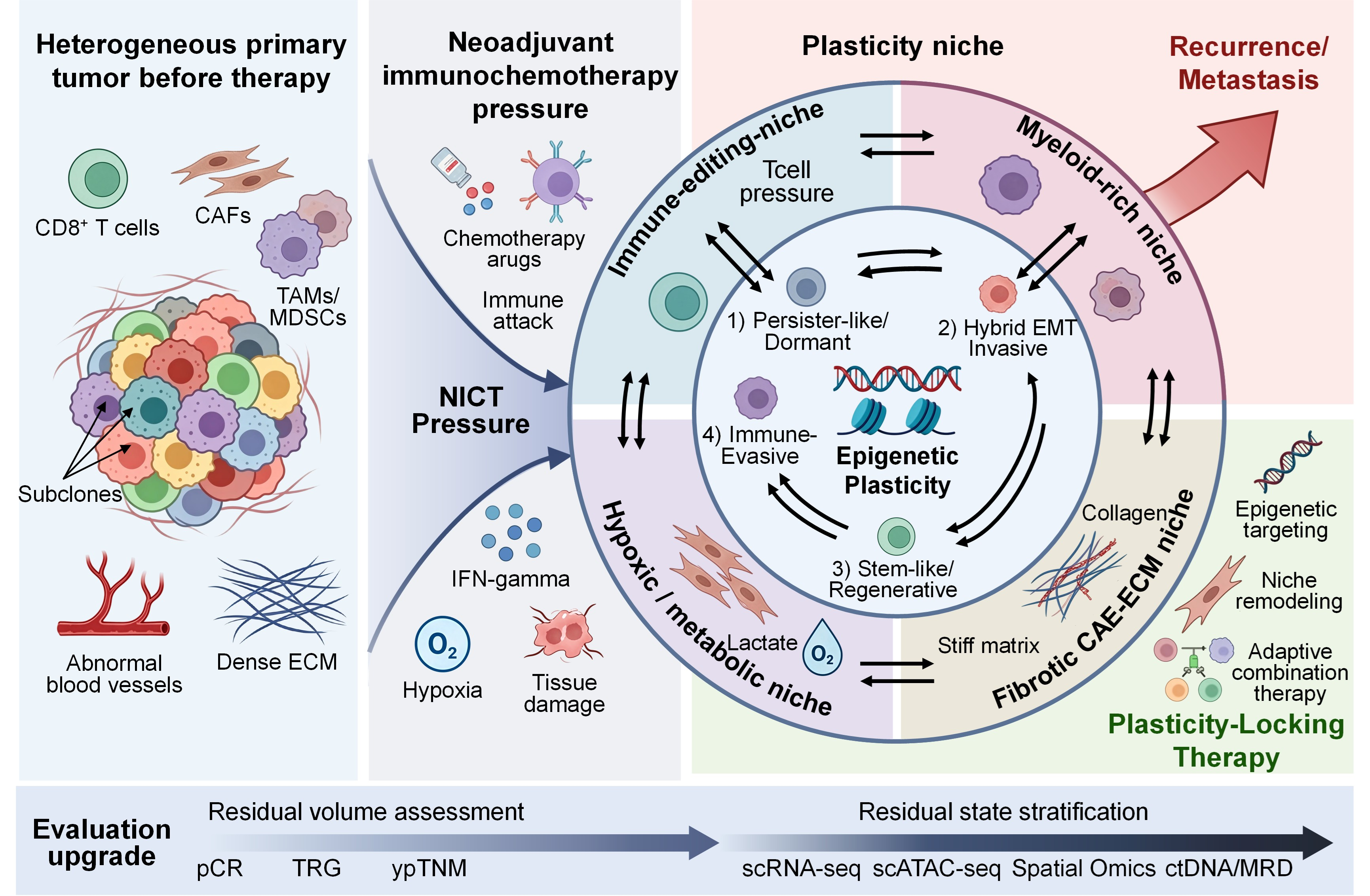
Neoadjuvant immunochemotherapy (NICT), mainly anti-PD-1/PD-L1 therapy combined with cytotoxic chemotherapy, significantly improved perioperative outcomes for resectable solid tumors such as lung cancer and breast cancer. But a large number of patients still had residual lesions and eventually relapsed. Residual tumor cells are not simply unremoved cellular debris, but represent a therapy-selected and therapy-amplified subset of a pre-existing heterogeneous and plastic tumor ecosystem. To avoid implying that therapy generates a new form of tumor plasticity de novo, we use the term “plasticity of non-apoptotic residual tumor cells” to describe the plastic behavior of viable malignant cells that survive treatment-induced cytotoxicity rather than entering apoptosis. In this review, we define the plasticity of non-apoptotic residual tumor cells as the capacity of residual malignant cells to preserve, switch, or re-enter phenotypic states such as dormancy, hybrid EMT, stem-like regeneration, and immune evasion under the combined influence of intrinsic tumor heterogeneity, systemic therapy pressure, and microenvironmental protection. Before the NICT-specific discussion, we outline general theoretical frameworks including therapeutic stress response, apoptosis-induced regeneration, genetic and non-genetic heterogeneity, as well as spatial heterogeneity of involved lymph nodes, so as to provide a more robust interpretation of residual lesion biology under NICT. Also, this review proposes that residual disease may be reconceptualized as a treatment-shaped plastic niche, whose biological behavior is jointly shaped by clonal selection, reversible phenotypic transformation, and microenvironmental ecological protection. We summarize several key states of residual tumor cells: persistent-like/resting state, hybrid EMT/invasive plasticity state, stem-like/regenerative state, and immune escape state, and elucidate the underlying epigenetic basis, including DNA methylation, histone modification, chromatin remodeling, and non-coding RNA network reprogramming. Meanwhile, niche factors such as immune stress, CAF/TAM enrichment, fibrotic matrix, hypoxia, and metabolic stress can further stabilize these states and promote the survival of relapse seeds. Based on this, we propose that future postoperative assessments should be upgraded from residual volume to a stratified residual state, and dynamically identified by combining single-cell omics, spatial pathology, and ctDNA/MRD monitoring. Furthermore, treatment strategies should shift from simply shrinking tumors to plasticity-locking therapy, that is, identifying, classifying, and blocking the plasticity escape pathways of residual lesions before they evolve into recurrence.

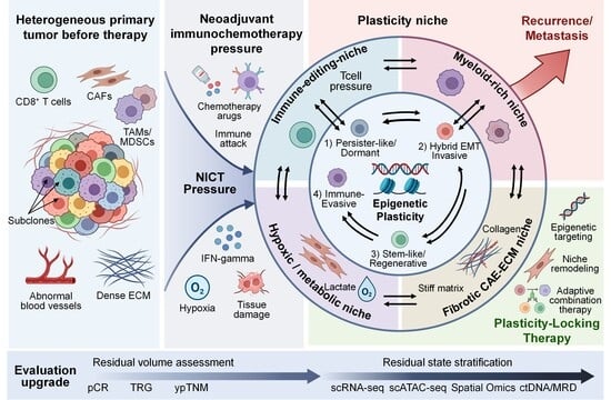
Graphical Abstract

1. Introduction

1.1. Clinical Significance of Neoadjuvant Immunochemotherapy and Residual Disease

Neoadjuvant therapy, particularly neoadjuvant immunochemotherapy (NICT), is reshaping the perioperative treatment landscape for resectable solid tumors, especially lung cancer and breast cancer [1]. However, even with significant tumor shrinkage, a considerable proportion of patients retain residual lesions postoperatively, eventually leading to tumor recurrence or metastasis [2]. This result suggests that a residual tumor is not simply unremoved cellular debris, but rather an adaptive survival population formed after treatment selection [3,4,5]. Current pathological assessment systems primarily focus on the volume and proportion of residual tumor cells, but fail to adequately capture the functional state of residual tumor cells and their dynamic interactions with the microenvironment [6].
The aims of neoadjuvant therapy are multifaceted: to downstage tumors and improve resectability, to eradicate micrometastatic disease that may already be present at diagnosis, to assess treatment sensitivity in vivo and thereby inform postoperative management, and in the immunotherapy era, to leverage the intact tumor as an in situ antigen source for priming systemic anti-tumor immunity [7,8]. NICT, which combines cytotoxic chemotherapy with immune checkpoint inhibition, has demonstrated significant improvements in pathological complete response (pCR) rates and event-free survival (EFS) across multiple tumor types without increasing perioperative risks. In resectable non-small-cell lung cancer (NSCLC), CheckMate 816 showed that neoadjuvant nivolumab plus chemotherapy increased pCR from 2.2% to 24.0% and improved EFS (HR 0.63) [9], while the NADIM II trial reported pCR rates of 36.3% versus 6.8% with chemotherapy alone [10]. The perioperative KEYNOTE-671 regimen similarly improved EFS (HR 0.58) in stage II–IIIB NSCLC [11]. Two additional perioperative phase III trials—AEGEAN (perioperative durvalumab plus chemotherapy) and CheckMate 77T (perioperative nivolumab plus chemotherapy)—further confirmed EFS benefits in resectable NSCLC [12,13]. In early triple-negative breast cancer (TNBC), KEYNOTE-522 demonstrated that adding pembrolizumab to neoadjuvant chemotherapy increased pCR from 51.2% to 64.8% and significantly improved EFS [14]. Importantly, systematic reviews and meta-analyses have confirmed that the addition of immunotherapy to neoadjuvant chemotherapy does not significantly increase surgical complications, delay surgery, or raise perioperative mortality [15].
Whether these improvements in pCR and EFS translate into overall survival (OS) benefit has been a central question. Recent evidence has now provided affirmative answers in key settings: the 5-year OS analysis of CheckMate 816 demonstrated a significant OS benefit with neoadjuvant nivolumab plus chemotherapy (HR for death 0.72; p = 0.048; 5-year OS 65.4% vs. 55.0%) [9], and the final OS analysis of KEYNOTE-522 confirmed significant OS improvement in early TNBC (60-month OS 86.6% vs. 81.7%; p = 0.002) [16]. However, mature OS data are still awaited for other tumor types where NICT is increasingly adopted, such as esophageal cancer (EC), gastric cancer (GC), and head and neck cancer (HNC). These emerging survival data further underscore the clinical importance of understanding residual disease biology; the patients who fail to achieve pCR and retain residual lesions represent the population in whom survival gains are lost, making the study of residual tumor plasticity a direct translational imperative.
The choice to focus specifically on NICT in this review is motivated by both clinical urgency and mechanistic rationale. Since 2022, NICT has been established as standard of care for resectable NSCLC, early TNBC, and increasingly for EC, GC, and HNC [10,11,14,17]. This rapid, practice-transforming adoption means that understanding the biology of residual disease after NICT is no longer a niche academic question, but a pressing translational imperative affecting tens of thousands of patients annually.
Moreover, NICT generates a dual selection landscape that distinguishes it from other neoadjuvant cytotoxic modalities [18,19]. We do not argue that apoptosis itself differs in NICT; apoptosis is apoptosis, and the intrinsic mitochondrial, extrinsic death receptor, and cytotoxic lymphocyte-mediated pathways described in Section 1.4 are shared across chemotherapy, radiotherapy, and targeted therapy [20,21,22,23,24]. Rather, what sets NICT apart is the selective pressure that determines which cells survive; chemotherapy alone selects for drug-tolerant persisters (DTPs) through pharmacological pressure (drug efflux, DNA repair, apoptosis evasion), whereas concurrent immune checkpoint inhibition imposes an additional immune-selective dimension: epigenetically mediated downregulation of MHC-I/B2M/TAP1-TAP2, IFN-γ pathway rewiring, and upregulation of alternative immune checkpoints (PD-L1, CD47, GAL9) that is absent in chemotherapy-only, radiotherapy, or targeted therapy settings [18,24]. Direct evidence for this distinction comes from the NEOSUMIT-01 randomized trial, which identified NICT-unique resistance mechanisms; an APOA1+ tumor cell-TREM2+ macrophage immunosuppressive axis and HLA-B*15:02-associated defective antigen presentation, that were absent in chemotherapy-alone resistance [25]. This immune-selective layer, superimposed on chemotherapy-driven persister selection, constitutes the specific feature that differentiates NICT from other neoadjuvant cytotoxic modalities and makes it a particularly informative paradigm for studying therapy-induced plasticity.
Despite above improvements, residual disease remains the most common outcome after NICT. Across NSCLC, pCR is achieved in only 24–36%, leaving 64–76% with residual lesions; in EC and GC, residual disease rates are 70–85%; even in TNBC, approximately 35% of KEYNOTE-522 patients retained residual tumor [9,10,14]. This is not merely a volumetric problem, and a meta-analysis of 43 trials (4100 patients) found that non-pCR after neoadjuvant immunotherapy is associated with approximately twice the risk of an EFS event (HR for pCR vs. non-pCR 0.48) and a ~45% higher risk of death (OS HR 0.55) across solid tumor types [26]. These data establish residual disease as one of the most prevalent and prognostically significant unmet needs in oncology, and provide the quantitative impetus for the mechanistic analysis that follows. Residual disease remains the major obstacle to durable cure after NICT.

1.2. Tumor Plasticity and the Biological Nature of Residual Tumor Cells

Emerging evidence suggests that the biological nature of residual tumor lesions is closely related to tumor plasticity, meaning that tumor cells can dynamically switch between stem-like, epithelial–mesenchymal transition (EMT)/hybrid EMT, and quiescence/reactivation to evade immune attack and treatment stress [27]. The plasticity of cancer stem cell-like cells, hypoxia-driven state transitions, and their coupling with treatment tolerance have been recognized as important theoretical frameworks for explaining recurrence [28]. Meanwhile, longitudinal transcriptomic studies further demonstrate that drug resistance-related programs often exist before treatment, and the treatment process is more like a screening and amplification of pre-existing tolerance states [29].
In this context, it is important to clarify what we mean by the plasticity of non-apoptotic residual tumor cells. Tumor plasticity is not a phenomenon that emerges only after NICT; rather, it is an intrinsic property of malignant tissues and is closely linked to intratumoral heterogeneity, clonal diversity, and non-genetic state transitions [30]. Systemic therapy does not necessarily create plasticity de novo, but can select pre-existing tolerant clones, accelerate reversible phenotypic switching, and stabilize adaptive states through treatment-induced microenvironmental remodeling [31].
Therefore, the term “plasticity of non-apoptotic residual tumor cells” is used here as an operational descriptor, not as a new biological category of plasticity. Rather, the framework’s conceptual contribution is to integrate genetic clonal selection, non-genetic adaptive plasticity, and microenvironmental ecological protection into a unified three-axis model; whereas established residual disease frameworks have typically addressed these axes in isolation [31,32], and to extend classical chemotherapy-era residual disease biology to include the immune-evasive state uniquely selected by NICT. It specifically refers to the plastic states of viable malignant cells that remain detectable after neoadjuvant treatment and have escaped treatment-induced apoptotic elimination [18,32,33]. These non-apoptotic residual cells may persist in persister-like/dormant, hybrid EMT/invasive, stem-like/regenerative, or immune-evasive states, thereby serving as potential relapse-competent reservoirs [34,35].

1.3. Microenvironmental and Epigenetic Regulation of Residual Cell States

At the microenvironment level, NICT can significantly reshape the tumor immune ecology. In EC, treatment response is closely related to the density of CD3+, PD-L1+ cells in the tumor area; furthermore, a more significant increase in CD8+ and CD20+ cells was found in patients who were responsive to treatment, while non-responders were more likely to exhibit characteristics of PD-L1 upregulation and immune exhaustion [3,4,36]. These changes suggest that residual lesions are not immune silence but are in a state of continuous immune selection and reprogramming.
Meanwhile, epigenetic reprogramming provides a reversible molecular basis for residual tumor plasticity. DNA methylation, histone modification, chromatin remodeling, and non-coding RNA networks can collectively regulate antigen presentation, stem programming, EMT, metabolic adaptation, and immune escape, enabling tumor cells to acquire short-term tolerance and long-term evolutionary advantages without altering their gene sequences [27,28,37,38,39]. Therefore, understanding the plasticity of residual tumors after NICT requires discussing microenvironmental selection pressures and epigenetic memory within the same framework.

1.4. General Biological Principles Underlying Therapy-Induced Residual Disease

Before specifically discussing NICT, a concise and general theoretical framework needs to be established to harmonize the terminology and mechanistic concepts. The initial response of tumors to systemic therapy is typically driven by broad therapy-induced stress responses, including DNA damage and replication stress, metabolic and redox reprogramming, as well as activation of the integrated stress response (ISR) and inflammatory signaling pathways [40,41]. These stress signals can lead to either irreversible cell death or adaptive reprogramming of surviving cells through stress-adaptive programs. Second, apoptosis should not be regarded merely as an endpoint of cellular elimination. Increasing evidence indicates that apoptotic cells can actively transmit pro-regenerative signals to neighboring viable tumor cells through “Phoenix Rising”-like paracrine mechanisms, involving pathways such as caspase-3 activation followed by iPLA2-COX-2-PGE2 signaling [20,42]. These processes may promote repopulation and regrowth of residual tumor cells after therapy. Third, regarding mechanisms of tumor cell elimination, immunochemotherapy-induced cytotoxicity is mediated through multiple parallel pathways, including perforin/granzyme release by cytotoxic lymphocytes, the extrinsic apoptotic pathway mediated by death receptors (Fas/TRAIL → caspase-8), and the intrinsic mitochondrial pathway (BAX/BAK → cytochrome-c → apoptosome → caspase-9 → caspase-3) [21,22]. The interplay between these death pathways and the TME may generate distinct paracrine effects and immunological consequences. Fourth, tumor genetic heterogeneity determines differential sensitivity of distinct clones to systemic therapies through processes of clonal selection. Importantly, metastatic dissemination is now recognized as an early event in tumor evolution, such that metastatic deposits, including involved lymph nodes and skip lesions may already be genetically heterogeneous before systemic therapy is initiated [43,44]. Systemic therapy further drives both genetic and epigenetic evolution of these pre-existing heterogeneous populations through treatment-induced selective pressure and epigenetic remodeling [31,45,46]. Consequently, achieving a durable cure in the neoadjuvant setting ultimately depends on managing this intrinsic tumor heterogeneity, not merely eradicating macroscopic residual lesions. In parallel, non-genetic mechanisms, including reversible DTP states, allow tumor cells to survive transient therapeutic pressure. These two mechanisms can coexist and collectively shape the biological characteristics of residual disease [40,41]. Finally, involved lymph nodes and so-called “skip” lesions may exhibit substantial spatial heterogeneity in both genomic composition and immune microenvironment. Therefore, clinical resection strategies, pathological evaluation, and postoperative treatment decisions should account for this spatial complexity [47,48].
Apoptosis should therefore be considered not merely as the opposite state of the viable residual cells discussed, but as both an upstream therapeutic event and a niche-shaping biological process [20,23,49]. In the chemotherapy component of NICT, DNA damage, replication stress, mitotic stress, and oxidative stress can converge on the intrinsic mitochondrial apoptotic pathway, involving BAX/BAK-mediated mitochondrial outer membrane permeabilization, cytochrome-c release, apoptosome formation, caspase-9 activation, and downstream caspase-3/7 execution [20,23]. In the immune component, PD-1/PD-L1 blockade does not directly induce tumor-cell apoptosis in the same manner as cytotoxic drugs; rather, it restores cytotoxic CD8+ T-cell and NK-cell activity, which can eliminate tumor cells through perforin/granzyme B-mediated apoptosis and through death receptor pathways such as Fas/FasL and TRAIL-DR4/DR5, followed by FADD recruitment, caspase-8 activation, and convergence on caspase-3/7 or mitochondrial amplification [24,50]. Thus, chemoimmunotherapy may engage intrinsic mitochondrial, extrinsic death receptor, and cytotoxic lymphocyte granule-associated apoptotic programs in treatment-sensitive tumor fractions [20,23,24]. Importantly, apoptotic tumor cells may also paradoxically support tumor repopulation by releasing paracrine regenerative signals [20,49]. Executioner caspases, especially caspase-3/7, can activate iPLA2 and downstream COX-2/PGE2 production, thereby promoting Wnt/β-catenin signaling, tissue repair-like responses, and compensatory proliferation in neighboring viable cells [20,49]. Therefore, in this review, non-apoptotic residual tumor cells are defined as the viable cellular compartment that retains plastic potential after therapy, whereas apoptotic tumor cells are considered an upstream source of cytotoxic selection, paracrine regrowth signals, and residual niche remodeling.

1.5. Scope and Objectives of the Review

Within this general framework, this review subsequently focuses on how the unique dual selective pressures imposed by NICT, combining chemotherapy and immune-mediated killing amplify and consolidate these fundamental mechanisms, ultimately shaping the biology of residual tumor disease.

2. Conceptual Model: Selection, Adaptation and Ecological Protection

The conceptual framework developed in this section is anchored on NICT, which serves as a particularly instructive model because it imposes simultaneous pharmacological and immune selection pressures, thereby enriching the full spectrum of plastic states, including immune-evasive phenotypes that are not selected for by chemotherapy alone. However, the core principles of clonal selection, adaptive plasticity, and ecological protection are broadly applicable to other neoadjuvant modalities, and we note where evidence from non-NICT contexts informs the model.
Tumor plasticity is a fundamental property of malignant tissues and a major source of intratumoral heterogeneity [51,52]. It allows cancer cells to shift among proliferative, dormant, invasive, stem-like, and immune-evasive states without requiring immediate acquisition of new driver mutations. In the setting of systemic therapy, this pre-existing plastic potential becomes clinically visible because treatment eliminates sensitive populations while selecting, accelerating, or stabilizing tolerant cellular states [18].
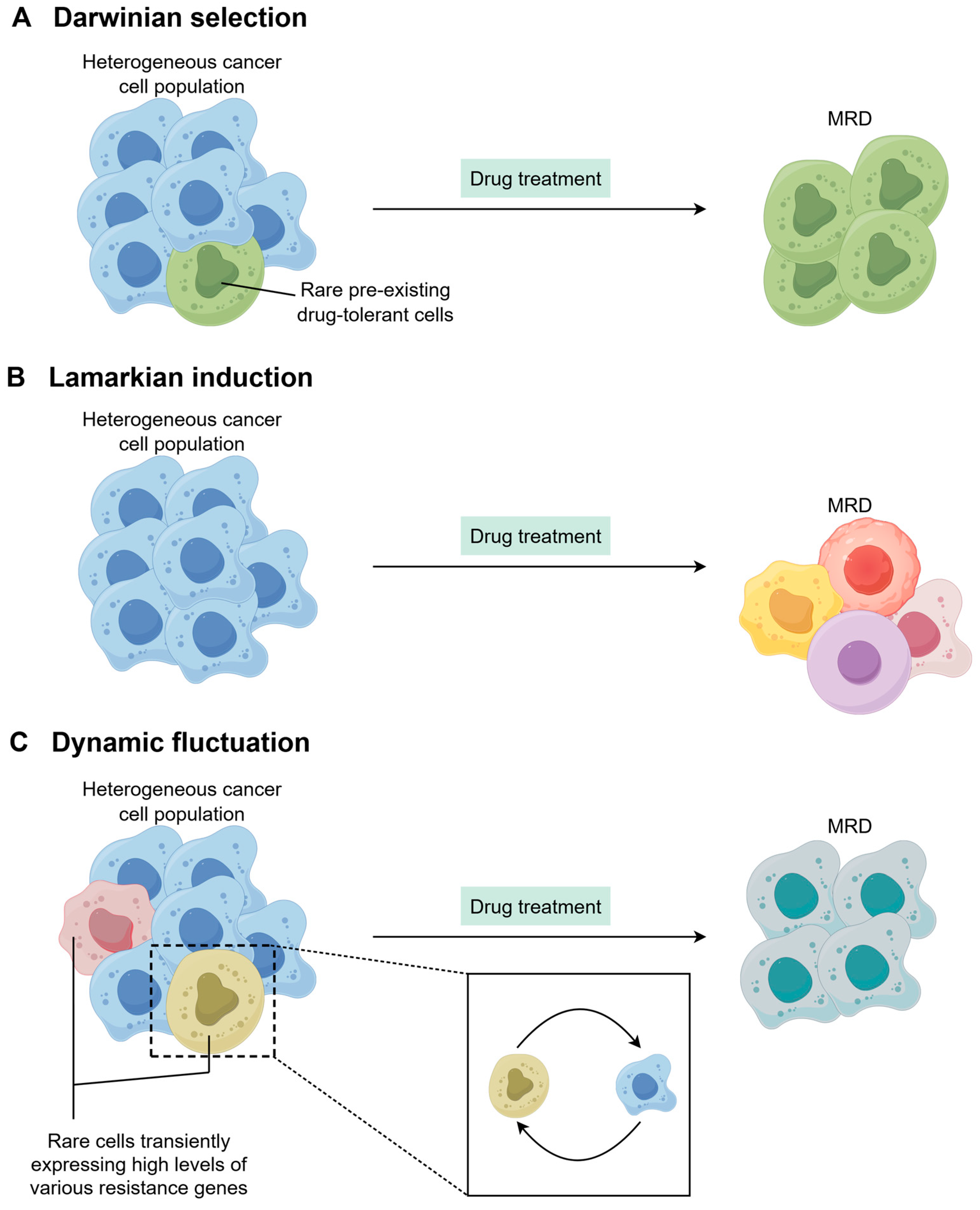
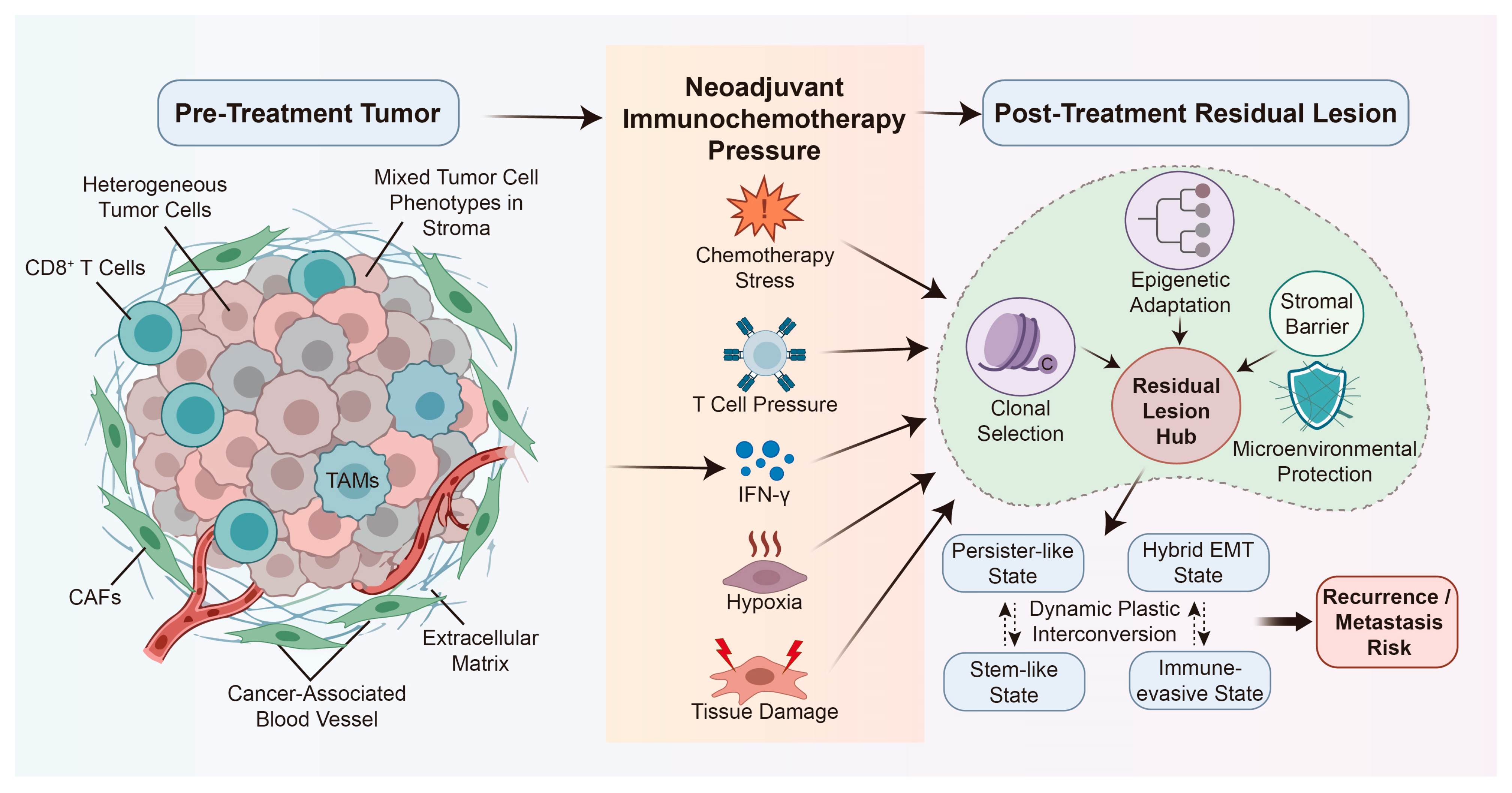
Accordingly, the plasticity discussed in this review should be understood as the plasticity of non-apoptotic residual tumor cells: viable malignant cells that have survived NICT rather than undergoing apoptosis. These cells are not assumed to have acquired a new plastic property after therapy; instead, therapy exposes, enriches, and stabilizes pre-existing or inducible plastic states within the residual disease compartment [53,54,55]. Furthermore, treatment resistance is often not explained by a single mechanism, but rather by the combined effects of genetic selection, non-genetic adaptation, and ecological protection [45]. The distinct scenarios through which therapy selects and stabilizes residual disease are conceptualized in Figure 1 [18].

2.1. Clonal Selection

Clonal selection represents the classic Darwinian Model of residual lesions, where tolerant or weakly tolerant clones that existed before treatment are selected and amplified under drug pressure. Treatment stress acts like environmental selection, preferentially eliminating treatment-sensitive tumor cell clones while allowing pre-existing drug-resistant clones to survive and eventually proliferate, becoming the basis for tumor recurrence [45]. Recent study further shows that under drug stress, different subclones can exhibit highly repetitive and predictable selection trajectories, suggesting that drug resistance does not occur randomly but depends on initial clonal structure and innate fitness differences [56].
Within this framework, some cells in the residual lesions do not need to acquire new driver mutations; instead, they survive directly due to their existing genetic makeup and high fitness. For NICT, this means that the “survivors” seen in postoperative residual tissue may not be newly developed resistance products during treatment, but rather clones that existed before treatment and were only revealed after treatment selection [15,57]. This explanation is significant for understanding incomplete pathological remission, postoperative relapse, and failed retreatment.

2.2. Adaptive Plasticity

In contrast, adaptive plasticity emphasizes that another type of residual cells is not stable drug resistance, but rather enters a reversible adaptive state under treatment stress [58]. These states include slow cycle, dormancy, stress tolerance, enhanced stemness, hybrid EMT, and low immune visibility, etc. [51,52] Their common feature is that cells temporarily reduce proliferation, enhance survival, and can return to a more primitive transcriptional state after the stress is relieved. Emerging evidence suggests that this adaptation is not entirely dependent on novel mutations, but is more likely driven by epigenetic reprogramming and switching of transcription factor networks [56].
Treatment stress, microenvironmental signals, and intrinsic epigenetic fluctuations can collectively induce tumor cells to switch between multiple states [52]; drug resistance can also be explained by a “one-to-many (epi)genotype-to-phenotype” mapping, meaning that the same genetic background can output multiple transcriptional states in different environments, a capability supported by stable epigenetic memory [56]. Therefore, the resistance observed in residual lesions does not necessarily signify real genetic immobilization, but may simply be a reversible, plastic adaptation that can rebound after treatment. This is why residual tumors often recur after initial shrinkage: they are not simply survived, but rather learned to survive under stress.

2.3. Ecological Protection

However, even with genetic selection advantages or epigenetic plasticity, residual tumor cells still require an environment that allows them to survive, this is called ecological protection. The survival of residual tumor cells relies not only on intrinsic cellular properties but also heavily on protective niches within the tumor microenvironment (TME) [59,60]. Cancer-associated fibroblasts (CAFs), tumor-associated macrophages (TAMs), myeloid-derived suppressor cells (MDSCs), regulatory T cells (Tregs), aberrant vasculature, hypoxia, extracellular matrix (ECM) barriers and metabolic stress jointly construct such protective niches for residual tumor cells [61].
CAFs can promote tumor cell migration, stemness, and treatment resistance through ECM remodeling, paracrine factors, and mechanotransmission; while immunosuppressive cell populations reduce immune clearance efficiency by weakening effector T cell activity, allowing residual cells to hide or remain in a low-responsive state [62,63]. Hypoxia and abnormal blood supply not only limit drug penetration but also drive EMT, metabolic reprogramming, and stress survival through hypoxia-inducible factor (HIF)-related processes. For residual lesions after NICT, the TME is not a passive background but an active “third-party force” involved in selection and protection: it both selects cells that can adapt to hypoxia, low nutrition, and immune stress, and helps these cells maintain reversible tolerance and recurrence potential [62,63,64].
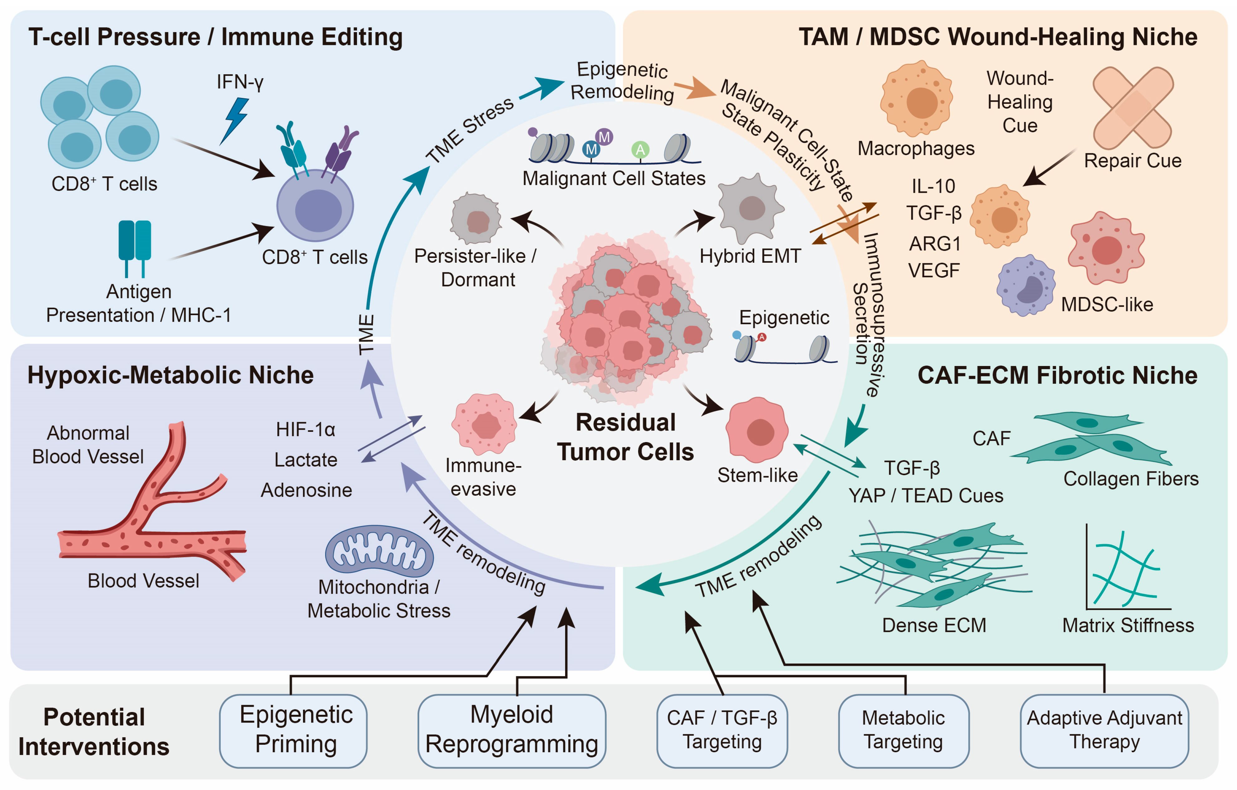
Therefore, the formation of residual lesions is not the result of a single clone or a single pathway, but rather the product of long-term co-evolution between the tumor cells themselves, their epigenetic state, and the surrounding niche (Figure 2).

3. Epigenetic Programs Underlying Residual Malignant Cell States

The four residual states described below:—persister-like/dormant, hybrid EMT/invasive plastic, stem-like/regenerative, and immune-evasive—are intended as analytical categories, not as rigid, mutually exclusive cell types. In reality, these states represent probabilistic attractors on a deformable epigenetic landscape: cells can occupy intermediate positions, co-express features of multiple states, and transition between states under changing selection pressures [65]. A pan-cancer single-cell analysis across 15 tumor types identified six gene modules whose expression defines recurrent cancer cell states, including stress response, EMT, and interferon response that constitute general features of cancer rather than properties of individual tumor types [66]. This convergence supports the cross-tumor-type applicability of the states described here. However, the precise molecular implementation of each state is constrained by the developmental origin of the tumor: a developmental constraint model proposes that cancer cells can only access states that are developmentally reachable from their cell of origin, which explains why, for example, EMT programs differ between epithelial and mesenchymal cancers, and why stem-like programs in lung adenocarcinoma recapitulate lung developmental hierarchies rather than intestinal or neural ones [67]. Furthermore, different genetic backgrounds can converge on the same catalog of states but alter their relative frequencies: a plastigenic process in which genetic alterations modulate state abundance rather than state identity [67]. Therefore, the four-state taxonomy should be interpreted as a working framework for organizing mechanistic insights, while recognizing that the boundaries between states are porous, the specific markers and regulators are tissue-context-dependent, and the dynamic interconversion between states is itself a biologically and clinically relevant property.

3.1. Persister-like and Dormant States

Post-treatment residual tumor cells often enter a DTP-like state or dormant state: these cells typically exhibit slow cell cycle/low proliferation, enhanced anti-apoptosis and stress tolerance, and transient, broad-spectrum, and reversible tolerance to multiple treatments [68,69]. When treatment is stopped or the microenvironmental stress changes, these cells can re-enter the proliferation program and drive relapse [70]. Therefore, this state can well explain the long-term existence and late relapse of minimal residual disease (MRD). Clinically, even after neoadjuvant therapy achieves complete pathological remission, residual tumor cells can still be detected in the resected specimen, suggesting that residual does not mean quiet and harmless, but rather resembles an adaptive reservoir temporarily suppressed by treatment [71,72].
At the epigenetic level, the stable maintenance of the pesrister/dormant state depends on reversible chromatin remodeling, rather than de novo mutations [72]. It is generally believed that these cells are often accompanied by enhanced repressive chromatin programs, including increased H3K27me3/H3K9me3, Polycomb-related silencing, chromatin compression, and widespread transcriptional silencing; moreover, histone deacetylase (HDAC)-mediated deacetylation is also considered one of the upstream mechanisms for maintaining low transcriptional activity [73,74,75].
Meanwhile, metabolic adaptation is essential for persister survival. DTP cells are not truly metabolic shut down, but maintain a minimum level of survival and subsequent resuscitation capacity through redox balance, lipid metabolism/oxidative phosphorylation (OXPHOS) reprogramming, and switching of other energy pathways [76,77]. Recent studies on residual disease in lung cancer further demonstrate that residual lesions can be maintained by specific signaling axes and synergize with persisters, indicating that “resting” itself is a highly organized adaptive program, rather than a simple proliferative halt [78]. Therefore, the persister-like/dormant state can be summarized as a reversible survival program shaped by epigenetic locking, transcriptional silencing, and metabolic resetting, and is one of the core relapse seeds of residual lesions.

3.2. Hybrid EMT and Invasive Plasticity

Hybrid EMT describes a cellular state in which cancer cells simultaneously co-express both epithelial and mesenchymal features, occupying a stable, functional equilibrium position along the epithelial–mesenchymal plasticity (EMP) landscape [79]. While “partial EMT” is sometimes used interchangeably, the two terms carry subtly different emphases: partial EMT underscores the incompleteness of the transition from an epithelial origin, whereas hybrid EMT emphasizes the functional co-existence of proliferative (epithelial) and invasive (mesenchymal) programs, a dual capacity that is uniquely advantageous for residual tumor cells (Table 1) [80]. Biologically, the hybrid state is not a default consequence of an interrupted transition but is actively maintained by core regulatory networks: the ZEB1/ZEB2–miR-200 and SNAIL1/SNAIL2–miR-34 double-negative feedback loops, coupled to epigenetic stabilizers such as H3K27ac remodeling at enhancers of EMT transcription factors [80,81]. EMP enables cells to traverse the epithelial–mesenchymal landscape bidirectionally, and the hybrid state represents the most fitness-optimized configuration for cells that must simultaneously preserve proliferative potential and acquire invasive, stem-like, and drug-tolerant properties [82,83,84,85]. The concept of EMT as a spectrum of intermediate states (rather than a binary switch) was first systematically articulated by Nieto et al., experimentally validated in vivo by Pastushenko et al. who identified multiple EMT subpopulations with distinct functional properties in skin cancer and mammary tumor models, and subsequently refined through consensus nomenclature [81,86]. Recent comprehensive reviews explicitly connect hybrid EMT to MRD, drug tolerance, and tumor dormancy, linking the concept directly to the themes of residual tumor plasticity [87,88,89,90].
Unlike the classic binary narrative of decreased E-cadherin and increased N-cadherin, residual tumors more often remain in a hybrid/partial EMT lineage: cells are not fully mesenchymalized, but retain some epithelial adhesion and proliferative capacity, and acquire migration, invasion, anti-apoptosis, and treatment resistance [97,98,99]. This mixed state is commonly found at the invasive front, tumor buds, and circulating tumor cell (CTC) clusters, and is associated with stronger tumor initiation, dissemination, and poor prognosis in various cancer types; in other words, the most dangerous aspect of residual cells is not complete EMT, but this reversible and switchable transitional state.
Mechanistically, hybrid EMT resembles enhancer-switching-driven transcriptional reprogramming rather than a simple flipping of a single marker [97]. Enhancers associated with invasion, stemness and stress adaptation acquire H3K27ac and establish a more open chromatin landscape [97]. AP-1, TEAD and ZEB1/SNAIL/TWIST collectively serve as core hubs for state transition [100]. These transcription factors do not function uniformly across all cancer types. Instead, they determine whether cells remain in an epithelial migratory state or progress toward a highly invasive mesenchymal program, contingent on specific tissue context and therapeutic stress. Notably, the YAP/TAZ-TEAD axis merits particular emphasis, which is tightly coupled with EMT, cancer stemness and invasiveness, and its dependency signature can predict TEAD dependence across platforms in multiple cancer types, suggesting this axis acts as a critical hub driving residual cells into an invasive plastic state [101,102].
Functionally, hybrid EMT often occurs concurrently with immune rejection and dissemination dominance. Studies in pancreatic cancer show that EMT-related transcription factors ZEB1 and SNAIL act as dual genetic and epigenetic regulators, driving Irf6 silencing and thus weakening tumor cell sensitivity to T cell killing and TNF-α-induced apoptosis [103]. In a neuroendocrine transdifferentiation model of prostate cancer, the CXCR4-LASP1-G9a-SNAIL axis further demonstrates that EMT is not a static marker change, but a state reconnection process amplified by both the microenvironment and epigenetics [104]. Also, background alterations such as p53 and LKB1 can also release the brakes on plasticity, making EMT/partial EMT more likely to coexist with chromatin instability, transdifferentiation, and invasiveness [105,106]. Therefore, in residual lesions after NICT, hybrid EMT/invasive plasticity is more suitable as a second-layer survival strategy: it enhances invasion and immune escape while preserving proliferative potential, pushing a few surviving cells into a transitional state most conducive to recurrence.

3.3. Stem-like and Regenerative States

Post-treatment tissue damage and inflammation repair can push some residual tumor cells into a stem-like/regenerative-like state. This state does not mean the existence of a fixed, static subset of cancer stem cells within the tumor; more accurately, it is a dynamic cellular state that can be induced by treatment stress and microenvironmental signals [107,108]. In this state, cells maintain a certain level of survival and proliferation capacity while acquiring stronger self-renewal, clonal reconstitution, and reversible differentiation, thus making them more likely to re-expand and participate in recurrence after treatment withdrawal. In other words, the stemness in residual lesions is more like a functional program under stress than a pre-set lineage endpoint. Mechanistically, this process is often reactivated by developmental signals such as Wnt, Notch, and Hedgehog, and works synergistically with core transcription factors such as SOX2, KLF4, and MYC [109]. Together, they propel residual cells back to a low-differentiation, highly plastic, and regenerative transcriptional state. Among these, YAP/TAZ, as the hub of mechanical and regenerative signals, is often located upstream in this network and functionally coupled with SOX2, supporting spheroidization, metabolic adaptation, and treatment tolerance [108,110]. In different tumor contexts, YAP/TAZ can also continuously amplify the regenerative-like transcriptional program through crosstalk with Notch, Wnt/β-catenin, and Hedgehog, enabling residual cells to retain certain epithelial characteristics while acquiring stronger regenerative and invasive potential [111].
At the epigenetic level, stem-like states are often accompanied by super-enhancer remodeling. Therapeutic stress can rewrite the enhancer accessibility of key transcription factors, reactivating a few core gene clusters and forming stable state memories. Recent studies have shown that SOX2 can inhibit or rearrange MYC transcription by altering the coactivator composition of the c-MYC super-enhancer; YAP/TAZ are also frequently incorporated into this enhancer-driven network, participating in maintaining stemness and regeneration programs [112,113]. Therefore, in residual lesions after NICT, the stem-like/regenerative-like state can be summarized as: therapeutic damage → regeneration signal activation → YAP/TAZ-Notch/Wnt/Hedgehog axis initiation → super-enhancer remodeling → stemness program reconstruction. This state is beneficial for short-term survival and provides the most plastic cellular reserve for subsequent relapse [107,112,114].

3.4. Immune-Evasive States

Another key survival strategy for residual tumor cells is to enter an immune-evasive/immune-low-visible state. This state is most directly related to immunotherapy, and its core characteristic is not simply escaping killing, but rather reducing the probability of being recognized: antigen processing and presentation capabilities decrease, MHC-I, B2M, and TAP1/TAP2-related pathways weaken, IFN-γ response is reprogrammed, and immunosuppressive molecules such as PD-L1 and CD47 are upregulated, ultimately creating a microenvironment unfriendly to CD8+ T cells and phagocytes [115,116,117]. In clinical samples, residual GC after anti-PD-1 therapy shows downregulation of HLA class I/MHC class II in tumor cells, suggesting that immune escape is not necessarily complete disappearance, but may be adaptive survival after reduced antigen visibility [118].
It is important to emphasize that this type of immune escape is not always driven by irreversible gene mutations. While abnormalities in pathways such as B2M, JAK1, and NLRC5 can indeed lead to stable antigen presentation defects or IFN-γ insensitivity, increasing evidence suggests that the low visibility of immunity in residual lesions is often maintained by chromatin state, transcription factor networks, and metabolic programs, and therefore has a degree of reversibility [119,120,121]. For example, epigenetic drugs can restore the expression of antigen presentation machinery such as MHC-I, B2M, PSMB8/9, and TAP1/TAP2, indicating that “turning off the presentation program” itself can be turned back on; however, this may also be accompanied by PD-L1 upregulation, suggesting that residual cells will rebalance between restoring antigenicity and maintaining inhibitory activity [122]. Similarly, IFN-γ may enhance anti-tumor immunity or induce tumor cells to remodel PD-L1, YTHDF2, or EMT-related programs, thereby converting inflammatory signals into an escape advantage [121,123].
Furthermore, immune escape is often closely coupled with EMT/hybrid EMT. In PD-L1-high NSCLC, the higher the EMT signature, the more likely there is a decrease in T cell infiltration, an increase in Treg and M2 macrophages, and a poorer response of immune checkpoint inhibitor (ICI); while EMT transcription factors such as TWIST1 and ZEB1 can directly promote PD-L1 expression, further amplifying the immune rejection effect [123,124]. This indicates that residual cells do not simply hide, but rather rewrite their immune interfaces simultaneously at the transcriptional and epigenetic levels, making them both more difficult to identify and more likely to push local TME into a cold tumor state [125,126].
Therefore, the immune-evasive state can be summarized as: weakened antigen presentation + IFN-γ pathway reprogramming + upregulation of immunosuppressive molecules + chemokine/EMT-driven T cell rejection. This state directly explains why some lesions, despite appearing to shrink after treatment, can still survive long-term under immune pressure and subsequently become a source of relapse (Table 2) [115,116,125]. Table 2 serves as a mechanistic reference summarizing the biological features of each residual cell state as characterized in post-treatment surgical specimens; it is not intended as a pre-treatment diagnostic panel. The translational counterpart: mapping each state to actionable therapeutic strategies will provide in Section 5.3.

4. Microenvironmental Niches Reinforcing Residual Plasticity

4.1. Immune Pressure and Immune Editing

Following neoadjuvant immunotherapy, residual tumor cells are not a passive group that luckily survives, but rather an adaptive subset that is reselected under continuous immune pressure. Cells easily recognized and killed by T cells are eliminated first, while those remaining are often cells with weaker antigen presentation, impaired IFN-γ responses, or those more prone to forming immune rejection phenotypes [115,127,128]. Therefore, the role of immunotherapy is not merely killing tumors, but also directly shaping the state of residual cells through immune editing, causing them to evolve towards lower visibility, lower immunogenicity, and greater plasticity.
The key to this process is not necessarily new irreversible gene mutations, but often the selection and reprogramming of existing cellular states. Classic alterations include weakening of the MHC-I/B2M/TAP1-TAP2-related antigen processing and presentation pathways, inactivation of the IFN-γ-dependent JAK/STAT axis, or epigenetic silencing of antigen presentation machinery, thereby reducing the ability of tumor cells to recognize CD8+ T cells [129,130]. Recent studies have further suggested that antigen visibility in residual tumors is not a static property, but rather changes dynamically under treatment stress; some cells can simultaneously carry B2M deficiency and IFN signaling defects, forming a more thorough T cell escape phenotype [127,131].
Meanwhile, immune pressure itself also reshapes the transcriptional state of tumor cells. Sustained IFN-γ exposure can enhance antigen presentation and inflammatory responses, and may also induce PD-L1 and LAG-3-related adaptive feedback, even pushing cells into a less differentiated and more tolerant state [132,133]. This means that while the immune system is clearing tumors, it is also “training” surviving cells to learn how to survive. In other words, immune pressure is not a one-way elimination mechanism, but a dual process of selection and state shaping: it determines both who is eliminated and in what state those remain [134,135].
Therefore, immune escape is not always a terminal event, but rather a plastic state formed under treatment stress. The reason why residual cells can persist is often not because they have completely lost their immune-related functions, but because they have transformed the immune system’s attack into a signal for self-state reprogramming, thereby gaining stronger long-term survival ability and subsequent relapse potential.

4.2. Myeloid-Rich Wound-Healing Niches

While chemotherapy and immunotherapy kill tumor cells, they also cause tissue damage, release of necrotic debris, and local inflammation, triggering a wound-healing-like myeloid response [136,137]. During this process, TAMs and MDSCs are often recruited and enriched, accompanied by the upregulation of repair/inhibitory molecules such as IL-10, TGF-β, ARG1, SPP1, and VEGF [137,138]. These signals promote angiogenesis, matrix remodeling, and fibrosis repair, while simultaneously suppressing effector T cell activity, thus transforming the process originally intended to clear damage into a barrier protecting residual tumor cells.
In fibrosis-associated hepatocellular carcinoma (HCC), studies have shown that TGF-β can drive monocyte differentiation into MDSCs and enhance immunosuppression and reduce T cell proliferation through the PPP1R15A-ARG1/S100A8/9 axis; inhibiting this axis can reduce M-MDSCs and enhance the efficacy of ICI therapy [138]. This suggests that the repair response activated after treatment is not necessarily a passive process of host self-recovery, but may be actively utilized by residual tumor cells, gradually transforming into an immune-protective niche. Therefore, this type of myeloid-rich wound-healing niche is essentially a repair-suppression complex state dominated by myeloid cells, providing a niche basis for the continued survival of residual tumors and subsequent recurrence [136].

4.3. CAF-ECM-Fibrotic Niches

CAF and ECM constitute the most typical physical-signal dual barrier in residual lesions. After treatment, ECM-producing myCAF/ECM-myCAF are often retained or remodeled in the residual area, promoting tumor cell survival and restricting CD8+ T cell entry through collagen deposition, matrix stiffening (ECM stiffening), and YAP1/TEAD activation [139,140]. In ovarian cancer, residual ANTXR1+ myofibroblasts after chemotherapy can suppress anti-tumor immunity through a YAP1-dependent mechanism; in breast cancer, ECM-myCAF/wound-myCAF/TGFβ-myCAF spatially co-localize with high TGF-β signaling, while ECM regulators can alter local TGF-β activity and affect CD8+ T cell infiltration [139,141]. These fibrotic niches are also often accompanied by enhanced chemokine programs (such as CXCL12) and LRRC15-related matrix programs, thereby further strengthening immune rejection, promoting hybrid EMT, and improving treatment tolerance [142,143]. Therefore, the CAF-ECM-fibrotic niche is not simply a scar-like background, but an immune isolation zone established by residual tumors using tissue repair processes, which can sustainably support survival and proliferation before recurrence [144].

4.4. Hypoxic and Metabolic Niches

Residual tumor regions are typically accompanied by abnormal blood vessels, persistent hypoxia, lactate accumulation, adenosine production, and nutrient competition, creating a local microenvironment characterized by hypoxic and metabolic stress. In these niches, HIF signaling, the AMPK-mTOR axis, and mitochondrial metabolic reprogramming work together to shift cells from a “proliferation-first” to a “survival-first” approach, further enhancing the plasticity and tolerance of residual cells [145,146]. More importantly, these metabolic changes are not merely energy adaptations but directly alter the supply of epigenetic substrates: NAD+ influences redox and deacetylation processes, acetyl-CoA determines histone acetylation levels, and α-KG and SAM connect demethylation and methylation reactions, respectively, thereby translating metabolic state into chromatin state [147,148].
Under hypoxic conditions, tumor cells often induce glycolysis, inhibit mitochondrial oxidation, and increase lactate production through HIF-1α [149,150]. Lactate is not only a metabolic waste product but also a signaling molecule and epigenetic regulator, promoting cell cycle maintenance, invasion programs, and treatment tolerance. Recent studies suggest that lactate accumulation can also participate in tumor state remodeling through histone lactylation, enabling cells to maintain higher transcriptional heterogeneity and reversibility in hypoxic and low-nutrient environments [150,151]. Meanwhile, hypoxia also affects NAD metabolism and residual mitochondrial function; the Complex I activity retained under hypoxia can maintain NAD+ regeneration and support α-KG-related metabolism, thereby influencing cell fate and adaptability [147,152].
Therefore, the key significance of the hypoxic and metabolic niche lies not in hypoxia itself, but in its transformation of microenvironmental stress into an inheritable epigenetic program. Residual tumor cells, through this program, can maintain a stem-like, persister-like, or invasive state under hypoxic, hyponutrient, and high lactate conditions, and proliferate again after the therapeutic stress is relieved [146,153]. This is precisely the logic this review aims to emphasize: metabolic stress converts microenvironmental adaptation into epigenetic plasticity (Figure 3).

4.5. Apoptosis-Induced Repopulation Signaling: Therapy as a Niche Sculptor

An underappreciated dimension of residual niche biology is that cytotoxic therapy itself directly generates pro-regenerative signals that promote the survival and expansion of residual tumor cells. This phenomenon, called “Phoenix Rising”, was first described by Huang et al., who demonstrated that caspase-3 activation in dying tumor cells triggers the iPLA2–COX-2–PGE2 axis, leading to STAT3-dependent proliferation of surviving tumor cells [49,154]. Importantly, higher levels of activated caspase-3 in tumor tissues correlated with significantly increased rates of recurrence and death in patients, providing direct clinical evidence for the relevance of this pathway. Kurtova et al. subsequently showed that Prostaglandin (PG) E2 released from chemotherapy-killed bladder cancer cells stimulates cancer stem cell proliferation and drives tumor repopulation between treatment cycles; pharmacological blockade of this axis with the cyclooxygenase-2 (COX-2). COX-2 inhibitor celecoxib abrogated chemoresistance [155].
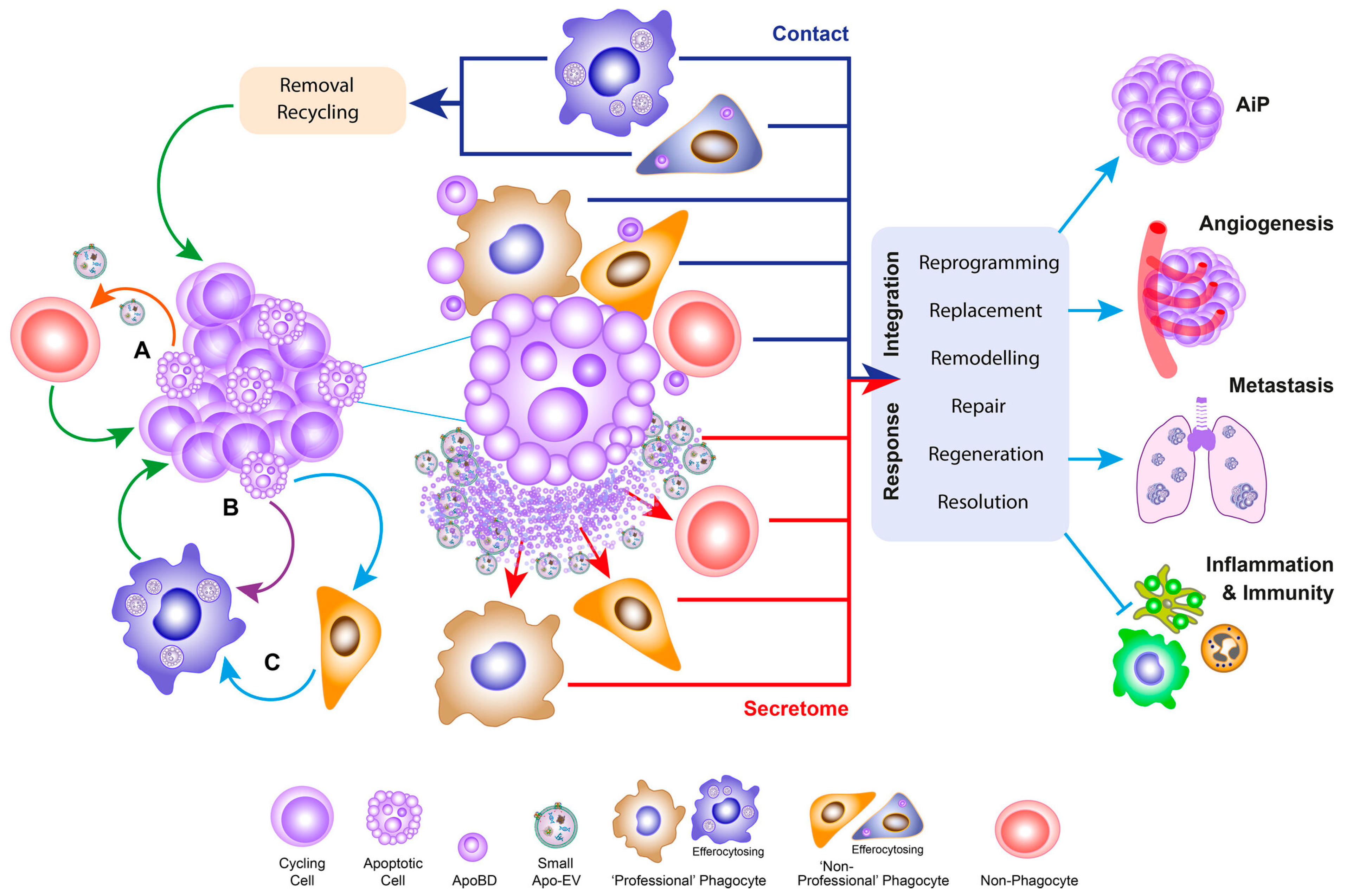
The apoptotic secretome extends well beyond PGE2, encompassing soluble mediators, damage-associated molecular patterns (DAMPs), cytokines/chemokines, lipids, and apoptotic extracellular vesicles/apoptotic bodies [156]. Dying cells can emit ATP and HMGB1, expose calreticulin on the cell surface, and present or release HSP-associated danger signals, which are sensed by receptors such as purinergic receptors, Toll-like receptors (TLRs), RAGE and CD91 to activate NF-κB-dependent inflammatory or reparative signaling in neighboring immune, stromal, and surviving tumor cells [157,158,159]. In parallel, apoptotic or therapy-damaged cells can promote compensatory proliferation and tumor repopulation through Wnt/Wingless-like mitogens, Hedgehog/Wnt pathway remodeling, ATP-driven macrophage amphiregulin production, and cytokine/chemokine-rich apoptotic bodies, thereby allowing tumors to hijack wound-healing-like repair programs after cytotoxic therapy [159,160,161]. The broader conceptual framework of apoptosis-driven remodeling of the TME is illustrated in Figure 4 of apoptosis-centered oncogenic signaling, in which apoptotic tumor cells communicate through soluble secretome components, apoptotic extracellular vesicles, and contact-dependent pathways to promote apoptosis-induced proliferation, angiogenesis, invasion, metastasis, reparative macrophage programs, and immune suppression [162].
In the NICT setting, this apoptosis-driven regeneration mechanism may be particularly relevant because both cytotoxic chemotherapy and immune-mediated tumor killing can increase the burden of dying tumor cells. Chemotherapy can induce apoptosis through DNA damage and DNA damage response pathways, whereas immune checkpoint blockade can enhance CD8+ T-cell cytotoxicity and tumor apoptosis, including granzyme/perforin-dependent killing and death receptor-associated apoptosis [163]. The resulting apoptotic debris and secretome may provide trophic and inflammatory cues to residual tumor cells. Preclinical studies show that dying tumor cells can stimulate tumor repopulation through caspase-3-dependent PGE2 production and through exosome-mediated support of tumor-repopulating cells [164]. Chemotherapy-induced COX-2/PGE2 signaling has also been shown to enhance cancer stemness, invasiveness, drug resistance, and repopulation in residual tumor cells [165]. Therefore, although direct clinical proof in NICT-treated residual lesions remains limited, the apoptotic secretome may contribute to the paradoxical emergence or selection of proliferative and stem-like residual tumor states after initial tumor shrinkage. This interpretation is consistent with evidence that cancer stem-like cells are enriched in post-treatment residual disease and with neoadjuvant immunotherapy datasets linking non-complete pathological response to higher residual tumor burden and stemness-associated features [166,167]. Targeting this apoptosis-driven regeneration loop: through COX-2/PGE2 inhibition, EP2/EP4 or EP4 antagonism, or selective modulation of DAMP pathways such as HMGB1 or ATP/P2 × 7R may represent a rational adjuvant strategy to limit post-NICT tumor repopulation [164,168,169]. However, because DAMPs can also support ICD and antitumor immunity, such approaches should be context- and timing-dependent rather than broad DAMP suppression.

5. Translational Implications: Assessing and Targeting the Residual State

5.1. From Residual Volume to Residual State

Traditional indicators such as pCR, MPR, TRG/TRS, ypTNM, and R0 resection have become the most commonly used efficacy endpoints after neoadjuvant therapy [170,171,172]. However, they essentially still primarily answer the question of “how much is left”, i.e., the residual tumor burden, rather than “what state the remaining tumor is in”. In resectable NSCLC, the need for standardized sampling and stratified interpretation in postoperative pathological evaluation has been emphasized, because residual lesions exhibit significant spatial heterogeneity [173]. In other words, simply summarizing the entire residual lesion based on the proportion of viable tumor is often insufficient to reflect its true biological consequences.
Therefore, assessments of neoadjuvant therapy should evolve from residual volume to residual-state profiling: in addition to residual quantity, it should also determine whether residual cells are persister-like, whether they retain hybrid EMT/stem-like programs, whether antigen presentation and IFN responses have been reprogrammed, and whether they are embedded in TAM-rich, CAF-rich, hypoxic, or immune-excluded niches [70,144,174]. What truly determines relapse potential is often not simply residual volume, but rather the type of plasticity program and microenvironment niche that sustains the residual cells.
From a translational perspective, circulating tumor DNA (ctDNA), molecular subtyping, and spatial pathology should be complementary rather than substitutive. ctDNA can indicate recurrence risk, microscopic residual mapping can improve recurrence prediction, and spatial omics can further reveal the cellular state and tissue structure of residual lesions [175,176,177]. Therefore, the ideal postoperative assessment framework in the future should include both residual volume and residual state, and determine subsequent adjuvant therapy, de-enhancing, or microenvironment reprogramming strategies accordingly [78,178]. While these individual modalities are already established, the framework’s translational contribution lies not in the tools themselves but in their integrated purpose: shifting from binary risk stratification to state-guided therapeutic selection, whereby the specific residual cell state directs plasticity-locking strategies that have no counterpart in current burden-based adjuvant decision-making, which is discussed in Section 5.3.

5.2. Technologies for Residual-State Profiling

The core value of these technologies lies not in their individual “more advanced” aspects, but in their shared ability to answer the same translational question: In what state are the residual tumor cells, and which microenvironmental niche protects them? Paired pre-treatment biopsies and post-treatment/residual lesion specimens provide the most basic timeline anchors, allowing us to directly compare the state transitions of the same patient before and after treatment; while single-cell RNA sequencing (scRNA-seq) further breaks down residual lesions from quantity into different cellular states, such as persister-like, stem-like, hybrid EMT-like, or immune-evasive programs [179,180]. Building upon this, scATAC-seq and single-cell multiome are significant because they connect state changes to chromatin accessibility and transcription factor networks, thus explaining why these residual states can be maintained, switched, or reprogrammed [181,182]. Spatial transcriptomics and multiplex immunohistochemistry (IHC) & immunofluorescence (IF) answer another crucial question: where do these states occur, and are they enclosed in TAM-rich, CAF-rich, hypoxic, or immune-excluded niches? Recently, in glioma and HCC residual disease, spatial omics has directly revealed the spatial coupling between myeloid cells, hypoxia, stem-like tumor cells, and CD8 exhaustion, suggesting that residual states are essentially a spatial phenomenon shaped by the microenvironment [180,183,184].
While ctDNA/MRD monitoring does not directly define cell state, it provides the most practical longitudinal readout: persistent ctDNA positivity post-surgery often indicates the presence of amplifiable residual clones, the risk of recurrence of which has been validated in both NSCLC and colon cancer [185,186]. Organoid-immune co-culture provides a causal validation platform for this framework: we can place patient-derived residual cells under specific immune stress, myeloid signaling, or metabolic conditions to test whether they maintain a persister state and identify reversible nodes [180,187]. Therefore, the closed loop of residual-state profiling should be “state definition—niche location—longitudinal tracking—functional validation”.

5.3. Residual-State-Guided Therapeutic Strategies

To sum up, it has a sense of concept that a “plasticity-locking therapy” strategy could be put forward. Its goal is not simply to continue killing more, but to prevent residual cells from escaping into a persister-like, stem-like, or immune-evasive state, locking residual lesions within a window where they are easier to eliminate, or at least less likely to recur [70,144].
First, epigenetic priming. Residual cells often reduce antigen visibility, weaken IFN responses, and gain greater plasticity through epigenetic reprogramming; these changes are particularly crucial in dormancy and therapy-persistent residual disease [70]. For example, HER2-low triple negative breast cancer shows HLA-related site hypermethylation accompanied by immune escape, while residual breast cancer cells can enter a non-hereditary dormancy/awakening state after long-term endocrine therapy [75,188]. Therefore, for residual lesions with low antigen-presentation, a “warm-up” strategy using related approaches of DNMT/HDAC/EZH2/BET/LSD1/CBP-p300 can be considered before ICI to restore IFN-driven immunogenicity and antigen presentation [189,190,191,192,193,194].
Second, niche targeting. Residual tumors do not survive solely through intracellular processes, they also depend on a protective niche comprising CAF, TAM, MDSC, TGF-β, VEGF, and metabolic immunosuppression. CAF-mediated rescue and vasculature-limited drug delivery are two major spatial mechanisms of residual disease, while fibrosis and the TGF-β axis can directly shape immune rejection [70,144]. Therefore, if residual lesions are CAF/TGFβ-high, anti-TGF-β or CAF-targeted combination therapy should be prioritized; if they are TAM/MDSC-rich, myeloid remodeling (such as CSF1R, CCR2/CXCR2, or adenosine pathway intervention) is more suitable.
Third, adaptive adjuvant therapy. Postoperative adjuvant therapy should not be stratified solely by ypTNM and TRG/TRS, but should incorporate a closed-loop decision-making process based on residual-state profiling and ctDNA/MRD. Longitudinally negative patients do not necessarily require intensive adjuvant therapy, and dynamic monitoring of MRD can support an adaptive framework of “monitoring–intervention–remonitoring”. Based on this, state-oriented postoperative strategies can be formed: antigen-presentation-low → epigenetic priming + ICI; CAF/TGFβ-high → anti-TGFβ/CAF targeting; TAM/MDSC-rich → myeloid reprogramming; IFN-high but exhausted → non-PD-1 checkpoints or co-stimulatory targets. The unit of future adjuvant therapy will no longer be just tumor burden, but residual state (Table 3).

5.4. Targeting State-Specific Therapeutic Vulnerabilities

Beyond the broad strategies of epigenetic priming and niche disruption discussed above, emerging evidence reveals that DTP cells acquire specific dependencies that create exploitable therapeutic vulnerabilities: a concept central to synthetic lethality approaches against residual disease.

5.4.1. Ferroptosis Vulnerability

The most validated DTP-specific vulnerability is their acquired dependency on glutathione peroxidase 4 (GPX4). Persister cells across multiple cancer types, including breast cancer, melanoma, lung cancer, and ovarian cancer, downregulate global antioxidant programs and become critically dependent on GPX4 to prevent lipid peroxidation and ferroptotic cell death [34]. GPX4 inhibition (e.g., RSL3, ML210) selectively kills persister cells while sparing drug-sensitive parental cells, and prevents tumor relapse in mouse models [34]. More recently, fentomycin-1, a lysosome-targeted compound that exploits the iron addiction of DTP cells, which upregulate CD44-mediated iron endocytosis, to catalyze lysosomal membrane lipid peroxidation and trigger ferroptosis selectively in CD44-high persister subpopulations, with validated efficacy in an immunocompetent breast cancer metastasis model [195]. These findings suggest that ferroptosis-inducing agents may represent a rational strategy to eradicate persister-like residual cells after NICT, particularly in patients with residual-state profiling indicating a dominant persister/dormant state.

5.4.2. Metabolic Synthetic Lethality

DTP cells can undergo metabolic reprogramming toward mitochondrial respiration/OXPHOS and lipid metabolism, including FAO, thereby creating targetable metabolic dependencies. This has been demonstrated in BRAF/MEK inhibitor-tolerant melanoma persisters and EGFR-TKI-induced lung cancer persister cells [196,197,198,199]. In addition, mitochondrial translation and mitochondrial biogenesis represent vulnerabilities of OXPHOS-dependent therapy-resistant or stem-like cancer cell states, as shown by studies using mitochondrial-targeted antibiotics such as tigecycline or doxycycline [200,201]. Finally, the ISR, particularly ISR-ATF4 signaling and PERK-dependent survival programs, has been linked to drug-tolerant or quiescent residual cancer cell states, supporting ISR/PERK-eIF2α-ATF4 signaling as a therapeutically targetable dependency [202,203,204].

5.4.3. DNA Damage Repair Synthetic Lethality

DTP cells exhibit elevated mutation rates and increased reliance on DNA damage tolerance pathways, creating synthetic lethality with ATR, CHK1, and CHK2 inhibitors. This approach exploits the replication stress inherent to the persister state [205,206,207,208].

5.4.4. Anti-Apoptotic Dependency

Notably, DTPs and immunotherapy persister cells (IPCs) share decreased mitochondrial apoptosis sensitivity and exhibit cross-resistance: DTPs resist T-cell killing, while IPCs resist drugs [209]. BH3 mimetics (e.g., navitoclax, venetoclax) can target the anti-apoptotic dependencies shared by both persister types, restoring sensitivity to chemotherapy and T-cell-mediated killing [209,210]. This cross-resistance is directly relevant to NICT, where residual cells survive both drug and immune pressure simultaneously.
These persister-specific vulnerabilities complement the broader plasticity-locking framework by providing concrete, mechanism-based therapeutic targets for each residual state (Table 3).

6. Perspective and Future Directions

6.1. Residual Disease as a Dynamic Therapeutic State

Residual disease after neoadjuvant therapy should be viewed not only as incomplete tumor eradication, but as a clinically informative window into therapy-induced adaptation. Importantly, residual tumor plasticity should not be interpreted as a process generated solely by neoadjuvant therapy. Rather, neoadjuvant therapy exposes and reshapes a pre-existing heterogeneous and plastic tumor ecosystem, leaving behind cellular states that are more capable of persistence, immune escape, and later relapse. Residual lesions can persist as biologically distinct compartments that retain relapse competence and may already inform adjuvant treatment decisions [211,212,213]. In this sense, the post-neoadjuvant residual lesion is not a passive remnant of treatment failure, but an active transitional state in which the evolutionary direction of the disease can still be modified.
A second major implication is that residual tumor state is shaped by the combined effects of epigenetic reprogramming and microenvironmental protection. Dormant breast cancer cells can enter heritable non-genetic adaptive states through epigenetic rewiring, while pancreatic cancer can acquire EMT-associated immune resistance through plasticity programs that directly reduce sensitivity to T-cell killing [75,103]. In parallel, residual disease is often maintained within stromal, hypoxic, and immune-modulatory niches that stabilize survival programs and preserve the capacity for later outgrowth [213,214]. These observations support a model in which relapse is not explained by residual volume alone, but by the interaction between cell-intrinsic plasticity and niche-level protection.
The translational challenge, therefore, is to move from measuring how much tumor remains to defining what residual state remains. Conventional endpoints such as pCR, TRG, ypTNM, and R0 resection remain essential, but they are insufficient on their own to explain why patients with similar pathological burden can have very different recurrence risks [211,215]. Residual-state profiling, combined with ctDNA/MRD monitoring and spatial pathology, could enable more rational postoperative risk stratification and help match adjuvant therapy to the biology of the remaining disease rather than to tumor size alone [213,215].

6.2. Current Limitations of the Residual-State Framework

A limitation of this review is its deliberate focus on NICT. While this choice maximizes mechanistic depth and clinical timeliness, it means that plasticity mechanisms unique to other neoadjuvant methods (such as targeted therapy resistance in HER2-positive breast cancer or radiation-induced plasticity) are not covered in comparable detail. Extending the “residual-state profiling” and “plasticity-locking” frameworks to these contexts represents an important future direction.
Second, the generalizability of the four-state taxonomy across tumor types and its relationship to cell state dynamics remains limited. The framework was constructed primarily from evidence in NSCLC, breast cancer, GC, and melanoma, tumor types in which NICT and neoadjuvant ICI trials have generated the richest single-cell and multi-omic datasets. Whether the same four states, with the same epigenetic drivers and microenvironmental dependencies, apply equally to tumors with different developmental origins (e.g., glioblastoma, sarcoma, or renal cell carcinoma) remains insufficiently tested. Cells may be drifting constantly in and out of certain states, which is not a weakness of the framework but a biologically grounded property: in the epigenetic landscape model, cancer increases landscape entropy, enabling stochastic transitions between attractor states that would be rarely occupied in normal cells [65]. Non-genetic resistance mechanisms, including reversible state transitions, are now recognized as a major and targetable form of therapeutic resistance [216]. The four-state taxonomy should therefore be understood not as a static classification but as a snapshot of metastable attractors: the clinically relevant question is not only which state a residual cell occupies at a given moment, but how rapidly and in which direction it transitions under therapeutic pressure. Current single-cell snapshot data can identify states but cannot fully resolve transition kinetics; trajectory inference and lineage tracing in paired pre- and post-treatment samples will be needed to map the transition probabilities between states and to identify the transition-state cells that may be most therapeutically vulnerable [217]. Acknowledging this dynamism does not undermine the utility of the four-state framework; rather, it highlights that the goal of plasticity-locking strategies should be to constrain transition capacity, not merely to eliminate cells in a given state.
A critical appraisal of the residual-state framework must confront a sobering reality: none of the four proposed cell states has been prospectively validated as a treatment-guiding biomarker in any solid tumor. This stands in sharp contrast to the rapid clinical translation of ctDNA-based MRD assessment. ctDNA-based MRD detection has demonstrated extraordinary prognostic power across multiple solid tumor types [175,215,218]. A 2026 consensus statement by the Asian Thoracic Oncology Research Group (ATORG) further underscores the growing clinical acceptance of ctDNA MRD in early stage NSCLC management [219]. Also, the partial EMT/cell-state biomarker remains gap. In contrast, biomarkers based on cell state (e.g., partial EMT gene expression signatures, hybrid EMT immunostaining) remain confined to retrospective tissue studies. While multiple retrospective analyses have correlated partial EMT signatures or tumor budding with patient outcomes in pancreatic, breast, and colorectal cancers, no prospective interventional trial has ever used a partial EMT or cell-state biomarker to assign therapy [85,220]. The reasons include spatial heterogeneity, temporal dynamics, lack of standardization, and difficulties in accessibility, unlike ctDNA, which can be longitudinally monitored via blood, cell-state assessment requires tissue, limiting dynamic application [173]. Therefore, the residual-state framework should be framed cautiously as a conceptual research model instead of a ready-to-use clinical tool. Its merits lie in guiding mechanistic studies on residual lesion recurrence, uncovering druggable preclinical pathways, and inspiring biomarker research. Clinical adoption will rely on blood-accessible surrogates (CTC profiles, cell-free RNA signatures, or ctDNA-derived residual state readouts) instead of direct tissue cell-state testing [175,218,221]. Specifically, the state-defining features described throughout this review constitute concrete candidate biomarkers for each residual state: MHC-I/B2M/TAP expression for the immune-evasive state (Section 3.4), CD44-high expression or GPX4-dependency for the persister/dormant state (Section 5.4.1) [34], partial EMT gene expression scores for the hybrid EMT state [85,220], and TGFβ signaling activity or αSMA-positive CAF density for the niche-protected state (Section 5.3). Their clinical implementation would follow a two-step path: first, retrospective validation in archived post-NICT surgical specimens using standardized IHC panels or spatial transcriptomic profiling; second, development of blood-accessible surrogates, particularly CTC-based cell-state profiling, which can capture EMT phenotype, PD-L1 expression, and antigen-presentation markers from circulating tumor cells [222], to enable longitudinal, non-invasive residual-state monitoring in prospective biomarker-defined trials. The model warrants modest interpretive framing until these surrogates gain prospective validation.
A further limitation of this review is the imbalance between the NICT-centric scope and the predominantly non-NICT origin of the underlying mechanistic evidence [223]. For the persister-like/dormant, stem-like/regenerative, and hybrid EMT/invasive-plasticity states, most foundational mechanisms were established in chemotherapy-treated, radiotherapy-treated, or pre-clinical model systems. Direct single-cell or spatial multi-omic profiling of NICT residual lesions is now emerging but remains limited in patient numbers and tumor types [224,225,226]. Where direct NICT evidence is absent, we have relied on mechanistic extrapolation from analogous neoadjuvant or adjuvant contexts, which we have flagged where applicable [227]. Future studies that prospectively profile NICT residual lesions with paired pre-treatment biopsies, using single-cell, spatial, and epigenomic platforms, will be essential to confirm that the four-state plasticity framework derived from mixed-treatment evidence holds in NICT-specific contexts, identify NICT-specific residual cell states (e.g., TREM2+ macrophage-driven immune evasion, APOA1+ tumor cell niches), and enable rational design of plasticity-locking adjuvant strategies [25].

6.3. Future Directions: Toward Residual-State-Guided Precision Therapy

An important complement to this review’s focus on residual disease is the question of what mechanisms drive pCR after NICT. From a biological perspective, pCR can be understood as the mirror image of the residual immune-evasive state: where residual disease is characterized by antigen presentation loss, IFN-γ pathway reprogramming, and immunosuppressive niche protection, pCR is associated with neoantigen-specific T-cell clonal expansion, enhanced MHC-I/B2M/TAP-dependent antigen presentation, robust IFN-γ response activation, and immunostimulatory niche conversion, including tertiary lymphoid structure (TLS) formation and M1 macrophage polarization [7,223]. This symmetry reinforces the centrality of immune-evasive plasticity as a determinant of treatment failure, and suggests that strategies designed to re-sensitize residual cells may recapitulate, at least in part, the immune biology of spontaneous CR.
A further question of translational importance is whether the residual plasticity mechanisms described here operate equivalently in micrometastatic deposits and spatially discontinuous (skip) lesions. Existing evidence suggests that response to NICT can be discordant between the primary tumor and regional lymph node metastases: in resectable NSCLC, complete nodal clearance (ypN0) occurs more frequently than complete primary tumor clearance (ypT0), and primary tumor pCR does not guarantee nodal clearance [228]. This discordance likely reflects fundamental differences in the local immune microenvironment: metastatic lymph nodes, particularly N2 stations, often display a less immune-activated phenotype than the primary tumor. Whether the taxonomy of residual plasticity states proposed here applies equally to skip lesions, bone marrow-disseminated tumor cells, and organ-specific micrometastases remains a critical unanswered question. Addressing it will require neoadjuvant trials that incorporate multi-site tissue sampling, spatial transcriptomic profiling of discontinuous tumor foci, and disseminated tumor cells’ characterization; this is a research agenda that extends beyond the current scope of this review but represents an essential next step for translating plasticity-informed strategies to the metastatic and MRD settings [179,184].
The next generation of neoadjuvant and adjuvant strategies may need to not only shrink tumors, but also expose, classify, and therapeutically lock residual tumor states before they evolve into relapse.

7. Conclusions

In conclusion, while residual disease after systemic therapy is a long-recognized clinical problem, the plasticity of non-apoptotic residual tumor cells framework adds value by integrating clonal selection, adaptive plasticity, and ecological protection into a unified, NICT-specific state taxonomy that bridges mechanistic biology with actionable residual-state profiling. Importantly, this term does not imply that residual cells possess a novel form of plasticity generated de novo by treatment. Rather, it refers to the therapy-selected, therapy-amplified, and niche-stabilized manifestation of intrinsic tumor plasticity within viable residual malignant cells that have escaped apoptotic elimination. However, the clinical utility of residual cell-state biomarkers remains substantially less mature than ctDNA-based MRD assessment. At present, partial EMT, hybrid EMT, stem-like, dormant, and immune-evasive states should be interpreted as mechanistic and exploratory features rather than validated clinical predictors. Future studies should determine whether integrating tissue-based residual-state profiling with ctDNA/MRD, pathological response, and spatial immune context can improve recurrence prediction beyond existing tools. Therapeutic strategies aimed at reversing or locking plastic states remain investigational and should be tested in biomarker-defined, prospectively designed studies before being considered clinically actionable.

Author Contributions

Conceptualization, W.M., R.L. and Q.L.; data curation, W.M., R.L., P.X. and B.X.; writing—original draft preparation, W.M., R.L., P.X. and B.X.; writing—review and editing, Q.L.; visualization, W.M.; supervision, Q.L.; project administration, Q.L.; funding acquisition, Q.L. All authors have read and agreed to the published version of the manuscript.

Funding

This research was funded by the Sichuan Science and Technology Program (2026NSFSC1914) and the National Clinical Research Center for Geriatrics, West China Hospital, Sichuan University (Z2023JC002).

Institutional Review Board Statement

Not applicable.

Informed Consent Statement

Not applicable.

Data Availability Statement

No new data were created or analyzed in this study. Data sharing is not applicable to this article.

Acknowledgments

We acknowledge our institution for allowing us to access literature resources for free. We acknowledge FigDraw.com for the help in figure generation. During the preparation of this manuscript, the authors used DeepSeek-V4-Pro for the purposes of English language polishing and grammar improvement. The authors have reviewed and edited the output and take full responsibility for the content of this publication.

Conflicts of Interest

The authors declare no conflicts of interest. The funders had no role in the design of the study; in the collection, analyses, or interpretation of data; in the writing of the manuscript; or in the decision to publish the results.

Abbreviations

The following abbreviations are used in this manuscript:
CAFsCancer-associated fibroblasts
COX-2Cyclooxygenase-2
CTCCirculating tumor cell
ctDNACirculating tumor DNA
DAMPsDamage-associated molecular patterns
DTPDrug-tolerant persister
ECEsophageal cancer
ECMExtracellular matrix
EFSEvent-free survival
EMPEpithelial–mesenchymal plasticity
EMTEpithelial–mesenchymal transition
FAOFatty acid oxidation
GCGastric cancer
GPX4Glutathione peroxidase 4
HCCHepatocellular carcinoma
HDACHistone deacetylase
HIFHypoxia-inducible factor
HNCHead and neck cancer
HRHazard ratio
HSPHeat-shock protein
ICIImmune checkpoint inhibitor
IFImmunofluorescence
IFNInterferon
IHCImmunohistochemistry
IPCsImmunotherapy persister cells
ISRIntegrated stress response
MDSCsMyeloid-derived suppressor cells
MRDMinimal residual disease
NICTNeoadjuvant immunochemotherapy
NSCLCNon-small cell lung cancer
OXPHOSOxidative phosphorylation
pCRPathological complete response
PGProstaglandin
OSOverall survival
RAGEReceptor for advanced glycation end products
scRNA-seqSingle-cell RNA sequencing
TAMsTumor-associated macrophages
TLRToll-like receptor
TLSTertiary lymphoid structure
TMETumor microenvironment
TNBCTriple-negative breast cancer
TregsRegulatory T cells

References

  1. Nair, K.G.; Kamath, S.D.; Chowattukunnel, N.; Krishnamurthi, S.S. Preoperative Strategies for Locally Advanced Colon Cancer. Curr. Treat. Options Oncol. 2024, 25, 376–388. [Google Scholar] [CrossRef] [Scilit] [PubMed]
  2. Leonce, C.; Saintigny, P.; Ortiz-Cuaran, S. Cell-Intrinsic Mechanisms of Drug Tolerance to Systemic Therapies in Cancer. Mol. Cancer Res. 2022, 20, 11–29. [Google Scholar] [CrossRef] [Scilit] [PubMed]
  3. Yehan, Z.; Sheng, Q.; Hong, Y.; Jiayu, L.; Jun, H.; Juan, J.; Min, S.; Jiaxin, Y.; Shangzhi, H.; Yi, W.; et al. To develop a prognostic model for neoadjuvant immunochemotherapy efficacy in esophageal squamous cell carcinoma by analyzing the immune microenvironment. Front. Immunol. 2024, 15, 1312380. [Google Scholar] [CrossRef] [Scilit] [PubMed]
  4. Liu, L.; Liu, Y.; Xu, L.; Ding, Y.; Han, J.; Wang, Q.; Chen, X.; Almhanna, K.; Han, C.; Wang, L. Tumor immune microenvironment remodeling and prognosis of patients with esophageal squamous cell carcinoma after neoadjuvant chemotherapy with and without immunotherapy: A retrospective cohort study. J. Thorac. Dis. 2024, 16, 3909–3922. [Google Scholar] [CrossRef] [Scilit] [PubMed]
  5. Zhai, W.; Zhang, C.; Duan, F.; Xie, J.; Dai, S.; Lin, Y.; Yan, Q.; Rao, B.; Li, L.; Zhou, Y.; et al. Dynamics of peripheral blood inflammatory index predict tumor pathological response and survival among patients with locally advanced non-small cell lung cancer who underwent neoadjuvant immunochemotherapy: A multi-cohort retrospective study. Front. Immunol. 2024, 15, 1422717. [Google Scholar] [CrossRef] [Scilit] [PubMed]
  6. Larios, F.; Gonzalez, M.R.; Ruiz-Arellanos, K.; Aquilino, E.S.G.; Pretell-Mazzini, J. Is Unplanned Excision of Soft Tissue Sarcomas Associated with Worse Oncological Outcomes?—A Systematic Review and Meta-Analysis. Cancers 2024, 16, 443. [Google Scholar] [CrossRef] [Scilit] [PubMed]
  7. Awada, G.; Cascone, T.; van der Heijden, M.S.; Blank, C.U.; Kok, M.; Chalabi, M. The rapidly evolving paradigm of neoadjuvant immunotherapy across cancer types. Nat. Cancer 2025, 6, 967–987. [Google Scholar] [CrossRef] [Scilit] [PubMed]
  8. Topalian, S.L.; Taube, J.M.; Pardoll, D.M. Neoadjuvant checkpoint blockade for cancer immunotherapy. Science 2020, 367, eaax0182. [Google Scholar] [CrossRef] [Scilit] [PubMed]
  9. Forde, P.M.; Spicer, J.D.; Provencio, M.; Mitsudomi, T.; Awad, M.M.; Wang, C.; Lu, S.; Felip, E.; Swanson, S.J.; Brahmer, J.R.; et al. Overall Survival with Neoadjuvant Nivolumab plus Chemotherapy in Lung Cancer. N. Engl. J. Med. 2025, 393, 741–752. [Google Scholar] [CrossRef] [Scilit] [PubMed]
  10. Provencio, M.; Nadal, E.; González-Larriba, J.L.; Martínez-Martí, A.; Bernabé, R.; Bosch-Barrera, J.; Casal-Rubio, J.; Calvo, V.; Insa, A.; Ponce, S.; et al. Perioperative Nivolumab and Chemotherapy in Stage III Non-Small-Cell Lung Cancer. N. Engl. J. Med. 2023, 389, 504–513. [Google Scholar] [CrossRef] [Scilit] [PubMed]
  11. Wakelee, H.; Liberman, M.; Kato, T.; Tsuboi, M.; Lee, S.H.; Gao, S.; Chen, K.N.; Dooms, C.; Majem, M.; Eigendorff, E.; et al. Perioperative Pembrolizumab for Early-Stage Non-Small-Cell Lung Cancer. N. Engl. J. Med. 2023, 389, 491–503. [Google Scholar] [CrossRef] [Scilit] [PubMed]
  12. Heymach, J.V.; Harpole, D.; Mitsudomi, T.; Taube, J.M.; Galffy, G.; Hochmair, M.; Winder, T.; Zukov, R.; Garbaos, G.; Gao, S.; et al. Perioperative Durvalumab for Resectable Non-Small-Cell Lung Cancer. N. Engl. J. Med. 2023, 389, 1672–1684. [Google Scholar] [CrossRef] [Scilit] [PubMed]
  13. Cascone, T.; Awad, M.M.; Spicer, J.D.; He, J.; Lu, S.; Sepesi, B.; Tanaka, F.; Taube, J.M.; Cornelissen, R.; Havel, L.; et al. Perioperative Nivolumab in Resectable Lung Cancer. N. Engl. J. Med. 2024, 390, 1756–1769. [Google Scholar] [CrossRef] [Scilit] [PubMed]
  14. Schmid, P.; Cortes, J.; Pusztai, L.; McArthur, H.; Kümmel, S.; Bergh, J.; Denkert, C.; Park, Y.H.; Hui, R.; Harbeck, N.; et al. Pembrolizumab for Early Triple-Negative Breast Cancer. N. Engl. J. Med. 2020, 382, 810–821. [Google Scholar] [CrossRef] [Scilit] [PubMed]
  15. Zhang, Q.; Duan, J.; Zhang, Y.; Yang, L.; Li, D. Perioperative or neo/adjuvant chemoimmunotherapy versus chemotherapy for resectable non-small cell lung cancer: A systematic review and network meta-analysis. Syst. Rev. 2025, 14, 24. [Google Scholar] [CrossRef] [Scilit] [PubMed]
  16. Schmid, P.; Cortes, J.; Dent, R.; McArthur, H.; Pusztai, L.; Kümmel, S.; Denkert, C.; Park, Y.H.; Hui, R.; Harbeck, N.; et al. Overall Survival with Pembrolizumab in Early-Stage Triple-Negative Breast Cancer. N. Engl. J. Med. 2024, 391, 1981–1991. [Google Scholar] [CrossRef] [Scilit] [PubMed]
  17. Forde, P.M.; Spicer, J.; Lu, S.; Provencio, M.; Mitsudomi, T.; Awad, M.M.; Felip, E.; Broderick, S.R.; Brahmer, J.R.; Swanson, S.J.; et al. Neoadjuvant Nivolumab plus Chemotherapy in Resectable Lung Cancer. N. Engl. J. Med. 2022, 386, 1973–1985. [Google Scholar] [CrossRef] [Scilit] [PubMed]
  18. Boumahdi, S.; de Sauvage, F.J. The great escape: Tumour cell plasticity in resistance to targeted therapy. Nat. Rev. Drug Discov. 2020, 19, 39–56. [Google Scholar] [CrossRef] [Scilit] [PubMed]
  19. Campisi, M.; Osaki, T.; Dryg, I.; Stornante, C.; Wolff, J.; Weirather, J.; Weaver, N.; Tarannum, M.; Gillanders, I.; Bers, A.; et al. Vascular STING activation facilitates NK cell anti-tumor immunity in small cell lung cancer. Cancer Cell 2026, 44, 858–878.e816. [Google Scholar] [CrossRef] [Scilit] [PubMed]
  20. Boland, K.; Flanagan, L.; Prehn, J.H. Paracrine control of tissue regeneration and cell proliferation by Caspase-3. Cell Death Dis. 2013, 4, e725. [Google Scholar] [CrossRef] [Scilit] [PubMed]
  21. Lim, S.Y.; Han, S.; Kim, Y.; Kim, H.; Kim, Y.; Park, J.A.; Yun, J.; Yong, S.B.; Won, Y.W.; Lim, K.S. Advanced breast cancer immunotherapy: Surface modification of NK cells for embedding antibody-drug conjugates. Biomed. Pharmacother. 2025, 189, 118245. [Google Scholar] [CrossRef] [Scilit] [PubMed]
  22. Kim, S.Y.; Maitra, R.; Goel, S. Multimodal immune activation abilities and characteristics of reovirus. Am. J. Transl. Res. 2021, 13, 14176–14185. [Google Scholar] [PubMed]
  23. Johnstone, R.W.; Ruefli, A.A.; Lowe, S.W. Apoptosis: A link between cancer genetics and chemotherapy. Cell 2002, 108, 153–164. [Google Scholar] [CrossRef] [Scilit] [PubMed]
  24. Tuomela, K.; Ambrose, A.R.; Davis, D.M. Escaping Death: How Cancer Cells and Infected Cells Resist Cell-Mediated Cytotoxicity. Front. Immunol. 2022, 13, 867098. [Google Scholar] [CrossRef] [Scilit] [PubMed]
  25. Zhao, Q.; Huang, R.; Wang, C.; Hu, J.; Nie, R.; Jian, R.; Wang, Y.; Wang, D.; Liang, C.; Chen, Y.; et al. Dissecting genetic and immune drivers of heterogeneous responses to neoadjuvant immunochemotherapy in gastric cancer. Cancer Cell 2026, 44, 809–830.e811. [Google Scholar] [CrossRef] [Scilit] [PubMed]
  26. Wei, C.; Sun, H.; Hu, J.; Ma, Z.; Cao, B. Association of pathological response with long-term survival outcomes after neoadjuvant immunotherapy: A meta-analysis. Int. Immunopharmacol. 2024, 133, 112078. [Google Scholar] [CrossRef] [Scilit] [PubMed]
  27. Sacco, J.L.; Gomez, E.W. Epithelial-Mesenchymal Plasticity and Epigenetic Heterogeneity in Cancer. Cancers 2024, 16, 3289. [Google Scholar] [CrossRef] [Scilit] [PubMed]
  28. Knopik-Skrocka, A.; Sempowicz, A.; Piwocka, O. Plasticity and resistance of cancer stem cells as a challenge for innovative anticancer therapies—Do we know enough to overcome this? EXCLI J. 2024, 23, 335–355. [Google Scholar] [CrossRef] [Scilit] [PubMed]
  29. Dadiani, M.; Friedlander, G.; Perry, G.; Balint-Lahat, N.; Gilad, S.; Morzaev-Sulzbach, D.; Shenoy, A.; Bossel Ben-Moshe, N.; Pavlovsky, A.; Bernstein-Molho, R.; et al. Chemoresistome mapping in individual breast cancer patients unravels diversity in dynamic transcriptional adaptation. Mol. Oncol. 2025, 19, 3665–3684. [Google Scholar] [CrossRef] [Scilit] [PubMed]
  30. Hanahan, D. Hallmarks of Cancer: New Dimensions. Cancer Discov. 2022, 12, 31–46. [Google Scholar] [CrossRef] [Scilit] [PubMed]
  31. Greaves, M.; Maley, C.C. Clonal evolution in cancer. Nature 2012, 481, 306–313. [Google Scholar] [CrossRef] [Scilit] [PubMed]
  32. Tarantino, P.; Hortobagyi, G.; Tolaney, S.M.; Mittendorf, E.A. Heterogeneity of Residual Disease After Neoadjuvant Systemic Therapy in Breast Cancer: A Review. JAMA Oncol. 2024, 10, 1578–1584. [Google Scholar] [CrossRef] [Scilit] [PubMed]
  33. Balko, J.M.; Giltnane, J.M.; Wang, K.; Schwarz, L.J.; Young, C.D.; Cook, R.S.; Owens, P.; Sanders, M.E.; Kuba, M.G.; Sánchez, V.; et al. Molecular profiling of the residual disease of triple-negative breast cancers after neoadjuvant chemotherapy identifies actionable therapeutic targets. Cancer Discov. 2014, 4, 232–245. [Google Scholar] [CrossRef] [Scilit] [PubMed]
  34. Hangauer, M.J.; Viswanathan, V.S.; Ryan, M.J.; Bole, D.; Eaton, J.K.; Matov, A.; Galeas, J.; Dhruv, H.D.; Berens, M.E.; Schreiber, S.L.; et al. Drug-tolerant persister cancer cells are vulnerable to GPX4 inhibition. Nature 2017, 551, 247–250. [Google Scholar] [CrossRef] [Scilit] [PubMed]
  35. Lupo, B.; Sassi, F.; Pinnelli, M.; Galimi, F.; Zanella, E.R.; Vurchio, V.; Migliardi, G.; Gagliardi, P.A.; Puliafito, A.; Manganaro, D.; et al. Colorectal cancer residual disease at maximal response to EGFR blockade displays a druggable Paneth cell-like phenotype. Sci. Transl. Med. 2020, 12, eaax8313. [Google Scholar] [CrossRef] [Scilit] [PubMed]
  36. He, Y.; Yang, D.; Lin, X.; Zhang, J.; Cheng, R.; Cao, L.; Yang, L.; Zhang, M.; Shi, X.; Jin, X.; et al. Neoadjuvant immunochemotherapy improves clinical outcomes of patients with esophageal cancer by mediating anti-tumor immunity of CD8+ T (Tc1) and CD16+ NK cells. Front. Immunol. 2024, 15, 1412693. [Google Scholar] [CrossRef] [Scilit] [PubMed]
  37. Ramesh, R.P.G.; Yasmin, H.; Ponnachan, P.; Al-Ramadi, B.; Kishore, U.; Joseph, A.M. Phenotypic heterogeneity and tumor immune microenvironment directed therapeutic strategies in pancreatic ductal adenocarcinoma. Front. Immunol. 2025, 16, 1573522. [Google Scholar] [CrossRef] [Scilit] [PubMed]
  38. Valcz, G.; Buzás, E.I.; Sebestyén, A.; Krenács, T.; Szállási, Z.; Igaz, P.; Molnár, B. Extracellular Vesicle-Based Communication May Contribute to the Co-Evolution of Cancer Stem Cells and Cancer-Associated Fibroblasts in Anti-Cancer Therapy. Cancers 2020, 12, 2324. [Google Scholar] [CrossRef] [Scilit] [PubMed]
  39. Forissier, V.; Wicinski, J.; Castagné, M.; Pinna, G.; Lin, S.; Grandon, A.; Bonnet, C.; Macario, M.; Castellano, R.; Valdenaire, S.; et al. The LRP4/YAP axis drives the radiation-tolerant persister (RTP) cell state in breast cancer. Theranostics 2025, 15, 7528–7544. [Google Scholar] [CrossRef] [Scilit] [PubMed]
  40. He, J.; Qiu, Z.; Fan, J.; Xie, X.; Sheng, Q.; Sui, X. Drug tolerant persister cell plasticity in cancer: A revolutionary strategy for more effective anticancer therapies. Signal Transduct. Target. Ther. 2024, 9, 209. [Google Scholar] [CrossRef] [Scilit] [PubMed]
  41. Wang, Z.; Wang, M.; Dong, B.; Wang, Y.; Ding, Z.; Shen, S. Drug-tolerant persister cells in cancer: Bridging the gaps between bench and bedside. Nat. Commun. 2025, 16, 10048. [Google Scholar] [CrossRef] [Scilit] [PubMed]
  42. Krasovec, G.; Horkan, H.R.; Quéinnec, É.; Chambon, J.P. The constructive function of apoptosis: More than a dead-end job. Front. Cell Dev. Biol. 2022, 10, 1033645. [Google Scholar] [CrossRef] [Scilit] [PubMed]
  43. Hosseini, H.; Obradović, M.M.S.; Hoffmann, M.; Harper, K.L.; Sosa, M.S.; Werner-Klein, M.; Nanduri, L.K.; Werno, C.; Ehrl, C.; Maneck, M.; et al. Early dissemination seeds metastasis in breast cancer. Nature 2016, 540, 552–558. [Google Scholar] [CrossRef] [Scilit] [PubMed]
  44. Hu, Z.; Ding, J.; Ma, Z.; Sun, R.; Seoane, J.A.; Scott Shaffer, J.; Suarez, C.J.; Berghoff, A.S.; Cremolini, C.; Falcone, A.; et al. Quantitative evidence for early metastatic seeding in colorectal cancer. Nat. Genet. 2019, 51, 1113–1122. [Google Scholar] [CrossRef] [Scilit] [PubMed]
  45. Fu, Y.C.; Liang, S.B.; Luo, M.; Wang, X.P. Intratumoral heterogeneity and drug resistance in cancer. Cancer Cell Int. 2025, 25, 103. [Google Scholar] [CrossRef] [Scilit] [PubMed]
  46. Naxerova, K. Evolutionary paths towards metastasis. Nat. Rev. Cancer 2025, 25, 545–560. [Google Scholar] [CrossRef] [Scilit] [PubMed]
  47. Wang, L.; Ye, G.; Xue, L.; Zhan, C.; Gu, J.; Xi, J.; Lin, Z.; Jiang, W.; Ge, D.; Wang, Q. Skip N2 Metastasis in Pulmonary Adenocarcinoma: Good Prognosis Similar to N1 Disease. Clin. Lung Cancer 2020, 21, e423–e434. [Google Scholar] [CrossRef] [Scilit] [PubMed]
  48. Guarnizo, A.; López Palacio, R.; Carrillo Bayona, J.A. Skip and Mediastinal Metastasis in Papillary Thyroid Cancer. Radiol. Imaging Cancer 2023, 5, e230018. [Google Scholar] [CrossRef] [Scilit] [PubMed]
  49. Huang, Q.; Li, F.; Liu, X.; Li, W.; Shi, W.; Liu, F.F.; O’Sullivan, B.; He, Z.; Peng, Y.; Tan, A.C.; et al. Caspase 3-mediated stimulation of tumor cell repopulation during cancer radiotherapy. Nat. Med. 2011, 17, 860–866. [Google Scholar] [CrossRef] [Scilit] [PubMed]
  50. Kumar, R.; Yu, F.; Zhen, Y.H.; Li, B.; Wang, J.; Yang, Y.; Ge, H.X.; Hu, P.S.; Xiu, J. PD-1 blockade restores impaired function of ex vivo expanded CD8(+) T cells and enhances apoptosis in mismatch repair deficient EpCAM(+)PD-L1(+) cancer cells. Onco Targets Ther. 2017, 10, 3453–3465. [Google Scholar] [CrossRef] [Scilit] [PubMed]
  51. Mehta, A.; Stanger, B.Z. Lineage Plasticity: The New Cancer Hallmark on the Block. Cancer Res. 2024, 84, 184–191. [Google Scholar] [CrossRef] [Scilit] [PubMed]
  52. Niu, X.; Liu, W.; Zhang, Y.; Liu, J.; Zhang, J.; Li, B.; Qiu, Y.; Zhao, P.; Wang, Z.; Wang, Z. Cancer plasticity in therapy resistance: Mechanisms and novel strategies. Drug Resist. Updat. 2024, 76, 101114. [Google Scholar] [CrossRef] [Scilit] [PubMed]
  53. Yuan, S.; Norgard, R.J.; Stanger, B.Z. Cellular Plasticity in Cancer. Cancer Discov. 2019, 9, 837–851. [Google Scholar] [CrossRef] [Scilit] [PubMed]
  54. Pérez-González, A.; Bévant, K.; Blanpain, C. Cancer cell plasticity during tumor progression, metastasis and response to therapy. Nat. Cancer 2023, 4, 1063–1082. [Google Scholar] [CrossRef] [Scilit] [PubMed]
  55. Gupta, P.B.; Pastushenko, I.; Skibinski, A.; Blanpain, C.; Kuperwasser, C. Phenotypic Plasticity: Driver of Cancer Initiation, Progression, and Therapy Resistance. Cell Stem Cell 2019, 24, 65–78. [Google Scholar] [CrossRef] [Scilit] [PubMed]
  56. Oliveira, E.A.; Milite, S.; Fernandez-Mateos, J.; Cresswell, G.D.; Yara-Romero, E.; Vlachogiannis, G.; Chen, B.; James, C.T.; Patruno, L.; Ascolani, G.; et al. Epigenetic Heritability of Cell Plasticity Drives Cancer Drug Resistance through a One-to-Many Genotype-to-Phenotype Paradigm. Cancer Res. 2025, 85, 2921–2938. [Google Scholar] [CrossRef] [Scilit] [PubMed]
  57. Matuschek, C.; Jazmati, D.; Bölke, E.; Tamaskovics, B.; Corradini, S.; Budach, W.; Krug, D.; Mohrmann, S.; Ruckhäberle, E.; Fehm, T.; et al. Post-Neoadjuvant Treatment Strategies in Breast Cancer. Cancers 2022, 14, 1246. [Google Scholar] [CrossRef] [Scilit] [PubMed]
  58. de Jong, J.J.; Reike, M.J.; Lotan, Y.; Seiler, R.; Davicioni, E.; Necchi, A.; Powles, T.; Black, P.C.; Szabados, B.; Gibb, E.A. Molecular Characterization of Residual Muscle-Invasive Bladder Cancer Identifies a Scar-Like Transcriptomic Profile with Favorable Prognosis after Neoadjuvant Therapy. Clin. Cancer Res. 2025, 31, 4174–4183. [Google Scholar] [CrossRef] [Scilit] [PubMed]
  59. Lejeune, M.; Reverté, L.; Sauras, E.; Gallardo, N.; Bosch, R.; Roso, A.; Petit, A.; Peg, V.; Riu, F.; García-Fontgivell, J.; et al. Prognostic Implications of the Residual Tumor Microenvironment after Neoadjuvant Chemotherapy in Triple-Negative Breast Cancer Patients without Pathological Complete Response. Cancers 2023, 15, 597. [Google Scholar] [CrossRef] [Scilit] [PubMed]
  60. Xing, X.; Shi, J.; Jia, Y.; Dou, Y.; Li, Z.; Dong, B.; Guo, T.; Cheng, X.; Li, X.; Du, H.; et al. Effect of neoadjuvant chemotherapy on the immune microenvironment in gastric cancer as determined by multiplex immunofluorescence and T cell receptor repertoire analysis. J. Immunother. Cancer 2022, 10, e003984. [Google Scholar] [CrossRef] [Scilit] [PubMed]
  61. Meng, W.; Huang, L.; Guo, J.; Xin, Q.; Liu, J.; Hu, Y. Innovative Nanomedicine Delivery: Targeting Tumor Microenvironment to Defeat Drug Resistance. Pharmaceutics 2024, 16, 1549. [Google Scholar] [CrossRef] [Scilit] [PubMed]
  62. Flynn, J.M.; Thadani, N.; Gallagher, E.E.; Azzaro, I.; Bodnar, C.M.; McCarty, C.P.; Romano, G.; Webster, M.R.; Capparelli, C. Plasticity and Functional Heterogeneity of Cancer-Associated Fibroblasts. Cancer Res. 2025, 85, 3378–3398. [Google Scholar] [CrossRef] [Scilit] [PubMed]
  63. Wang, H.; Li, J.; Du, F.; Deng, H. Cancer stem cells: Bridging microenvironmental interactions and clinical therapy. Clin. Transl. Med. 2025, 15, e70406. [Google Scholar] [CrossRef] [Scilit] [PubMed]
  64. Fiorini, E.; Malinova, A.; Schreyer, D.; Pasini, D.; Bevere, M.; Alessio, G.; Rosa, D.; D’Agosto, S.; Azzolin, L.; Milite, S.; et al. MYC ecDNA promotes intratumour heterogeneity and plasticity in PDAC. Nature 2025, 640, 811–820. [Google Scholar] [CrossRef] [Scilit] [PubMed]
  65. Feinberg, A.P.; Levchenko, A. Epigenetics as a mediator of plasticity in cancer. Science 2023, 379, eaaw3835. [Google Scholar] [CrossRef] [Scilit] [PubMed]
  66. Barkley, D.; Moncada, R.; Pour, M.; Liberman, D.A.; Dryg, I.; Werba, G.; Wang, W.; Baron, M.; Rao, A.; Xia, B.; et al. Cancer cell states recur across tumor types and form specific interactions with the tumor microenvironment. Nat. Genet. 2022, 54, 1192–1201. [Google Scholar] [CrossRef] [Scilit] [PubMed]
  67. Patel, A.S.; Yanai, I. A developmental constraint model of cancer cell states and tumor heterogeneity. Cell 2024, 187, 2907–2918. [Google Scholar] [CrossRef] [Scilit] [PubMed]
  68. Wang, P.; Ke, B.; Ma, G. Drug-tolerant persister cancer cells. J. Natl. Cancer Cent. 2024, 4, 1–5. [Google Scholar] [CrossRef] [Scilit] [PubMed]
  69. Chen, Y.C.; Gowda, K.; Amin, S.; Schell, T.D.; Sharma, A.K.; Robertson, G.P. Pharmacological agents targeting drug-tolerant persister cells in cancer. Pharmacol. Res. 2024, 203, 107163. [Google Scholar] [CrossRef] [Scilit] [PubMed]
  70. Sun, X.; Wu, L.F.; Altschuler, S.J.; Hata, A.N. Targeting therapy-persistent residual disease. Nat. Cancer 2024, 5, 1298–1304. [Google Scholar] [CrossRef] [Scilit] [PubMed]
  71. Walker, R.C.; Harrington, J.; Breininger, S.P.; Pickering, O.; Hill, S.L.; Sharpe, B.P.; Grace, B.; Reddin, I.; Rajak, R.; Manousopoulou, A.; et al. Residual cancer cells after apparent complete pathological response to neoadjuvant therapy in oesophageal adenocarcinoma. Br. J. Surg. 2024, 111, znae103. [Google Scholar] [CrossRef] [Scilit] [PubMed]
  72. Collignon, E. Unveiling the role of cellular dormancy in cancer progression and recurrence. Curr. Opin. Oncol. 2024, 36, 74–81. [Google Scholar] [CrossRef] [Scilit] [PubMed]
  73. Glancy, E.; Choy, N.; Eckersley-Maslin, M.A. Bivalent chromatin: A developmental balancing act tipped in cancer. Biochem. Soc. Trans. 2024, 52, 217–229. [Google Scholar] [CrossRef] [Scilit] [PubMed]
  74. Rinke, J.; Chase, A.; Cross, N.C.P.; Hochhaus, A.; Ernst, T. EZH2 in Myeloid Malignancies. Cells 2020, 9, 1639. [Google Scholar] [CrossRef] [Scilit] [PubMed]
  75. Rosano, D.; Sofyali, E.; Dhiman, H.; Ghirardi, C.; Ivanoiu, D.; Heide, T.; Vingiani, A.; Bertolotti, A.; Pruneri, G.; Canale, E.; et al. Long-term Multimodal Recording Reveals Epigenetic Adaptation Routes in Dormant Breast Cancer Cells. Cancer Discov. 2024, 14, 866–889. [Google Scholar] [CrossRef] [Scilit] [PubMed]
  76. Nie, M.; Hu, Z. Metabolic orchestration of drug-tolerant persister cells in cancer. Life Med. 2024, 3, lnae040. [Google Scholar] [CrossRef] [Scilit] [PubMed]
  77. Chen, H.L.; Jin, W.L. Diapause-like Drug-Tolerant Persister State: The Key to Nirvana Rebirth. Medicina 2024, 60, 228. [Google Scholar] [CrossRef] [Scilit] [PubMed]
  78. Haderk, F.; Chou, Y.T.; Cech, L.; Fernández-Méndez, C.; Yu, J.; Olivas, V.; Meraz, I.M.; Barbosa Rabago, D.; Kerr, D.L.; Gomez, C.; et al. Focal adhesion kinase-YAP signaling axis drives drug-tolerant persister cells and residual disease in lung cancer. Nat. Commun. 2024, 15, 3741. [Google Scholar] [CrossRef] [Scilit] [PubMed]
  79. Sample, R.A.; Nogueira, M.F.; Mitra, R.D.; Puram, S.V. Epigenetic regulation of hybrid epithelial-mesenchymal cell states in cancer. Oncogene 2023, 42, 2237–2248. [Google Scholar] [CrossRef] [Scilit] [PubMed]
  80. Haerinck, J.; Goossens, S.; Berx, G. The epithelial-mesenchymal plasticity landscape: Principles of design and mechanisms of regulation. Nat. Rev. Genet. 2023, 24, 590–609. [Google Scholar] [CrossRef] [Scilit] [PubMed]
  81. Nieto, M.A.; Huang, R.Y.; Jackson, R.A.; Thiery, J.P. EMT: 2016. Cell 2016, 166, 21–45. [Google Scholar] [CrossRef] [Scilit] [PubMed]
  82. Nieto, M.A.; Cano, A. The epithelial-mesenchymal transition under control: Global programs to regulate epithelial plasticity. Semin. Cancer Biol. 2012, 22, 361–368. [Google Scholar] [CrossRef] [Scilit] [PubMed]
  83. Hong, T.; Watanabe, K.; Ta, C.H.; Villarreal-Ponce, A.; Nie, Q.; Dai, X. An Ovol2-Zeb1 Mutual Inhibitory Circuit Governs Bidirectional and Multi-step Transition between Epithelial and Mesenchymal States. PLoS Comput. Biol. 2015, 11, e1004569. [Google Scholar] [CrossRef] [Scilit] [PubMed]
  84. Jolly, M.K.; Boareto, M.; Huang, B.; Jia, D.; Lu, M.; Ben-Jacob, E.; Onuchic, J.N.; Levine, H. Implications of the Hybrid Epithelial/Mesenchymal Phenotype in Metastasis. Front. Oncol. 2015, 5, 155. [Google Scholar] [CrossRef] [Scilit] [PubMed]
  85. Grosse-Wilde, A.; Fouquier d’Hérouël, A.; McIntosh, E.; Ertaylan, G.; Skupin, A.; Kuestner, R.E.; del Sol, A.; Walters, K.A.; Huang, S. Stemness of the hybrid Epithelial/Mesenchymal State in Breast Cancer and Its Association with Poor Survival. PLoS ONE 2015, 10, e0126522. [Google Scholar] [CrossRef] [Scilit] [PubMed]
  86. Pastushenko, I.; Brisebarre, A.; Sifrim, A.; Fioramonti, M.; Revenco, T.; Boumahdi, S.; Van Keymeulen, A.; Brown, D.; Moers, V.; Lemaire, S.; et al. Identification of the tumour transition states occurring during EMT. Nature 2018, 556, 463–468. [Google Scholar] [CrossRef] [Scilit] [PubMed]
  87. Rodriguez-Tirado, C.; Sosa, M.S. How much do we know about the metastatic process? Clin. Exp. Metastasis 2024, 41, 275–299. [Google Scholar] [CrossRef] [Scilit] [PubMed]
  88. Fontana, R.; Mestre-Farrera, A.; Yang, J. Update on Epithelial-Mesenchymal Plasticity in Cancer Progression. Annu. Rev. Pathol. 2024, 19, 133–156. [Google Scholar] [CrossRef] [Scilit] [PubMed]
  89. Aouad, P.; Quinn, H.M.; Berger, A.; Brisken, C. Tumor dormancy: EMT beyond invasion and metastasis. Genesis 2024, 62, e23552. [Google Scholar] [CrossRef] [Scilit] [PubMed]
  90. Moore, P.C.; Henderson, K.W.; Classon, M. The epigenome and the many facets of cancer drug tolerance. Adv. Cancer Res. 2023, 158, 1–39. [Google Scholar] [CrossRef] [Scilit] [PubMed]
  91. Jolly, M.K.; Tripathi, S.C.; Jia, D.; Mooney, S.M.; Celiktas, M.; Hanash, S.M.; Mani, S.A.; Pienta, K.J.; Ben-Jacob, E.; Levine, H. Stability of the hybrid epithelial/mesenchymal phenotype. Oncotarget 2016, 7, 27067–27084. [Google Scholar] [CrossRef] [Scilit] [PubMed]
  92. Schliekelman, M.J.; Taguchi, A.; Zhu, J.; Dai, X.; Rodriguez, J.; Celiktas, M.; Zhang, Q.; Chin, A.; Wong, C.H.; Wang, H.; et al. Molecular portraits of epithelial, mesenchymal, and hybrid States in lung adenocarcinoma and their relevance to survival. Cancer Res. 2015, 75, 1789–1800. [Google Scholar] [CrossRef] [Scilit] [PubMed]
  93. Jia, D.; Jolly, M.K.; Boareto, M.; Parsana, P.; Mooney, S.M.; Pienta, K.J.; Levine, H.; Ben-Jacob, E. OVOL guides the epithelial-hybrid-mesenchymal transition. Oncotarget 2015, 6, 15436–15448. [Google Scholar] [CrossRef] [Scilit] [PubMed]
  94. Genna, A.; Vanwynsberghe, A.M.; Villard, A.V.; Pottier, C.; Ancel, J.; Polette, M.; Gilles, C. EMT-Associated Heterogeneity in Circulating Tumor Cells: Sticky Friends on the Road to Metastasis. Cancers 2020, 12, 1632. [Google Scholar] [CrossRef] [Scilit] [PubMed]
  95. Jia, D.; George, J.T.; Tripathi, S.C.; Kundnani, D.L.; Lu, M.; Hanash, S.M.; Onuchic, J.N.; Jolly, M.K.; Levine, H. Testing the gene expression classification of the EMT spectrum. Phys. Biol. 2019, 16, 025002. [Google Scholar] [CrossRef] [Scilit] [PubMed]
  96. Najafi, A.; Jolly, M.K.; George, J.T. Population dynamics of EMT elucidates the timing and distribution of phenotypic intra-tumoral heterogeneity. iScience 2023, 26, 106964. [Google Scholar] [CrossRef] [Scilit] [PubMed]
  97. Sinha, D.; Saha, P.; Samanta, A.; Bishayee, A. Emerging Concepts of Hybrid Epithelial-to-Mesenchymal Transition in Cancer Progression. Biomolecules 2020, 10, 1561. [Google Scholar] [CrossRef] [Scilit] [PubMed]
  98. Mullins, R.D.Z.; Pal, A.; Barrett, T.F.; Heft Neal, M.E.; Puram, S.V. Epithelial-Mesenchymal Plasticity in Tumor Immune Evasion. Cancer Res. 2022, 82, 2329–2343. [Google Scholar] [CrossRef] [Scilit] [PubMed]
  99. Lien, H.C.; Yu, H.C.; Yu, W.H.; Lin, S.F.; Chen, T.W.; Chen, I.C.; Hsiao, L.P.; Yeh, L.C.; Li, Y.C.; Lo, C.; et al. Characteristics and transcriptional regulators of spontaneous epithelial-mesenchymal transition in genetically unperturbed patient-derived non-spindled breast carcinoma. Breast Cancer Res. 2024, 26, 130. [Google Scholar] [CrossRef] [Scilit] [PubMed]
  100. Kanai, R.; Norton, E.; Stern, P.; Hynes, R.O.; Lamar, J.M. Identification of a Gene Signature That Predicts Dependence upon YAP/TAZ-TEAD. Cancers 2024, 16, 852. [Google Scholar] [CrossRef] [Scilit] [PubMed]
  101. Zaafour, A.; Seeneevassen, L.; Nguyen, T.L.; Genevois, C.; Nicolas, N.; Sifré, E.; Giese, A.; Porcheron, C.; Descarpentrie, J.; Dubus, P.; et al. Inhibition of proprotein convertases activity results in repressed stemness and invasiveness of cancer stem cells in gastric cancer. Gastric Cancer 2024, 27, 292–307. [Google Scholar] [CrossRef] [Scilit] [PubMed]
  102. Hillen, H.; Candi, A.; Vanderhoydonck, B.; Kowalczyk, W.; Sansores-Garcia, L.; Kesikiadou, E.C.; Van Huffel, L.; Spiessens, L.; Nijs, M.; Soons, E.; et al. A Novel Irreversible TEAD Inhibitor, SWTX-143, Blocks Hippo Pathway Transcriptional Output and Causes Tumor Regression in Preclinical Mesothelioma Models. Mol. Cancer Ther. 2024, 23, 3–13. [Google Scholar] [CrossRef] [Scilit] [PubMed]
  103. Kim, I.K.; Diamond, M.S.; Yuan, S.; Kemp, S.B.; Kahn, B.M.; Li, Q.; Lin, J.H.; Li, J.; Norgard, R.J.; Thomas, S.K.; et al. Plasticity-induced repression of Irf6 underlies acquired resistance to cancer immunotherapy in pancreatic ductal adenocarcinoma. Nat. Commun. 2024, 15, 1532. [Google Scholar] [CrossRef] [Scilit] [PubMed]
  104. Liu, L.; Rascón, I.A.; Lin, D.; Ni, Y.; Dong, X.; Xue, H.; Lin, Y.Y.; Haegert, A.; Sar, F.; Peacock, J.W.; et al. CXCR4-LASP1-G9a-SNAIL axis drives NEPC transdifferentiation via induction of EMT and downregulation of REST. Cell Genom. 2025, 5, 100916. [Google Scholar] [CrossRef] [Scilit] [PubMed]
  105. Carstens, J.L.; Lovisa, S. Epithelial-to-mesenchymal transition drives cancer genomic instability. J. Exp. Clin. Cancer Res. 2025, 44, 135. [Google Scholar] [CrossRef] [Scilit] [PubMed]
  106. Peura, J.; Johnson, C.; Pitarresi, J.R. To EMT or Not to EMT: Ablation of Mesenchymal Tumor Cell Lineages Reveals the Essential Role of EMT in Pancreatic Cancer Initiation and Evolution. Cancer Res. 2025, 85, 2146–2148. [Google Scholar] [CrossRef] [Scilit] [PubMed]
  107. Li, S.; Yang, K.; Jin, Z.; Yan, P.; Wei, Y.; Li, J.; Xu, M.; Guo, X.; Xing, Q.; Zhang, H.; et al. Recent advances in bladder cancer stem cells (BCSCs): A descriptive review of emerging therapeutic targets. iScience 2025, 28, 112720. [Google Scholar] [CrossRef] [Scilit] [PubMed]
  108. Talukdar, P.D.; Pramanik, K.; Gatti, P.; Mukherjee, P.; Ghosh, D.; Roy, H.; Germain, M.; Chatterji, U. Precise targeting of transcriptional co-activators YAP/TAZ annihilates chemoresistant brCSCs by alteration of their mitochondrial homeostasis. Signal Transduct. Target. Ther. 2025, 10, 61. [Google Scholar] [CrossRef] [Scilit] [PubMed]
  109. Huang, X.; Chen, W.; Wang, Y.; Shytikov, D.; Wang, Y.; Zhu, W.; Chen, R.; He, Y.; Yang, Y.; Guo, W. Canonical and noncanonical NOTCH signaling in the nongenetic resistance of cancer: Distinct and concerted control. Front. Med. 2025, 19, 23–52. [Google Scholar] [CrossRef] [Scilit] [PubMed]
  110. Fernández-Parejo, N.; Lorenzo-Martín, L.F.; García-Pedrero, J.M.; Rodrigo, J.P.; Dosil, M.; Bustelo, X.R. VAV2 orchestrates the interplay between regenerative proliferation and ribogenesis in both keratinocytes and oral squamous cell carcinoma. Sci. Rep. 2024, 14, 4060. [Google Scholar] [CrossRef] [Scilit] [PubMed]
  111. Pham, T.T.Q.; Kuo, Y.C.; Chang, W.L.; Weng, H.J.; Huang, Y.H. Double-sided niche regulation in skin stem cell and cancer: Mechanisms and clinical applications. Mol. Cancer 2025, 24, 147. [Google Scholar] [CrossRef] [Scilit] [PubMed]
  112. Ormsbee Golden, B.D.; Gonzalez, D.V.; Yochum, G.S.; Coulter, D.W.; Rizzino, A. SOX2 represses c-MYC transcription by altering the co-activator landscape of the c-MYC super-enhancer and promoter regions. J. Biol. Chem. 2024, 300, 107642. [Google Scholar] [CrossRef] [Scilit] [PubMed]
  113. Warren, R.; Klinkhammer, K.; Lyu, H.; Knopp, J.; Yuan, T.; Yao, C.; Stripp, B.; De Langhe, S.P. Cell competition drives bronchiolization and pulmonary fibrosis. Nat. Commun. 2024, 15, 10624. [Google Scholar] [CrossRef] [Scilit] [PubMed]
  114. Sabău, A.H.; Tinca, A.C.; Niculescu, R.; Cocuz, I.G.; Cozac-Szöke, A.R.; Lazar, B.A.; Chiorean, D.M.; Budin, C.E.; Cotoi, O.S. Cancer Stem Cells in Melanoma: Drivers of Tumor Plasticity and Emerging Therapeutic Strategies. Int. J. Mol. Sci. 2025, 26, 7419. [Google Scholar] [CrossRef] [Scilit] [PubMed]
  115. Verdys, P.; Johansen, A.Z.; Gupta, A.; Presti, M.; Dionisio, E.; Madsen, D.H.; Curioni-Fontecedro, A.; Donia, M. Acquired resistance to immunotherapy in solid tumors. Trends Mol. Med. 2025, 31, 1008–1020. [Google Scholar] [CrossRef] [Scilit] [PubMed]
  116. Yang, M.; Cui, M.; Sun, Y.; Liu, S.; Jiang, W. Mechanisms, combination therapy, and biomarkers in cancer immunotherapy resistance. Cell Commun. Signal. 2024, 22, 338. [Google Scholar] [CrossRef] [Scilit] [PubMed]
  117. Chuang, C.H.; Zhen, Y.Y.; Ma, J.Y.; Lee, T.H.; Hung, H.Y.; Wu, C.C.; Wang, P.H.; Huang, C.T.; Huang, M.S.; Hsiao, M.; et al. CD47-mediated immune evasion in early-stage lung cancer progression. Biochem. Biophys. Res. Commun. 2024, 720, 150066. [Google Scholar] [CrossRef] [Scilit] [PubMed]
  118. Matsuida, H.; Mimura, K.; Nakajima, S.; Saito, K.; Hayashishita, S.; Takiguchi, C.; Nirei, A.; Kikuchi, T.; Hanayama, H.; Okayama, H.; et al. Residual Tumor Resection After Anti-PD-1 Therapy: A Promising Treatment Strategy for Overcoming Immune Evasive Phenotype Induced by Anti-PD-1 Therapy in Gastric Cancer. Cells 2025, 14, 1212. [Google Scholar] [CrossRef] [Scilit] [PubMed]
  119. Chariou, P.L.; Minnar, C.M.; Tandon, M.; Guest, M.R.; Chari, R.; Schlom, J.; Gameiro, S.R. Generation of murine tumor models refractory to αPD-1/-L1 therapies due to defects in antigen processing/presentation or IFNγ signaling using CRISPR/Cas9. PLoS ONE 2024, 19, e0287733. [Google Scholar] [CrossRef] [Scilit]
  120. Shukla, A.; Cayarga, A.A.; Lucier, J.F.; Santharam, M.A.; Quenum, A.J.I.; Ihsan, A.U.; Lévesque, D.; Boisvert, F.M.; Ramanathan, S.; Ilangumaran, S. Essential Role of NLRC5 in Cancer Immune Surveillance and Cancer Immunoediting. Scand. J. Immunol. 2025, 102, e70047. [Google Scholar] [CrossRef] [Scilit] [PubMed]
  121. Xiao, S.; Ma, S.; Sun, B.; Pu, W.; Duan, S.; Han, J.; Hong, Y.; Zhang, J.; Peng, Y.; He, C.; et al. The tumor-intrinsic role of the m(6)A reader YTHDF2 in regulating immune evasion. Sci. Immunol. 2024, 9, eadl2171. [Google Scholar] [CrossRef] [Scilit] [PubMed]
  122. Murtadha, A.H.; Sharudin, N.A.; Azahar, I.I.M.; Che Has, A.T.; Mokhtar, N.F. Upregulation of MHC I Antigen Processing Machinery Gene Expression in Breast Cancer Cells by Trichostatin A. Mol. Biol. 2024, 58, 121–125. [Google Scholar] [CrossRef] [Scilit]
  123. Jeong, H.; Koh, J.; Kim, S.; Song, S.G.; Lee, S.H.; Jeon, Y.; Lee, C.H.; Keam, B.; Lee, S.H.; Chung, D.H.; et al. Epithelial-mesenchymal transition induced by tumor cell-intrinsic PD-L1 signaling predicts a poor response to immune checkpoint inhibitors in PD-L1-high lung cancer. Br. J. Cancer 2024, 131, 23–36. [Google Scholar] [CrossRef] [Scilit] [PubMed]
  124. Yu, X.; Xu, J. TWIST1 Drives Cytotoxic CD8+ T-Cell Exhaustion through Transcriptional Activation of CD274 (PD-L1) Expression in Breast Cancer Cells. Cancers 2024, 16, 1973. [Google Scholar] [CrossRef] [Scilit] [PubMed]
  125. Pan, Y.; Yuan, C.; Zeng, C.; Sun, C.; Xia, L.; Wang, G.; Chen, X.; Zhang, B.; Liu, J.; Ding, Z.Y. Cancer stem cells and niches: Challenges in immunotherapy resistance. Mol. Cancer 2025, 24, 52. [Google Scholar] [CrossRef] [Scilit] [PubMed]
  126. Alsaafeen, B.H.; Ali, B.R.; Elkord, E. Resistance mechanisms to immune checkpoint inhibitors: Updated insights. Mol. Cancer 2025, 24, 20. [Google Scholar] [CrossRef] [Scilit] [PubMed]
  127. Nielsen, M.; Presti, M.; Sztupinszki, Z.; Jensen, A.W.P.; Draghi, A.; Chamberlain, C.A.; Schina, A.; Yde, C.W.; Wojcik, J.; Szallasi, Z.; et al. Coexisting Alterations of MHC Class I Antigen Presentation and IFNγ Signaling Mediate Acquired Resistance of Melanoma to Post-PD-1 Immunotherapy. Cancer Immunol. Res. 2022, 10, 1254–1262. [Google Scholar] [CrossRef] [Scilit] [PubMed]
  128. Paschen, A.; Melero, I.; Ribas, A. Central Role of the Antigen-Presentation and Interferon-γ Pathways in Resistance to Immune Checkpoint Blockade. Annu. Rev. Cancer Biol. 2022, 6, 85–102. [Google Scholar] [CrossRef] [Scilit]
  129. Huber, F.; Bassani-Sternberg, M. Defects in antigen processing and presentation: Mechanisms, immune evasion and implications for cancer vaccine development. Nat. Rev. Immunol. 2026, 26, 23–34. [Google Scholar] [CrossRef] [Scilit] [PubMed]
  130. Wang, Y.; Chen, Z.; Liang, K.; Wang, W.; Hu, Z.; Mao, Y.; Liang, X.; Jiang, L.; Liu, Z.; Ma, Z. AGO2 mediates immunotherapy failure via suppressing tumor IFN-gamma response-dependent CD8(+) T cell immunity. Cell Rep. 2025, 44, 115445. [Google Scholar] [CrossRef] [Scilit] [PubMed]
  131. Kalbasi, A.; Tariveranmoshabad, M.; Hakimi, K.; Kremer, S.; Campbell, K.M.; Funes, J.M.; Vega-Crespo, A.; Parisi, G.; Champekar, A.; Nguyen, C.; et al. Uncoupling interferon signaling and antigen presentation to overcome immunotherapy resistance due to JAK1 loss in melanoma. Sci. Transl. Med. 2020, 12, eabb0152. [Google Scholar] [CrossRef] [Scilit] [PubMed]
  132. Ribas, A. Basic rules to respond to PD-1 blockade cancer immunotherapy. J. Immunother. Cancer 2025, 13, e012096. [Google Scholar] [CrossRef] [Scilit] [PubMed]
  133. Wawrzyniak, P.; Hartman, M.L. Dual role of interferon-gamma in the response of melanoma patients to immunotherapy with immune checkpoint inhibitors. Mol. Cancer 2025, 24, 89. [Google Scholar] [CrossRef] [Scilit] [PubMed]
  134. Yan, C.; Wang, X.F. Tumor immune evasion: Systemic immunosuppressive networks beyond the local microenvironment. Proc. Natl. Acad. Sci. USA 2025, 122, e2502597122. [Google Scholar] [CrossRef] [Scilit]
  135. Kumar, R.I.; Jain, K.; Rai, K.R.; Arora, P.; Gururajan, H.; Sarkar, K. Function of antigen-presenting cells in non-small-cell lung cancer (NSCLC). Med. Oncol. 2025, 42, 162. [Google Scholar] [CrossRef] [Scilit] [PubMed]
  136. Mundhe, D.; Erez, N. Time to heal: Inhibiting fibrosis prevents glioblastoma recurrence. Trends Cancer 2024, 10, 987–989. [Google Scholar] [CrossRef] [Scilit] [PubMed]
  137. Wang, Z.; Du, X.; Xing, X.; Xie, W.; Xin, H.; Liu, W. Myeloid-Derived Suppressor Cells: Orchestrators of Tumor Immune Evasion and Therapeutic Vulnerabilities. Mol. Cancer Res. 2025, 23, 829–838. [Google Scholar] [CrossRef] [Scilit] [PubMed]
  138. Liu, X.; Liu, M.; Wu, H.; Tang, W.; Yang, W.; Chan, T.T.H.; Zhang, L.; Chen, S.; Xiong, Z.; Liang, J.; et al. PPP1R15A-expressing monocytic MDSCs promote immunosuppressive liver microenvironment in fibrosis-associated hepatocellular carcinoma. JHEP Rep. 2024, 6, 101087. [Google Scholar] [CrossRef] [Scilit] [PubMed]
  139. Licaj, M.; Mhaidly, R.; Kieffer, Y.; Croizer, H.; Bonneau, C.; Meng, A.; Djerroudi, L.; Mujangi-Ebeka, K.; Hocine, H.R.; Bourachot, B.; et al. Residual ANTXR1+ myofibroblasts after chemotherapy inhibit anti-tumor immunity via YAP1 signaling pathway. Nat. Commun. 2024, 15, 1312. [Google Scholar] [CrossRef] [Scilit] [PubMed]
  140. Croizer, H.; Mhaidly, R.; Kieffer, Y.; Gentric, G.; Djerroudi, L.; Leclere, R.; Pelon, F.; Robley, C.; Bohec, M.; Meng, A.; et al. Deciphering the spatial landscape and plasticity of immunosuppressive fibroblasts in breast cancer. Nat. Commun. 2024, 15, 2806. [Google Scholar] [CrossRef] [Scilit] [PubMed]
  141. Honda, C.K.; Kurozumi, S.; Fujii, T.; Pourquier, D.; Khellaf, L.; Boissiere, F.; Horiguchi, J.; Oyama, T.; Shirabe, K.; Colinge, J.; et al. Cancer-associated fibroblast spatial heterogeneity and EMILIN1 expression in the tumor microenvironment modulate TGF-β activity and CD8(+) T-cell infiltration in breast cancer. Theranostics 2024, 14, 1873–1885. [Google Scholar] [CrossRef] [Scilit]
  142. Sulaiman, R.; Koirala, N.; Aske, J.C.; Lin, X.; Rojas-Espaillat, L.; Starks, D.; Dale, A.; Gaster, K.; De, P.; Dey, N. A landscape of patient-derived cancer-associated fibroblast signals in endometrial cancers. Am. J. Cancer Res. 2024, 14, 467–489. [Google Scholar] [CrossRef] [Scilit] [PubMed]
  143. Krishnamurty, A.T.; Shyer, J.A.; Thai, M.; Gandham, V.; Buechler, M.B.; Yang, Y.A.; Pradhan, R.N.; Wang, A.W.; Sanchez, P.L.; Qu, Y.; et al. LRRC15(+) myofibroblasts dictate the stromal setpoint to suppress tumour immunity. Nature 2022, 611, 148–154. [Google Scholar] [CrossRef] [Scilit] [PubMed]
  144. Milne, A.; Marusyk, A.; Maini, P.K.; Anderson, A.R.A.; Picco, N. The role of environmentally mediated drug resistance in facilitating the spatial distribution of residual disease. Commun. Biol. 2025, 8, 1189. [Google Scholar] [CrossRef] [Scilit] [PubMed]
  145. Zhang, T.Q.; Lv, Q.Y.; Jin, W.L. The cellular-centered view of hypoxia tumor microenvironment: Molecular mechanisms and therapeutic interventions. Biochim. Biophys. Acta Rev. Cancer 2024, 1879, 189137. [Google Scholar] [CrossRef] [Scilit]
  146. Mancini, C.; Lori, G.; Pranzini, E.; Taddei, M.L. Metabolic challengers selecting tumor-persistent cells. Trends Endocrinol. Metab. 2024, 35, 263–276. [Google Scholar] [CrossRef] [Scilit] [PubMed]
  147. Burtscher, J.; Dünnwald, T.; Paglia, G. Modulation of NAD metabolism by oxygen availability. Free Radic. Biol. Med. 2025, 238, 673–681. [Google Scholar] [CrossRef] [Scilit] [PubMed]
  148. Egger, A.S.; Rauch, E.; Sharma, S.; Kipura, T.; Hotze, M.; Mair, T.; Hohenegg, A.; Kobler, P.; Heiland, I.; Kwiatkowski, M. Linking metabolism and histone acetylation dynamics by integrated metabolic flux analysis of Acetyl-CoA and histone acetylation sites. Mol. Metab. 2024, 90, 102032. [Google Scholar] [CrossRef] [Scilit] [PubMed]
  149. Liu, Y.; Suhail, Y.; Novin, A.; Afzal, J.; Pant, A.; Kshitiz. Lactate in breast cancer cells is associated with evasion of hypoxia-induced cell cycle arrest and adverse patient outcome. Hum. Cell 2024, 37, 768–781. [Google Scholar] [CrossRef] [Scilit] [PubMed]
  150. Wang, D.; Du, G.; Chen, X.; Wang, J.; Liu, K.; Zhao, H.; Cheng, C.; He, Y.; Jing, N.; Xu, P.; et al. Zeb1-controlled metabolic plasticity enables remodeling of chromatin accessibility in the development of neuroendocrine prostate cancer. Cell Death Differ. 2024, 31, 779–791. [Google Scholar] [CrossRef] [Scilit]
  151. Sui, Y.; Shen, Z.; Wang, Z.; Feng, J.; Zhou, G. Lactylation in cancer: Metabolic mechanism and therapeutic strategies. Cell Death Discov. 2025, 11, 68. [Google Scholar] [CrossRef] [Scilit] [PubMed]
  152. Chinopoulos, C. Complex I activity in hypoxia: Implications for oncometabolism. Biochem. Soc. Trans. 2024, 52, 529–538. [Google Scholar] [CrossRef] [Scilit] [PubMed]
  153. Drapela, S.; Garcia, B.M.; Gomes, A.P.; Correia, A.L. Metabolic landscape of disseminated cancer dormancy. Trends Cancer 2025, 11, 321–333. [Google Scholar] [CrossRef] [Scilit] [PubMed]
  154. Meng, W.; Luo, Y.; Zhao, L.; Zhang, Y.; Liu, J.; Li, S.; Du, Y.; Li, H. Bibliometric study on the utilization of sorafenib in hepatocellular carcinoma. Front. Oncol. 2024, 14, 1507608. [Google Scholar] [CrossRef] [Scilit] [PubMed]
  155. Kurtova, A.V.; Xiao, J.; Mo, Q.; Pazhanisamy, S.; Krasnow, R.; Lerner, S.P.; Chen, F.; Roh, T.T.; Lay, E.; Ho, P.L.; et al. Blocking PGE2-induced tumour repopulation abrogates bladder cancer chemoresistance. Nature 2015, 517, 209–213. [Google Scholar] [CrossRef] [Scilit] [PubMed]
  156. Vella, C.L.; Fonseka, P.; Grant, E.J.; Rutter, S.F.; Abeid, D.Y.; Ozkocak, D.C.; Nguyen, T.K.; Vinh, A.; Paone, S.; Drummond, G.R.; et al. Endothelial cell-derived apoptotic bodies modulate innate and adaptive immune responses during inflammation. Cell Commun. Signal. 2025, 23, 418. [Google Scholar] [CrossRef] [Scilit] [PubMed]
  157. Feng, S.R.; Dong, E.F.; Gao, Z.; Xu, M.R.; Kwangwari, P.; Sun, C.Q.; Chen, J.F.; Shi, Y.H.; Liu, W.R.; Wu, W.X.; et al. Autophagy-promoted immunogenic cell death elicited by tyrosine kinase inhibitor orchestrates a synergistic immunotherapeutic microenvironment in hepatocellular carcinoma. Exp. Hematol. Oncol. 2026. [Google Scholar] [CrossRef] [Scilit] [PubMed]
  158. Harkness, T.; Sedlacek, A.L.; Shah, K.; Juergens, A.M.; Greenberger, J.; Mukherjee, A.; Binder, R.J. Disruption of CD91 association with AXL and Fgr abrogates HSP-mediated signaling and cancer immunosurveillance. Oncoimmunology 2025, 14, 2581875. [Google Scholar] [CrossRef] [Scilit] [PubMed]
  159. Lucas, C.D.; Medina, C.B.; Bruton, F.A.; Dorward, D.A.; Raymond, M.H.; Tufan, T.; Etchegaray, J.I.; Barron, B.; Oremek, M.E.M.; Arandjelovic, S.; et al. Pannexin 1 drives efficient epithelial repair after tissue injury. Sci. Immunol. 2022, 7, eabm4032. [Google Scholar] [CrossRef] [Scilit] [PubMed]
  160. Jiang, M.; Yu, Y.; Jiao, Z.; Wu, T.; Ning, K.; Yang, Z.; Chen, W.; Yang, A. Dying cells as architects of the stem cell niche: A conserved mechanism driving tissue regeneration and tumor therapy resistance. Cell Commun. Signal. 2026, 24, 220. [Google Scholar] [CrossRef] [Scilit] [PubMed]
  161. Verma, D.; Sarkar, B.; Singh, J.; Singh, A.; Mutsuddi, M.; Mukherjee, A. Loss of non-muscle myosin II Zipper leads to apoptosis-induced compensatory proliferation in Drosophila. Biochim. Biophys. Acta Mol. Cell Res. 2025, 1872, 119939. [Google Scholar] [CrossRef] [Scilit] [PubMed]
  162. Gregory, C.D. Hijacking homeostasis: Regulation of the tumor microenvironment by apoptosis. Immunol. Rev. 2023, 319, 100–127. [Google Scholar] [CrossRef] [Scilit] [PubMed]
  163. Feng, Y.; Lin, Z.; Hu, W.; Wang, X.; Zhang, Z.; Lan, X.; Zhang, X. Utilizing PET Imaging To Visualize Immune Activation and Tumor Apoptosis. Mol. Pharm. 2025, 22, 2122–2130. [Google Scholar] [CrossRef] [Scilit] [PubMed]
  164. Jiang, M.J.; Chen, Y.Y.; Dai, J.J.; Gu, D.N.; Mei, Z.; Liu, F.R.; Huang, Q.; Tian, L. Dying tumor cell-derived exosomal miR-194-5p potentiates survival and repopulation of tumor repopulating cells upon radiotherapy in pancreatic cancer. Mol. Cancer 2020, 19, 68. [Google Scholar] [CrossRef] [Scilit] [PubMed]
  165. Liu, J.; Chang, B.; Li, Q.; Xu, L.; Liu, X.; Wang, G.; Wang, Z.; Wang, L. Redox-Responsive Dual Drug Delivery Nanosystem Suppresses Cancer Repopulation by Abrogating Doxorubicin-Promoted Cancer Stemness, Metastasis, and Drug Resistance. Adv. Sci. 2019, 6, 1801987. [Google Scholar] [CrossRef] [Scilit] [PubMed]
  166. Reisenauer, K.N.; Aroujo, J.; Tao, Y.; Ranganathan, S.; Romo, D.; Taube, J.H. Therapeutic vulnerabilities of cancer stem cells and effects of natural products. Nat. Prod. Rep. 2023, 40, 1432–1456. [Google Scholar] [CrossRef] [Scilit] [PubMed]
  167. Zhao, J.; Shen, M.; Xu, X.; Ji, S.; Xu, S.; Xu, L.; Yang, Y.; Ye, M.; Lu, Y.; Wang, P.; et al. Proteomics and single cell profiling identify keratin driven preexisting immunity influences lung squamous carcinoma neoadjuvant therapy. Cancer Lett. 2025, 633, 218000. [Google Scholar] [CrossRef] [Scilit] [PubMed]
  168. Zhang, Y.; Yang, X.; Cheng, Z.; He, M.; He, J.; Wang, W.; Quan, K.; Ma, S.; Wu, J.; Chai, X.; et al. Targeting EP2/EP4-driven expansion of suppressive VSIG4(high) macrophages overcomes immunotherapy resistance in colorectal cancer. Cell Rep. 2026, 45, 117450. [Google Scholar] [CrossRef] [Scilit] [PubMed]
  169. Liu, Y.; Liu, L.; Wu, B.; Zhang, J.; Zhao, C.; Lu, B.; Hu, Y.; Ouyang, W.; Guo, Z.; Hu, R.; et al. HMGB1 blockade attenuates cardiac injury induced by radiotherapy combined with PD-1 inhibitor while maintaining the anti-tumor efficacy. Apoptosis 2026, 31, 149. [Google Scholar] [CrossRef] [Scilit] [PubMed]
  170. Saqi, A.; Leslie, K.O.; Moreira, A.L.; Lantuejoul, S.; Shu, C.A.; Rizvi, N.A.; Sonett, J.R.; Tajima, K.; Sun, S.W.; Gitlitz, B.J.; et al. Assessing Pathologic Response in Resected Lung Cancers: Current Standards, Proposal for a Novel Pathologic Response Calculator Tool, and Challenges in Practice. JTO Clin. Res. Rep. 2022, 3, 100310. [Google Scholar] [CrossRef] [Scilit] [PubMed]
  171. Meng, W.; Yang, B.; Huang, B.; Chen, C.; Zhu, J.; Jian, D.; Zhong, L.; Wang, D.; Li, C.; Bai, L. Impact of preoperative transcatheter rectal arterial chemoembolization with concurrent chemoradiotherapy on surgery and prognosis of patients with locally advanced rectal cancer. J. Surg. Oncol. 2021, 124, 1451–1458. [Google Scholar] [CrossRef] [Scilit] [PubMed]
  172. Meng, W.; Zhu, Y.; Wang, J.; Gan, J.; Liu, J.; Wang, D.; Li, W.; Li, C. Intra-arterial Delivery of Tislelizumab plus Transarterial Chemoembolization for Rectal Cancer: A Novel Regimen to Achieve Sphincter Preservation and Prevent Anastomotic Leakage. MedComm 2025, 6, e70456. [Google Scholar] [CrossRef] [Scilit] [PubMed]
  173. Elhanani, O.; Ben-Uri, R.; Keren, L. Spatial profiling technologies illuminate the tumor microenvironment. Cancer Cell 2023, 41, 404–420. [Google Scholar] [CrossRef] [Scilit] [PubMed]
  174. Qiu, G.; Tang, Y.; Zuo, J.; Wu, H.; Wan, Y. Deciphering spatially confined immune evasion niches in osteosarcoma with 3-D spatial transcriptomics: A literature review. Front. Oncol. 2025, 15, 1640645. [Google Scholar] [CrossRef] [Scilit] [PubMed]
  175. Elliott, M.J.; Howarth, K.; Main, S.; Fuentes Antrás, J.; Echelard, P.; Dou, A.; Amir, E.; Nadler, M.B.; Shah, E.; Yu, C.; et al. Ultrasensitive Detection and Monitoring of Circulating Tumor DNA Using Structural Variants in Early-Stage Breast Cancer. Clin. Cancer Res. 2025, 31, 1520–1532. [Google Scholar] [CrossRef] [Scilit] [PubMed]
  176. Park, Y.; Han, Y.B.; Kim, J.; Kang, M.; Lee, B.; Ahn, E.S.; Han, S.; Kim, H.; Na, H.Y.; Han, H.S.; et al. Microscopic tumor mapping of post-neoadjuvant therapy pancreatic cancer specimens to predict post-surgical recurrence: A prospective cohort study. Pancreatology 2024, 24, 562–571. [Google Scholar] [CrossRef] [Scilit] [PubMed]
  177. Meng, W.; Pan, L.; Huang, L.; Li, Q.; Sun, Y. Applications of image-guided locoregional transarterial chemotherapy in patients with inoperable colorectal cancer: A review. Front. Oncol. 2024, 14, 1464242. [Google Scholar] [CrossRef] [Scilit] [PubMed]
  178. Chen, J.H.; Gainor, J.F. Spatial biology captures the effects of neoadjuvant chemo-immunotherapy in lung cancer. Nat. Genet. 2025, 57, 6–8. [Google Scholar] [CrossRef] [Scilit] [PubMed]
  179. Gulati, G.S.; D’Silva, J.P.; Liu, Y.; Wang, L.; Newman, A.M. Profiling cell identity and tissue architecture with single-cell and spatial transcriptomics. Nat. Rev. Mol. Cell Biol. 2025, 26, 11–31. [Google Scholar] [CrossRef] [Scilit] [PubMed]
  180. Miller, T.E.; El Farran, C.A.; Couturier, C.P.; Chen, Z.; D’Antonio, J.P.; Verga, J.; Villanueva, M.A.; Gonzalez Castro, L.N.; Tong, Y.E.; Saadi, T.A.; et al. Programs, origins and immunomodulatory functions of myeloid cells in glioma. Nature 2025, 640, 1072–1082. [Google Scholar] [CrossRef] [Scilit] [PubMed]
  181. Fu, S.; Wang, S.; Si, D.; Li, G.; Gao, Y.; Liu, Q. Benchmarking single-cell multi-modal data integrations. Nat. Methods 2025, 22, 2437–2448. [Google Scholar] [CrossRef] [Scilit] [PubMed]
  182. Metzner, E.; Southard, K.M.; Norman, T.M. Multiome Perturb-seq unlocks scalable discovery of integrated perturbation effects on the transcriptome and epigenome. Cell Syst. 2025, 16, 101161. [Google Scholar] [CrossRef] [Scilit] [PubMed]
  183. Chen, X.; Sun, S.; Li, S.; Yu, S.; Chen, J.; Chen, X. Attenuated immune surveillance during squamous cell transformation of pancreatic adenosquamous cancer defines new therapeutic opportunity for cancer interception. J. Immunother. Cancer 2025, 13, e012066. [Google Scholar] [CrossRef] [Scilit] [PubMed]
  184. Lemaitre, L.; Adeniji, N.; Suresh, A.; Reguram, R.; Zhang, J.; Park, J.; Reddy, A.; Trevino, A.E.; Mayer, A.T.; Deutzmann, A.; et al. Spatial analysis reveals targetable macrophage-mediated mechanisms of immune evasion in hepatocellular carcinoma minimal residual disease. Nat. Cancer 2024, 5, 1534–1556. [Google Scholar] [CrossRef] [Scilit] [PubMed]
  185. Oh, Y.; Yoon, S.M.; Lee, J.; Park, J.H.; Lee, S.; Hong, T.; Chung, L.I.; Sudhaman, S.; Riddell, T.; Palsuledesai, C.C.; et al. Personalized, tumor-informed, circulating tumor DNA assay for detecting minimal residual disease in non-small cell lung cancer patients receiving curative treatments. Thorac. Cancer 2024, 15, 1095–1102. [Google Scholar] [CrossRef] [Scilit] [PubMed]
  186. Martín-Arana, J.; Gimeno-Valiente, F.; Henriksen, T.V.; García-Micó, B.; Martínez-Castedo, B.; Gambardella, V.; Martínez-Ciarpaglini, C.; Palomar, B.; Huerta, M.; Camblor, D.G.; et al. Whole-exome tumor-agnostic ctDNA analysis enhances minimal residual disease detection and reveals relapse mechanisms in localized colon cancer. Nat. Cancer 2025, 6, 1000–1016. [Google Scholar] [CrossRef] [Scilit] [PubMed]
  187. Ferreira, N.; Alves, F.; Markus, A. Organoid-Immune Cell Co-culture for Stable Live Imaging. In Methods in Molecular Biology; Springer: New York, NY, USA, 2025. [Google Scholar] [CrossRef] [Scilit] [PubMed]
  188. Ding, X.H.; Xiao, Y.; Chen, F.; Liu, C.L.; Fu, T.; Shao, Z.M.; Jiang, Y.Z. The HLA-I landscape confers prognosis and antitumor immunity in breast cancer. Brief. Bioinform. 2024, 25, bbae151. [Google Scholar] [CrossRef] [Scilit] [PubMed]
  189. Xu, M.; Hong, J.J.; Zhang, X.; Sun, M.; Liu, X.; Kang, J.; Stack, H.; Fang, W.; Lei, H.; Lacoste, X.; et al. Targeting SWI/SNF ATPases reduces neuroblastoma cell plasticity. Embo J. 2024, 43, 4522–4541. [Google Scholar] [CrossRef] [Scilit] [PubMed]
  190. Goyal, A.; Bauer, J.; Hey, J.; Papageorgiou, D.N.; Stepanova, E.; Daskalakis, M.; Scheid, J.; Dubbelaar, M.; Klimovich, B.; Schwarz, D.; et al. DNMT and HDAC inhibition induces immunogenic neoantigens from human endogenous retroviral element-derived transcripts. Nat. Commun. 2023, 14, 6731. [Google Scholar] [CrossRef] [Scilit] [PubMed]
  191. Zingg, D.; Arenas-Ramirez, N.; Sahin, D.; Rosalia, R.A.; Antunes, A.T.; Haeusel, J.; Sommer, L.; Boyman, O. The Histone Methyltransferase Ezh2 Controls Mechanisms of Adaptive Resistance to Tumor Immunotherapy. Cell Rep. 2017, 20, 854–867. [Google Scholar] [CrossRef] [Scilit] [PubMed]
  192. Melief, J.; Baldran-Groves, L.; Gerault, M.A.; Liang, Y.Y.; Pasca, S.; de Los Santos, M.C.; Wickström, S.; Lövgren, T.; Ramos, A.D.; Larsson, L.G.; et al. Pleiotropic effects of BET inhibition broadly boost tumor immunogenicity to CD8(+) T cells. Oncoimmunology 2026, 15, 2658916. [Google Scholar] [CrossRef] [Scilit] [PubMed]
  193. Sheng, W.; LaFleur, M.W.; Nguyen, T.H.; Chen, S.; Chakravarthy, A.; Conway, J.R.; Li, Y.; Chen, H.; Yang, H.; Hsu, P.H.; et al. LSD1 Ablation Stimulates Anti-tumor Immunity and Enables Checkpoint Blockade. Cell 2018, 174, 549–563.e519. [Google Scholar] [CrossRef] [Scilit] [PubMed]
  194. Zhou, Y.; Bastian, I.N.; Long, M.D.; Dow, M.; Li, W.; Liu, T.; Ngu, R.K.; Antonucci, L.; Huang, J.Y.; Phung, Q.T.; et al. Activation of NF-κB and p300/CBP potentiates cancer chemoimmunotherapy through induction of MHC-I antigen presentation. Proc. Natl. Acad. Sci. USA 2021, 118, e2025840118. [Google Scholar] [CrossRef] [Scilit] [PubMed]
  195. Cañeque, T.; Baron, L.; Müller, S.; Carmona, A.; Colombeau, L.; Versini, A.; Solier, S.; Gaillet, C.; Sindikubwabo, F.; Sampaio, J.L.; et al. Activation of lysosomal iron triggers ferroptosis in cancer. Nature 2025, 642, 492–500. [Google Scholar] [CrossRef] [Scilit] [PubMed]
  196. Shen, S.; Faouzi, S.; Souquere, S.; Roy, S.; Routier, E.; Libenciuc, C.; André, F.; Pierron, G.; Scoazec, J.Y.; Robert, C. Melanoma Persister Cells Are Tolerant to BRAF/MEK Inhibitors via ACOX1-Mediated Fatty Acid Oxidation. Cell Rep. 2020, 33, 108421. [Google Scholar] [CrossRef] [Scilit] [PubMed]
  197. Aloia, A.; Müllhaupt, D.; Chabbert, C.D.; Eberhart, T.; Flückiger-Mangual, S.; Vukolic, A.; Eichhoff, O.; Irmisch, A.; Alexander, L.T.; Scibona, E.; et al. A Fatty Acid Oxidation-dependent Metabolic Shift Regulates the Adaptation of BRAF-mutated Melanoma to MAPK Inhibitors. Clin. Cancer Res. 2019, 25, 6852–6867. [Google Scholar] [CrossRef] [Scilit] [PubMed]
  198. Zhang, Y.; Zhang, X.; Yang, X.; Chen, X.; Wang, Y.; Hu, J.; Liu, R.; Luo, X. EGFR-TKIs Induced DPP4 Drives Metabolic Reprogramming of Persister Cells in Lung Cancer. Adv. Sci. 2025, 12, e06950. [Google Scholar] [CrossRef] [Scilit] [PubMed]
  199. Meng, W.; Liu, Y.; Zhang, H.; Wang, M.; Mu, X.; Zhang, Z.; Tie, Y. Dynamic Time-Resolved Remodeling of the Immune Microenvironment After Resistance to BRAF/MEK Inhibitors in Melanoma: Mechanisms, Biomarkers, and Emerging Therapeutic Strategies. Int. J. Mol. Sci. 2026, 27, 4484. [Google Scholar] [CrossRef] [Scilit] [PubMed]
  200. Valipour, B.; Davari, S.; Farahzadi, R.; Pourrasol, S.; Mehran, N.; Dizaji Asl, K.; Altaha, S.M.; Hojjati, Z.; Nozad Charoudeh, H. Inhibition of mitochondria induces apoptosis and reduces telomere length and activity in acute myeloid leukemia stem cells. Cell Biochem. Funct. 2023, 41, 1477–1487. [Google Scholar] [CrossRef] [Scilit] [PubMed]
  201. Ózsvári, B.; Magalhães, L.G.; Latimer, J.; Kangasmetsa, J.; Sotgia, F.; Lisanti, M.P. A Myristoyl Amide Derivative of Doxycycline Potently Targets Cancer Stem Cells (CSCs) and Prevents Spontaneous Metastasis, Without Retaining Antibiotic Activity. Front. Oncol. 2020, 10, 1528. [Google Scholar] [CrossRef] [Scilit] [PubMed]
  202. Kalkavan, H.; Chen, M.J.; Crawford, J.C.; Quarato, G.; Fitzgerald, P.; Tait, S.W.G.; Goding, C.R.; Green, D.R. Sublethal cytochrome c release generates drug-tolerant persister cells. Cell 2022, 185, 3356–3374.e3322. [Google Scholar] [CrossRef] [Scilit] [PubMed]
  203. Calvo, V.; Zheng, W.; Adam-Artigues, A.; Staschke, K.A.; Huang, X.; Cheung, J.F.; Nobre, A.R.; Fujisawa, S.; Liu, D.; Fumagalli, M.; et al. A PERK-Specific Inhibitor Blocks Metastatic Progression by Limiting Integrated Stress Response-Dependent Survival of Quiescent Cancer Cells. Clin. Cancer Res. 2023, 29, 5155–5172. [Google Scholar] [CrossRef] [Scilit] [PubMed]
  204. Xiao, C.; Li, Y.; Liu, Y.; Dong, R.; He, X.; Lin, Q.; Zang, X.; Wang, K.; Xia, Y.; Kong, L. Overcoming Cancer Persister Cells by Stabilizing the ATF4 Promoter G-quadruplex. Adv. Sci. 2024, 11, e2401748. [Google Scholar] [CrossRef] [Scilit] [PubMed]
  205. Lopez-Gonzalez, J.S.; Perez-Medina, M.; Galicia-Velasco, M.; Aguilar-Cazares, D. Drug-tolerant persister cells in cancer: A scoping review of definitions, models, and molecular mechanisms. Front. Oncol. 2026, 16, 1771061. [Google Scholar] [CrossRef] [Scilit] [PubMed]
  206. Huntoon, C.J.; Flatten, K.S.; Wahner Hendrickson, A.E.; Huehls, A.M.; Sutor, S.L.; Kaufmann, S.H.; Karnitz, L.M. ATR inhibition broadly sensitizes ovarian cancer cells to chemotherapy independent of BRCA status. Cancer Res. 2013, 73, 3683–3691. [Google Scholar] [CrossRef] [Scilit] [PubMed]
  207. Ferrao, P.T.; Bukczynska, E.P.; Johnstone, R.W.; McArthur, G.A. Efficacy of CHK inhibitors as single agents in MYC-driven lymphoma cells. Oncogene 2012, 31, 1661–1672. [Google Scholar] [CrossRef] [Scilit] [PubMed]
  208. Smith, J.; Tho, L.M.; Xu, N.; Gillespie, D.A. The ATM-Chk2 and ATR-Chk1 pathways in DNA damage signaling and cancer. Adv. Cancer Res. 2010, 108, 73–112. [Google Scholar] [CrossRef] [Scilit] [PubMed]
  209. Davern, M.; Turner, C.J.; Griffin, D.; Bencsics, L.; Chan, B.C.; Kung, J.Y.; Olson, M.L.; Walker Williams, C.; Soni, S.; Krotee, L.; et al. Drug-tolerant persisters and immunotherapy persister cells exhibit cross-resistance and share common survival mechanisms. Cell Death Differ. 2025, 33, 1398–1415. [Google Scholar] [CrossRef] [Scilit] [PubMed]
  210. Saxena, K.; Hung, S.H.; Ryu, E.; Singh, S.; Zhang Tatarata, Q.; Zeng, Z.; Wang, Z.; Konopleva, M.Y.; Yee, C. BH3 mimetics augment cytotoxic T cell killing of acute myeloid leukemia via mitochondrial apoptotic mechanism. Cell Death Discov. 2025, 11, 120. [Google Scholar] [CrossRef] [Scilit] [PubMed]
  211. Agostinetto, E.; Buisseret, L.; Salgado, R.; Kok, M.; Ignatiadis, M. Residual disease post neoadjuvant chemo-immunotherapy in early triple-negative breast cancer: Does it help tailor adjuvant treatment? Ann. Oncol. 2024, 35, 409–411. [Google Scholar] [CrossRef] [Scilit] [PubMed]
  212. Schlam, I.; Dower, J.; Lynce, F. Addressing Residual Disease in HER2-Positive and Triple-Negative Breast Cancer: What Is Next? Curr. Oncol. Rep. 2024, 26, 336–345. [Google Scholar] [CrossRef] [Scilit] [PubMed]
  213. Llovet, J.M.; Pinyol, R.; Yarchoan, M.; Singal, A.G.; Marron, T.U.; Schwartz, M.; Pikarsky, E.; Kudo, M.; Finn, R.S. Adjuvant and neoadjuvant immunotherapies in hepatocellular carcinoma. Nat. Rev. Clin. Oncol. 2024, 21, 294–311. [Google Scholar] [CrossRef] [Scilit] [PubMed]
  214. Stillger, M.N.; Kurowski, K.; Bronsert, P.; Brombacher, E.; Kreutz, C.; Werner, M.; Tang, L.; Timme-Bronsert, S.; Schilling, O. Neoadjuvant chemo- or chemo-radiation-therapy of pancreatic ductal adenocarcinoma differentially shift ECM composition, complement activation, energy metabolism and ribosomal proteins of the residual tumor mass. Int. J. Cancer 2024, 154, 2162–2175. [Google Scholar] [CrossRef] [Scilit] [PubMed]
  215. Abbosh, C.; Hodgson, D.; Doherty, G.J.; Gale, D.; Black, J.R.M.; Horn, L.; Reis-Filho, J.S.; Swanton, C. Implementing circulating tumor DNA as a prognostic biomarker in resectable non-small cell lung cancer. Trends Cancer 2024, 10, 643–654. [Google Scholar] [CrossRef] [Scilit] [PubMed]
  216. Marine, J.C.; Dawson, S.J.; Dawson, M.A. Non-genetic mechanisms of therapeutic resistance in cancer. Nat. Rev. Cancer 2020, 20, 743–756. [Google Scholar] [CrossRef] [Scilit] [PubMed]
  217. Bhat, G.R.; Sethi, I.; Sadida, H.Q.; Rah, B.; Mir, R.; Algehainy, N.; Albalawi, I.A.; Masoodi, T.; Subbaraj, G.K.; Jamal, F.; et al. Cancer cell plasticity: From cellular, molecular, and genetic mechanisms to tumor heterogeneity and drug resistance. Cancer Metastasis Rev. 2024, 43, 197–228. [Google Scholar] [CrossRef] [Scilit] [PubMed]
  218. Henriksen, T.V.; Demuth, C.; Frydendahl, A.; Nors, J.; Nesic, M.; Rasmussen, M.H.; Reinert, T.; Larsen, O.H.; Jaensch, C.; Løve, U.S.; et al. Unraveling the potential clinical utility of circulating tumor DNA detection in colorectal cancer-evaluation in a nationwide Danish cohort. Ann. Oncol. 2024, 35, 229–239. [Google Scholar] [CrossRef] [Scilit] [PubMed]
  219. Tan, A.C.; Liao, B.C.; Li, M.; Lee, D.; Uehara, Y.; Thamlikitkul, L.; Zhang, J.T.; Zheng, M.; Lee, C.K.; Pavlakis, N.; et al. Consensus Statement on ctDNA Minimal Residual Disease Testing in Early Stage NSCLC: A Delphi Study by the Asian Thoracic Oncology Research Group. J. Thorac. Oncol. 2026. [Google Scholar] [CrossRef] [Scilit] [PubMed]
  220. Tan, T.Z.; Miow, Q.H.; Miki, Y.; Noda, T.; Mori, S.; Huang, R.Y.; Thiery, J.P. Epithelial-mesenchymal transition spectrum quantification and its efficacy in deciphering survival and drug responses of cancer patients. EMBO Mol. Med. 2014, 6, 1279–1293. [Google Scholar] [CrossRef] [Scilit] [PubMed]
  221. Taniguchi, H.; Nakamura, Y.; Kotani, D.; Yukami, H.; Mishima, S.; Sawada, K.; Shirasu, H.; Ebi, H.; Yamanaka, T.; Aleshin, A.; et al. CIRCULATE-Japan: Circulating tumor DNA-guided adaptive platform trials to refine adjuvant therapy for colorectal cancer. Cancer Sci. 2021, 112, 2915–2920. [Google Scholar] [CrossRef] [Scilit] [PubMed]
  222. Dai, C.S.; Mishra, A.; Edd, J.; Toner, M.; Maheswaran, S.; Haber, D.A. Circulating tumor cells: Blood-based detection, molecular biology, and clinical applications. Cancer Cell 2025, 43, 1399–1422. [Google Scholar] [CrossRef] [Scilit] [PubMed]
  223. Pauken, K.E.; Alhalabi, O.; Goswami, S.; Sharma, P. Neoadjuvant immune checkpoint therapy: Enabling insights into fundamental human immunology and clinical benefit. Cancer Cell 2025, 43, 623–640. [Google Scholar] [CrossRef] [Scilit] [PubMed]
  224. Yang, Z.; Tian, H.; Chen, X.; Li, B.; Bai, G.; Cai, Q.; Xu, J.; Guo, W.; Wang, S.; Peng, Y.; et al. Single-cell sequencing reveals immune features of treatment response to neoadjuvant immunochemotherapy in esophageal squamous cell carcinoma. Nat. Commun. 2024, 15, 9097. [Google Scholar] [CrossRef] [Scilit] [PubMed]
  225. Hu, J.; Zhang, L.; Xia, H.; Yan, Y.; Zhu, X.; Sun, F.; Sun, L.; Li, S.; Li, D.; Wang, J.; et al. Tumor microenvironment remodeling after neoadjuvant immunotherapy in non-small cell lung cancer revealed by single-cell RNA sequencing. Genome Med. 2023, 15, 14. [Google Scholar] [CrossRef] [Scilit] [PubMed]
  226. Liu, Z.; Yang, Z.; Wu, J.; Zhang, W.; Sun, Y.; Zhang, C.; Bai, G.; Yang, L.; Fan, H.; Chen, Y.; et al. A single-cell atlas reveals immune heterogeneity in anti-PD-1-treated non-small cell lung cancer. Cell 2025, 188, 3081–3096.e3019. [Google Scholar] [CrossRef] [Scilit] [PubMed]
  227. Wang, H.; Sun, H.; Yi, S.; Jin, Y.; Lu, Y.; Zuo, R.; Yang, Y.; Dong, Z.; Guo, Y.; Pan, Z.; et al. Residual disease subtyping predicts survival and guides adjuvant immunotherapy in esophageal squamous cell carcinoma after neoadjuvant chemoimmunotherapy. Front. Immunol. 2026, 17, 1742272. [Google Scholar] [CrossRef] [Scilit] [PubMed]
  228. Zhai, W.Y.; Zhao, Z.R.; Chen, S.; Yu, H.; Lin, Y.B.; Wang, Y.Z.; Long, H. Response of primary tumor and lymph node in non-small cell lung cancer after neoadjuvant immunotherapy: A pooled analysis. J. Immunother. Cancer 2022, 10, e005160. [Google Scholar] [CrossRef] [Scilit] [PubMed]
Figure 1. Multiple theoretical frameworks have been put forward to explain how non-apoptotic, drug-tolerant residual tumor cells persist following anticancer interventions. (A) The Darwinian selection model holds that a tiny subset of drug-tolerant cells already resides within tumors prior to antitumor treatment, and only this subpopulation is capable of surviving drug exposure. (B) According to the Lamarkian induction hypothesis, drug exposure triggers epigenetic alterations in heterogeneous cancer cell populations, which then generate drug-tolerant cell populations; multiple distinct drug-tolerant phenotypes may form simultaneously and persist together within residual tumor lesions. (C) The third proposed mechanism states that cancer cells display variable expression levels of resistance-related biomarkers. When treatment commences, rare tumor cells with abundant expression of diverse resistance genes manage to survive, followed by transcriptional reprogramming that locks in stable expression profiles of resistance gene signatures. Notably, these distinct models do not contradict one another and can co-occur. As one illustrative example, researchers hypothesize that pre-existing rare cancer cell subpopulations can withstand chemotherapy, then undergo differentiation to acquire a drug-tolerant phenotype. MRD, minimal residual disease.
Biomolecules 16 01065 g001
Figure 2. Non-apoptotic residual tumor cells within a therapy-shaped plasticity niche after NICT. The left side depicts the pre-treatment tumor ecosystem: composed of polyclonal heterogeneous tumor cells, CD8+ T cells, tumor-associated macrophages (TAMs), cancer-associated fibroblasts (CAFs), aberrant blood vessels, and fibrotic matrix. The middle section shows the convergence of multiple pressures exerted by NICT to the right, including chemotherapy stress, T-cell cytotoxic pressure, persistent interferon (IFN)-γ signaling, hypoxia, and tissue damage/wound-healing cues. The right side illustrates a residual plasticity niche in which pre-existing and inducible plastic states are selected, amplified, and stabilized by therapy-induced pressures, driven by three synergistic mechanisms: clonal selection, epigenetic adaptation, and microenvironmental protection, resulting in four dominant residual states: persistent-like/dormant, hybrid EMT/invasive-plastic, stem-like/regenerative, and immune-evasive. Bidirectional connections between these states indicate reversible transitions, collectively increasing recurrence/metastasis risk.
Biomolecules 16 01065 g002
Figure 3. Reciprocal feedback between epigenetically plastic tumor cells and adaptive microenvironmental niches. In the middle: Residual tumor cells are labeled with four plasticity states: persister-like/dormant, hybrid EMT, stem-like, and immune-evasive. Peripheral niches: T cell stress/immune editing, TAM/MDSC wound healing niche, CAF-ECM fibrosis niche, and hypoxia-metabolic niche. Bidirectional arrows indicate a closed loop: TME stress → epigenetic remodeling → malignant cell state plasticity → immunosuppressive secretion group → TME remodeling. The bottom lists potential interventions: epigenetic priming, myeloid reprogramming, CAF/TGF-β targeting, metabolic targeting, and adaptive adjuvant therapy.
Biomolecules 16 01065 g003
Figure 4. Overview of the potential oncogenic effects of apoptosis in tumor tissues. Representation of intercellular signals from apoptotic cells in a growing tumor, which are mediated either through the apoptotic secretome, including small apoptotic extracellular vesicle (ApoEV) production (lower center, red pathways), or via contact-dependent means (upper center, dark blue pathways). Response signals are integrated into pro-oncogenic effector programs (right) such as trophic apoptosis-induced proliferation (AiP), angiogenesis, invasion, enhanced migration and metastasis, resolution and anti-inflammatory signaling as well as suppression of pro-inflammatory effects and of innate and adaptive anti-tumor immunity. Recycling of digested apoptotic cell-derived components (top left) also very likely helps support tumor growth. Macrophages probably play critical roles in many of these responses and comprise the major professional efferocytic cells of the TME and probably respond, independently of efferocytosis to contact-mediated and secretory communication from apoptotic cells too. Notably, virtually any cell in the malignant tissue has the potential to respond to the various signals emanating from apoptotic cells, some acting as professional efferocytes. In this way, it is proposed that intricate, intercellular circuits (left), founded in apoptosis, can be established to support disease development. Illustrative, theoretical examples of such circuits are shown in which the endpoint in each case is pro-oncogenic (green arrows). In circuit A (orange arrow), contactless stimulation of a non-phagocyte (e.g., lymphocyte) of the TME is shown. In B (purple arrow), the response is that of an efferocytosing macrophage, while C is more complex (blue arrows) depicting communication between non-professional and professional phagocytes. (Reproduced with permission from Ref. [162]. 2023 John Wiley & Sons Ltd.).
Biomolecules 16 01065 g004
Table 1. Comparison of hybrid EMT and complete EMT/partial EMT.
ItemHybrid EMT (Hybrid/Intermediate State)Partial EMT (Broad Definition)/Complete EMTReferences
DefinitionA well-defined intermediate epithelial–mesenchymal state in which cells simultaneously express epithelial and mesenchymal markers. Regulatory circuits (e.g., miR-200/ZEB) are maintained at intermediate co-expression levels.Partial EMT: a broad term describing any incomplete EMT process.
Complete EMT: cells that have reached or closely resemble a fully mesenchymal phenotype.
[84,91]
Molecular markersRetained E-cadherin, increased Vimentin, intermediate miR-200 and ZEB expression; frequently associated with H3K27ac-mediated chromatin remodeling.Complete EMT: marked downregulation of E-cadherin and high Vimentin expression.
Partial EMT (broad usage): marker combinations are variable and not standardized.
[79,92]
StabilityCan be stabilized by phenotypic stability factors (PSFs) such as OVOL and GRHL2; capable of existing as a stable state while retaining phenotypic plasticity and rapid switching ability.Complete EMT is generally more stably locked into a mesenchymal phenotype.
Partial EMT is often transient and represents an intermediate transition state.
[91,93]
Functional consequencesPromotes collective migration, circulating tumor cell (CTC) clusters, enhanced stemness, increased therapy resistance, and greater invasive potential.Complete EMT favors single-cell migration.
Partial EMT (broad usage) is associated with diverse functional phenotypes depending on the degree of transition.
[84,94]
Detection methodsSingle-cell transcriptomics, EMT scoring, combined immunohistochemistry (E-cadherin + Vimentin), and spatial omics for identifying invasive fronts and tumor budding.Conventional pathology or imaging cannot reliably distinguish these states; molecular profiling and single-cell analyses are generally required for accurate characterization.[95,96]
Table 2. Epigenetic programs underlying residual malignant cell states after therapy.
Residual Malignant Cell StateCore PhenotypeDominant Epigenetic ProgramKey Regulators/PathwaysBiological ConsequenceTherapeutic Implication
Persister-like and dormant stateSlow-cycling or quiescent cells; enhanced stress tolerance; reversible broad drug toleranceChromatin compaction, transcriptional silencing, increased repressive histone marks such as H3K9me3 and H3K27me3, HDAC-dependent deacetylationHDACs, EZH2/EZH1, KDM5/KDM6, Polycomb programs, metabolic rewiring linked to OXPHOS, redox balance, and autophagyMaintains MRD and enables late relapse after treatment withdrawal or microenvironmental changeTarget chromatin repression, dormancy exit, and survival metabolism; combine epigenetic therapy with cytotoxic or targeted agents
Hybrid EMT and invasive plasticityPartial EMT/mixed epithelial–mesenchymal state; preserved proliferation with increased migration, invasion, and anti-apoptotic capacityEnhancer switching, increased H3K27ac at invasion- and plasticity-associated loci, transcriptional rewiring rather than full lineage conversionYAP/TAZ-TEAD, AP-1, ZEB1, SNAIL, TWIST, Wnt/Notch crosstalkPromotes invasion, dissemination, therapy tolerance, and immune exclusion while retaining enough epithelial traits for continued growthBlock plasticity circuits, super-enhancer activity, and YAP/TAZ-driven invasive programs
Stem-like and regenerative stateSelf-renewal, clonogenic expansion, and reversible dedifferentiation; often induced by injury, inflammation, and tissue repair signalsSuper-enhancer remodeling, activation of developmental/transcriptional programs, open chromatin at stemness lociWnt, Notch, Hedgehog, SOX2, KLF4, MYC, YAP/TAZRe-establishes a regenerative-like program that fuels clonal repopulation and recurrenceTarget regeneration-associated transcriptional circuits and enhancer dependencies; prevent reactivation of stemness programs
Immune-evasive stateReduced antigen visibility, impaired immune recognition, immune-suppressive microenvironmentEpigenetic repression of antigen presentation and IFN-response programs; chromatin-mediated rewiring of immune signalingMHC-I/B2M/TAP1/TAP2 downregulation, IFN-γ response rewiring, PD-L1, CD47, GAL9, chemokine remodelingEnables escape from cytotoxic T cells and myeloid clearance; supports immune escape and persistence under immunotherapyRestore antigen presentation, reverse immune silencing, and combine epigenetic drugs with immunotherapy
Table 3. Residual-state-guided plasticity-locking therapeutic strategies after neoadjuvant therapy.
Strategy CategoryTargetCore LogicApplicable Residual State
Epigenetic primingDNMT, HDAC, EZH2, BET, LSD1, CBP/p300Restore antigen presentation, enhance IFN response, induce viral mimicry, and suppress EMT/stemness programsAntigen-presentation-low, IFN-low
Niche targetingTGF-β, VEGF, CSF1R, CCR2/CXCR2, adenosine, CAF/TAMDisrupt the protective niche that sustains residual tumor cellsCAF/TGFβ-high, TAM/MDSC-rich
Adaptive adjuvant therapyctDNA/MRD + residual-state profilingEscalate postoperative therapy based on biological residual states rather than ypTNM/TRG aloneAll high-risk residual states
Ferroptosis inductionGPX4, System Xc-, lysosomal iron (Fento-1), ACSL4Exploit DTP-acquired antioxidant dependency; selectively kill persister cells via lipid peroxidationPersister-like/dormant state; stem-like state
Metabolic synthetic lethalityFAO (ACOX1, CPT1A/etomoxir), OXPHOS, PERK/ISR, ATR/CHK1, BH3 mimetics (navitoclax)Exploit metabolic reprogramming, replication stress, and anti-apoptotic dependency unique to DTP cellsPersister-like/dormant state; immune-evasive state (BH3 mimetics for cross-resistance)
Disclaimer/Publisher’s Note: The statements, opinions and data contained in all publications are solely those of the individual author(s) and contributor(s) and not of MDPI and/or the editor(s). MDPI and/or the editor(s) disclaim responsibility for any injury to people or property resulting from any ideas, methods, instructions or products referred to in the content.