Next Issue
Volume 10, January
Previous Issue
Volume 9, September


Diagnostics, Volume 9, Issue 4 (December 2019) – 103 articles

Cover Story (view full-size image): 
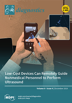
This project demonstrated the successful ability to use commercially available conference devices to remotely guide nonmedically trained adults to perform an ultrasound exam for acute cardiac, pulmonary, and abdominal assessment. This proof of concept study suggests a novel, low-cost system that allows the examiner to communicate hands-free. Further studies should evaluate how this described system can be utilized to expand the role of teleultrasound, with a particular focus on resource limited settings. View this paper.
  • Issues are regarded as officially published after their release is announced to the table of contents alert mailing list.
  • You may sign up for e-mail alerts to receive table of contents of newly released issues.
  • PDF is the official format for papers published in both, html and pdf forms. To view the papers in pdf format, click on the "PDF Full-text" link, and use the free Adobe Reader to open them.
Order results
Result details
Section
Select all
Export citation of selected articles as:
18 pages, 1440 KiB  
Article
Safety Study of Sodium Pentosan Polysulfate for Adult Patients with Mucopolysaccharidosis Type II
by Kenji Orii, Alaena Lim, Shunji Tomatsu, Molly Stapleton, Yasuyuki Suzuki, Calogera M. Simonaro, Edward H. Schuchman, Toshiyuki Fukao and Tadashi Matsumoto
Diagnostics 2019, 9(4), 226; https://doi.org/10.3390/diagnostics9040226 - 17 Dec 2019
Cited by 10 | Viewed by 4267
Abstract
Current therapies for the mucopolysaccharidoses (MPS) do not effectively address skeletal and neurological manifestations. Pentosan polysulfate (PPS) is an alternative treatment strategy that has been shown to improve bone architecture, mobility, and neuroinflammation in MPS animals. The aims of this study were to [...] Read more.
Current therapies for the mucopolysaccharidoses (MPS) do not effectively address skeletal and neurological manifestations. Pentosan polysulfate (PPS) is an alternative treatment strategy that has been shown to improve bone architecture, mobility, and neuroinflammation in MPS animals. The aims of this study were to a) primarily establish the safety of weekly PPS injections in attenuated MPS II, b) assess the efficacy of treatment on MPS pathology, and c) define appropriate clinical endpoints and biomarkers for future clinical trials. Subcutaneous injections were administered to three male Japanese patients for 12 weeks. Enzyme replacement therapy was continued in two of the patients while they received PPS and halted for two months in one patient before starting PPS. During treatment, one patient experienced an elevation of alanine transaminase, and another patient experienced convulsions; however, these incidences were non-cumulative and unrelated to PPS administration, respectively. Overall, the drug was well-tolerated in all patients, and no serious drug-related adverse events were noted. Generally, PPS treatment led to an increase in several parameters of shoulder range of motion and decrease of the inflammatory cytokines, MIF and TNF-α, which are potential clinical endpoints and biomarkers, respectively. Changes in urine and serum glycosaminoglycans were inconclusive. Overall, this study demonstrates the safety of using PPS in adults with MPS II and suggests the efficacy of PPS on MPS pathology with the identification of potential clinical endpoints and biomarkers. Full article
(This article belongs to the Special Issue Mucopolysaccharidoses: Diagnosis, Treatment, and Management)
Show Figures

Figure 1

17 pages, 3805 KiB  
Review
Imaging Modalities for Diagnosis of Deep Pelvic Endometriosis: Comparison between Trans-Vaginal Sonography, Rectal Endoscopy Sonography and Magnetic Resonance Imaging. A Head-to-Head Meta-Analysis
by Marco Noventa, Marco Scioscia, Michele Schincariol, Francesco Cavallin, Giovanni Pontrelli, Bruna Virgilio, Salvatore Giovanni Vitale, Antonio Simone Laganà, Francesco Dessole, Erich Cosmi, Donato D’Antona, Alessandra Andrisani, Carlo Saccardi, Amerigo Vitagliano and Guido Ambrosini
Diagnostics 2019, 9(4), 225; https://doi.org/10.3390/diagnostics9040225 - 17 Dec 2019
Cited by 50 | Viewed by 4004
Abstract
Objectives: A meta-analysis, with a head-to-head approach, was carried out to compare the three most common techniques for a deep pelvic endometriosis (DPE) diagnosis. We focused on: transvaginal-sonography (TVS), magnetic-resonance imaging (MRI), and rectal-endoscopy-sonography (RES). Methods: Electronic databases were searched from their inception [...] Read more.
Objectives: A meta-analysis, with a head-to-head approach, was carried out to compare the three most common techniques for a deep pelvic endometriosis (DPE) diagnosis. We focused on: transvaginal-sonography (TVS), magnetic-resonance imaging (MRI), and rectal-endoscopy-sonography (RES). Methods: Electronic databases were searched from their inception until December 2018. All prospective and well-defined retrospective studies carried out in tertiary referral centers were considered. This review was reported following the Preferred Reporting Items for Systematic Reviews and Meta-Analyses (PRISMA) and Synthesizing Evidence from Diagnostic Accuracy Tests (SEDATE) guidelines. We considered only papers in which at least two imaging modalities were compared in the same set of patients (head-to-head approach). Meta-analysis of diagnostic test accuracy (DTA) was performed separately for each location of interest. Bivariate or univariate approach has been applied when appropriate. We analyze the DTA of TVS vs. MRI, TVS vs. RES, and MRI vs. RES. Results: Our meta-analysis (17 studies included) showed high-to-moderate DTA of TVS for all endometriosis locations (apart from recto-vaginal septum (RVS)) that were not statistically different from MRI and RES for those localized in the posterior compartment. RES results were more accurate than MRI for RS lesions but less accurate than TVS for other pelvic locations, except for RVS. Conclusions: All approaches provide good accuracy with specific strong points. Ultrasonography demonstrated a diagnostic accuracy not inferior to MRI and RES; therefore, it must be considered the primary approach for DPE diagnosis. MRI has to be considered as a valuable approach in settings where highly skilled sonographers are not available. Keypoints: (1) We confirmed the non-inferiority of TVS compared to MRI and RES for the diagnosis of specific pelvic anatomic location of endometriosis lesions. (2) Ultrasonography could be considered the primary approach for DPE diagnosis (less invasive than RES and less expensive than MRI). (3) MRI has to be considered as a valuable approach in settings where skilled sonographers are not available. Full article
(This article belongs to the Section Medical Imaging and Theranostics)
Show Figures

Figure 1

10 pages, 1538 KiB  
Article
Blood Ketone Bodies and Breath Acetone Analysis and Their Correlations in Type 2 Diabetes Mellitus
by Valentine Saasa, Mervyn Beukes, Yolandy Lemmer and Bonex Mwakikunga
Diagnostics 2019, 9(4), 224; https://doi.org/10.3390/diagnostics9040224 - 17 Dec 2019
Cited by 76 | Viewed by 9220
Abstract
Analysis of volatile organic compounds in the breath for disease detection and monitoring has gained momentum and clinical significance due to its rapid test results and non-invasiveness, especially for diabetes mellitus (DM). Studies have suggested that breath gases, including acetone, may be related [...] Read more.
Analysis of volatile organic compounds in the breath for disease detection and monitoring has gained momentum and clinical significance due to its rapid test results and non-invasiveness, especially for diabetes mellitus (DM). Studies have suggested that breath gases, including acetone, may be related to simultaneous blood glucose (BG) and blood ketone levels in adults with types 2 and 1 diabetes. Detecting altered concentrations of ketones in the breath, blood and urine may be crucial for the diagnosis and monitoring of diabetes mellitus. This study assesses the efficacy of a simple breath test as a non-invasive means of diabetes monitoring in adults with type 2 diabetes mellitus. Human breath samples were collected in Tedlar™ bags and analyzed by headspace solid-phase microextraction and gas chromatography-mass spectrometry (HS-SPME/GC-MS). The measurements were compared with capillary BG and blood ketone levels (β-hydroxybutyrate and acetoacetate) taken at the same time on a single visit to a routine hospital clinic in 30 subjects with type 2 diabetes and 28 control volunteers. Ketone bodies of diabetic subjects showed a significant increase when compared to the control subjects; however, the ketone levels were was controlled in both diabetic and non-diabetic volunteers. Worthy of note, a statistically significant relationship was found between breath acetone and blood acetoacetate (R = 0.89) and between breath acetone and β-hydroxybutyrate (R = 0.82). Full article
(This article belongs to the Special Issue Point-of-Care Detection Devices for Healthcare)
Show Figures

Graphical abstract

12 pages, 2738 KiB  
Article
Four-Dimensional Flow Magnetic Resonance Imaging for Assessment of Velocity Magnitudes and Flow Patterns in The Human Carotid Artery Bifurcation: Comparison with Computational Fluid Dynamics
by Minh Tri Ngo, Chul In Kim, Jinmu Jung, Gyung Ho Chung, Dong Hwan Lee and Hyo Sung Kwak
Diagnostics 2019, 9(4), 223; https://doi.org/10.3390/diagnostics9040223 - 13 Dec 2019
Cited by 18 | Viewed by 4715
Abstract
Purpose: Knowledge of the hemodynamics in the vascular system is important to understand and treat vascular pathology. The present study aimed to evaluate the hemodynamics in the human carotid artery bifurcation measured by four-dimensional (4D) flow magnetic resonance imaging (MRI) as compared to [...] Read more.
Purpose: Knowledge of the hemodynamics in the vascular system is important to understand and treat vascular pathology. The present study aimed to evaluate the hemodynamics in the human carotid artery bifurcation measured by four-dimensional (4D) flow magnetic resonance imaging (MRI) as compared to computational fluid dynamics (CFD). Methods: This protocol used MRI data of 12 healthy volunteers for the 3D vascular models and 4D flow MRI measurements for the boundary conditions in CFD simulation. We compared the velocities measured at the carotid bifurcation and the 3D velocity streamlines of the carotid arteries obtained by these two methods. Results: There was a good agreement for both maximum and minimum velocity values between the 2 methods for velocity magnitude at the bifurcation plane. However, on the 3D blood flow visualization, secondary flows, and recirculation regions are of poorer quality when visualized through the 4D flow MRI. Conclusion: 4D flow MRI and CFD show reasonable agreement in demonstrated velocity magnitudes at the carotid artery bifurcation. However, the visualization of blood flow at the recirculation regions and the assessment of secondary flow characteristics should be enhanced for the use of 4D flow MRI in clinical situations. Full article
(This article belongs to the Section Medical Imaging and Theranostics)
Show Figures

Figure 1

14 pages, 3754 KiB  
Article
Nanoparticle-Based Biosensing Assay for Universally Accessible Low-Cost TB Detection with Comparable Sensitivity as Culture
by Ruben Kenny Briceno, Shane Ryan Sergent, Santiago Moises Benites and Evangelyn C. Alocilja
Diagnostics 2019, 9(4), 222; https://doi.org/10.3390/diagnostics9040222 - 13 Dec 2019
Cited by 14 | Viewed by 3741
Abstract
Tuberculosis (TB) is the leading cause of death globally, surpassing HIV. Furthermore, multidrug-resistant and extensively drug-resistant TB have become global public health threats. Care of TB patients starts with quality, accessible, and affordable diagnosis. The study presents a novel technique called nanoparticle-based colorimetric [...] Read more.
Tuberculosis (TB) is the leading cause of death globally, surpassing HIV. Furthermore, multidrug-resistant and extensively drug-resistant TB have become global public health threats. Care of TB patients starts with quality, accessible, and affordable diagnosis. The study presents a novel technique called nanoparticle-based colorimetric biosensing assay (NCBA) based on the principles of magnetically activated cell enrichment. A total of 1108 sputum samples were subjected to sputum smear microscopy (SSM), NCBA, and standard culture. SSM and NCBA were completed in 20 min; culture was completed in 8 weeks. Results show that NCBA has matching sensitivity of 100.0% and specificity of 99.7% compared to the gold standard culture method at a cost of $0.50/test based on Peruvian conditions. Sputum smear microscopy has 63.87% sensitivity compared to culture. NCBA has the potential of being used in local health clinics as it only requires a microscope that is widely available in many rural areas. Because NCBA could detect low levels of bacterial load comparable to culture, it could be used for rapid and early TB-onset detection. The gain in time is critical as TB is airborne and highly infectious, minimizing contact exposure. Early detection could lead to early treatment, while the patient’s immune system is still high. The low cost makes NCBA affordable and accessible to those who need them the most. Full article
(This article belongs to the Special Issue Biosensors-Based Diagnostics)
Show Figures

Figure 1

15 pages, 2158 KiB  
Review
Imaging Measurement of Whole Gut Transit Time in Paediatric and Adult Functional Gastrointestinal Disorders: A Systematic Review and Narrative Synthesis
by Hayfa Sharif, David Devadason, Nichola Abrehart, Rebecca Stevenson and Luca Marciani
Diagnostics 2019, 9(4), 221; https://doi.org/10.3390/diagnostics9040221 - 13 Dec 2019
Cited by 14 | Viewed by 5659
Abstract
Background: functional gastrointestinal disorders (FGID) are common conditions in children and adults, often associated with abnormalities of whole gut transit. Currently, transit tests can be performed using several imaging methods, including tracking of radiopaque markers, gamma scintigraphy with the use of radioisotopes, magnetic [...] Read more.
Background: functional gastrointestinal disorders (FGID) are common conditions in children and adults, often associated with abnormalities of whole gut transit. Currently, transit tests can be performed using several imaging methods, including tracking of radiopaque markers, gamma scintigraphy with the use of radioisotopes, magnetic tracking methods, tracking of movement of wireless motility capsules, and emerging magnetic resonance imaging (MRI) approaches. Objectives: to review recent literature on diagnostic imaging techniques used to investigate whole gut transit in FGIDs. Methods: a systematic review was carried out. The different techniques are described briefly, with particular emphasis on contemporary literature and new developments, particularly in the field of MRI. Conclusions: emerging MRI capsule marker methods are promising new tools to study whole gut transit in FGIDs. Full article
(This article belongs to the Section Medical Imaging and Theranostics)
Show Figures

Figure 1

11 pages, 569 KiB  
Article
Cardiovascular Magnetic Resonance Identifies High-Risk Systemic Sclerosis Patients with Normal Echocardiograms and Provides Incremental Prognostic Value
by George Markousis-Mavrogenis, Vasiliki-Kalliopi Bournia, Stylianos Panopoulos, Loukia Koutsogeorgopoulou, George Kanoupakis, Dimitrios Apostolou, Gikas Katsifis, Michail Polychroniadis, Theodoros Dimitroulas, Genovefa Kolovou, George D. Kitas, Sophie I. Mavrogeni and Petros P. Sfikakis
Diagnostics 2019, 9(4), 220; https://doi.org/10.3390/diagnostics9040220 - 11 Dec 2019
Cited by 23 | Viewed by 2839
Abstract
Background: Acute cardiac events are a significant contributor to mortality in systemic sclerosis (SSc). However, echocardiographic evaluation may be deceptively normal during an acute presentation. We hypothesized that in diffuse SSc patients presenting with acute cardiac events and a normal echocardiogram, cardiovascular magnetic [...] Read more.
Background: Acute cardiac events are a significant contributor to mortality in systemic sclerosis (SSc). However, echocardiographic evaluation may be deceptively normal during an acute presentation. We hypothesized that in diffuse SSc patients presenting with acute cardiac events and a normal echocardiogram, cardiovascular magnetic resonance (CMR) would have incremental diagnostic/prognostic value. Methods. 50 consecutive diffuse SSc patients with normal echocardiograms were evaluated using a 1.5T system. A total of 27 (63%) had experienced an acute cardiac event three to tendays before CMR evaluation (rhythm disturbances, angina pectoris, shortness of breath). Left/right ventricular (LV/RV) volumes and ejection fractions (EF), as well as LV mass, the T2-signal ratio, early/late gadolinium enhancement (EGE/LGE), native/post-contrast T1-mapping, T2-mapping and extracellular volume fraction (ECV) were compared between the event and no-event groups. Results: No differences were identified in LV/RV volumes/EF/mass. In logistic regression analyses, independent predictors of belonging to the event group were EGE (odds ratio (95% CI): 1.55 (1.06–2.26), p = 0.024), LGE (1.81 (1.23–2.67), p = 0.003), T2 mapping (1.20 (1.06–1.36), p = 0.004) and native/post-contrast T1 mapping (1.17 (1.04–1.32), p = 0.007 and 0.86 (0.75–0.98), p = 0.025). At a median follow-up of ~1.2 years, 42% vs. 11% of the event/no-event group respectively reached a combined endpoint of event occurrence/recurrence or cardiovascular mortality. Of the independent predictors resulting from logistic regression analyses, only LGE (hazard ratio (95% CI): 1.20 (1.11–1.30), p < 0.001), T2-mapping (1.07 (1.01–1.14), p = 0.025) and native T1-mapping (1.08 (1.01–1.15), p = 0.017) independently predicted the combined endpoint. Conclusions: A normal echocardiogram does not preclude myocardial lesions in diffuse SSc patients, which can be detected by CMR especially in symptomatic patients. Full article
Show Figures

Figure 1

17 pages, 729 KiB  
Article
A Hierarchical Machine Learning Model to Discover Gleason Grade-Specific Biomarkers in Prostate Cancer
by Osama Hamzeh, Abedalrhman Alkhateeb, Julia Zhuoran Zheng, Srinath Kandalam, Crystal Leung, Govindaraja Atikukke, Dora Cavallo-Medved, Nallasivam Palanisamy and Luis Rueda
Diagnostics 2019, 9(4), 219; https://doi.org/10.3390/diagnostics9040219 - 11 Dec 2019
Cited by 27 | Viewed by 4253
Abstract
(1) Background:One of the most common cancers that affect North American men and men worldwide is prostate cancer. The Gleason score is a pathological grading system to examine the potential aggressiveness of the disease in the prostate tissue. Advancements in computing and next-generation [...] Read more.
(1) Background:One of the most common cancers that affect North American men and men worldwide is prostate cancer. The Gleason score is a pathological grading system to examine the potential aggressiveness of the disease in the prostate tissue. Advancements in computing and next-generation sequencing technology now allow us to study the genomic profiles of patients in association with their different Gleason scores more accurately and effectively. (2) Methods: In this study, we used a novel machine learning method to analyse gene expression of prostate tumours with different Gleason scores, and identify potential genetic biomarkers for each Gleason group. We obtained a publicly-available RNA-Seq dataset of a cohort of 104 prostate cancer patients from the National Center for Biotechnology Information’s (NCBI) Gene Expression Omnibus (GEO) repository, and categorised patients based on their Gleason scores to create a hierarchy of disease progression. A hierarchical model with standard classifiers in different Gleason groups, also known as nodes, was developed to identify and predict nodes based on their mRNA or gene expression. In each node, patient samples were analysed via class imbalance and hybrid feature selection techniques to build the prediction model. The outcome from analysis of each node was a set of genes that could differentiate each Gleason group from the remaining groups. To validate the proposed method, the set of identified genes were used to classify a second dataset of 499 prostate cancer patients collected from cBioportal. (3) Results: The overall accuracy of applying this novel method to the first dataset was 93.3%; the method was further validated to have 87% accuracy using the second dataset. This method also identified genes that were not previously reported as potential biomarkers for specific Gleason groups. In particular, PIAS3 was identified as a potential biomarker for Gleason score 4 + 3 = 7, and UBE2V2 for Gleason score 6. (4) Insight: Previous reports show that the genes predicted by this newly proposed method strongly correlate with prostate cancer development and progression. Furthermore, pathway analysis shows that both PIAS3 and UBE2V2 share similar protein interaction pathways, the JAK/STAT signaling process. Full article
(This article belongs to the Special Issue Next-Generation Sequencing in Tumor Diagnosis and Treatment)
Show Figures

Figure 1

10 pages, 1041 KiB  
Article
Impact of Implementing Antenatal Syphilis Point-of-Care Testing on Maternal Mortality in KwaZulu-Natal, South Africa: An Interrupted Time Series Analysis
by Tivani P. Mashamba-Thompson, Paul K. Drain, Desmond Kuupiel and Benn Sartorius
Diagnostics 2019, 9(4), 218; https://doi.org/10.3390/diagnostics9040218 - 10 Dec 2019
Cited by 2 | Viewed by 3230
Abstract
Background: Syphilis infection has been associated with an increased risk of HIV infection during pregnancy which poses greater risk for maternal mortality, and antenatal syphilis point-of-care (POC) testing has been introduced to improve maternal and child health outcomes. There is limited evidence on [...] Read more.
Background: Syphilis infection has been associated with an increased risk of HIV infection during pregnancy which poses greater risk for maternal mortality, and antenatal syphilis point-of-care (POC) testing has been introduced to improve maternal and child health outcomes. There is limited evidence on the impact of syphilis POC testing on maternal outcomes in high HIV prevalent settings. We used syphilis POC testing as a model to evaluate the impact of POC diagnostics on the improvement of maternal mortality in KwaZulu-Natal, South Africa. Methods: We extracted 132 monthly data points on the number of maternal deaths in facilities and number of live births in facilities for 12 tertiary healthcare facilities in KwaZulu-Natal (KZN), South Africa from 2004 to 2014 from District Health Information System (DHIS) health facility archived. We employed segmented Poisson regression analysis of interrupted time series to assess the impact of the exposure on maternal mortality ratio (MMR) before and after the implementation of antenatal syphilis POC testing. We processed and analyzed data using Stata Statistical Software: Release 13. (Stata, Corp LP, College Station, TX, USA). Results: The provincial average annual maternal mortality ratio (MMR) was estimated at 176.09 ± 43.92 ranging from a minimum of 68.48 to maximum of 225.49 per 100,000 live births. The data comprised 36 temporal points before the introduction of syphilis POC test exposure and 84 after the introduction in primary health care clinics in KZN. The average annual MMR for KZN from 2004 to 2014 was estimated at 176.09 ± 43.92. A decrease in MMR level was observed during 2008 after syphilis POC test implementation, followed by a rise during 2009. Analysis of the MMR trend estimates a significant 1.5% increase in MMR trends during the period before implementation and 1.3% increase after implementation of syphilis POC testing (p < 0.001). Conclusion: Although our finding suggests a brief reduction in the MMR trend after the implementation of antenatal syphilis POC testing, a continued increase in syphilis rates is seen in KwaZulu-Natal, South Africa. The study used one of the most powerful quasi-experimental research methods, segmented Poisson regression analysis of interrupted time series to model the impact of syphilis POC on maternal outcome. The study finding requires confirmation by use of more rigorous primary study design. Full article
Show Figures

Figure 1

14 pages, 4338 KiB  
Review
Arrhythmogenic Inflammatory Cardiomyopathy in Autoimmune Rheumatic Diseases: A Challenge for Cardio-Rheumatology
by Sophie I. Mavrogeni, George Markousis-Mavrogenis, Constantina Aggeli, Dimitris Tousoulis, George D. Kitas, Genovefa Kolovou, Efstathios K. Iliodromitis and Petros P. Sfikakis
Diagnostics 2019, 9(4), 217; https://doi.org/10.3390/diagnostics9040217 - 10 Dec 2019
Cited by 11 | Viewed by 3890
Abstract
Ventricular arrhythmia (VA) in autoimmune rheumatic diseases (ARD) is an expression of autoimmune inflammatory cardiomyopathy (AIC), caused by structural, electrical, or inflammatory heart disease, and has a serious impact on a patient’s outcome. Myocardial scar of ischemic or nonischemic origin through a re-entry [...] Read more.
Ventricular arrhythmia (VA) in autoimmune rheumatic diseases (ARD) is an expression of autoimmune inflammatory cardiomyopathy (AIC), caused by structural, electrical, or inflammatory heart disease, and has a serious impact on a patient’s outcome. Myocardial scar of ischemic or nonischemic origin through a re-entry mechanism facilitates the development of VA. Additionally, autoimmune myocardial inflammation, either isolated or as a part of the generalized inflammatory process, also facilitates the development of VA through arrhythmogenic autoantibodies and inflammatory channelopathies. The clinical presentation of AIC varies from oligo-asymptomatic presentation to severe VA and sudden cardiac death (SCD). Both positron emission tomography (PET) and cardiovascular magnetic resonance (CMR) can diagnose AIC early and be useful tools for the assessment of therapies during follow-ups. The AIC treatment should be focused on the following: (1) early initiation of cardiac medication, including ACE-inhibitors, b-blockers, and aldosterone antagonists; (2) early initiation of antirheumatic medication, depending on the underlying disease; and (3) potentially implantable cardioverter–defibrillator (ICD) and/or ablation therapy in patients who are at high risk for SCD. Full article
Show Figures

Graphical abstract

21 pages, 1203 KiB  
Review
Noncoding RNAs and Liquid Biopsy in Lung Cancer: A Literature Review
by Antonia Haranguș, Ioana Berindan-Neagoe, Doina Adina Todea, Ioan Șimon and Mărioara Șimon
Diagnostics 2019, 9(4), 216; https://doi.org/10.3390/diagnostics9040216 - 09 Dec 2019
Cited by 7 | Viewed by 3131
Abstract
Lung cancer represents a genetically heterogeneous disease with low survival rates. Recent data have evidenced key roles of noncoding RNAs in lung cancer initiation and progression. These functional RNA molecules that can act as both oncogenes and tumor suppressors may become future biomarkers [...] Read more.
Lung cancer represents a genetically heterogeneous disease with low survival rates. Recent data have evidenced key roles of noncoding RNAs in lung cancer initiation and progression. These functional RNA molecules that can act as both oncogenes and tumor suppressors may become future biomarkers and more efficient therapeutic targets. In the precision medicine era, circulating nucleic acids have the potential to reshape the management and prognosis of cancer patients. Detecting genomic alterations and level variations of circulating nucleic acids in liquid biopsy samples represents a noninvasive method for portraying tumor burden. Research is currently trying to validate the potential role of liquid biopsy in lung cancer screening, prognosis, monitoring of disease progression, and treatment response. However, this method requires complex detection assays, and implementation of plasma genotyping in clinical practice continues to be hindered by discrepancies that arise when compared to tissue genotyping. Understanding the genomic landscape of lung cancer is essential in order to provide useful and innovative research in the age of patient-tailored therapy. In this landscape, the noncoding RNAs play a crucial role due to their target genes that dramatically influence the tumor microenvironment and the response to therapy. This article addresses present and future possible roles of liquid biopsy in lung cancer. It also discusses how the complex role of noncoding RNAs in lung tumorigenesis could influence the management of this pathology. Full article
(This article belongs to the Section Pathology and Molecular Diagnostics)
Show Figures

Graphical abstract

8 pages, 3457 KiB  
Article
Correlation between Spot Sign and Intracranial Hemorrhage Expansion on Dual-Phase CT Angiography
by Hyesoo Kim, Ja Hong Goo, Hyo Sung Kwak, Seung Bae Hwang and Gyung Ho Chung
Diagnostics 2019, 9(4), 215; https://doi.org/10.3390/diagnostics9040215 - 07 Dec 2019
Cited by 1 | Viewed by 3842
Abstract
Purpose: Expansion of intracranial hemorrhage (ICH) is an important predictor of poor clinical outcome. ICH expansion can be predicted with a spot sign on computed tomographic angiography (CTA). We aimed to evaluate the correlation between spot signs on CTA and ICH expansion on [...] Read more.
Purpose: Expansion of intracranial hemorrhage (ICH) is an important predictor of poor clinical outcome. ICH expansion can be predicted with a spot sign on computed tomographic angiography (CTA). We aimed to evaluate the correlation between spot signs on CTA and ICH expansion on dual-phase CTA. Methods: Patients with spontaneous ICH between January 2017 and April 2019 who underwent an initial CT, dual-phase CTA, and a subsequent CT were retrospectively identified. ICH expansion was defined as volume growth of >33% or >6 mL. We analyzed the presence and change in size of the spot sign in the first phase and second phase CTA. Also, we divided the morphological status of the spot sign, such as a dot-like lesion or linear contrast extravasation, in the first and second phase CTA. Results: A total of 206 patients, including 38 (18.5%) with ICH expansion and 45 (21.8%) with a spot sign, qualified for analysis. Of patients with a spot sign, 26 (57.8%) had ICH expansion on subsequent CT. Increased size of a spot sign in second-phase CTA was more frequent in the ICH expansion group than in the no-expansion group (96.2% vs. 52.6%, p < 0.001). First visualization of a spot sign in the second phase was more common in the no-expansion group than in the ICH expansion group (47.4% vs. 3.8%, p < 0.001). The morphological patterns of a spot sign between the two groups were not significantly different. Conclusion: Spot signs on dual-phase CTA have different sizes and morphological patterns. Increased size of a spot sign in the second phase of CTA can help identify patients at risk for ICH expansion. Full article
(This article belongs to the Section Medical Imaging and Theranostics)
Show Figures

Figure 1

14 pages, 243 KiB  
Review
Peri-Implantitis Diagnosis and Prognosis Using Biomarkers in Peri-Implant Crevicular Fluid: A Narrative Review
by Hatem Alassy, Praveen Parachuru and Larry Wolff
Diagnostics 2019, 9(4), 214; https://doi.org/10.3390/diagnostics9040214 - 07 Dec 2019
Cited by 53 | Viewed by 5551
Abstract
Dental implant diseases, peri-implantitis (PI) and peri-implant mucositis (PIM), have shown wide prevalence in recent studies. Despite the prevalence, diagnosing peri-implant disease (PID) remains challenging as common diagnostic methods of periodontal probing and radiographs may be inaccurate. These methods only document pre-existing destruction [...] Read more.
Dental implant diseases, peri-implantitis (PI) and peri-implant mucositis (PIM), have shown wide prevalence in recent studies. Despite the prevalence, diagnosing peri-implant disease (PID) remains challenging as common diagnostic methods of periodontal probing and radiographs may be inaccurate. These methods only document pre-existing destruction rather than current disease activity. Furthermore, there is no current model to predict the progression of PID. Though a predictive model is lacking, biomarkers may offer some potential. Biomarkers are commonly used in medicine to objectively determine disease state, or responses to a therapeutic intervention. Gingival crevicular fluid (GCF) biomarkers have moderate diagnostic validity in periodontitis. Biomarkers in peri-implant crevicular fluid (PICF) also show promising results in regard to their diagnostic and prognostic value. The aim of this review is to summarize the current knowledge of PICF biomarkers in the diagnosis of PID and evaluate their validity to predict disease progression. This review found that PICF studies utilize different methods of sampling and interpretation with varying validity (sensitivity and specificity). A number of promising diagnostic techniques were identified. Commercially available chair-side tests for MMP-8 to diagnose periodontal disease and PID activity are now available. Future directions include proteomics and metabolomics for accurate, site-specific diagnosis and prediction of PID progression. Although more research is needed, this review concludes that the assessment of proinflammatory cytokines (IL-1β, TNFα, MMP-8) in the PICF may be of value to diagnose PI and PIM but current research remains insufficient to indicate whether biomarkers predict peri-implant disease progression. Full article
(This article belongs to the Special Issue Periodontal Point-of-Care Tests)
16 pages, 5078 KiB  
Article
Shear Wave Elastography in Diagnosing Secondary Hyperparathyroidism
by Laura Cotoi, Florin Borcan, Ioan Sporea, Dana Amzar, Oana Schiller, Adalbert Schiller, Cristina Adriana Dehelean, Gheorghe Nicusor Pop and Dana Stoian
Diagnostics 2019, 9(4), 213; https://doi.org/10.3390/diagnostics9040213 - 05 Dec 2019
Cited by 8 | Viewed by 3588
Abstract
This study evaluates the diagnostic value of two-dimensional shear wave elastography (2 D-SWE) technique in the evaluation of hyperplastic parathyroid glands in cases with secondary and tertiary hyperparathyroidism. A total of 59 patients (end-stage renal disease, under supplemental dialysis program) with visible parathyroid [...] Read more.
This study evaluates the diagnostic value of two-dimensional shear wave elastography (2 D-SWE) technique in the evaluation of hyperplastic parathyroid glands in cases with secondary and tertiary hyperparathyroidism. A total of 59 patients (end-stage renal disease, under supplemental dialysis program) with visible parathyroid hyperplastic glands on ultrasound, confirmed by biochemical assay and scintigraphy, were enrolled; they were examined on grayscale ultrasound and 2 D shear wave elastography. We determined the elasticity index (EI) in the parathyroid gland, thyroid parenchyma and surrounding muscles, and the elasticity ratio of hyperplastic parathyroid glands compared to muscle, specifically sternocleidomastoid muscle. Patients presented fibrocystic bone disease with secondary hyperparathyroidism induced by end-stage chronic kidney disease; being on prolonged chronic dialysis therapy, they had positive sestamibi scintigraphy and high values of serum parathormone (1141.04 pg/mL). Nodules placed posterior to the thyroid capsule that were cystic, had a hypoechoic aspect, and were homogenous with an independent afferent artery were found. Mean EI in the parathyroid gland was 7.83 kPa, the median value in thyroid parenchyma was 13.76 kPa, and mean muscle EI value was 15.78 kPa. The observed mean parathyroid/muscle SWE ratio was 0.5356 and the value for parathyroid/normal thyroid parenchyma was 0.5995. Using receiver operating characteristic (ROC) analysis, we found that EI below 9.74 kPa correctly identifies parathyroid tissue, with a sensitivity of 94.8%, specificity of 90.7%, and accuracy of 92.26% when compared to normal thyroid tissue. Compared with the muscle tissue, we identified that EI below 9.98 kPa has a sensitivity, specificity, and accuracy of 93.8%, 90.7%, and 91.75%, respectively. Ultrasound elastography is a helpful tool in identifying parathyroid hyperplasia in patients with chronic kidney disease. A cutoff value of 9.98 kPa can be used in 2 D-SWE for accurate diagnosis of parathyroid disease. Full article
(This article belongs to the Special Issue Elastography)
Show Figures

Graphical abstract

13 pages, 257 KiB  
Article
The Effect of Zoledronic Acid on Serum Biomarkers among Patients with Chronic Low Back Pain and Modic Changes in Lumbar Magnetic Resonance Imaging
by Katri Koivisto, Jaro Karppinen, Marianne Haapea, Jyri Järvinen, Eero Kyllönen, Osmo Tervonen, Jaakko Niinimäki, Mauro Alini, Jeffrey Lotz, Stefan Dudli, Dino Samartzis, Juha Risteli, Marja-Leena Majuri, Harri Alenius and Sibylle Grad
Diagnostics 2019, 9(4), 212; https://doi.org/10.3390/diagnostics9040212 - 04 Dec 2019
Cited by 9 | Viewed by 3488
Abstract
The aim of the current study was to compare changes in serum biomarkers, including inflammatory mediators, signaling molecules, growth factors and markers of bone turnover after a single intravenous infusion of 5 mg zoledronic acid (ZA, a long-acting bisphosphonate; n = 20) or [...] Read more.
The aim of the current study was to compare changes in serum biomarkers, including inflammatory mediators, signaling molecules, growth factors and markers of bone turnover after a single intravenous infusion of 5 mg zoledronic acid (ZA, a long-acting bisphosphonate; n = 20) or placebo (n = 20) among patients with Modic changes (MC) and chronic low back pain in a randomized controlled design. The MCs were classified into M1, predominating M1, predominating M2, and M2. We measured the serum concentrations of 39 biomarkers at baseline, and one month and one year after treatment. After Benjamini–Hochberg (B–H) correction, we observed significant differences in three biomarkers over one year: Interferon-γ-inducible protein (IP-10) had risen in the ZA group (p = 0.005), whereas alkaline phosphatase (AFOS) and intact procollagen I N-terminal propeptide (iPINP) had significantly decreased in the ZA group, but had not changed in the placebo group (p < 0.001 for both). Change in iPINP correlated with change in the volume of all MC and M1 lesions. ZA downregulated bone turnover markers as expected and, surprisingly, increased the chemokine IP-10 relative to placebo treatment. This adds to our knowledge of the effects of ZA on MC and the biomarkers that signal this process. Full article
(This article belongs to the Section Pathology and Molecular Diagnostics)
7 pages, 407 KiB  
Protocol
Clinical Significance of MicroRNAs in Patients with Sepsis: Protocol for a Systematic Review and Meta-Analysis
by Daisuke Hasegawa, Kazuma Yamakawa, Kohei Taniguchi, Shuhei Murao and Osamu Nishida
Diagnostics 2019, 9(4), 211; https://doi.org/10.3390/diagnostics9040211 - 03 Dec 2019
Cited by 4 | Viewed by 2538
Abstract
Sepsis is a dysregulated immune response that leads to organ dysfunction and has high mortality rates despite recent therapeutic advancements. Accurate diagnosis and risk stratification are important for effective sepsis treatment; however, no decisive diagnostic or prognostic biomarkers are currently available. To understand [...] Read more.
Sepsis is a dysregulated immune response that leads to organ dysfunction and has high mortality rates despite recent therapeutic advancements. Accurate diagnosis and risk stratification are important for effective sepsis treatment; however, no decisive diagnostic or prognostic biomarkers are currently available. To understand whether microRNA (miRNA) might be useful biomarkers of sepsis, we aim to assess the diagnostic and prognostic accuracy of three miRNAs (122, 150, and 223) in sepsis patients via a meta-analysis of relevant published data. We will search electronic bibliographic databases (MEDLINE, EMBASE, and the Cochrane Central Register of Controlled Trials) for pertinent retrospective and prospective studies in October 2019. Two reviewers will evaluate the collected titles, abstracts, and full articles, and extract the data. We will assess the included studies using the Quality Assessment of Diagnostic Accuracy Studies-2 tool. If feasible, we will use bivariate random effects and hierarchical summary receiver operating characteristic (ROC) models to estimate summary ROCs, pooled sensitivity and specificity values, and the corresponding 95% confidence intervals. We will evaluate heterogeneity via clinical and methodological subgroup and sensitivity analyses. This systematic review will clarify the diagnostic and prognostic accuracy of select miRNAs in sepsis. It may also identify knowledge gaps in sepsis’ diagnosis and prognosis. Full article
(This article belongs to the Section Pathology and Molecular Diagnostics)
Show Figures

Figure 1

18 pages, 3404 KiB  
Article
Ultrasound and Clinical Preoperative Characteristics for Discrimination Between Ovarian Metastatic Colorectal Cancer and Primary Ovarian Cancer: A Case-Control Study
by Maciej Stukan, Juan Luis Alcazar, Jacek Gębicki, Elizabeth Epstein, Marcin Liro, Alexandra Sufliarska, Sebastian Szubert, Stefano Guerriero, Elena Ioana Braicu, Mariusz Szajewski, Małgorzata Pietrzak-Stukan and Daniela Fischerova
Diagnostics 2019, 9(4), 210; https://doi.org/10.3390/diagnostics9040210 - 01 Dec 2019
Cited by 7 | Viewed by 4264
Abstract
The aim of this study was to describe the clinical and sonographic features of ovarian metastases originating from colorectal cancer (mCRC), and to discriminate mCRC from primary ovarian cancer (OC). We conducted a multi-institutional, retrospective study of consecutive patients with ovarian mCRC who [...] Read more.
The aim of this study was to describe the clinical and sonographic features of ovarian metastases originating from colorectal cancer (mCRC), and to discriminate mCRC from primary ovarian cancer (OC). We conducted a multi-institutional, retrospective study of consecutive patients with ovarian mCRC who had undergone ultrasound examination using the International Ovarian Tumor Analysis (IOTA) terminology, with the addition of evaluating signs of necrosis and abdominal staging. A control group included patients with primary OC. Clinical and ultrasound data, subjective assessment (SA), and an assessment of different neoplasias in the adnexa (ADNEX) model were evaluated. Fisher’s exact and Student’s t-tests, the area under the receiver–operating characteristic curve (AUC), and classification and regression trees (CART) were used to conduct statistical analyses. In total, 162 patients (81 with OC and 81 with ovarian mCRC) were included. None of the patients with OC had undergone chemotherapy for CRC in the past, compared with 40% of patients with ovarian mCRC (p < 0.001). The ovarian mCRC tumors were significantly larger, a necrosis sign was more frequently present, and tumors had an irregular wall or were fixed less frequently; ascites, omental cake, and carcinomatosis were less common in mCRC than in primary OC. In a subgroup of patients with ovarian mCRC who had not undergone treatment for CRC in anamnesis, tumors were larger, and had fewer papillations and more locules compared with primary OC. The highest AUC for the discrimination of ovarian mCRC from primary OC was for CART (0.768), followed by SA (0.735) and ADNEX calculated with CA-125 (0.680). Ovarian mCRC and primary OC can be distinguished based on patient anamnesis, ultrasound pattern recognition, a proposed decision tree model, and an ADNEX model with CA-125 levels. Full article
(This article belongs to the Section Medical Imaging and Theranostics)
Show Figures

Figure 1

18 pages, 2941 KiB  
Article
Long Term Survival in Patients Suffering from Glio-blastoma Multiforme: A Single-Center Observational Cohort Study
by Daniele Armocida, Alessandro Pesce, Federico Di Giammarco, Alessandro Frati, Antonio Santoro and Maurizio Salvati
Diagnostics 2019, 9(4), 209; https://doi.org/10.3390/diagnostics9040209 - 30 Nov 2019
Cited by 23 | Viewed by 3528
Abstract
Background: Glioblastomas (GBM) are generally burdened, to date, by a dismal prognosis, although long term survivors have a relatively significant incidence. Our specific aim was to determine the exact impact of many surgery-, patient- and tumor-related variables on survival parameters. Methods: The surgical, [...] Read more.
Background: Glioblastomas (GBM) are generally burdened, to date, by a dismal prognosis, although long term survivors have a relatively significant incidence. Our specific aim was to determine the exact impact of many surgery-, patient- and tumor-related variables on survival parameters. Methods: The surgical, radiological and clinical outcomes of patients have been retrospectively reviewed for the present study. All the patients have been operated on in our institution and classified according their overall survival in long term survivors (LTS) and short term survivors (STS). A thorough review of our surgical series was conducted to compare the oncologic results of the patients in regard to: (1) surgical-(2) molecular and (3) treatment-related features. Results: A total of 177 patients were included in the final cohort. Extensive statistical analysis by means of univariate, multivariate and survival analyses disclosed a survival advantage for patients presenting a younger age, a smaller lesion and a better functional status at presentation. From the histochemical point of view, Ki67 (%) was the strongest predictor of better oncologic outcomes. A stepwise analysis of variance outlines the existence of eight prognostic subgroups according to the molecular patterns of Ki67 overexpression and epidermal growth factor receptor (EGFR), p53 and isocitrate dehydrogenase (IDH) mutations. Conclusions: On the grounds of our statistical analyses we can affirm that the following factors were significant predictors of survival advantage: Karnofsky performance status (KPS), age, volume of the lesion, motor disorder at presentation and/or a Ki67 overexpression. In our experience, LTS is associated with a gross total resection (GTR) of tumor correlated with EGFR and p53 mutations with regardless of localization, and poorly correlated to dimension. We suppose that performing a standard molecular analysis (IDH, EGFR, p53 and Ki67) is not sufficient to predict the behavior of a GBM in regards to overall survival (OS), nor to provide a deeper understanding of the meaning of the different genetic alterations in the DNA of cancer cells. A fine molecular profiling is feasible to precisely stratify the prognosis of GBM patients. Full article
(This article belongs to the Special Issue Molecular Diagnostics of CNS Tumours)
Show Figures

Figure 1

11 pages, 2138 KiB  
Article
Systematic Approach for Drug Repositioning of Anti-Epileptic Drugs
by Younhee Ko, Chulho Lee, Youngmok Lee and Jin-Sung Lee
Diagnostics 2019, 9(4), 208; https://doi.org/10.3390/diagnostics9040208 - 30 Nov 2019
Cited by 5 | Viewed by 3241
Abstract
Epilepsy is a central neurological disorder affecting individuals of all ages and causing unpredictable seizures. In spite of the improved efficacy of new antiepileptic drugs and novel therapy, there are still approximately 20%~30% of patients, who have either intractable or uncontrolled seizures. The [...] Read more.
Epilepsy is a central neurological disorder affecting individuals of all ages and causing unpredictable seizures. In spite of the improved efficacy of new antiepileptic drugs and novel therapy, there are still approximately 20%~30% of patients, who have either intractable or uncontrolled seizures. The epilepsy drug–target network (EDT) is constructed and successfully demonstrates the characteristics and efficacy of popularly used AEDs through the identification of causative genes for 60 epilepsy patients. We discovered that the causative genes of most intractable patients were not the targets of existing AEDs, as well as being very far from the etiological mechanisms of existing AEDs in the functional networks. We show that the existence of new drugs that target the causative genes of intractable epilepsy patients, which will be potential candidates for refractory epilepsy patients. Our systematic approach demonstrates a new possibility for drug repositioning through the combination of the drug-target and functional networks. Full article
(This article belongs to the Section Machine Learning and Artificial Intelligence in Diagnostics)
Show Figures

Figure 1

14 pages, 435 KiB  
Review
The Performance of Deep Learning Algorithms on Automatic Pulmonary Nodule Detection and Classification Tested on Different Datasets That Are Not Derived from LIDC-IDRI: A Systematic Review
by Dana Li, Bolette Mikela Vilmun, Jonathan Frederik Carlsen, Elisabeth Albrecht-Beste, Carsten Ammitzbøl Lauridsen, Michael Bachmann Nielsen and Kristoffer Lindskov Hansen
Diagnostics 2019, 9(4), 207; https://doi.org/10.3390/diagnostics9040207 - 29 Nov 2019
Cited by 42 | Viewed by 5682
Abstract
The aim of this study was to systematically review the performance of deep learning technology in detecting and classifying pulmonary nodules on computed tomography (CT) scans that were not from the Lung Image Database Consortium and Image Database Resource Initiative (LIDC-IDRI) database. Furthermore, [...] Read more.
The aim of this study was to systematically review the performance of deep learning technology in detecting and classifying pulmonary nodules on computed tomography (CT) scans that were not from the Lung Image Database Consortium and Image Database Resource Initiative (LIDC-IDRI) database. Furthermore, we explored the difference in performance when the deep learning technology was applied to test datasets different from the training datasets. Only peer-reviewed, original research articles utilizing deep learning technology were included in this study, and only results from testing on datasets other than the LIDC-IDRI were included. We searched a total of six databases: EMBASE, PubMed, Cochrane Library, the Institute of Electrical and Electronics Engineers, Inc. (IEEE), Scopus, and Web of Science. This resulted in 1782 studies after duplicates were removed, and a total of 26 studies were included in this systematic review. Three studies explored the performance of pulmonary nodule detection only, 16 studies explored the performance of pulmonary nodule classification only, and 7 studies had reports of both pulmonary nodule detection and classification. Three different deep learning architectures were mentioned amongst the included studies: convolutional neural network (CNN), massive training artificial neural network (MTANN), and deep stacked denoising autoencoder extreme learning machine (SDAE-ELM). The studies reached a classification accuracy between 68–99.6% and a detection accuracy between 80.6–94%. Performance of deep learning technology in studies using different test and training datasets was comparable to studies using same type of test and training datasets. In conclusion, deep learning was able to achieve high levels of accuracy, sensitivity, and/or specificity in detecting and/or classifying nodules when applied to pulmonary CT scans not from the LIDC-IDRI database. Full article
(This article belongs to the Special Issue Artificial Intelligence in Diagnostics)
Show Figures

Figure 1

9 pages, 792 KiB  
Article
Sarcopenic Factors May Have No Impact on Outcomes in Ovarian Cancer Patients
by Naomi Nakayama, Kentaro Nakayama, Kohei Nakamura, Sultana Razia and Satoru Kyo
Diagnostics 2019, 9(4), 206; https://doi.org/10.3390/diagnostics9040206 - 28 Nov 2019
Cited by 17 | Viewed by 2787
Abstract
Although the prognostic value of sarcopenic factors, such as loss of muscle mass and quality, have been widely reported in patients with cancer during the last decade, the value in those with ovarian cancer remains unclear. Therefore, this study evaluated the prognostic impact [...] Read more.
Although the prognostic value of sarcopenic factors, such as loss of muscle mass and quality, have been widely reported in patients with cancer during the last decade, the value in those with ovarian cancer remains unclear. Therefore, this study evaluated the prognostic impact of sarcopenic factors in patients with ovarian cancer. We retrospectively evaluated the data of 94 ovarian cancer patients who underwent surgery and chemotherapy at the Shimane University Hospital between March 2006 and 2013. Preoperative computed tomography scan at the level of the third lumbar vertebra was used to evaluate skeletal muscle volume and quality based on the skeletal muscle index (SMI) and intramuscular adipose tissue content (IMAC), respectively. The impact of preoperative SMI and IMAC on outcomes was subsequently investigated. Low SMI and high IMAC were not significantly associated with disease-free survival (p = 0.329 and p = 0.3370, respectively) or poor overall survival (p = 0.921 and p = 0.988, respectively). Neither preoperative low muscle volume nor low muscle quality was a poor prognostic factor in ovarian cancer. Full article
(This article belongs to the Special Issue Ovarian Cancer: Characteristics, Screening, Diagnosis and Treatment)
Show Figures

Figure 1

10 pages, 1673 KiB  
Article
Diet-Induced Rat Model of Gradual Development of Non-Alcoholic Fatty Liver Disease (NAFLD) with Lipopolysaccharides (LPS) Secretion
by Dominika Maciejewska, Agnieszka Łukomska, Karolina Dec, Karolina Skonieczna-Żydecka, Izabela Gutowska, Marta Skórka-Majewicz, Daniel Styburski, Kamila Misiakiewicz-Has, Anna Pilutin, Joanna Palma, Katarzyna Sieletycka, Wojciech Marlicz and Ewa Stachowska
Diagnostics 2019, 9(4), 205; https://doi.org/10.3390/diagnostics9040205 - 27 Nov 2019
Cited by 21 | Viewed by 4099
Abstract
Background: Non-alcoholic fatty liver disease (NAFLD) is one of the most common liver disorders in industrialized Western countries. The prevalence of the disease is estimated to range from 4% to 46% worldwide. The aim of study was to develop an animal model with [...] Read more.
Background: Non-alcoholic fatty liver disease (NAFLD) is one of the most common liver disorders in industrialized Western countries. The prevalence of the disease is estimated to range from 4% to 46% worldwide. The aim of study was to develop an animal model with gradual NAFLD development. Methods: Sprague-Dawley rats were fed a high-fat and high-cholesterol (HFHCh) diet. The rats from the study and control groups were sacrificed after 2, 4, 8, 12, 16, and 20 weeks of dietary exposure. Results: Analysis of biochemical parameters showed that after only two weeks, ALT and cholesterol concentration in serum were elevated. After 4 weeks, TNF-α and HOMA-IR were significantly higher compared to the control group. NAFLD progression started after 12 weeks of diet-weight gain and increased LPS secretions were noticed. During the experiment, rats induced steatosis (from stage 0/1 after 4 weeks to stage 2/3 after 20 weeks), inflammation (from stage 0/1 after 4 weeks to stage 1/2 after 20 weeks), and fibrosis (from stage 1 after 12 weeks to stage 2 after 20 weeks). Conclusion: We can assume that the presented model based on the HFHCh diet induced gradual development of NAFLD. We confirmed that the animal NAFLD model increases LPS secretions during disease progression. Full article
(This article belongs to the Special Issue Animal Models for Studying and Screening Human Diseases)
Show Figures

Figure 1

11 pages, 1204 KiB  
Article
Rapid Extraction Method of Mycobacterium ulcerans DNA from Clinical Samples of Suspected Buruli Ulcer Patients
by Michael Frimpong, Hubert Senanu Ahor, Samuel Asamoah Sakyi, Bernadette Agbavor, Emmanuel Akowuah and Richard Odame Phillips
Diagnostics 2019, 9(4), 204; https://doi.org/10.3390/diagnostics9040204 - 26 Nov 2019
Cited by 6 | Viewed by 7083
Abstract
Isothermal amplification techniques such as recombinase polymerase amplification (RPA) and loop-mediated isothermal amplification (LAMP) for diagnosing Buruli ulcer, a necrotic skin disease caused by Mycobacterium ulcerans, have renewed hope for the molecular diagnosis of clinically suspected Buruli ulcer cases in endemic districts. [...] Read more.
Isothermal amplification techniques such as recombinase polymerase amplification (RPA) and loop-mediated isothermal amplification (LAMP) for diagnosing Buruli ulcer, a necrotic skin disease caused by Mycobacterium ulcerans, have renewed hope for the molecular diagnosis of clinically suspected Buruli ulcer cases in endemic districts. If these techniques are applied at district-level hospitals or clinics, they will help facilitate early case detection with prompt treatment, thereby reducing disability and associated costs of disease management. The accuracy as well as the application of these molecular techniques at point of need is dependent on simple and fast DNA extraction. We have modified and tested a rapid extraction protocol for use with an already developed recombinase polymerase amplification assay. The entire procedure from “sample in, extraction and DNA amplification” was conducted in a mobile suitcase laboratory within 40 min. The DNA extraction procedure was performed within 15 min, with only two manipulation/pipetting steps needed. The diagnostic sensitivity and specificity of this extraction protocol together with M. ulcerans RPA in comparison with standard DNA extraction with real-time PCR was 87% (n = 26) and 100% (n = 13), respectively. We have established a simple, fast and efficient protocol for the extraction and detection of M. ulcerans DNA in clinical samples that is adaptable to field conditions. Full article
(This article belongs to the Section Point-of-Care Diagnostics and Devices)
Show Figures

Figure 1

11 pages, 1987 KiB  
Article
Two Potential Clinical Applications of Origami-Based Paper Devices
by Zong-Keng Kuo, Tsui-Hsuan Chang, Yu-Shin Chen, Chao-Min Cheng and Chia-Ying Tsai
Diagnostics 2019, 9(4), 203; https://doi.org/10.3390/diagnostics9040203 - 26 Nov 2019
Cited by 3 | Viewed by 2853
Abstract
Detecting small amounts of analyte in clinical practice is challenging because of deficiencies in specimen sample availability and unsuitable sampling environments that prevent reliable sampling. Paper-based analytical devices (PADs) have successfully been used to detect ultralow amounts of analyte, and origami-based PADs (O-PADs) [...] Read more.
Detecting small amounts of analyte in clinical practice is challenging because of deficiencies in specimen sample availability and unsuitable sampling environments that prevent reliable sampling. Paper-based analytical devices (PADs) have successfully been used to detect ultralow amounts of analyte, and origami-based PADs (O-PADs) offer advantages that may boost the overall potential of PADs in general. In this study, we investigated two potential clinical applications for O-PADs. The first O-PAD we investigated was an origami-based enzyme-linked immunosorbent assay (ELISA) system designed to detect different concentrations of rabbit IgG. This device was designed with four wing structures, each of which acted as a reagent loading zone for pre-loading ELISA reagents, and a central test sample loading zone. Because this device has a low limit of detection (LOD), it may be suitable for detecting IgG levels in tears from patients with a suspected viral infection (such as herpes simplex virus (HSV)). The second O-PAD we investigated was designed to detect paraquat levels to determine potential poisoning. To use this device, we sequentially folded each of two separate reagent zones, one preloaded with NaOH and one preloaded with ascorbic acid (AA), over the central test zone, and added 8 µL of sample that then flowed through each reagent zone and onto the central test zone. The device was then unfolded to read the results on the test zone. The three folded layers of paper provided a moist environment not achievable with conventional paper-based ELISA. Both O-PADs were convenient to use because reagents were preloaded, and results could be observed and analyzed with image analysis software. O-PADs expand the testing capacity of simpler PADs while leveraging their characteristic advantages of convenience, cost, and ease of use, particularly for point-of-care diagnosis. Full article
(This article belongs to the Special Issue Point-of-Care Detection Devices for Healthcare)
Show Figures

Graphical abstract

13 pages, 1298 KiB  
Article
Protein Carbonyl Content Is a Predictive Biomarker of Eccentric Left Ventricular Hypertrophy in Hemodialysis Patients
by Zorica M. Dimitrijevic, Sonja S. Salinger Martinovic, Valentina N. Nikolic and Tatjana P. Cvetkovic
Diagnostics 2019, 9(4), 202; https://doi.org/10.3390/diagnostics9040202 - 25 Nov 2019
Cited by 7 | Viewed by 5073
Abstract
High prevalence of left ventricular hypertrophy (LVH) and elevated oxidative stress are associated with poor outcomes in chronic hemodialysis patients. Abnormal left ventriculаr geomеtry and different geometric patterns play an important role as well. Our study analyzed the role of oxidative stress on [...] Read more.
High prevalence of left ventricular hypertrophy (LVH) and elevated oxidative stress are associated with poor outcomes in chronic hemodialysis patients. Abnormal left ventriculаr geomеtry and different geometric patterns play an important role as well. Our study analyzed the role of oxidative stress on myocardial remodeling in these patients. Plasma malondialdehyde (MDA), protein carbonyl (PC) content, and total antioxidative capacity (TAC) were investigated in 104 hemodialysis patients together with transthoracic echocardiography. Compared to patients with normal ventricular geometry, patients with LVH had increased MDA and PC plasma concentration. Multivariate analysis demonstrated that protein carbonyls, as biomarkers of oxidative protein modification, were an independent predictor of eccentric hypertrophy (eLVH), including higher LV end-diastolic diameter and LV end-diastolic volume, (β = 0.32 and β = 0.28, p < 0.001 for both). The incidence of eLVH increased progressively from the lowest to the highest baseline PC tertile (p < 0.001 for the trend) and the subjects in the former group showed a 76% greater risk of developing eLVH compared to their counterparts. After further adjustment for the potential mediators, PCs carried eLVH odds (95% confidence interval (CI)) of 1.256 (0.998–1.514), per standard deviation increase. High plasma protein carbonyls levels are a significant independent predictor of eccentric LVH in chronic hemodialysis patients. Full article
(This article belongs to the Section Pathology and Molecular Diagnostics)
Show Figures

Figure 1

5 pages, 190 KiB  
Editorial
Pediatrics: An Evolving Concept for the 21st Century
by Consolato M. Sergi
Diagnostics 2019, 9(4), 201; https://doi.org/10.3390/diagnostics9040201 - 25 Nov 2019
Cited by 3 | Viewed by 2030
Abstract
Pediatrics is rapidly evolving, and the diagnostic tools are expanding the spectrum of diagnoses that can be identified at the bedside [...] Full article
(This article belongs to the Special Issue Diagnosis and Management of Pediatric Diseases)
8 pages, 3607 KiB  
Communication
Overall Survival and Adjuvant Therapy in Women with Ovarian Carcinosarcoma: A Single-Institution Experience
by Aaron Nizam, Bethany Bustamante, Weiwei Shan, Karin K. Shih, Jill S. Whyte, Antoinette Sakaris, Lisa dos Santos, Marina Frimer, Andrew W. Menzin, Alexander Truskinovsky and Gary L. Goldberg
Diagnostics 2019, 9(4), 200; https://doi.org/10.3390/diagnostics9040200 - 22 Nov 2019
Cited by 10 | Viewed by 2877
Abstract
Background: Carcinosarcoma of the ovary (CSO) is a rare and aggressive variant of ovarian cancer. Due to the rare nature of the disease there is insufficient evidence to make recommendations regarding standard management and overall prognosis. Methods: An Institutional Review Board-approved study identified [...] Read more.
Background: Carcinosarcoma of the ovary (CSO) is a rare and aggressive variant of ovarian cancer. Due to the rare nature of the disease there is insufficient evidence to make recommendations regarding standard management and overall prognosis. Methods: An Institutional Review Board-approved study identified all our patients with CSO between January 2011 and May 2018. Demographic and outcome measures were abstracted from the medical records and tumor board files. Cox proportional hazard models, log rank tests, and comparisons of means were used to calculate significance (p < 0.05). Results: 27 women with CSO were identified. The median age at diagnosis was 65 years (range 48–91). Five women (18%) presented with early stage disease (Stage I or II) and 22 patients (82%) presented with late stage III or IV disease. Twenty patients (74%) received intravenous platinum-based combination chemotherapy. Seven patients did not receive chemotherapy during their treatment course. The median overall survival was 23 months (range 2–68 months). Overall survival was not significantly worsened by the stage of disease at diagnosis. There was no difference in survival based on the age at diagnosis, tobacco status or ethnicity (p > 0.05). Conclusion: This is one of the largest single institution experiences with CSO. The majority of our patients presented with advanced stage disease and received adjuvant platinum-based chemotherapy after cytoreductive surgery. The median overall survival of 23 months was not affected by the stage of the disease. The optimal management of this rare disease needs further study with collaborative, prospective multi-institutional trials. Full article
(This article belongs to the Special Issue Ovarian Cancer: Characteristics, Screening, Diagnosis and Treatment)
Show Figures

Figure 1

7 pages, 513 KiB  
Case Report
Diagnostic Approach to Traumatic Axonal Injury of the Spinothalamic Tract in Individual Patients with Mild Traumatic Brain Injury
by Sung Ho Jang and Han Do Lee
Diagnostics 2019, 9(4), 199; https://doi.org/10.3390/diagnostics9040199 - 21 Nov 2019
Cited by 9 | Viewed by 2755
Abstract
Objectives: We investigated an approach for the diagnosis of traumatic axonal injury (TAI) of the spinothalamic tract (STT) that was based on diffusion tensor tractography (DTT) results and a statistical comparison of individual patients who showed central pain following mild traumatic brain injury [...] Read more.
Objectives: We investigated an approach for the diagnosis of traumatic axonal injury (TAI) of the spinothalamic tract (STT) that was based on diffusion tensor tractography (DTT) results and a statistical comparison of individual patients who showed central pain following mild traumatic brain injury (mTBI) with the control group. Methods: Five right-handed female patients in their forties and with central pain following mTBI and 12 age-, sex-, and handedness-matched healthy control subjects were recruited. After DTT reconstruction of the STT, we analyzed the STT in terms of three DTT parameters (fractional anisotropy (FA), mean diffusivity (MD), and fiber number (FN)) and its configuration (narrowing and tearing). To assess narrowing, we determined the area of the STT on an axial slice of the subcortical white matter. Results: the FN values were significantly lower in at least one hemisphere of each patient when compared to those of the control subjects (p < 0.05). Significant decrements from the STT area in the control group were observed in at least one hemisphere of each patient (p < 0.05). Regarding configurational analysis, the STT showed narrowing and/or partial tearing in at least one hemisphere of each of the five patients. Conclusions: Herein, we demonstrate a DTT-based approach for the diagnosis of TAI of the STT. The approach involves a statistical comparison between DTT parameters of individual patients who show central pain following mTBI and those of an age-, gender-, and handedness-matched control group. We think that the method described in this study can be useful in the diagnosis of TAI of the STT in individual mTBI patients. Full article
(This article belongs to the Section Medical Imaging and Theranostics)
Show Figures

Figure 1

9 pages, 921 KiB  
Article
Evaluation of Hepatic Steatosis in Rodents by Time-Domain Nuclear Magnetic Resonance
by João A.B. Pedroso, João Paulo Camporez, Luciana T. Belpiede, Rafaela S. Pinto, José Cipolla-Neto and Jose Donato, Jr.
Diagnostics 2019, 9(4), 198; https://doi.org/10.3390/diagnostics9040198 - 20 Nov 2019
Cited by 2 | Viewed by 2635
Abstract
Devices that analyze body composition of rodents by time-domain nuclear magnetic resonance (TD-NMR) are becoming popular in research centers that study metabolism. Theoretically, TD-NMR devices can also evaluate lipid content in isolated tissues. However, the accuracy of TD-NMR to determine hepatic steatosis in [...] Read more.
Devices that analyze body composition of rodents by time-domain nuclear magnetic resonance (TD-NMR) are becoming popular in research centers that study metabolism. Theoretically, TD-NMR devices can also evaluate lipid content in isolated tissues. However, the accuracy of TD-NMR to determine hepatic steatosis in the liver of small laboratory animals has not been evaluated in detail. We observed that TD-NMR was able to detect increased lipid content in the liver of rats consuming high-fat diet (HFD) for 12 weeks and in genetically obese (Lepob/ob and Leprdb/db) mice. The lipid content determined by TD-NMR showed a positive correlation with triglyceride content measured by colorimetric assays. In contrast, TD-NMR did not detect hepatic steatosis in C57BL/6 mice consuming HFD for 4 or 12 weeks, despite their obesity and increased liver triglyceride content. These findings indicate that tissue mass and the severity of hepatic steatosis affect the sensitivity of TD-NMR to detect liver lipid content. Full article
(This article belongs to the Section Medical Imaging and Theranostics)
Show Figures

Figure 1

9 pages, 229 KiB  
Article
Is the Fatty Acids Profile in Blood a Good Predictor of Liver Changes? Correlation of Fatty Acids Profile with Fatty Acids Content in the Liver
by Dominika Maciejewska, Joanna Palma, Karolina Dec, Karolina Skonieczna-Żydecka, Izabela Gutowska, Małgorzata Szczuko, Karolina Jakubczyk and Ewa Stachowska
Diagnostics 2019, 9(4), 197; https://doi.org/10.3390/diagnostics9040197 - 19 Nov 2019
Cited by 7 | Viewed by 2940
Abstract
Background: Existing data show a correlation between the profile of fatty acids, liver, and blood. Therefore, the aim of our study was to investigate the correlation between the fatty acids profile in blood pallets and the liver. Methods: The experiment was performed on [...] Read more.
Background: Existing data show a correlation between the profile of fatty acids, liver, and blood. Therefore, the aim of our study was to investigate the correlation between the fatty acids profile in blood pallets and the liver. Methods: The experiment was performed on 60 eight-week-old male Sprague-Dawley rats. The study group (n = 30, 5 groups, 6 rats each) received a cholesterol diet; the control group (n = 30, 5 groups, 6 rats each) received standard food for laboratory rats. The rats from both the study and control groups were sacrificed after 2, 4, 8, 12, and 16 weeks of dietary exposure. The fatty acids profile was measured using gas chromatography (GC). Results: In both the control and study group, the highest correlations were observed in palmitoleic acid (RHO = 0.68), heptadecanoic acid (RHO = 0.65), vaccenic acid (RHO = 0.72), eicosapentaenoic acid (RHO = 0.68), docosapentaenoic acid (RHO = 0.77), and docosahexaenoic (RHO = 0.77). Among liver indexes, the highest correlations were desaturase-18 (0.61). Conclusions: Fatty acids profile is a sensitive marker of the development of potentially pathological changes in the liver. The potential markers of fatty liver are: oleic acid, vaccenic acid, EPA, DHA, docosapentaenoic acid, and desaturase index (SCD-18 index). Full article
(This article belongs to the Section Pathology and Molecular Diagnostics)
Previous Issue
Next Issue
Back to TopTop