Next Issue
Volume 10, February
Previous Issue
Volume 9, December


Diagnostics, Volume 10, Issue 1 (January 2020) – 54 articles

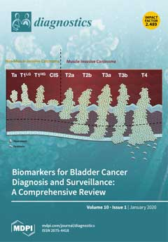
Cover Story (view full-size image): Bladder cancer (BC) is rising in its incidence and prevalence and is accompanied by a high morbidity and mortality. Diagnosis and monitoring of BC is mainly performed through invasive tests (cystoscopies), which are highly uncomfortable for the patient and not exempt of comorbidities. There is an imperative need for non-invasive testing for early detection, disease monitoring, and treatment response in BC. In this review, we compare different tests based on molecular biomarkers and evaluate their potential role in BC diagnosis, follow-up, and treatment response monitoring. View this paper.
  • Issues are regarded as officially published after their release is announced to the table of contents alert mailing list.
  • You may sign up for e-mail alerts to receive table of contents of newly released issues.
  • PDF is the official format for papers published in both, html and pdf forms. To view the papers in pdf format, click on the "PDF Full-text" link, and use the free Adobe Reader to open them.
Order results
Result details
Section
Select all
Export citation of selected articles as:
15 pages, 5333 KiB  
Article
Disabled Homolog 2 (DAB2) Protein in Tumor Microenvironment Correlates with Aggressive Phenotype in Human Urothelial Carcinoma of the Bladder
by Yoshitaka Itami, Makito Miyake, Sayuri Ohnishi, Yoshihiro Tatsumi, Daisuke Gotoh, Shunta Hori, Yousuke Morizawa, Kota Iida, Kenta Ohnishi, Yasushi Nakai, Takeshi Inoue, Satoshi Anai, Nobumichi Tanaka, Tomomi Fujii, Keiji Shimada, Hideki Furuya, Vedbar S. Khadka, Youping Deng and Kiyohide Fujimoto
Diagnostics 2020, 10(1), 54; https://doi.org/10.3390/diagnostics10010054 - 20 Jan 2020
Cited by 13 | Viewed by 4202
Abstract
Disabled homolog-2 (DAB2) has been reported to be a tumor suppressor gene. However, a number of contrary studies suggested that DAB2 promotes tumor invasion in urothelial carcinoma of the bladder (UCB). Here, we investigated the clinical role and biological function of [...] Read more.
Disabled homolog-2 (DAB2) has been reported to be a tumor suppressor gene. However, a number of contrary studies suggested that DAB2 promotes tumor invasion in urothelial carcinoma of the bladder (UCB). Here, we investigated the clinical role and biological function of DAB2 in human UCB. Immunohistochemical staining analysis for DAB2 was carried out on UCB tissue specimens. DAB2 expression levels were compared with clinicopathological factors. DAB2 was knocked-down by small interfering RNA (siRNA) transfection, and then its effects on cell proliferation, invasion, and migration, and changes to epithelial-mesenchymal transition (EMT)-related proteins were evaluated. In our in vivo assays, tumor-bearing athymic nude mice subcutaneously inoculated with human UCB cells (MGH-U-3 or UM-UC-3) were treated by DAB2-targeting siRNA. Higher expression of DAB2 was associated with higher clinical T category, high tumor grade, and poor oncological outcome. The knock-down of DAB2 decreased both invasion and migration ability and expression of EMT-related proteins. Significant inhibitory effects on tumor growth and invasion were observed in xenograft tumors of UM-UC-3 treated by DAB2-targeting siRNA. Our findings suggested that DAB2 expression was associated with poor prognosis through increased oncogenic properties including tumor proliferation, migration, invasion, and enhancement of EMT in human UCB. Full article
(This article belongs to the Special Issue Urogenital Cancers: Diagnostic, Predictive, and Prognostic Markers)
Show Figures

Figure 1

9 pages, 229 KiB  
Editorial
Acknowledgement to Reviewers of Diagnostics in 2019
by Diagnostics Editorial Office
Diagnostics 2020, 10(1), 53; https://doi.org/10.3390/diagnostics10010053 - 20 Jan 2020
Viewed by 2178
Abstract
The editorial team greatly appreciates the reviewers who have dedicated their considerable time and expertise to the journal’s rigorous editorial process over the past 12 months, regardless of whether the papers are finally published or not [...] Full article
16 pages, 588 KiB  
Review
Mucinous Cancer of the Ovary: Overview and Current Status
by Abdulaziz Babaier and Prafull Ghatage
Diagnostics 2020, 10(1), 52; https://doi.org/10.3390/diagnostics10010052 - 19 Jan 2020
Cited by 67 | Viewed by 11659
Abstract
Mucinous ovarian cancer (MOC) is a rare subtype of epithelial ovarian carcinoma (EOC). Whereas all EOC subtypes are addressed in the same way, MOC is a distinct entity. Appreciating the pathological features and genomic profile of MOC may result in the improvement in [...] Read more.
Mucinous ovarian cancer (MOC) is a rare subtype of epithelial ovarian carcinoma (EOC). Whereas all EOC subtypes are addressed in the same way, MOC is a distinct entity. Appreciating the pathological features and genomic profile of MOC may result in the improvement in management and, hence, the prognosis. Distinguishing primary MOC from metastatic mucinous carcinoma can be challenging but is essential. Early-stage MOC carries an excellent prognosis, with advanced disease having a poor outcome. Surgical management plays an essential role in the early stage and in metastatic disease. Chemotherapy is usually administered for stage II MOC and beyond. The standard gynecology protocol is frequently used, but gastrointestinal regimens have also been administered. As MOC is associated with multiple molecular alterations, targeted therapy could be the answer to treat this disease. Full article
(This article belongs to the Special Issue Ovarian Cancer: Characteristics, Screening, Diagnosis and Treatment)
Show Figures

Figure 1

17 pages, 1979 KiB  
Article
The Influence of Serum Sample Storage Conditions on Selected Laboratory Parameters Related to Oxidative Stress: A Preliminary Study
by Lilla Pawlik-Sobecka, Katarzyna Sołkiewicz, Izabela Kokot, Aleksandra Kiraga, Sylwia Płaczkowska, Agnieszka Matylda Schlichtinger and Ewa Maria Kratz
Diagnostics 2020, 10(1), 51; https://doi.org/10.3390/diagnostics10010051 - 19 Jan 2020
Cited by 14 | Viewed by 5817
Abstract
The present work aims at accessing the stability of biological material stored for diagnostic and scientific purposes. The influence of the temperature, storage time, and cyclic thawing on concentration stability of selected oxidative stress parameters in human serum was investigated. The study group [...] Read more.
The present work aims at accessing the stability of biological material stored for diagnostic and scientific purposes. The influence of the temperature, storage time, and cyclic thawing on concentration stability of selected oxidative stress parameters in human serum was investigated. The study group consisted of 20 serum samples collected from healthy volunteers aged 18–52. The parameters whose reference ranges were not determined and to which validated determination methods did not correspond were examined by manual methods (FRAP and AOPP). Automatic methods were used to determine routine laboratory tests (albumin, total protein, bilirubin, uric acid) using the Konelab 20i® analyzer. The samples were stored at various temperatures (room temperature, 4 °C, −20 °C, −80 °C) for max 6 months and were subjected to cyclic thawing at 1 month intervals. In order to check whether any differences between the concentrations of the studied parameters existed when the samples were stored in various conditions, the paired Student t-test or Wilcoxon test and comparison to desirable bias were applied. Based on the obtained results, it was found that the temperature and time of serum sample storage significantly affected the stability of the analyzed parameters and determined different shelf lives of serum samples for oxidative stress examination. Therefore, continuing the investigation concerning the impact of storage conditions on various serum parameters seems justified due to the discrepancy between the individual results obtained by different researchers and the inconsistencies between the results of scientific research and the applicable recommendations. Full article
(This article belongs to the Section Pathology and Molecular Diagnostics)
Show Figures

Figure 1

13 pages, 1903 KiB  
Article
Widefield Swept Source OCTA in Retinitis Pigmentosa
by Rodolfo Mastropasqua, Rossella D’Aloisio, Chiara De Nicola, Giada Ferro, Alfonso Senatore, Daniele Libertini, Guido Di Marzio, Marta Di Nicola, Giuseppe Di Martino, Luca Di Antonio and Lisa Toto
Diagnostics 2020, 10(1), 50; https://doi.org/10.3390/diagnostics10010050 - 19 Jan 2020
Cited by 16 | Viewed by 3772
Abstract
(1) Background: To evaluate superficial capillary plexus (SCP), deep capillary plexus (DCP), choriocapillaris (CC), perfusion density (PD), and vessel length density (VLD) in macular and near/mid periphery regions in patients with retinitis pigmentosa (RP) using widefield swept source optical coherence tomography angiography (WSS-OCTA). [...] Read more.
(1) Background: To evaluate superficial capillary plexus (SCP), deep capillary plexus (DCP), choriocapillaris (CC), perfusion density (PD), and vessel length density (VLD) in macular and near/mid periphery regions in patients with retinitis pigmentosa (RP) using widefield swept source optical coherence tomography angiography (WSS-OCTA). (2) Methods: Twelve RP patients (20 eyes) and 20 age-matched subjects (20 eyes) were imaged with the SS-OCTA system (PLEX Elite 9000, Carl Zeiss Meditec Inc., Dublin, CA, USA). Quantitative analysis was performed in the macular and peripheral regions. The main outcome measures were SCP, DCP, CC, PD, and VLD in central and peripheral areas. (3) Results: Mean visual acuity, central macular thickness, and microperimetry were significantly reduced in RP patients compared to normal subjects (p < 0.05). The perfusion density and VLD of SCP, DCP, and CC were significantly reduced in RP patients compared to normal controls both in the central and peripheral retina (p < 0.05). A significant direct correlation was found in RP patients between PD of the 1.5 mm central retina both in DCP and CC and microperimetry at 4° and 8°. (4) Conclusions: Widefield SS-OCTA shows an impairment of retinal and choroidal perfusion density and vessel length density in central and peripheral retina of RP patients. The reduction of flow features correlates with the macular function. Full article
(This article belongs to the Section Medical Imaging and Theranostics)
Show Figures

Figure 1

10 pages, 500 KiB  
Article
Cardiac Catheterization versus Echocardiography for Monitoring Pulmonary Pressure: A Prospective Study in Patients with Connective Tissue Disease-Associated Pulmonary Arterial Hypertension
by Vasiliki Kalliopi Bournia, Iraklis Tsangaris, Loukianos Rallidis, Dimitrios Konstantonis, Frantzeska Frantzeskaki, Anastasia Anthi, Stylianos E. Orfanos, Eftychia Demerouti, Panagiotis Karyofillis, Vassilis Voudris, Katerina Laskari, Stylianos Panopoulos, Panayiotis G. Vlachoyiannopoulos and Petros P. Sfikakis
Diagnostics 2020, 10(1), 49; https://doi.org/10.3390/diagnostics10010049 - 19 Jan 2020
Cited by 7 | Viewed by 3633
Abstract
Standard echocardiography is important for pulmonary arterial hypertension (PAH) screening in patients with connective tissue disease (CTD), but PAH diagnosis and monitoring require cardiac catheterization. Herein, using cardiac catheterization as reference, we tested the hypothesis that follow-up echocardiography is adequate for clinical decision-making [...] Read more.
Standard echocardiography is important for pulmonary arterial hypertension (PAH) screening in patients with connective tissue disease (CTD), but PAH diagnosis and monitoring require cardiac catheterization. Herein, using cardiac catheterization as reference, we tested the hypothesis that follow-up echocardiography is adequate for clinical decision-making in these patients. We prospectively studied 69 consecutive patients with CTD-associated PAH. Invasive baseline pulmonary artery systolic pressure (PASP) was 60.19 ± 16.33 mmHg (mean ± SD) and pulmonary vascular resistance (PVR) was 6.44 ± 2.95WU. All patients underwent hemodynamic and echocardiographic follow-up after 9.47 ± 7.29 months; 27 patients had a third follow-up after 17.2 ± 7.4 months from baseline. We examined whether clinically meaningful hemodynamic deterioration of follow-up catheterization-derived PASP (i.e., > 10% increase) could be predicted by simultaneous echocardiography. Echocardiography predicted hemodynamic PASP deterioration with 59% sensitivity, 85% specificity, and 63/83% positive/negative predictive value, respectively. In multivariate analysis, successful echocardiographic prediction correlated only with higher PVR in previous catheterization (p = 0.05, OR = 1.235). Notably, in patients having baseline PVR > 5.45 WU, echocardiography had both sensitivity and positive predictive values of 73%, and both specificity and negative predictive value of 91% for detecting hemodynamic PASP deterioration. In selected patients with CTD-PAH echocardiography can predict PASP deterioration with high specificity and negative predictive value. Additional prospective studies are needed to confirm that better patient selection can increase the ability of standard echocardiography to replace repeat catheterization. Full article
Show Figures

Figure 1

11 pages, 769 KiB  
Article
High Genetic Diversity and No Evidence of Clonal Relation in Synchronous Thyroid Carcinomas Associated with Hashimoto’s Thyroiditis: A Next-Generation Sequencing Analysis
by Csaba Molnár, Emese Sarolta Bádon, Attila Mokánszki, Anikó Mónus, Lívia Beke, Ferenc Győry, Endre Nagy and Gábor Méhes
Diagnostics 2020, 10(1), 48; https://doi.org/10.3390/diagnostics10010048 - 17 Jan 2020
Cited by 6 | Viewed by 3204
Abstract
The close association between pre-existing Hashimoto’s thyroiditis and thyroid cancer is well established. The simultaneous occurrence of multiple neoplastic foci within the same organ suggests a common genotoxic effect potentially contributing to carcinogenesis, the nature of which is still not clear. Next-generation sequencing [...] Read more.
The close association between pre-existing Hashimoto’s thyroiditis and thyroid cancer is well established. The simultaneous occurrence of multiple neoplastic foci within the same organ suggests a common genotoxic effect potentially contributing to carcinogenesis, the nature of which is still not clear. Next-generation sequencing (NGS) provides a potent tool to demonstrate and compare the mutational profile of the independent neoplastic foci. Our collection of 47 cases with thyroid carcinoma and Hashimoto’s thyroiditis included 14 with at least two tumorous foci. Detailed histological analysis highlighted differences in histomorphology, immunoprofile, and biological characteristics. Further, a 67-gene NGS panel was applied to demonstrate the mutational diversity of the synchronic tumors. Significant differences could be detected with a wide spectrum of pathogenic gene variants involved (ranging between 5 and 18, cutoff >5.0 variant allele frequencies (VAF)). Identical gene variants represented in both synchronous tumors of the same thyroid gland were found in only two cases (BRAF and JAK3 genes). An additional set of major driver mutations was identified at variable allele frequencies in a highly individual setup suggesting a clear clonal independence. The different BRAF statuses in coincident thyroid carcinoma foci within the same organ outline a special challenge for molecular follow-up and therapeutic decision-making. Full article
(This article belongs to the Special Issue Next-Generation Sequencing in Tumor Diagnosis and Treatment)
Show Figures

Figure 1

14 pages, 858 KiB  
Article
HLA Allele and Haplotype Frequencies in Three Urban Mexican Populations: Genetic Diversity for the Approach of Genomic Medicine
by Alma D. Del Angel-Pablo, Ana Itzel Juárez-Martín, Gloria Pérez-Rubio, Enrique Ambrocio-Ortiz, Luis A. López-Flores, Angel E. Camarena and Ramcés Falfán-Valencia
Diagnostics 2020, 10(1), 47; https://doi.org/10.3390/diagnostics10010047 - 16 Jan 2020
Cited by 6 | Viewed by 4043
Abstract
Genetic variability defends us against pathogen-driven antigens; human leucocyte antigens (HLA) is the immunological system in charge of this work. The Mexican mestizo population arises mainly from the mixture of three founder populations; Amerindian, Spaniards, and a smaller proportion of the African population. [...] Read more.
Genetic variability defends us against pathogen-driven antigens; human leucocyte antigens (HLA) is the immunological system in charge of this work. The Mexican mestizo population arises mainly from the mixture of three founder populations; Amerindian, Spaniards, and a smaller proportion of the African population. We describe allele and haplotype frequencies of HLA class I (-A and -B) and class II (-DRB1 and -DQB1), which were analyzed by PCR-SSP in Mexican mestizo from three urban populations of Mexico: Chihuahua-Chihuahua City (n = 88), Mexico City-Tlalpan (n = 330), and Veracruz-Xalapa (n = 84). The variability of the allele HLA class I and class II among the three regions of Mexico are in four alleles: HLA-A*24:02 (36.39%), -B*35:01 (16.04%), -DRB1*04:07 (17.33%), and -DQB1*03:02 (31.47%), these alleles have been previously described in some indigenous populations. We identified 5 haplotypes with a frequency >1%: HLA-A*02:01-B*35:01-DRB1*08:02-DQB1*04:02, A*68:01-B*39:01-DRB1*08:02-DQB1*04:02, A*02:01-B*35:01-DRB1*04:07-DQB1*03:02, A*68:01-B*39:01-DRB1*04:07-DQB1*03:02, and A*01:01-B*08:01-DRB1*03:01-DQB1*02:01. Also, the haplotype A*02:01-B*35:01-DRB1*08:02-DQB1*04:02 was identified in Tlalpan and Xalapa regions. Haplotype A*01:01-B*08:01-DRB1*03:01-DQB1*02:01 was found only in Tlalpan and Chihuahua. In the Xalapa region, the most frequent haplotype was A*24:02-B*35:01-DRB1*04:07-DQB1*03:02. These alleles and haplotypes have been described in Amerindian populations. Our data are consistent with previous studies and contribute to the analysis of the variability in the Mexican population. Full article
(This article belongs to the Section Pathology and Molecular Diagnostics)
Show Figures

Figure 1

12 pages, 899 KiB  
Article
Assessment of Activity of Daily Life in Mucopolysaccharidosis Type II Patients with Hematopoietic Stem Cell Transplantation
by Yasuyuki Suzuki, Madeleine Taylor, Kenji Orii, Toshiyuki Fukao, Tadao Orii and Shunji Tomatsu
Diagnostics 2020, 10(1), 46; https://doi.org/10.3390/diagnostics10010046 - 16 Jan 2020
Cited by 9 | Viewed by 3897
Abstract
The effectiveness of hematopoietic stem cell transplantation (HSCT) for mucopolysaccharidosis type II (MPS II, Hunter disease) remains controversial although recent studies have shown HSCT provides more clinical impact. This study aims to evaluate the long-term effectiveness of HSCT using the activity of daily [...] Read more.
The effectiveness of hematopoietic stem cell transplantation (HSCT) for mucopolysaccharidosis type II (MPS II, Hunter disease) remains controversial although recent studies have shown HSCT provides more clinical impact. This study aims to evaluate the long-term effectiveness of HSCT using the activity of daily living (ADL) scores in patients with MPS II. Sixty-nine severely affected MPS II patients (19 patients who received HSCT and 50 untreated patients) and 40 attenuated affected patients (five with HSCT and 35 untreated) were investigated by a simplified ADL questionnaire. The frequency of clinical findings and the scores of ADL (verbal, gross motor, and the level of care) were analyzed statistically. The mean age of onset of 19 severely affected patients who received HSCT was 1.40 years ± 1.06, which is not statistically different from that of 50 untreated patients (p = 0.11). Macroglossia, frequent airway infection, hepatosplenomegaly, joint contracture, and sleep apnea were less frequent in the HSCT-treated group of severe MPS II patients. The severe phenotype HSCT treated group reported a statistically significant higher score of verbal function and gross motor function between the ages of 10 and 15 years and a higher level of care score between 10 and 20 years. Patients with the attenuated phenotype showed high ADL scores, and all of five HSCT treated patients reported a lower frequency of frequent airway infection, coarse skin, umbilical/inguinal hernia, hepatosplenomegaly, heart valve disorders, and carpal tunnel. In conclusion, HSCT is effective, resulting in improvements in clinical features and ADL in patients with MPS II. HSCT should be re-reviewed as a therapeutic option for MPS II patients. Full article
(This article belongs to the Special Issue Mucopolysaccharidoses: Diagnosis, Treatment, and Management)
Show Figures

Figure 1

9 pages, 1848 KiB  
Case Report
Papillary Thyroid Cancer in a Struma Ovarii in a 17-Year-Old Nulliparous Patient: A Case Report
by Agnieszka Gonet, Rafał Ślusarczyk, Danuta Gąsior-Perczak, Artur Kowalik, Janusz Kopczyński and Aldona Kowalska
Diagnostics 2020, 10(1), 45; https://doi.org/10.3390/diagnostics10010045 - 15 Jan 2020
Cited by 14 | Viewed by 5034
Abstract
Introduction: Struma ovarii accounts for 2% of mature teratomas. Struma ovarii is diagnosed when thyroid tissue accounts for >50% of the teratoma. Malignant transformation is rare, occurring in <5% of struma ovarii cases. Case presentation: A 17-year-old patient was diagnosed with papillary thyroid [...] Read more.
Introduction: Struma ovarii accounts for 2% of mature teratomas. Struma ovarii is diagnosed when thyroid tissue accounts for >50% of the teratoma. Malignant transformation is rare, occurring in <5% of struma ovarii cases. Case presentation: A 17-year-old patient was diagnosed with papillary thyroid cancer in struma ovarii. The patient exhibited menstrual disorders. Abdominal and pelvic CT revealed a 17 cm mass in the left adnexa. Laparoscopic removal of the left adnexa with enucleation of right ovarian cysts was performed. Histopathological diagnosis was a follicular variant papillary carcinoma measuring 23 mm in diameter. Immunohistochemical positive expression of CK19, TTF-1, and thyroglobulin (Tg) confirmed the diagnosis. Molecular analysis detected the BRAF K601E mutation in ovarian tumor tissues. Preoperative serum Tg concentration was >300 ng/mL, which decreased to 38.2 ng/mL after gynecological surgery with undetectable anti-Tg antibodies. The patient underwent total thyroidectomy with no cancer detected on histopathological examination. The patient was treated with I-131 and showed no recurrence 4 years after the diagnosis. Conclusions: Malignant struma ovarii is diagnosed by surgery. Because papillary carcinoma in struma ovarii is rare and there are no guidelines regarding the management of this type of cancer, therapeutic decisions should be made individually based on clinical and pathological data. Full article
(This article belongs to the Special Issue New Insights in Thyroid Diagnostics)
Show Figures

Graphical abstract

16 pages, 10274 KiB  
Article
Cross-Dataset Evaluation of Deep Learning Networks for Uterine Cervix Segmentation
by Peng Guo, Zhiyun Xue, L. Rodney Long and Sameer Antani
Diagnostics 2020, 10(1), 44; https://doi.org/10.3390/diagnostics10010044 - 14 Jan 2020
Cited by 18 | Viewed by 8855
Abstract
Evidence from recent research shows that automatic visual evaluation (AVE) of photographic images of the uterine cervix using deep learning-based algorithms presents a viable solution for improving cervical cancer screening by visual inspection with acetic acid (VIA). However, a significant performance determinant in [...] Read more.
Evidence from recent research shows that automatic visual evaluation (AVE) of photographic images of the uterine cervix using deep learning-based algorithms presents a viable solution for improving cervical cancer screening by visual inspection with acetic acid (VIA). However, a significant performance determinant in AVE is the photographic image quality. While this includes image sharpness and focus, an important criterion is the localization of the cervix region. Deep learning networks have been successfully applied for object localization and segmentation in images, providing impetus for studying their use for fine contour segmentation of the cervix. In this paper, we present an evaluation of two state-of-the-art deep learning-based object localization and segmentation methods, viz., Mask R-convolutional neural network (CNN) and MaskX R-CNN, for automatic cervix segmentation using three datasets. We carried out extensive experimental tests and algorithm comparisons on each individual dataset and across datasets, and achieved performance either notably higher than, or comparable to, that reported in the literature. The highest Dice and intersection-over-union (IoU) scores that we obtained using Mask R-CNN were 0.947 and 0.901, respectively. Full article
(This article belongs to the Section Medical Imaging and Theranostics)
Show Figures

Figure 1

14 pages, 1711 KiB  
Review
Hyperthermic Intraperitoneal Chemotherapy in Ovarian Cancer
by McKayla J. Riggs, Prakash K. Pandalai, Joseph Kim and Charles S. Dietrich
Diagnostics 2020, 10(1), 43; https://doi.org/10.3390/diagnostics10010043 - 14 Jan 2020
Cited by 27 | Viewed by 6794
Abstract
Hyperthermic intraperitoneal chemotherapy (HIPEC) in conjunction with cytoreductive surgery (CRS) holds promise as an adjunctive treatment strategy in malignancies affecting the peritoneal surface, effectively targeting remaining microscopic residual tumor. HIPEC increases concentrations of chemotherapy directly within the peritoneal cavity compared with the intravenous [...] Read more.
Hyperthermic intraperitoneal chemotherapy (HIPEC) in conjunction with cytoreductive surgery (CRS) holds promise as an adjunctive treatment strategy in malignancies affecting the peritoneal surface, effectively targeting remaining microscopic residual tumor. HIPEC increases concentrations of chemotherapy directly within the peritoneal cavity compared with the intravenous route and reduces the systemic side effects associated with prolonged adjuvant intraperitoneal exposure. Furthermore, hyperthermia increases tissue penetration and is synergistic with the therapeutic chemotherapy agents used. In ovarian cancer, evidence is building for its use in both primary and recurrent scenarios. In this review, we examine the history of HIPEC, the techniques used, and the available data guiding its use in primary and recurrent ovarian cancer. Full article
(This article belongs to the Special Issue Ovarian Cancer: Characteristics, Screening, Diagnosis and Treatment)
Show Figures

Figure 1

11 pages, 1088 KiB  
Article
Development of a Flow-Free Automated Colorimetric Detection Assay Integrated with Smartphone for Zika NS1
by Md Alamgir Kabir, Hussein Zilouchian, Mazhar Sher and Waseem Asghar
Diagnostics 2020, 10(1), 42; https://doi.org/10.3390/diagnostics10010042 - 14 Jan 2020
Cited by 26 | Viewed by 8302
Abstract
The Zika virus (ZIKV) is an emerging flavivirus transmitted to humans by Aedes mosquitoes that can potentially cause microcephaly, Guillain–Barré Syndrome, and other birth defects. Effective vaccines for Zika have not yet been developed. There is a necessity to establish an easily deployable, [...] Read more.
The Zika virus (ZIKV) is an emerging flavivirus transmitted to humans by Aedes mosquitoes that can potentially cause microcephaly, Guillain–Barré Syndrome, and other birth defects. Effective vaccines for Zika have not yet been developed. There is a necessity to establish an easily deployable, high-throughput, low-cost, and disposable point-of-care (POC) diagnostic platform for ZIKV infections. We report here an automated magnetic actuation platform suitable for a POC microfluidic sandwich enzyme-linked immunosorbent assay (ELISA) using antibody-coated superparamagnetic beads. The smartphone integrated immunoassay is developed for colorimetric detection of ZIKV nonstructural protein 1 (NS1) antigen using disposable chips to accommodate the reactions inside the chip in microliter volumes. An in-house-built magnetic actuator platform automatically moves the magnetic beads through different aqueous phases. The assay requires a total of 9 min to automatically control the post-capture washing, horseradish peroxidase (HRP) conjugated secondary antibody probing, washing again, and, finally, color development. By measuring the saturation intensity of the developed color from the smartphone captured video, the presented assay provides high sensitivity with a detection limit of 62.5 ng/mL in whole plasma. These results advocate a great promise that the platform would be useful for the POC diagnosis of Zika virus infection in patients and can be used in resource-limited settings. Full article
(This article belongs to the Special Issue Mobile Diagnosis 2.0)
Show Figures

Graphical abstract

17 pages, 2017 KiB  
Article
Shear-Wave Elastography Variability Analysis and Relation with Kidney Allograft Dysfunction: A Single-Center Study
by Sorana D. Bolboacă, Florin Ioan Elec, Alina Daciana Elec, Adriana Milena Muntean, Mihai Adrian Socaciu, Gheorghita Iacob, Răzvan Zaro, Alexandra-Ioana Andrieș, Ramona Maria Bădulescu, Radu Mihai Ignat, Mihaela Iancu and Radu Ion Badea
Diagnostics 2020, 10(1), 41; https://doi.org/10.3390/diagnostics10010041 - 13 Jan 2020
Cited by 21 | Viewed by 4089
Abstract
Shear-wave elastography (SWE) showed the absence or presence of significant differences among stable kidney allograft function and allograft dysfunction. We evaluated the variability of kidney allograft stiffness in relation to allograft dysfunction, respectively, in terms of a correlation of stiffness with patients’ characteristics. [...] Read more.
Shear-wave elastography (SWE) showed the absence or presence of significant differences among stable kidney allograft function and allograft dysfunction. We evaluated the variability of kidney allograft stiffness in relation to allograft dysfunction, respectively, in terms of a correlation of stiffness with patients’ characteristics. A single-center prospective study on patients who had undergone renal transplantation was conducted between October 2017 and November 2018. Patients were clinically classified as having a stable allograft function or allograft dysfunction. SWE examinations performed by the same radiologist with a LOGIQ E9 were evaluated. Ten measurements were done for Young’s modulus (kPa) at the level of allograft cortex and another ten at the level of medulla. Eighty-three SWE examinations from 63 patients, 69 stable allografts, and 14 allografts with dysfunction were included in the analysis. The intra-examinations stiffness showed high variability, with the quantile covariation coefficient ranging from 2.21% to 45.04%. The inter-examinations stiffness showed heterogeneity (from 28.66% to 42.38%). The kidney allograft cortex stiffness showed significantly higher values in cases with dysfunction (median = 28.70 kPa, interquartile range (IQR) = (25.68–31.98) kPa) as compared to those with stable function (median = 20.99 kPa, interquartile range = (16.08–27.68) kPa; p-value = 0.0142). Allograft tissue stiffness (both cortex and medulla) was significantly negatively correlated with body mass index (−0.44, p-value < 0.0001 for allograft cortex and −0.42, p-value = 0.0001 for allograft medulla), and positively correlated with Proteinuria/Creatinuria ratio (0.33, p-value = 0.0021 for allograft cortex and 0.28, p-value = 0.0105 for allograft medulla) but remained statistically significant only in cases with stable function. The cortical tissue stiffness proved significantly higher values for patients with allograft dysfunction as compared to patients with stable function, but to evolve as an additional tool for the evaluation of patients with a kidney transplant and to change the clinical practice, more extensive studies are needed. Full article
(This article belongs to the Special Issue Elastography)
Show Figures

Graphical abstract

11 pages, 1629 KiB  
Article
Timely-Automatic Procedure for Estimating the Endocardial Limits of the Left Ventricle Assessed Echocardiographically in Clinical Practice
by Darian M. Onchis, Codruta Istin, Cristina Tudoran, Mariana Tudoran and Pedro Real
Diagnostics 2020, 10(1), 40; https://doi.org/10.3390/diagnostics10010040 - 13 Jan 2020
Cited by 1 | Viewed by 2949
Abstract
In this paper, we propose an analytical rapid method to estimate the endocardial borders of the left ventricular walls on echocardiographic images for prospective clinical integration. The procedure was created as a diagnostic support tool for the clinician and it is based on [...] Read more.
In this paper, we propose an analytical rapid method to estimate the endocardial borders of the left ventricular walls on echocardiographic images for prospective clinical integration. The procedure was created as a diagnostic support tool for the clinician and it is based on the use of the anisotropic generalized Hough transform. Its application is guided by a Gabor-like filtering for the approximate delimitation of the region of interest without the need for computing further anatomical characteristics. The algorithm is applying directly a deformable template on the predetermined filtered region and therefore it is responsive and straightforward implementable. For accuracy considerations, we have employed a support vector machine classifier to determine the confidence level of the automated marking. The clinical tests were performed at the Cardiology Clinic of the County Emergency Hospital Timisoara and they improved the physicians perception in more than 50% of the cases. The report is concluded with medical discussions. Full article
(This article belongs to the Section Medical Imaging and Theranostics)
Show Figures

Figure 1

19 pages, 9947 KiB  
Review
Biomarkers for Bladder Cancer Diagnosis and Surveillance: A Comprehensive Review
by Rui Batista, Nuno Vinagre, Sara Meireles, João Vinagre, Hugo Prazeres, Ricardo Leão, Valdemar Máximo and Paula Soares
Diagnostics 2020, 10(1), 39; https://doi.org/10.3390/diagnostics10010039 - 13 Jan 2020
Cited by 82 | Viewed by 10406
Abstract
Bladder cancer (BC) ranks as the sixth most prevalent cancer in the world, with a steady rise in its incidence and prevalence, and is accompanied by a high morbidity and mortality. BC is a complex disease with several molecular and pathological pathways, thus [...] Read more.
Bladder cancer (BC) ranks as the sixth most prevalent cancer in the world, with a steady rise in its incidence and prevalence, and is accompanied by a high morbidity and mortality. BC is a complex disease with several molecular and pathological pathways, thus reflecting different behaviors depending on the clinical staging of the tumor and molecular type. Diagnosis and monitoring of BC is mainly performed by invasive tests, namely periodic cystoscopies; this procedure, although a reliable method, is highly uncomfortable for the patient and it is not exempt of comorbidities. Currently, there is no formal indication for the use of molecular biomarkers in clinical practice, even though there are several tests available. There is an imperative need for a clinical non-invasive testing for early detection, disease monitoring, and treatment response in BC. In this review, we aim to assess and compare different tests based on molecular biomarkers and evaluate their potential role as new molecules for bladder cancer diagnosis, follow-up, and treatment response monitoring. Full article
(This article belongs to the Special Issue Urogenital Cancers: Diagnostic, Predictive, and Prognostic Markers)
Show Figures

Graphical abstract

17 pages, 804 KiB  
Article
The Panel of 12 Cell-Free MicroRNAs as Potential Biomarkers in Prostate Neoplasms
by Maria Yu. Konoshenko, Evgeniy A. Lekchnov, Olga E. Bryzgunova, Ivan A. Zaporozhchenko, Sergey V. Yarmoschuk, Oksana A. Pashkovskaya, Svetlana V. Pak and Pavel P. Laktionov
Diagnostics 2020, 10(1), 38; https://doi.org/10.3390/diagnostics10010038 - 10 Jan 2020
Cited by 25 | Viewed by 3638
Abstract
Prostate cancer is a global biological, medical, and social issue aggravated by the lack of reliable, highly specific, and sensitive non-invasive tests for diagnosis and staging of prostate cancer. One prospective source of biomarkers are the cell-free miRNAs present in various biological fluids. [...] Read more.
Prostate cancer is a global biological, medical, and social issue aggravated by the lack of reliable, highly specific, and sensitive non-invasive tests for diagnosis and staging of prostate cancer. One prospective source of biomarkers are the cell-free miRNAs present in various biological fluids. In the present study, we validated the diagnostic potential of cell-free miRNAs: miR-19b, miR-22, miR-92a, miR-378, miR-425, miR-30e, miR-31, miR-125b, miR-200b, miR-205, miR-375, and miR-660; we estimated the required sample size and the minimal miRNA set for a subsequent large-scale validation study. Relative expression of 12 miRNA combined in 31 ratios was investigated in three fractions of biological fluids (urine extracellular vesicles, clarified urine, and plasma) obtained from patients with prostate cancer (n = 10), benign prostate hyperplasia (n = 8), and healthy volunteers (n = 11). Eight of the miRNAs found in urine vesicles (miR-19b, miR-30e, miR-31, miR-92a, miR-125, miR-200, miR-205, and miR-660) showed great promise and when combined into six ratios (miR-125b/miR-30e, miR-200/miR-30e, miR-205/miR-30e, miR-31/miR-30e, miR-660/miR-30e, and miR-19b/miR-92a) could classify patients with prostate cancer, benign prostate hyperplasia, and healthy donors with 100% specificity, 100% sensitivity, and with a high degree of reliability for most donors. Full article
(This article belongs to the Special Issue Diagnostic Biomarkers in Prostate Cancer 2020)
Show Figures

Graphical abstract

14 pages, 1125 KiB  
Article
Laryngeal, Tracheal, and Bronchial Disease in the Mucopolysaccharidoses: Endoscopic Study
by Paulo Pires de Mello, Anneliese Lopes Barth, Danielle de Araujo Torres, Mariana Pires de Mello Valente and Dafne Dain Gandelman Horovitz
Diagnostics 2020, 10(1), 37; https://doi.org/10.3390/diagnostics10010037 - 10 Jan 2020
Cited by 5 | Viewed by 3128
Abstract
Mucopolysaccharidoses (MPS) are genetically determined diseases, leading to a deficiency of enzymes in the glycosaminoglycan (GAG) degradation pathway. The accumulation of GAG occurs in connective tissue in various organs and systems of the body, including the larynx, trachea, and bronchi. Respiratory symptoms are [...] Read more.
Mucopolysaccharidoses (MPS) are genetically determined diseases, leading to a deficiency of enzymes in the glycosaminoglycan (GAG) degradation pathway. The accumulation of GAG occurs in connective tissue in various organs and systems of the body, including the larynx, trachea, and bronchi. Respiratory symptoms are common and severe in these patients, and respiratory disease is a frequent cause of death. A cross-sectional study with flexible bronchoscopy was conducted in 30 MPS patients (6 MPS I, 8 MPS II, 2 MPS III, 3 MPS IV-A, and 11 MPS VI). Only four patients (13.33%) had a normal airway; nine (30%) had mild to moderate disease, 12 (40%) moderate to severe, and five patients (16.67%) had severe disease. Of particular interest, neuronopathic MPS II had the largest proportion of tracheostomized patients who died due to respiratory complications; in MPS IV-A, all patients had significant tracheobronchial deformity with associated tracheomalacia, despite lacking laryngeal involvement. Laryngotracheobronchial disease (LTBD) was associated to longer disease history and was significantly more severe in older patients. Longer use of enzyme replacement therapy did not prevent the progression of LTBD, although the age of therapy introduction may be a crucial factor in lower airway involvement. Full article
(This article belongs to the Special Issue Mucopolysaccharidoses: Diagnosis, Treatment, and Management)
Show Figures

Figure 1

5 pages, 554 KiB  
Interesting Images
Lyme Neuroborreliosis in a Patient with Breast Cancer: MRI and PET/CT Findings
by Mathilde Ørbæk, Camilla Klausen, Anne-Mette Lebech and Helene Mens
Diagnostics 2020, 10(1), 36; https://doi.org/10.3390/diagnostics10010036 - 9 Jan 2020
Cited by 1 | Viewed by 4968
Abstract
We present a case demonstrating the performance of different radiographical and nuclear medicine imaging modalities in the diagnostic work-up of a patient with Lyme neuroborreliosis. The patient presented in late summer 2019 with radicular pains followed by a foot drop and peripheral facial [...] Read more.
We present a case demonstrating the performance of different radiographical and nuclear medicine imaging modalities in the diagnostic work-up of a patient with Lyme neuroborreliosis. The patient presented in late summer 2019 with radicular pains followed by a foot drop and peripheral facial palsy, both right-sided. Due to a history of breast cancer, disseminated malignant disease was initially suspected. Bone metastasis was ruled out by skeletal scintigraphy. Magnetic resonance imaging (MRI) of the neuroaxis and a whole body 18F-FDG PET-CT was performed within 48 hours. The MRI revealed a strong contrast enhancement of the conus medullaris and fibers of the cauda equina, while the 18F-FDG PET/CT was without pathological findings. Examination of cerebrospinal fluid led to the definitive diagnosis of Lyme neuroborreliosis with monocytic pleocytosis and a positive intrathecal test for Borrelia burgdorferi. The patient became pain-free after 10 days of ceftriaxone, and the paralysis slowly regressed the following month. This case highlights the difficulty of the diagnosis of Lyme neuroborreliosis and discusses the relevant imaging findings. Full article
(This article belongs to the Section Medical Imaging and Theranostics)
Show Figures

Figure 1

8 pages, 15377 KiB  
Communication
First Glance of Molecular Profile of Atypical Cellular Angiofibroma/Cellular Angiofibroma with Sarcomatous Transformation by Next Generation Sequencing
by Yi-Che Chang Chien, Attila Mokánszki, Hsuan-Ying Huang, Raimundo Geronimo Silva, Jr., Chien-Chin Chen, Lívia Beke, Anikó Mónus and Gábor Méhes
Diagnostics 2020, 10(1), 35; https://doi.org/10.3390/diagnostics10010035 - 9 Jan 2020
Cited by 5 | Viewed by 3651
Abstract
Cellular angiofibroma is a rare benign mesenchymal neoplasm most commonly occurring in the vulvovaginal region in women and the inguinoscrotal region in men with specific genetic deletion involved in the RB1 gene in chromosome 13q14 region. Atypical cellular angiofibroma and cellular angiofibroma with [...] Read more.
Cellular angiofibroma is a rare benign mesenchymal neoplasm most commonly occurring in the vulvovaginal region in women and the inguinoscrotal region in men with specific genetic deletion involved in the RB1 gene in chromosome 13q14 region. Atypical cellular angiofibroma and cellular angiofibroma with sarcomatous transformation are recently described variants showing worrisome morphological features and strong, diffuse p16 expression. Nevertheless, the molecular profile of these tumor entities is largely unknown. We carried out a next generation sequencing (NGS) study from six cases of atypical cellular angiofibroma and cellular angiofibroma with sarcomatous transformation. We were able to identify oncogenic TP53 gene mutations (33%) which may contribute to pathogenesis also resulting in p16 overexpression. In addition, RB1 gene alterations generally present were identified. Since it is a recently described and rare entity, the whole molecular signaling pathway is still largely obscured and the analysis of larger cohorts is needed to elucidate this issue. Full article
(This article belongs to the Special Issue Next-Generation Sequencing in Tumor Diagnosis and Treatment)
Show Figures

Figure 1

10 pages, 932 KiB  
Article
UV Fluorescence-Based Determination of Urinary Advanced Glycation End Products in Patients with Chronic Kidney Disease
by Mieke Steenbeke, Sander De Bruyne, Elisabeth Van Aken, Griet Glorieux, Wim Van Biesen, Jonas Himpe, Gilles De Meester, Marijn Speeckaert and Joris Delanghe
Diagnostics 2020, 10(1), 34; https://doi.org/10.3390/diagnostics10010034 - 9 Jan 2020
Cited by 14 | Viewed by 3953
Abstract
Advanced glycation end products (AGEs) are a class of proteins or lipids that are non-enzymatically glycated and oxidized after contact with aldose sugars. The accumulation of AGEs results in carbonyl stress, which is characteristic for diabetes mellitus, uremia, atherosclerosis and vascular dysfunction. In [...] Read more.
Advanced glycation end products (AGEs) are a class of proteins or lipids that are non-enzymatically glycated and oxidized after contact with aldose sugars. The accumulation of AGEs results in carbonyl stress, which is characteristic for diabetes mellitus, uremia, atherosclerosis and vascular dysfunction. In recent decades, several innovative methods have been developed to measure the concentration of AGEs in blood or urine. In the present study, we evaluated the use of UV fluorescence as an alternative tool to detect urinary AGEs in four groups of well characterized chronic kidney disease (CKD) patients over a wide range of kidney insufficiency and in a group of healthy subjects. Using an excitation wavelength of 365 nm, the fluorescence spectra of urinary AGEs were recorded in the 400–620 nm emission range. When considering the emission peaks at 440 nm and 490 nm, a significantly higher AGE-specific fluorescence intensity was detected in CKD patients compared to healthy subjects (p < 0.0001 and p = 0.0001, respectively). The urinary creatinine adjusted fluorescence emission spectra in the group of CKD patients with diabetes mellitus were comparable with those of CKD patients without diabetes mellitus. Creatinine-adjusted fluorescence emission spectra were highest in CKD patients with proteinuria, moderate in CKD patients without proteinuria and lowest in healthy controls (p < 0.0001 at both emission wavelengths). In a multiple regression analysis, age, CRP and insulin treatment were predictors of fluorescence intensity at the emission wavelength of 440 nm. Age and insulin treatment were predictors at 490 nm. The presented method is a simple, cheap, alternative method to monitor the AGE-load in the CKD population. Full article
(This article belongs to the Section Pathology and Molecular Diagnostics)
Show Figures

Figure 1

10 pages, 906 KiB  
Article
High-Performance Liquid Chromatography as a Novel Method for the Determination of α-Defensins in Synovial Fluid for Diagnosis of Orthopedic Infections
by Pavel Melicherčík, Eva Klapková, Karel Kotaška, David Jahoda, Ivan Landor and Václav Čeřovský
Diagnostics 2020, 10(1), 33; https://doi.org/10.3390/diagnostics10010033 - 9 Jan 2020
Cited by 13 | Viewed by 4115
Abstract
The α-defensins (AD) present in synovial fluid have been regarded as constituting the most accurate periprosthetic joint infection (PJI) biomarker. The methods most commonly used for estimating AD as a biomarker are the qualitative Synovasure® PJI tests, based on the technique of [...] Read more.
The α-defensins (AD) present in synovial fluid have been regarded as constituting the most accurate periprosthetic joint infection (PJI) biomarker. The methods most commonly used for estimating AD as a biomarker are the qualitative Synovasure® PJI tests, based on the technique of lateral flow, and quantitative enzyme-linked immunosorbent assay (ELISA). Here, we propose a novel test based on detecting α-defensins in synovial fluid by high-performance liquid chromatography (HPLC). Synovial fluid was collected from 157 patients diagnosed with PJI, infectious arthritis (IA), arthrosis, reactive arthritis, and rheumatoid arthritis. AD concentrations in the fluid were determined by HPLC, and these same samples were used for additional diagnostic analyses. The results were statistically processed to calculate cutoff concentrations for PJI and IA. HPLC testing showed a sensitivity of 94% and a specificity of 92% for diagnosis of PJI, and a sensitivity of 97% and a specificity of 87% for diagnosis of IA. Using HPLC, we detected in synovial fluid a combination of three α-defensins: human neutrophil peptides HNP1, HNP2, and HNP3. All measured AD concentration values shown in this work refer to the sum of these three individual concentrations. Our study shows that the HPLC method meets the conditions for measuring precise concentrations of the sum of AD and can be recommended as part of a diagnostic array for PJI and IA diagnostics. By this method, we have verified that higher levels of AD in synovial fluid can also be seen in rheumatoid illnesses, crystal arthropathies, and reactive arthritis. Full article
(This article belongs to the Section Pathology and Molecular Diagnostics)
Show Figures

Figure 1

13 pages, 626 KiB  
Article
Percussion Entropy Analysis of Synchronized ECG and PPG Signals as a Prognostic Indicator for Future Peripheral Neuropathy in Type 2 Diabetic Subjects
by Hai-Cheng Wei, Na Ta, Wen-Rui Hu, Sheng-Ying Wang, Ming-Xia Xiao, Xiao-Jing Tang, Jian-Jung Chen and Hsien-Tsai Wu
Diagnostics 2020, 10(1), 32; https://doi.org/10.3390/diagnostics10010032 - 9 Jan 2020
Cited by 15 | Viewed by 4159
Abstract
Diabetic peripheral neuropathy (DPN) is one of the most common chronic complications of diabetes. It has become an essential public health crisis, especially for care in the home. Synchronized electrocardiogram (ECG) and photoplethysmography (PPG) signals were obtained from healthy non-diabetic (n = [...] Read more.
Diabetic peripheral neuropathy (DPN) is one of the most common chronic complications of diabetes. It has become an essential public health crisis, especially for care in the home. Synchronized electrocardiogram (ECG) and photoplethysmography (PPG) signals were obtained from healthy non-diabetic (n = 37) and diabetic (n = 85) subjects without peripheral neuropathy, recruited from the diabetic outpatient clinic. The conventional parameters, including low-/high-frequency power ratio (LHR), small-scale multiscale entropy index (MEISS), large-scale multiscale entropy index (MEILS), electrocardiogram-based pulse wave velocity (PWVmean), and percussion entropy index (PEI), were computed as baseline and were then followed for six years after the initial PEI measurement. Three new diabetic subgroups with different PEI values were identified for the goodness-of-fit test and Cox proportional Hazards model for relative risks analysis. Finally, Cox regression analysis showed that the PEI value was significantly and independently associated with the risk of developing DPN after adjustment for some traditional risk factors for diabetes (relative risks = 4.77, 95% confidence interval = 1.87 to 6.31, p = 0.015). These findings suggest that the PEI is an important risk parameter for new-onset DPN as a result of a chronic complication of diabetes and, thus, a smaller PEI value can provide valid information that may help identify type 2 diabetic patients at a greater risk of future DPN. Full article
(This article belongs to the Special Issue Point-of-Care Detection Devices for Healthcare)
Show Figures

Figure 1

5 pages, 651 KiB  
Case Report
Duodenal Ulceration in a Child with Coeliac Disease
by Polina S. Lototskaya, Marina A. Manina, Aleksandr S. Tertychnyy, Andrey A. Zamyatnin, Jr. and Svetlana I. Erdes
Diagnostics 2020, 10(1), 31; https://doi.org/10.3390/diagnostics10010031 - 9 Jan 2020
Cited by 1 | Viewed by 3307
Abstract
Coeliac disease (CD) is a gluten-dependent inflammatory disease of the small bowel that affects up to 1% of the global population. Herein, the presence of ulcers, erosions, or strictures in the duodenum for non-advanced cases of CD is a rarity. Case report: We [...] Read more.
Coeliac disease (CD) is a gluten-dependent inflammatory disease of the small bowel that affects up to 1% of the global population. Herein, the presence of ulcers, erosions, or strictures in the duodenum for non-advanced cases of CD is a rarity. Case report: We present a clinical case of a 17-year-old girl, who from the age of 9, had suffered from erosive Helicobacter pylori (HP)-associated gastritis and erosive duodenitis. At 16, she was diagnosed with a duodenal ulcer, complicated by cicatricial deformity of the bulb. While an atypical course in the development of the disease had led to the initial delay in diagnosis, a serum study and an intestinal biopsy confirmed CD. Discussion: A recent study found an elevated rate of peptic ulcer disease in patients with CD. From literature searches, comorbid HP infection and CD have indeed been widely reported, whereas cases highlighting the prevalence of CD-associated peptic ulcers have been observed and reported in only a few instances. Consequently, greater awareness is warranted and must be exercised for identifying the origins of ulcerative lesions that may be CD-related or -derived. Full article
(This article belongs to the Section Pathology and Molecular Diagnostics)
Show Figures

Figure 1

13 pages, 1100 KiB  
Article
The Genes—Candidates for Prognostic Markers of Metastasis by Expression Level in Clear Cell Renal Cell Cancer
by Natalya Apanovich, Maria Peters, Pavel Apanovich, Danzan Mansorunov, Anna Markova, Vsevolod Matveev and Alexander Karpukhin
Diagnostics 2020, 10(1), 30; https://doi.org/10.3390/diagnostics10010030 - 8 Jan 2020
Cited by 12 | Viewed by 4067
Abstract
The molecular prognostic markers of metastasis are important for personalized approaches to clear cell renal cell carcinoma (ccRCC) treatment but markers for practical use are still missing. To address this gap we studied the expression of ten genes—CA9, NDUFA4L2, VWF [...] Read more.
The molecular prognostic markers of metastasis are important for personalized approaches to clear cell renal cell carcinoma (ccRCC) treatment but markers for practical use are still missing. To address this gap we studied the expression of ten genes—CA9, NDUFA4L2, VWF, IGFBP3, BHLHE41, EGLN3, SAA1, CSF1R, C1QA, and FN1—through RT-PCR, in 56 ccRCC patients without metastases and with metastases. All of these, excluding CSF1R, showed differential and increased (besides SAA1) expression in non-metastasis tumors. The gene expression levels in metastasis tumors were decreased, besides CSF1R, FN1 (not changed), and SAA1 (increased). There were significant associations of the differentially expressed genes with ccRCC metastasis by ROC analysis and the Fisher exact test. The association of the NDUFA4L2, VWF, EGLN3, SAA1, and C1QA expression with ccRCC metastasis is shown for the first time. The CA9, NDUFA4L2, BHLHE4, and EGLN3 were distinguished as the strongest candidates for ccRCC metastasis biomarkers. We used an approach that presupposed that the metastasis marker was the expression levels of any three genes from the selected panel and received sensitivity (88%) and specificity (73%) levels with a relative risk of RR > 3. In conclusion, a panel of selected genes—the candidates in biomarkers of ccRCC metastasis—was created for the first time. The results might shed some light on the ccRCC metastasis processes. Full article
(This article belongs to the Special Issue Urogenital Cancers: Diagnostic, Predictive, and Prognostic Markers)
Show Figures

Figure 1

6 pages, 1447 KiB  
Case Report
Anaplastic Thyroid Carcinoma Histologically Mimicking a Plasmacytoma
by Joshua H. Mo, Donald Tan, Xiaohui S. Zhao, Tjoson Tjoa and Beverly Y. Wang
Diagnostics 2020, 10(1), 29; https://doi.org/10.3390/diagnostics10010029 - 8 Jan 2020
Viewed by 5406
Abstract
Anaplastic (undifferentiated) thyroid carcinoma (ATC) is a rare malignancy which may arise from transformation of a pre-existing differentiated carcinoma. We report the unique case where a lesion of thyroid origin presented with the histological features of mature plasma cells. Immunohistochemistry confirmed the lesion [...] Read more.
Anaplastic (undifferentiated) thyroid carcinoma (ATC) is a rare malignancy which may arise from transformation of a pre-existing differentiated carcinoma. We report the unique case where a lesion of thyroid origin presented with the histological features of mature plasma cells. Immunohistochemistry confirmed the lesion to be an anaplastic thyroid carcinoma arising from papillary thyroid carcinoma. A tumor mimicking a malignancy of a different cellular origin can lead clinicians to incorrect treatment approaches. Careful correlation with clinical details and knowledge of these unique presentations is important for reaching the correct diagnosis. Full article
(This article belongs to the Special Issue Imaging-Histopathology Correlation)
Show Figures

Figure 1

15 pages, 3953 KiB  
Article
Enhanced Colorimetric Signal for Accurate Signal Detection in Paper-Based Biosensors
by Dorin Harpaz, Evgeni Eltzov, Timothy S. E. Ng, Robert S. Marks and Alfred I. Y. Tok
Diagnostics 2020, 10(1), 28; https://doi.org/10.3390/diagnostics10010028 - 7 Jan 2020
Cited by 25 | Viewed by 13850
Abstract
Paper-based colorimetric biosensors combine the use of paper with colorimetric signal detection. However, they usually demonstrate lower sensitivities because a signal amplification procedure has not been used. Stopping the reaction of colorimetric signal generation is often used in lab-based assays in order to [...] Read more.
Paper-based colorimetric biosensors combine the use of paper with colorimetric signal detection. However, they usually demonstrate lower sensitivities because a signal amplification procedure has not been used. Stopping the reaction of colorimetric signal generation is often used in lab-based assays in order to amplify and stabilize the colorimetric signal for detection. In this study, the generation of a stopped colorimetric signal was examined for accurate and enhanced signal detection in paper-based biosensors. The colorimetric reaction in biosensors is usually based on the interaction between the enzyme horseradish peroxidase (HRP) and a selected chromogenic substrate. The two most commonly used HRP substrates, 3,3’,5,5’-tetramethylbenzidine (TMB) and 2’-azinobis (3-ethylbenzothiazoline-6-sulfonic-acid) (ABTS), were compared in terms of their ability to generate a stopped colorimetric signal on membrane. The stopped colorimetric signal was visible for TMB but not for ABTS. Moreover, the generation of stopped colorimetric signal was dependent on the presence of polyvinylidene-difluoride (PVDF) membrane as the separation layer. With PVDF the colorimetric signal (color intensity) was higher (TMB: 126 ± 6 and ABTS: 121 ± 9) in comparison to without PVDF (TMB: 110 ± 2 and ABTS: 102 ± 4). The TMB stopped colorimetric signal demonstrated a more stable signal detection with lower standard deviation values. To conclude, a stopped colorimetric signal can be generated in paper-based biosensors for enhanced and accurate signal detection. Full article
(This article belongs to the Special Issue Miniaturized Technologies for Point-of-Care)
Show Figures

Graphical abstract

23 pages, 2821 KiB  
Article
Deep Learning Techniques for Automatic Detection of Embryonic Neurodevelopmental Disorders
by Omneya Attallah, Maha A. Sharkas and Heba Gadelkarim
Diagnostics 2020, 10(1), 27; https://doi.org/10.3390/diagnostics10010027 - 7 Jan 2020
Cited by 47 | Viewed by 4962
Abstract
The increasing rates of neurodevelopmental disorders (NDs) are threatening pregnant women, parents, and clinicians caring for healthy infants and children. NDs can initially start through embryonic development due to several reasons. Up to three in 1000 pregnant women have embryos with brain defects; [...] Read more.
The increasing rates of neurodevelopmental disorders (NDs) are threatening pregnant women, parents, and clinicians caring for healthy infants and children. NDs can initially start through embryonic development due to several reasons. Up to three in 1000 pregnant women have embryos with brain defects; hence, the primitive detection of embryonic neurodevelopmental disorders (ENDs) is necessary. Related work done for embryonic ND classification is very limited and is based on conventional machine learning (ML) methods for feature extraction and classification processes. Feature extraction of these methods is handcrafted and has several drawbacks. Deep learning methods have the ability to deduce an optimum demonstration from the raw images without image enhancement, segmentation, and feature extraction processes, leading to an effective classification process. This article proposes a new framework based on deep learning methods for the detection of END. To the best of our knowledge, this is the first study that uses deep learning techniques for detecting END. The framework consists of four stages which are transfer learning, deep feature extraction, feature reduction, and classification. The framework depends on feature fusion. The results showed that the proposed framework was capable of identifying END from embryonic MRI images of various gestational ages. To verify the efficiency of the proposed framework, the results were compared with related work that used embryonic images. The performance of the proposed framework was competitive. This means that the proposed framework can be successively used for detecting END. Full article
(This article belongs to the Section Machine Learning and Artificial Intelligence in Diagnostics)
Show Figures

Figure 1

7 pages, 2847 KiB  
Article
Early Response Assessment to Targeted Therapy Using 3′-deoxy-3′[(18)F]-Fluorothymidine (18F-FLT) PET/CT in Lung Cancer
by Kalevi Kairemo, Elmer B. Santos, Homer A. Macapinlac and Vivek Subbiah
Diagnostics 2020, 10(1), 26; https://doi.org/10.3390/diagnostics10010026 - 6 Jan 2020
Cited by 12 | Viewed by 4129
Abstract
Although 2-deoxy-2-[18F]-fluoro-D-glucose positron emission tomography/computed tomography (18F-FDG PET/CT) is a sensitive nuclear medicine modality, specificity for characterizing lung cancer is limited. Tumor proliferation and early response to molecularly targeted therapy could be visualized using 3′-deoxy-3′[(18)F]-fluorothymidine (18F-FLT) PET/CT. The superiority [...] Read more.
Although 2-deoxy-2-[18F]-fluoro-D-glucose positron emission tomography/computed tomography (18F-FDG PET/CT) is a sensitive nuclear medicine modality, specificity for characterizing lung cancer is limited. Tumor proliferation and early response to molecularly targeted therapy could be visualized using 3′-deoxy-3′[(18)F]-fluorothymidine (18F-FLT) PET/CT. The superiority of 18F-FLT PET/CT over 18F-FDG PET/CT in early therapeutic monitoring has been well described in patients after chemotherapy, radiotherapy, and/or chemo/radiotherapy. In thispilot study, we explorethe use of 18F-FLT PET/CT as an early response evaluation modality in patients with lung cancerand provide specific case studies of patients with small cell lung cancer and non-small cell lung cancer who received novel targeted therapies. Early response for c-MET inhibitor was observed in four weeks and for MDM2 inhibitor in nine days. Full article
(This article belongs to the Special Issue Imaging of Early Response in Cancer Management 2019)
Show Figures

Graphical abstract

9 pages, 4046 KiB  
Article
TTF-1/p63-Positive Poorly Differentiated NSCLC: A Histogenetic Hypothesis from the Basal Reserve Cell of the Terminal Respiratory Unit
by Daniela Cabibi, Sandro Bellavia, Antonino Giulio Giannone, Nadia Barraco, Calogero Cipolla, Anna Martorana, Vito Rodolico, Massimo Cajozzo and Ada Maria Florena
Diagnostics 2020, 10(1), 25; https://doi.org/10.3390/diagnostics10010025 - 6 Jan 2020
Cited by 15 | Viewed by 6765
Abstract
TTF-1 is expressed in the alveolar epithelium and in the basal cells of distal terminal bronchioles. It is considered the most sensitive and specific marker to define the adenocarcinoma arising from the terminal respiratory unit (TRU). TTF-1, CK7, CK5/6, p63 and p40 are [...] Read more.
TTF-1 is expressed in the alveolar epithelium and in the basal cells of distal terminal bronchioles. It is considered the most sensitive and specific marker to define the adenocarcinoma arising from the terminal respiratory unit (TRU). TTF-1, CK7, CK5/6, p63 and p40 are useful for typifying the majority of non-small-cell lung cancers, with TTF and CK7 being typically expressed in adenocarcinomas and the latter three being expressed in squamous cell carcinoma. As tumors with coexpression of both TTF-1 and p63 in the same cells are rare, we describe different cases that coexpress them, suggesting a histogenetic hypothesis of their origin. We report 10 cases of poorly differentiated non-small-cell lung carcinoma (PD-NSCLC). Immunohistochemistry was performed by using TTF-1, p63, p40 (ΔNp63), CK5/6 and CK7. EGFR and BRAF gene mutational analysis was performed by using real-time PCR. All the cases showed coexpression of p63 and TTF-1. Six of them showing CK7+ and CK5/6− immunostaining were diagnosed as “TTF-1+ p63+ adenocarcinoma”. The other cases of PD-NSCLC, despite the positivity for CK5/6, were diagnosed as “adenocarcinoma, solid variant”, in keeping with the presence of TTF-1 expression and p40 negativity. A “wild type” genotype of EGFR was evidenced in all cases. TTF1 stained positively the alveolar epithelium and the basal reserve cells of TRU, with the latter also being positive for p63. The coexpression of p63 and TTF-1 could suggest the origin from the basal reserve cells of TRU and represent the capability to differentiate towards different histogenetic lines. More aggressive clinical and morphological features could characterize these “basal-type tumors” like those in the better known “basal-like” cancer of the breast. Full article
(This article belongs to the Section Pathology and Molecular Diagnostics)
Show Figures

Figure 1

Previous Issue
Next Issue
Back to TopTop