Next Issue
Volume 8, August
Previous Issue
Volume 8, June


Cells, Volume 8, Issue 7 (July 2019) – 128 articles

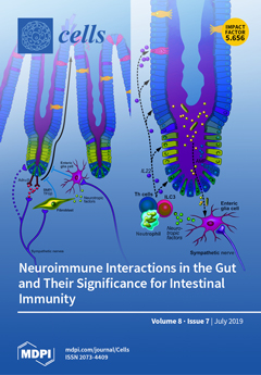
Cover Story (view full-size image): The sympathetic and parasympathetic nervous systems are essential modulators of the intestinal immune system, with the enteric nervous system acting as a potential interface. It remains, however, to be elucidated which specific nerves, neurotransmitters, neuropeptides, and receptors are accountable and which immune cells are involved. Adrenergic and cholinergic neuronal activity can further modulate the homeostasis of specialized epithelial cells in the crypt stem cell niche and hence also impact the mucosal barrier and the intestinal microbiota. The autonomic nervous system is therefore a potential therapeutic target for intestinal inflammatory diseases, and the advancement of new neuromodulatory techniques such as bioelectronic implantable devices must be pursued. View this paper.
  • Issues are regarded as officially published after their release is announced to the table of contents alert mailing list.
  • You may sign up for e-mail alerts to receive table of contents of newly released issues.
  • PDF is the official format for papers published in both, html and pdf forms. To view the papers in pdf format, click on the "PDF Full-text" link, and use the free Adobe Reader to open them.
Order results
Result details
Section
Select all
Export citation of selected articles as:
17 pages, 3140 KiB  
Article
Ensemble of Deep Recurrent Neural Networks for Identifying Enhancers via Dinucleotide Physicochemical Properties
by Kok Keng Tan, Nguyen Quoc Khanh Le, Hui-Yuan Yeh and Matthew Chin Heng Chua
Cells 2019, 8(7), 767; https://doi.org/10.3390/cells8070767 - 23 Jul 2019
Cited by 26 | Viewed by 4558
Abstract
Enhancers are short deoxyribonucleic acid fragments that assume an important part in the genetic process of gene expression. Due to their possibly distant location relative to the gene that is acted upon, the identification of enhancers is difficult. There are many published works [...] Read more.
Enhancers are short deoxyribonucleic acid fragments that assume an important part in the genetic process of gene expression. Due to their possibly distant location relative to the gene that is acted upon, the identification of enhancers is difficult. There are many published works focused on identifying enhancers based on their sequence information, however, the resulting performance still requires improvements. Using deep learning methods, this study proposes a model ensemble of classifiers for predicting enhancers based on deep recurrent neural networks. The input features of deep ensemble networks were generated from six types of dinucleotide physicochemical properties, which had outperformed the other features. In summary, our model which used this ensemble approach could identify enhancers with achieved sensitivity of 75.5%, specificity of 76%, accuracy of 75.5%, and MCC of 0.51. For classifying enhancers into strong or weak sequences, our model reached sensitivity of 83.15%, specificity of 45.61%, accuracy of 68.49%, and MCC of 0.312. Compared to the benchmark result, our results had higher performance in term of most measurement metrics. The results showed that deep model ensembles hold the potential for improving on the best results achieved to date using shallow machine learning methods. Full article
(This article belongs to the Special Issue Biocomputing and Synthetic Biology in Cells)
Show Figures

Figure 1

22 pages, 5465 KiB  
Article
Microglial Phenotyping in Neurodegenerative Disease Brains: Identification of Reactive Microglia with an Antibody to Variant of CD105/Endoglin
by Douglas G. Walker, Lih-Fen Lue, Thomas G. Beach and Ikuo Tooyama
Cells 2019, 8(7), 766; https://doi.org/10.3390/cells8070766 - 23 Jul 2019
Cited by 10 | Viewed by 6846
Abstract
Inflammation is considered a key pathological process in neurodegenerative diseases, including Alzheimer’s disease (AD) and Parkinson’s disease (PD), but there are still mechanisms not understood. In the brain, most microglia are performing essential homeostatic functions, but can also respond to pathogenic stimuli by [...] Read more.
Inflammation is considered a key pathological process in neurodegenerative diseases, including Alzheimer’s disease (AD) and Parkinson’s disease (PD), but there are still mechanisms not understood. In the brain, most microglia are performing essential homeostatic functions, but can also respond to pathogenic stimuli by producing harmful pro-inflammatory cytokines or free radicals. Distinguishing between damaging and homeostatic microglia in human diseased brain tissues is a challenge. This report describes findings using a monoclonal antibody to CD105/Endoglin (R&D Systems MAB1097) that identifies subtypes of activated microglia. CD105/Endoglin is a co-receptor for transforming growth factor beta (TGFβ) receptor that antagonizes TGFβ signaling. CD105/Endoglin is a marker for vascular endothelial cells, but was originally identified as a marker for activated macrophages. This antibody did not identify endothelial cells in brain sections, only microglia-like cells. In this study, we examined with this antibody tissue section from middle temporal gyrus derived from human brains from normal control subjects with low-plaque pathology, high-plaque pathology, and AD cases, and also substantia nigra samples from control and PD cases, in conjunction with antibodies to markers of pathology and microglia. In low-plaque pathology cases, CD105-positive microglia were mostly absent, but noticeably increased with increasing pathology. CD105-positive cells strongly colocalized with amyloid-beta plaques, but not phosphorylated tau positive tangles. In substantia nigra, strong microglial CD105 staining was observed in microglia associated with degenerating dopaminergic neurons and neuromelanin. In PD cases with few surviving dopaminergic neurons, this staining had decreased. By Western blot, this antibody identified polypeptide bands of 70 kDa in brain samples, and samples from microglia, macrophages, and brain endothelial cells. In comparison with other tested CD105 antibodies, this antibody did not recognize the glycosylated forms of CD105 on Western blots. Overall, the data indicate that this antibody and this marker could have utility for subtyping of microglia in pathologically-involved tissue. Full article
(This article belongs to the Special Issue Microglia in Neurodegenerative Diseases)
Show Figures

Graphical abstract

21 pages, 1680 KiB  
Article
Comparison of the RNA Content of Extracellular Vesicles Derived from Paracoccidioides brasiliensis and Paracoccidioides lutzii
by Roberta Peres da Silva, Larissa G. V. Longo, Julia P. C. da Cunha, Tiago J. P. Sobreira, Marcio L. Rodrigues, Helisson Faoro, Samuel Goldenberg, Lysangela R. Alves and Rosana Puccia
Cells 2019, 8(7), 765; https://doi.org/10.3390/cells8070765 - 23 Jul 2019
Cited by 34 | Viewed by 4531
Abstract
Paracoccidioides brasiliensis and P. lutzii cause human paracoccidioidomycosis. We have previously characterized the <200-nt RNA sub-populations contained in fungal extracellular vesicles (EVs) from P. brasiliensis Pb18 and other pathogenic fungi. We have presently used the RNA-seq strategy to compare the <200- and >200-nt [...] Read more.
Paracoccidioides brasiliensis and P. lutzii cause human paracoccidioidomycosis. We have previously characterized the <200-nt RNA sub-populations contained in fungal extracellular vesicles (EVs) from P. brasiliensis Pb18 and other pathogenic fungi. We have presently used the RNA-seq strategy to compare the <200- and >200-nt RNA fractions contained in EVs isolated from culture supernatants of P. brasiliensis Pb18, Pb3, and P. lutzii Pb01. Shared mRNA sequences were related to protein modification, translation, and DNA metabolism/biogenesis, while those related to transport and oxidation-reduction were exclusive to Pb01. The presence of functional full-length mRNAs was validated by in vitro translation. Among small non-coding (nc)RNA, 15 were common to all samples; small nucleolar (sno)RNAs were enriched in P. brasiliensis EVs, whereas for P. lutzii there were similar proportions of snoRNA, rRNA, and tRNA. Putative exonic sRNAs were highly abundant in Pb18 EVs. We also found sRNA sequences bearing incomplete microRNA structures mapping to exons. RNA-seq data suggest that extracellular fractions containing Pb18 EVs can modulate the transcriptome of murine monocyte-derived dendritic cells in a transwell system. Considering that sRNA classes are involved in transcription/translation modulation, our general results may indicate that differences in virulence among fungal isolates can be related to their distinct EV-RNA content. Full article
(This article belongs to the Section Intracellular and Plasma Membranes)
Show Figures

Figure 1

12 pages, 925 KiB  
Review
Mesenchymal Stem Cells for Perianal Crohn’s Disease
by Michele Carvello, Amy Lightner, Takayuki Yamamoto, Paulo Gustavo Kotze and Antonino Spinelli
Cells 2019, 8(7), 764; https://doi.org/10.3390/cells8070764 - 23 Jul 2019
Cited by 69 | Viewed by 9308
Abstract
Perianal fistulizing Crohn’s disease (PFCD) is associated with significant morbidity and might negatively impact the quality of life of CD patients. In the last two decades, the management of PFCD has evolved in terms of the multidisciplinary approach involving gastroenterologists and colorectal surgeons. [...] Read more.
Perianal fistulizing Crohn’s disease (PFCD) is associated with significant morbidity and might negatively impact the quality of life of CD patients. In the last two decades, the management of PFCD has evolved in terms of the multidisciplinary approach involving gastroenterologists and colorectal surgeons. However, the highest fistula healing rates, even combining surgical and anti-TNF agents, reaches 50% of treated patients. More recently, the administration of mesenchymal stem cells (MSCs) have shown notable promising results in the treatment of PFCD. The aim of this review is to describe the rationale and the possible mechanism of action of MSC application for PFCD and the most recent results of randomized clinical trials. Furthermore, the unmet needs of the current administration process and the expected next steps to improve the outcomes will be addressed. Full article
Show Figures

Figure 1

12 pages, 2175 KiB  
Article
Impact of Q141K on the Transport of Epidermal Growth Factor Receptor Tyrosine Kinase Inhibitors by ABCG2
by Yutaka Inoue, Takashi Morita, Mari Onozuka, Ken-ichi Saito, Kazumi Sano, Kazuhiko Hanada, Masami Kondo, Yoichi Nakamura, Tohru Kishino, Hiroshi Nakagawa and Yoji Ikegami
Cells 2019, 8(7), 763; https://doi.org/10.3390/cells8070763 - 23 Jul 2019
Cited by 6 | Viewed by 4051
Abstract
The ATP-binding cassette transporter ABCG2 is expressed in various organs, such as the small intestine, liver, and kidney, and influences the pharmacokinetics of drugs that are its substrates. ABCG2 is also expressed by cancer cells and mediates resistance to anticancer agents by promoting [...] Read more.
The ATP-binding cassette transporter ABCG2 is expressed in various organs, such as the small intestine, liver, and kidney, and influences the pharmacokinetics of drugs that are its substrates. ABCG2 is also expressed by cancer cells and mediates resistance to anticancer agents by promoting the efflux of these drugs. In the present study, we investigated the interactions between epidermal growth factor receptor tyrosine kinase inhibitors and ABCG2 by MTT assay, intracellular drug accumulation assay, and FACS. This study showed that four epidermal growth factor receptor tyrosine kinase inhibitors (EGFR TKIs) (gefitinib, erlotinib, lapatinib, and afatinib) were transported from tumor cells as substrates of ABCG2. Q141K is a common single-nucleotide polymorphism of ABCG2 in Asians. We demonstrated that the extracellular efflux of gefitinib, erlotinib, and lapatinib was reduced by Q141K, whereas afatinib transport was not affected. In addition, all four EGFR TKIs inhibited the transport of other substrates by both wild-type and variant ABCG2 at 0.1 μM concentrations. Accordingly, epidermal growth factor receptor tyrosine kinase inhibitors may induce interactions with other drugs that are substrates of ABCG2, and single-nucleotide polymorphisms of ABCG2 may influence both the pharmacokinetics and efficacy of these anticancer agents. Full article
(This article belongs to the Special Issue ABC Transporters: From Basic Functions to Diseases)
Show Figures

Figure 1

22 pages, 2851 KiB  
Article
Regulation of Acetylcholine Quantal Release by Coupled Thrombin/BDNF Signaling in Mouse Motor Synapses
by Alexander Gaydukov, Polina Bogacheva, Ekaterina Tarasova, Anastasia Molchanova, Anna Miteva, Ekaterina Pravdivceva and Olga Balezina
Cells 2019, 8(7), 762; https://doi.org/10.3390/cells8070762 - 22 Jul 2019
Cited by 11 | Viewed by 4061
Abstract
The aim of this study was to compare the acute effects of thrombin and brain-derived neurotrophic factor (BDNF) on spontaneous miniature endplate potentials (MEPPs) and multiquantal evoked endplate potentials (EPPs) in mouse neuromuscular junctions (NMJs) of m. diaphragma and m. EDL. Intracellular microelectrode [...] Read more.
The aim of this study was to compare the acute effects of thrombin and brain-derived neurotrophic factor (BDNF) on spontaneous miniature endplate potentials (MEPPs) and multiquantal evoked endplate potentials (EPPs) in mouse neuromuscular junctions (NMJs) of m. diaphragma and m. EDL. Intracellular microelectrode recordings of MEPPs and EPPs were used to evaluate the changes in acetylcholine (ACh) release in mature and newly-formed mouse NMJs. Thrombin (1 nM) increased the amplitude of MEPPs and EPPs by 25–30% in mature and newly-formed NMJs. This effect was due to an enhanced loading of synaptic vesicles with ACh and increase of ACh quantal size, since it was fully prevented by blocking of vesicular ACh transporter. It was also prevented by tropomyosin-related kinase B (TrkB) receptors inhibitor ANA12. Exogenous BDNF (1 nM) mimicked thrombin effect and increased the amplitude of MEPPs and EPPs by 25–30%. It required involvement of protein kinase A (PKA) and mitogen-activated protein kinase (MEK1/2)-mediated pathway, but not phospholipase C (PLC). Blocking A2A adenosine receptors by ZM241385 abolished the effect of BDNF, whereas additional stimulation of A2A receptors by CGS21680 increased MEPP amplitudes, which was prevented by MEK1/2 inhibitor U0126. At mature NMJs, BDNF enhanced MEPPs frequency by 30–40%. This effect was selectively prevented by inhibition of PLC, but not PKA or MEK1/2. It is suggested that interrelated effects of thrombin/BDNF in mature and newly-formed NMJs are realized via enhancement of vesicular ACh transport and quantal size increase. BDNF-induced potentiation of synaptic transmission involves the functional coupling between A2A receptor-dependent active PKA and neurotrophin-triggered MAPK pathway, as well as PLC-dependent increase in frequency of MEPPs. Full article
Show Figures

Figure 1

15 pages, 7764 KiB  
Article
Tumor Angiogenic Inhibition Triggered Necrosis (TAITN) in Oral Cancer
by Saori Yoshida, Hotaka Kawai, Takanori Eguchi, Shintaro Sukegawa, May Wathone Oo, Chang Anqi, Kiyofumi Takabatake, Keisuke Nakano, Kuniaki Okamoto and Hitoshi Nagatsuka
Cells 2019, 8(7), 761; https://doi.org/10.3390/cells8070761 - 22 Jul 2019
Cited by 17 | Viewed by 5943
Abstract
CXCR4 is a chemokine receptor crucial in tumor progression, although the angiogenic role of CXCR4 in oral squamous cell carcinoma (OSCC) has not been investigated. Here we show that CXCR4 is crucial for tumor angiogenesis, thereby supporting tumor survival in OSCC. Immunohistochemistry on [...] Read more.
CXCR4 is a chemokine receptor crucial in tumor progression, although the angiogenic role of CXCR4 in oral squamous cell carcinoma (OSCC) has not been investigated. Here we show that CXCR4 is crucial for tumor angiogenesis, thereby supporting tumor survival in OSCC. Immunohistochemistry on human clinical specimens revealed that CXCR4 and a tumor vasculature marker CD34 were co-distributed in tumor vessels in human OSCC specimens. To uncover the effects of CXCR4 inhibition, we treated the OSCC-xenografted mice with AMD3100, so-called plerixafor, an antagonist of CXCR4. Notably, we found a unique pathophysiological structure defined as tumor angiogenic inhibition triggered necrosis (TAITN), which was induced by the CXCR4 antagonism. Treatment with AMD3100 increased necrotic areas with the induction of hypoxia-inducible factor-1α in the xenografted tumors, suggesting that AMD3100-induced TAITN was involved in hypoxia and ischemia. Taken together, we demonstrated that CXCR4 plays a crucial role in tumor angiogenesis required for OSCC progression, whereas TAITN induced by CXCR4 antagonism could be an effective anti-angiogenic therapeutic strategy in OSCC treatment. Full article
(This article belongs to the Special Issue Angiogenesis in Cancer)
Show Figures

Graphical abstract

23 pages, 6926 KiB  
Article
Neurological Enhancement Effects of Melatonin against Brain Injury-Induced Oxidative Stress, Neuroinflammation, and Neurodegeneration via AMPK/CREB Signaling
by Shafiq Ur Rehman, Muhammad Ikram, Najeeb Ullah, Sayed Ibrar Alam, Hyun Young Park, Haroon Badshah, Kyonghwan Choe and Myeong Ok Kim
Cells 2019, 8(7), 760; https://doi.org/10.3390/cells8070760 - 21 Jul 2019
Cited by 118 | Viewed by 10904
Abstract
Oxidative stress and energy imbalance strongly correlate in neurodegenerative diseases. Repeated concussion is becoming a serious public health issue with uncontrollable adverse effects in the human population, which involve cognitive dysfunction and even permanent disability. Here, we demonstrate that traumatic brain injury (TBI) [...] Read more.
Oxidative stress and energy imbalance strongly correlate in neurodegenerative diseases. Repeated concussion is becoming a serious public health issue with uncontrollable adverse effects in the human population, which involve cognitive dysfunction and even permanent disability. Here, we demonstrate that traumatic brain injury (TBI) evokes oxidative stress, disrupts brain energy homeostasis, and boosts neuroinflammation, which further contributes to neuronal degeneration and cognitive dysfunction in the mouse brain. We also demonstrate that melatonin (an anti-oxidant agent) treatment exerts neuroprotective effects, while overcoming oxidative stress and energy depletion and reducing neuroinflammation and neurodegeneration. Male C57BL/6N mice were used as a model for repetitive mild traumatic brain injury (rmTBI) and were treated with melatonin. Protein expressions were examined via Western blot analysis, immunofluorescence, and ELISA; meanwhile, behavior analysis was performed through a Morris water maze test, and Y-maze and beam-walking tests. We found elevated oxidative stress, depressed phospho-5′AMP-activated protein kinase (p-AMPK) and phospho- CAMP-response element-binding (p-CREB) levels, and elevated p-NF-κB in rmTBI mouse brains, while melatonin treatment significantly regulated p-AMPK, p-CREB, and p-NF-κB in the rmTBI mouse brain. Furthermore, rmTBI mouse brains showed a deregulated mitochondrial system, abnormal amyloidogenic pathway activation, and cognitive functions which were significantly regulated by melatonin treatment in the mice. These findings provide evidence, for the first time, that rmTBI induces brain energy imbalance and reduces neuronal cell survival, and that melatonin treatment overcomes energy depletion and protects against brain damage via the regulation of p-AMPK/p-CREB signaling pathways in the mouse brain. Full article
(This article belongs to the Special Issue Melatonin in Human Health and Diseases)
Show Figures

Figure 1

19 pages, 6275 KiB  
Article
The Intrinsic GDP/GTP Exchange Activities of Cdc42 and Rac1 Are Critical Determinants for Their Specific Effects on Mobilization of the Actin Filament System
by Pontus Aspenström
Cells 2019, 8(7), 759; https://doi.org/10.3390/cells8070759 - 21 Jul 2019
Cited by 18 | Viewed by 4958
Abstract
The Rho GTPases comprise a subfamily of the Ras superfamily of small GTPases. Their importance in regulation of cell morphology and cell migration is well characterized. According to the prevailing paradigm, Cdc42 regulates the formation of filopodia, Rac1 regulates the formation of lamellipodia, [...] Read more.
The Rho GTPases comprise a subfamily of the Ras superfamily of small GTPases. Their importance in regulation of cell morphology and cell migration is well characterized. According to the prevailing paradigm, Cdc42 regulates the formation of filopodia, Rac1 regulates the formation of lamellipodia, and RhoA triggers the assembly of focal adhesions. However, this scheme is clearly an oversimplification, as the Rho subfamily encompasses 20 members with diverse effects on a number of vital cellular processes, including cytoskeletal dynamics and cell proliferation, migration, and invasion. This article highlights the importance of the catalytic activities of the classical Rho GTPases Cdc42 and Rac1, in terms of their specific effects on the dynamic reorganization of the actin filament system. GTPase-deficient mutants of Cdc42 and Rac1 trigger the formation of broad lamellipodia and stress fibers, and fast-cycling mutations trigger filopodia formation and stress fiber dissolution. The filopodia response requires the involvement of the formin family of actin nucleation promotors. In contrast, the formation of broad lamellipodia induced by GTPase-deficient Cdc42 and Rac1 is mediated through Arp2/3-dependent actin nucleation. Full article
Show Figures

Figure 1

19 pages, 3643 KiB  
Article
Copine A Interacts with Actin Filaments and Plays a Role in Chemotaxis and Adhesion
by Matthew J. Buccilli, April N. Ilacqua, Mingxi Han, Andrew A. Banas, Elise M. Wight, Hanqian Mao, Samantha P. Perry, Tasha S. Salter, David R. Loiselle, Timothy A.J. Haystead and Cynthia K. Damer
Cells 2019, 8(7), 758; https://doi.org/10.3390/cells8070758 - 21 Jul 2019
Cited by 5 | Viewed by 4385
Abstract
Copines make up a family of calcium-dependent, phospholipid-binding proteins found in numerous eukaryotic organisms. Copine proteins consist of two C2 domains at the N-terminus followed by an A domain similar to the von Willebrand A domain found in integrins. We are studying copine [...] Read more.
Copines make up a family of calcium-dependent, phospholipid-binding proteins found in numerous eukaryotic organisms. Copine proteins consist of two C2 domains at the N-terminus followed by an A domain similar to the von Willebrand A domain found in integrins. We are studying copine protein function in the model organism, Dictyostelium discoideum, which has six copine genes, cpnA-cpnF. Previous research showed that cells lacking the cpnA gene exhibited a cytokinesis defect, a contractile vacuole defect, and developmental defects. To provide insight into the role of CpnA in these cellular processes, we used column chromatography and immunoprecipitation to isolate proteins that bind to CpnA. These proteins were identified by mass spectrometry. One of the proteins identified was actin. Purified CpnA was shown to bind to actin filaments in a calcium-dependent manner in vitro. cpnA cells exhibited defects in three actin-based processes: chemotaxis, cell polarity, and adhesion. These results suggest that CpnA plays a role in chemotaxis and adhesion and may do so by interacting with actin filaments. Full article
(This article belongs to the Section Cell Motility and Adhesion)
Show Figures

Figure 1

9 pages, 778 KiB  
Review
The IL4I1 Enzyme: A New Player in the Immunosuppressive Tumor Microenvironment
by Valérie Molinier-Frenkel, Armelle Prévost-Blondel and Flavia Castellano
Cells 2019, 8(7), 757; https://doi.org/10.3390/cells8070757 - 20 Jul 2019
Cited by 43 | Viewed by 7769
Abstract
The high metabolic needs of T lymphocytes in response to activation make them particularly vulnerable to modifications of their biochemical milieu. Immunosuppressive enzymes produced in the tumor microenvironment modify nutrient availability by catabolizing essential or semi-essential amino acids and producing toxic catabolites, thus [...] Read more.
The high metabolic needs of T lymphocytes in response to activation make them particularly vulnerable to modifications of their biochemical milieu. Immunosuppressive enzymes produced in the tumor microenvironment modify nutrient availability by catabolizing essential or semi-essential amino acids and producing toxic catabolites, thus participating in the local sabotage of the antitumor immune response. L-amino-acid oxidases are FAD-bound enzymes found throughout evolution, from bacteria to mammals, and are often endowed with anti-infectious properties. IL4I1 is a secreted L-phenylalanine oxidase mainly produced by inflammatory antigen-presenting cells—in particular, macrophages present in T helper type 1 granulomas and in various types of tumors. In the last decade, it has been shown that IL4I1 is involved in the fine control of B- and T-cell adaptive immune responses. Preclinical models have revealed its role in cancer immune evasion. Recent clinical data highlight IL4I1 as a new potential prognostic marker in human melanoma. As a secreted enzyme, IL4I1 may represent an easily targetable molecule for cancer immunotherapy. Full article
(This article belongs to the Special Issue Tumor Microenvironment: Interaction and Metabolism)
Show Figures

Figure 1

10 pages, 8751 KiB  
Article
Myeloid Dendritic Cells Are Enriched in Lymph Node Tissue of Early Rheumatoid Arthritis Patients but not in At Risk Individuals
by T.H. Ramwadhdoebe, M.I. Ramos, K.I. Maijer, K.P. van Lienden, M. Maas, D.M. Gerlag, P.P. Tak, M.C. Lebre and L.G.M. van Baarsen
Cells 2019, 8(7), 756; https://doi.org/10.3390/cells8070756 - 20 Jul 2019
Cited by 7 | Viewed by 4558
Abstract
Lymph nodes (LNs) are highly organized structures where specific immune responses are initiated by dendritic cells (DCs). We investigated the frequency and distribution of human myeloid (mDCs) and plasmacytoid (pDCs) in LNs and blood during the earliest phases of rheumatoid arthritis (RA). We [...] Read more.
Lymph nodes (LNs) are highly organized structures where specific immune responses are initiated by dendritic cells (DCs). We investigated the frequency and distribution of human myeloid (mDCs) and plasmacytoid (pDCs) in LNs and blood during the earliest phases of rheumatoid arthritis (RA). We included 22 RA-risk individuals positive for IgM rheumatoid factor and/or anti-citrullinated protein antibodies, 16 biological-naïve RA patients and 8 healthy controls (HCs). DC subsets (CD1c+ mDCs and CD304+ pDCs) in LN tissue and paired peripheral blood were analyzed using flow cytometry and confocal microscopy. In blood of RA patients a significant decreased frequency of pDCs was found, with a similar trend for mDCs. In contrast, mDC frequencies were higher in RA compared with HCs and RA-risk individuals, especially in LN. Frequency of mDCs seemed higher in LNs compared to paired blood samples in all donors, while pDCs were higher in LNs only in RA patients. As expected, both mDCs and pDCs localized mainly in T-cell areas of LN tissue. In conclusion, compared with RA-risk individuals, mDCs and pDCs were enriched in the LN tissue of early-RA patients, while their frequency in RA-risk individuals was comparable to HCs. This may suggest that other antigen-presenting cells are responsible for initial breaks of tolerance, while mDCs and pDCs are involved in sustaining inflammation. Full article
(This article belongs to the Special Issue The Molecular and Cellular Basis for Rheumatoid Arthritis)
Show Figures

Figure 1

13 pages, 1910 KiB  
Article
S100-EPISPOT: A New Tool to Detect Viable Circulating Melanoma Cells
by Laure Cayrefourcq, Aurélie De Roeck, Caroline Garcia, Pierre-Emmanuel Stoebner, Fanny Fichel, Françoise Garima, Françoise Perriard, Jean-Pierre Daures, Laurent Meunier and Catherine Alix-Panabières
Cells 2019, 8(7), 755; https://doi.org/10.3390/cells8070755 - 20 Jul 2019
Cited by 24 | Viewed by 4279
Abstract
Metastatic melanoma is one of the most aggressive and drug-resistant cancers with very poor overall survival. Circulating melanoma cells (CMCs) were first described in 1991. However, there is no general consensus on the clinical utility of CMC detection, largely due to conflicting results [...] Read more.
Metastatic melanoma is one of the most aggressive and drug-resistant cancers with very poor overall survival. Circulating melanoma cells (CMCs) were first described in 1991. However, there is no general consensus on the clinical utility of CMC detection, largely due to conflicting results linked to the use of heterogeneous patient populations and different detection methods. Here, we developed a new EPithelial ImmunoSPOT (EPISPOT) assay to detect viable CMCs based on their secretion of the S100 protein (S100-EPISPOT). Then, we compared the results obtained with the S100-EPISPOT assay and the CellSearch® CMC kit using blood samples from a homogeneous population of patients with metastatic melanoma. We found that S100-EPISPOT sensitivity was significantly higher than that of CellSearch®. Specifically, the percentage of patients with ≥2 CMCs was significantly higher using S100-EPISPOT than CellSearch® (48% and 21%, respectively; p = 0.0114). Concerning CMC prognostic value, only the CellSearch® results showed a significant association with overall survival (p = 0.006). However, due to the higher sensitivity of the new S100-EPISPOT assay, it would be interesting to determine whether this functional test could be used in patients with non-metastatic melanoma for the early detection of tumor relapse and for monitoring the treatment response. Full article
Show Figures

Figure 1

12 pages, 2041 KiB  
Article
Transcription Factors Indirectly Regulate Genes through Nuclear Colocalization
by Zhiming Dai
Cells 2019, 8(7), 754; https://doi.org/10.3390/cells8070754 - 20 Jul 2019
Cited by 6 | Viewed by 3737
Abstract
Various types of data, including genomic sequences, transcription factor (TF) knockout data, TF-DNA interaction and expression profiles, have been used to decipher TF regulatory mechanisms. However, most of the genes affected by knockout of a particular TF are not bound by that factor. [...] Read more.
Various types of data, including genomic sequences, transcription factor (TF) knockout data, TF-DNA interaction and expression profiles, have been used to decipher TF regulatory mechanisms. However, most of the genes affected by knockout of a particular TF are not bound by that factor. Here, I showed that this interesting result can be partially explained by considering the nuclear positioning of TF knockout affected genes and TF bound genes. I found that a statistically significant number of TF knockout affected genes show nuclear colocalization with genes bound by the corresponding TF. Although these TF knockout affected genes are not directly bound by the corresponding TF; the TF tend to be in the same cellular component with the TFs that directly bind these genes. TF knockout affected genes show co-expression and tend to be involved in the same biological process with the spatially adjacent genes that are bound by the corresponding TF. These results demonstrate that TFs can regulate genes through nuclear colocalization without direct DNA binding, complementing the conventional view that TFs directly bind DNA to regulate genes. My findings will have implications in understanding TF regulatory mechanisms. Full article
(This article belongs to the Special Issue Bioinformatics and Computational Biology 2019)
Show Figures

Figure 1

18 pages, 6711 KiB  
Article
Annexin A1 Contained in Extracellular Vesicles Promotes the Activation of Keratinocytes by Mesoglycan Effects: An Autocrine Loop Through FPRs
by Emanuela Pessolano, Raffaella Belvedere, Valentina Bizzarro, Paola Franco, Iolanda De Marco, Francesco Petrella, Amalia Porta, Alessandra Tosco, Luca Parente, Mauro Perretti and Antonello Petrella
Cells 2019, 8(7), 753; https://doi.org/10.3390/cells8070753 - 19 Jul 2019
Cited by 31 | Viewed by 4461
Abstract
We have recently demonstrated that mesoglycan, a fibrinolytic compound, may be a promising pro-healing drug for skin wound repair. We showed that mesoglycan induces migration, invasion, early differentiation, and translocation to the membrane of keratinocytes, as well as the secretion of annexin A1 [...] Read more.
We have recently demonstrated that mesoglycan, a fibrinolytic compound, may be a promising pro-healing drug for skin wound repair. We showed that mesoglycan induces migration, invasion, early differentiation, and translocation to the membrane of keratinocytes, as well as the secretion of annexin A1 (ANXA1), further involved in keratinocytes activation. These events are triggered by the syndecan-4 (SDC4)/PKCα pathway. SDC4 also participates to the formation and secretion of microvesicles (EVs) which may contribute to wound healing. EVs were isolated from HaCaT cells, as human immortalized keratinocytes, and then characterised by Western blotting, Field Emission-Scanning Electron Microscopy, and Dynamic Light Scattering. Their autocrine effects were investigated by Wound-Healing/invasion assays and confocal microscopy to analyse cell motility and differentiation, respectively. Here, we found that the mesoglycan increased the release of EVs which amplify its same effects. ANXA1 contained in the microvesicles is able to promote keratinocytes motility and differentiation by acting on Formyl Peptide Receptors (FPRs). Thus, the extracellular form of ANXA1 may be considered as a link to intensify the effects of mesoglycan. In this study, for the first time, we have identified an interesting autocrine loop ANXA1/EVs/FPRs in human keratinocytes, induced by mesoglycan. Full article
Show Figures

Figure 1

16 pages, 1546 KiB  
Review
Dual Roles of the AMP-Activated Protein Kinase Pathway in Angiogenesis
by Yuanjun Li, Ruipu Sun, Junrong Zou, Ying Ying and Zhijun Luo
Cells 2019, 8(7), 752; https://doi.org/10.3390/cells8070752 - 19 Jul 2019
Cited by 72 | Viewed by 8845
Abstract
Angiogenesis plays important roles in development, stress response, wound healing, tumorigenesis and cancer progression, diabetic retinopathy, and age-related macular degeneration. It is a complex event engaging many signaling pathways including vascular endothelial growth factor (VEGF), Notch, transforming growth factor-beta/bone morphogenetic proteins (TGF-β/BMPs), and [...] Read more.
Angiogenesis plays important roles in development, stress response, wound healing, tumorigenesis and cancer progression, diabetic retinopathy, and age-related macular degeneration. It is a complex event engaging many signaling pathways including vascular endothelial growth factor (VEGF), Notch, transforming growth factor-beta/bone morphogenetic proteins (TGF-β/BMPs), and other cytokines and growth factors. Almost all of them eventually funnel to two crucial molecules, VEGF and hypoxia-inducing factor-1 alpha (HIF-1α) whose expressions could change under both physiological and pathological conditions. Hypoxic conditions stabilize HIF-1α, while it is upregulated by many oncogenic factors under normaxia. HIF-1α is a critical transcription activator for VEGF. Recent studies have shown that intracellular metabolic state participates in regulation of sprouting angiogenesis, which may involve AMP-activated protein kinase (AMPK). Indeed, AMPK has been shown to exert both positive and negative effects on angiogenesis. On the one hand, activation of AMPK mediates stress responses to facilitate autophagy which stabilizes HIF-1α, leading to increased expression of VEGF. On the other hand, AMPK could attenuate angiogenesis induced by tumor-promoting and pro-metastatic factors, such as the phosphoinositide 3-kinase /protein kinase B (Akt)/mammalian target of rapamycin (PI3K/Akt/mTOR), hepatic growth factor (HGF), and TGF-β/BMP signaling pathways. Thus, this review will summarize research progresses on these two opposite effects and discuss the mechanisms behind the discrepant findings. Full article
(This article belongs to the Special Issue Angiogenesis in Cancer)
Show Figures

Figure 1

14 pages, 2066 KiB  
Article
HLA-DQA1 and HLA-DQB1 Alleles, Conferring Susceptibility to Celiac Disease and Type 1 Diabetes, Are More Expressed Than Non-Predisposing Alleles and Are Coordinately Regulated
by Federica Farina, Stefania Picascia, Laura Pisapia, Pasquale Barba, Serena Vitale, Adriana Franzese, Enza Mozzillo, Carmen Gianfrani and Giovanna Del Pozzo G
Cells 2019, 8(7), 751; https://doi.org/10.3390/cells8070751 - 19 Jul 2019
Cited by 32 | Viewed by 6687
Abstract
HLA DQA1*05 and DQB1*02 alleles encoding the DQ2.5 molecule and HLA DQA1*03 and DQB1*03 alleles encoding DQ8 molecules are strongly associated with celiac disease (CD) and type 1 diabetes (T1D), two common autoimmune diseases (AD). We previously demonstrated that DQ2.5 genes showed a [...] Read more.
HLA DQA1*05 and DQB1*02 alleles encoding the DQ2.5 molecule and HLA DQA1*03 and DQB1*03 alleles encoding DQ8 molecules are strongly associated with celiac disease (CD) and type 1 diabetes (T1D), two common autoimmune diseases (AD). We previously demonstrated that DQ2.5 genes showed a higher expression with respect to non-CD associated alleles in heterozygous DQ2.5 positive (HLA DR1/DR3) antigen presenting cells (APC) of CD patients. This differential expression affected the level of the encoded DQ2.5 molecules on the APC surface and established the strength of gluten-specific CD4+ T cells response. Here, we expanded the expression analysis of risk alleles in patients affected by T1D or by T1D and CD comorbidity. In agreement with previous findings, we found that DQ2.5 and DQ8 risk alleles are more expressed than non-associated alleles also in T1D patients and favor the self-antigen presentation. To investigate the mechanism causing the high expression of risk alleles, we focused on HLA DQA1*05 and DQB1*02 alleles and, by ectopic expression of a single mRNA, we modified the quantitative equilibrium among the two transcripts. After transfection of DR7/DR14 B-LCL with HLA-DQA1*05 cDNA, we observed an overexpression of the endogenous DQB1*02 allele. The DQ2.5 heterodimer synthesized was functional and able to present gluten antigens to cognate CD4+ T cells. Our results indicated that the high expression of alpha and beta transcripts, encoding for the DQ2.5 heterodimeric molecules, was strictly coordinated by a mechanism acting at a transcriptional level. These findings suggested that, in addition to the predisposing HLA-DQ genotype, also the expression of risk alleles contributed to the establishment of autoimmunity. Full article
(This article belongs to the Special Issue Major Histocompatibility Complex (MHC) in Health and Disease)
Show Figures

Figure 1

14 pages, 3956 KiB  
Brief Report
Autoantibodies Specific to ERα are Involved in Tamoxifen Resistance in Hormone Receptor Positive Breast Cancer
by Angela Maselli, Stefania Parlato, Rossella Puglisi, Carla Raggi, Massimo Spada, Daniele Macchia, Giada Pontecorvi, Elisabetta Iessi, Maria Teresa Pagano, Francesca Cirulli, Lucia Gabriele, Alessandra Carè, Patrizia Vici, Laura Pizzuti, Maddalena Barba, Paola Matarrese, Marina Pierdominici and Elena Ortona
Cells 2019, 8(7), 750; https://doi.org/10.3390/cells8070750 - 19 Jul 2019
Cited by 7 | Viewed by 3679
Abstract
Tamoxifen resistance is a major hurdle in the treatment of estrogen receptor (ER)-positive breast cancer. The mechanisms of tamoxifen resistance are not fully understood although several underlying molecular events have been suggested. Recently, we identified autoantibodies reacting with membrane-associated ERα (anti-ERα Abs) in [...] Read more.
Tamoxifen resistance is a major hurdle in the treatment of estrogen receptor (ER)-positive breast cancer. The mechanisms of tamoxifen resistance are not fully understood although several underlying molecular events have been suggested. Recently, we identified autoantibodies reacting with membrane-associated ERα (anti-ERα Abs) in sera of breast cancer patients, able to promote tumor growth. Here, we investigated whether anti-ERα Abs purified from sera of ER-positive breast cancer patients could contribute to tamoxifen resistance. Anti-ERα Abs inhibited tamoxifen-mediated effects on cell cycle and proliferation in MCF-7 cells. Moreover, anti-ERα Abs hampered the tamoxifen-mediated reduction of tumor growth in SCID mice xenografted with breast tumor. Notably, simvastatin-mediated disaggregation of lipid rafts, where membrane-associated ERα is embedded, restored tamoxifen sensitivity, preventing anti-ERα Abs effects. In conclusion, detection of serum anti-ERα Abs may help predict tamoxifen resistance and concur to appropriately inform therapeutic decisions concerning hormone therapy in ER-positive breast cancer patients. Full article
(This article belongs to the Special Issue Estrogen Receptor Expression and Function in Health and Disease)
Show Figures

Figure 1

27 pages, 1109 KiB  
Review
Dicarbonyl Stress at the Crossroads of Healthy and Unhealthy Aging
by Cecilia Nigro, Alessia Leone, Francesca Fiory, Immacolata Prevenzano, Antonella Nicolò, Paola Mirra, Francesco Beguinot and Claudia Miele
Cells 2019, 8(7), 749; https://doi.org/10.3390/cells8070749 - 19 Jul 2019
Cited by 54 | Viewed by 7682
Abstract
Dicarbonyl stress occurs when dicarbonyl metabolites (i.e., methylglyoxal, glyoxal and 3-deoxyglucosone) accumulate as a consequence of their increased production and/or decreased detoxification. This toxic condition has been associated with metabolic and age-related diseases, both of which are characterized by a pro-inflammatory and pro-oxidant [...] Read more.
Dicarbonyl stress occurs when dicarbonyl metabolites (i.e., methylglyoxal, glyoxal and 3-deoxyglucosone) accumulate as a consequence of their increased production and/or decreased detoxification. This toxic condition has been associated with metabolic and age-related diseases, both of which are characterized by a pro-inflammatory and pro-oxidant state. Methylglyoxal (MGO) is the most reactive dicarbonyl and the one with the highest endogenous flux. It is the precursor of the major quantitative advanced glycated products (AGEs) in physiological systems, arginine-derived hydroimidazolones, which accumulate in aging and dysfunctional tissues. The aging process is characterized by a decline in the functional properties of cells, tissues and whole organs, starting from the perturbation of crucial cellular processes, including mitochondrial function, proteostasis and stress-scavenging systems. Increasing studies are corroborating the causal relationship between MGO-derived AGEs and age-related tissue dysfunction, unveiling a previously underestimated role of dicarbonyl stress in determining healthy or unhealthy aging. This review summarizes the latest evidence supporting a causal role of dicarbonyl stress in age-related diseases, including diabetes mellitus, cardiovascular disease and neurodegeneration. Full article
(This article belongs to the Special Issue Glycation and Dicarbonyl Stress in Aging and Disease)
Show Figures

Graphical abstract

19 pages, 310 KiB  
Review
Golgi Fragmentation in Neurodegenerative Diseases: Is There a Common Cause?
by José Ángel Martínez-Menárguez, Mónica Tomás, Narcisa Martínez-Martínez and Emma Martínez-Alonso
Cells 2019, 8(7), 748; https://doi.org/10.3390/cells8070748 - 19 Jul 2019
Cited by 40 | Viewed by 6513
Abstract
In most mammalian cells, the Golgi complex forms a continuous ribbon. In neurodegenerative diseases, the Golgi ribbon of a specific group of neurons is typically broken into isolated elements, a very early event which happens before clinical and other pathological symptoms become evident. [...] Read more.
In most mammalian cells, the Golgi complex forms a continuous ribbon. In neurodegenerative diseases, the Golgi ribbon of a specific group of neurons is typically broken into isolated elements, a very early event which happens before clinical and other pathological symptoms become evident. It is not known whether this phenomenon is caused by mechanisms associated with cell death or if, conversely, it triggers apoptosis. When the phenomenon was studied in diseases such as Parkinson’s and Alzheimer’s or amyotrophic lateral sclerosis, it was attributed to a variety of causes, including the presence of cytoplasmatic protein aggregates, malfunctioning of intracellular traffic and/or alterations in the cytoskeleton. In the present review, we summarize the current findings related to these and other neurodegenerative diseases and try to search for clues on putative common causes. Full article
(This article belongs to the Special Issue Membrane Traffic in Health and Disease)
17 pages, 1383 KiB  
Review
Macrophage-Mediated Subversion of Anti-Tumour Immunity
by Valeria Quaranta and Michael C. Schmid
Cells 2019, 8(7), 747; https://doi.org/10.3390/cells8070747 - 19 Jul 2019
Cited by 60 | Viewed by 8820
Abstract
Despite the incredible clinical benefits obtained by the use of immune checkpoint blockers (ICBs), resistance is still common for many types of cancer. Central for ICBs to work is activation and infiltration of cytotoxic CD8+ T cells following tumour-antigen recognition. However, it [...] Read more.
Despite the incredible clinical benefits obtained by the use of immune checkpoint blockers (ICBs), resistance is still common for many types of cancer. Central for ICBs to work is activation and infiltration of cytotoxic CD8+ T cells following tumour-antigen recognition. However, it is now accepted that even in the case of immunogenic tumours, the effector functions of CD8+ T cells are highly compromised by the presence of an immunosuppressive tumour microenvironment (TME) at the tumour site. Tumour-associated macrophages (TAMs) are among the most abundant non-malignant stromal cell types within the TME and they are crucial drivers of tumour progression, metastasis and resistance to therapy. TAMs are able to regulate either directly or indirectly various aspects of tumour immunity, including T cell recruitment and functions. In this review we discuss the mechanisms by which TAMs subvert CD8+ T cell immune surveillance and how their targeting in combination with ICBs represents a very powerful therapeutic strategy. Full article
Show Figures

Figure 1

15 pages, 3432 KiB  
Article
Roscovitine Attenuates Microglia Activation and Monocyte Infiltration via p38 MAPK Inhibition in the Rat Frontoparietal Cortex Following Status Epilepticus
by Ji-Eun Kim, Hana Park, Seo-Hyeon Choi, Min-Jeong Kong and Tae-Cheon Kang
Cells 2019, 8(7), 746; https://doi.org/10.3390/cells8070746 - 19 Jul 2019
Cited by 24 | Viewed by 4451
Abstract
Under physiological conditions, microglia are unique immune cells resident in the brain that is isolated from the systemic immune system by brain-blood barrier. Following status epilepticus (SE, a prolonged seizure activity), microglia are rapidly activated and blood-derived monocytes that infiltrate the brain; therefore, [...] Read more.
Under physiological conditions, microglia are unique immune cells resident in the brain that is isolated from the systemic immune system by brain-blood barrier. Following status epilepticus (SE, a prolonged seizure activity), microglia are rapidly activated and blood-derived monocytes that infiltrate the brain; therefore, the regulations of microglia activation and monocyte infiltration are one of the primary therapeutic strategies for inhibition of undesirable consequences from SE. Roscovitine, a potent (but not selective) cyclin-dependent kinase 5 (CDK5) inhibitor, has been found to exert anti-inflammatory and microglia-inhibiting actions in several in vivo models, although the underlying mechanisms have not been clarified. In the present study, roscovitine attenuated SE-induces monocyte infiltration without vasogenic edema formation in the frontoparietal cortex (FPC), accompanied by reducing expressions of monocyte chemotactic protein-1 (MCP-1) and lysosome-associated membrane protein 1 (LAMP1) in resident microglia, while it did not affect microglia transformation to amoeboid form. Furthermore, roscovitine ameliorated the up-regulation of p38 mitogen-activated protein kinase (p38 MAPK) phosphorylation, but not nuclear factor-κB-S276 phosphorylation. Similar to roscovitine, SB202190, a p38 MAPK inhibitor, mitigated monocyte infiltration and microglial expressions of MCP-1 and LAMP1 in the FPC following SE. Therefore, these findings suggest for the first time that roscovitine may inhibit SE-induced neuroinflammation via regulating p38 MAPK-mediated microglial responses. Full article
(This article belongs to the Special Issue Study around Neuroinflammation)
Show Figures

Figure 1

18 pages, 6839 KiB  
Article
Melatonin and (−)-Epigallocatechin-3-Gallate: Partners in Fighting Cancer
by Lingyun Zhang, Yufeng He, Ximing Wu, Guangshan Zhao, Ke Zhang, Chung S. Yang, Russel J. Reiter and Jinsong Zhang
Cells 2019, 8(7), 745; https://doi.org/10.3390/cells8070745 - 19 Jul 2019
Cited by 23 | Viewed by 4384
Abstract
We have demonstrated previously that melatonin attenuates hepatotoxicity triggered by high doses of (−)-epigallocatechin-3-gallate (EGCG) in mice. The current work investigated the influence of melatonin on the oncostatic activity of EGCG in two cancer cell lines, wherein melatonin induced an opposite response of [...] Read more.
We have demonstrated previously that melatonin attenuates hepatotoxicity triggered by high doses of (−)-epigallocatechin-3-gallate (EGCG) in mice. The current work investigated the influence of melatonin on the oncostatic activity of EGCG in two cancer cell lines, wherein melatonin induced an opposite response of p21. In human tongue cancer TCA8113 cells, melatonin-induced p21 and EGCG-mediated formation of quinoproteins were positively associated with the oncostatic effects of melatonin and EGCG. Melatonin-stimulated an increase in p21 which was correlated with a pronounced nuclear translocation of thioredoxin 1 and thioredoxin reductase 1, both of which are known to induce p21 via promoting p53 trans-activation. Melatonin did not influence the EGCG-mediated increase of quinoprotein formation nor did EGCG impair melatonin-induced p21 up-regulation. Co-treatment with both agents enhanced the cell-killing effect as well as the inhibitory activities against cell migration and colony formation. It is known that p21 also plays a powerful anti-apoptotic role in some cancer cells and confers these cells with a survival advantage, making it a target for therapeutic suppression. In human hepatocellular carcinoma HepG2 cells, melatonin suppressed p21 along with the induction of pro-survival proteins, PI3K and COX-2. However, EGCG prevented against melatonin-induced PI3K and COX-2, and melatonin probably sensitized HepG2 cells to EGCG cytotoxicity via down-regulating p21, Moreover, COX-2 and HO-1 were significantly reduced only by the co-treatment, and melatonin aided EGCG to achieve an increased inhibition on Bcl2 and NFκB. These events occurring in the co-treatment collectively resulted in an enhanced cytotoxicity. In addition, the co-treatment also enhanced the inhibitory activities against cell migration and colony formation. Overall, the results gathered from these two cancer cell lines with a divergent p21 response to melatonin show that the various oncostatic activities of melatonin and EGCG together are more robust than each agent alone, suggesting that they may be useful partners in fighting cancer. Full article
(This article belongs to the Special Issue Melatonin in Human Health and Diseases)
Show Figures

Figure 1

17 pages, 3456 KiB  
Article
Large Scale Profiling of Protein Isoforms Using Label-Free Quantitative Proteomics Revealed the Regulation of Nonsense-Mediated Decay in Moso Bamboo (Phyllostachys edulis)
by Xiaolan Yu, Yongsheng Wang, Markus V. Kohnen, Mingxin Piao, Min Tu, Yubang Gao, Chentao Lin, Zecheng Zuo and Lianfeng Gu
Cells 2019, 8(7), 744; https://doi.org/10.3390/cells8070744 - 19 Jul 2019
Cited by 11 | Viewed by 5130
Abstract
Moso bamboo is an important forest species with a variety of ecological, economic, and cultural values. However, the gene annotation information of moso bamboo is only based on the transcriptome sequencing, lacking the evidence of proteome. The lignification and fiber in moso bamboo [...] Read more.
Moso bamboo is an important forest species with a variety of ecological, economic, and cultural values. However, the gene annotation information of moso bamboo is only based on the transcriptome sequencing, lacking the evidence of proteome. The lignification and fiber in moso bamboo leads to a difficulty in the extraction of protein using conventional methods, which seriously hinders research on the proteomics of moso bamboo. The purpose of this study is to establish efficient methods for extracting the total proteins from moso bamboo for following mass spectrometry-based quantitative proteome identification. Here, we have successfully established a set of efficient methods for extracting total proteins of moso bamboo followed by mass spectrometry-based label-free quantitative proteome identification, which further improved the protein annotation of moso bamboo genes. In this study, 10,376 predicted coding genes were confirmed by quantitative proteomics, accounting for 35.8% of all annotated protein-coding genes. Proteome analysis also revealed the protein-coding potential of 1015 predicted long noncoding RNA (lncRNA), accounting for 51.03% of annotated lncRNAs. Thus, mass spectrometry-based proteomics provides a reliable method for gene annotation. Especially, quantitative proteomics revealed the translation patterns of proteins in moso bamboo. In addition, the 3284 transcript isoforms from 2663 genes identified by Pacific BioSciences (PacBio) single-molecule real-time long-read isoform sequencing (Iso-Seq) was confirmed on the protein level by mass spectrometry. Furthermore, domain analysis of mass spectrometry-identified proteins encoded in the same genomic locus revealed variations in domain composition pointing towards a functional diversification of protein isoform. Finally, we found that part transcripts targeted by nonsense-mediated mRNA decay (NMD) could also be translated into proteins. In summary, proteomic analysis in this study improves the proteomics-assisted genome annotation of moso bamboo and is valuable to the large-scale research of functional genomics in moso bamboo. In summary, this study provided a theoretical basis and technical support for directional gene function analysis at the proteomics level in moso bamboo. Full article
Show Figures

Figure 1

4 pages, 177 KiB  
Editorial
Advances in Autophagy, Tissue Injury, and Homeostasis: Cells Special Issue
by Pei-Hui Lin
Cells 2019, 8(7), 743; https://doi.org/10.3390/cells8070743 - 19 Jul 2019
Cited by 4 | Viewed by 2812
Abstract
Macroautophagy (hereafter referred to as autophagy, a word derived from Greek meaning “auto-digestion”) is a lysosome-dependent quality control process to degrade and turnover damaged or senescent organelles and proteins for cellular renewal [...] Full article
(This article belongs to the Special Issue Autophagy in Tissue Injury and Homeostasis)
11 pages, 1605 KiB  
Article
Physical Exercise Modulates miR-21-5p, miR-129-5p, miR-378-5p, and miR-188-5p Expression in Progenitor Cells Promoting Osteogenesis
by Maria Teresa Valenti, Michela Deiana, Samuele Cheri, Monica Dotta, Francesco Zamboni, Daniele Gabbiani, Federico Schena, Luca Dalle Carbonare and Monica Mottes
Cells 2019, 8(7), 742; https://doi.org/10.3390/cells8070742 - 19 Jul 2019
Cited by 43 | Viewed by 5707
Abstract
Physical exercise is known to promote beneficial effects on overall health, counteracting risks related to degenerative diseases. MicroRNAs (miRNAs), short non-coding RNAs affecting the expression of a cell’s transcriptome, can be modulated by different stimuli. Yet, the molecular effects on osteogenic differentiation triggered [...] Read more.
Physical exercise is known to promote beneficial effects on overall health, counteracting risks related to degenerative diseases. MicroRNAs (miRNAs), short non-coding RNAs affecting the expression of a cell’s transcriptome, can be modulated by different stimuli. Yet, the molecular effects on osteogenic differentiation triggered by miRNAs upon physical exercise are not completely understood. In this study, we recruited 20 male amateur runners participating in a half marathon. Runners’ sera, collected before (PRE RUN) and after (POST RUN) the run, were added to cultured human mesenchymal stromal cells. We then investigated their effects on the modulation of selected miRNAs and the consequential effects on osteogenic differentiation. Our results showed an increased expression of miRNAs promoting osteogenic differentiation (miR-21-5p, miR-129-5p, and miR-378-5p) and a reduced expression of miRNAs involved in the adipogenic differentiation of progenitor cells (miR-188-5p). In addition, we observed the downregulation of PTEN and SMAD7 expression along with increased AKT/pAKT and SMAD4 protein levels in MSCs treated with POST RUN sera. The consequent upregulation of RUNX2 expression was also proven, highlighting the molecular mechanisms by which miR-21-5p promotes osteogenic differentiation. In conclusion, our work proposes novel data, which demonstrate how miRNAs may regulate the osteogenic commitment of progenitor cells in response to physical exercise. Full article
(This article belongs to the Collection Regulatory Functions of microRNAs)
Show Figures

Graphical abstract

28 pages, 2849 KiB  
Article
Protein Quality Control Activation and Microtubule Remodeling in Hypertrophic Cardiomyopathy
by Larissa M. Dorsch, Maike Schuldt, Cristobal G. dos Remedios, Arend F. L. Schinkel, Peter L. de Jong, Michelle Michels, Diederik W. D. Kuster, Bianca J. J. M. Brundel and Jolanda van der Velden
Cells 2019, 8(7), 741; https://doi.org/10.3390/cells8070741 - 18 Jul 2019
Cited by 21 | Viewed by 5975
Abstract
Hypertrophic cardiomyopathy (HCM) is the most common inherited cardiac disorder. It is mainly caused by mutations in genes encoding sarcomere proteins. Mutant forms of these highly abundant proteins likely stress the protein quality control (PQC) system of cardiomyocytes. The PQC system, together with [...] Read more.
Hypertrophic cardiomyopathy (HCM) is the most common inherited cardiac disorder. It is mainly caused by mutations in genes encoding sarcomere proteins. Mutant forms of these highly abundant proteins likely stress the protein quality control (PQC) system of cardiomyocytes. The PQC system, together with a functional microtubule network, maintains proteostasis. We compared left ventricular (LV) tissue of nine donors (controls) with 38 sarcomere mutation-positive (HCMSMP) and 14 sarcomere mutation-negative (HCMSMN) patients to define HCM and mutation-specific changes in PQC. Mutations in HCMSMP result in poison polypeptides or reduced protein levels (haploinsufficiency, HI). The main findings were (1) several key PQC players were more abundant in HCM compared to controls, (2) after correction for sex and age, stabilizing heat shock protein (HSP)B1, and refolding, HSPD1 and HSPA2 were increased in HCMSMP compared to controls, (3) α-tubulin and acetylated α-tubulin levels were higher in HCM compared to controls, especially in HCMHI, (4) myosin-binding protein-C (cMyBP-C) levels were inversely correlated with α-tubulin, and (5) α-tubulin levels correlated with acetylated α-tubulin and HSPs. Overall, carrying a mutation affects PQC and α-tubulin acetylation. The haploinsufficiency of cMyBP-C may trigger HSPs and α-tubulin acetylation. Our study indicates that proliferation of the microtubular network may represent a novel pathomechanism in cMyBP-C haploinsufficiency-mediated HCM. Full article
(This article belongs to the Special Issue The Role of Proteostasis Derailment in Cardiac Diseases)
Show Figures

Graphical abstract

19 pages, 3372 KiB  
Article
Induction of Acquired Resistance towards EGFR Inhibitor Gefitinib in a Patient-Derived Xenograft Model of Non-Small Cell Lung Cancer and Subsequent Molecular Characterization
by Julia Schueler, Cordula Tschuch, Kerstin Klingner, Daniel Bug, Anne-Lise Peille, Leanne de Koning, Eva Oswald, Hagen Klett and Wolfgang Sommergruber
Cells 2019, 8(7), 740; https://doi.org/10.3390/cells8070740 - 18 Jul 2019
Cited by 13 | Viewed by 4621
Abstract
In up to 30% of non-small cell lung cancer (NSCLC) patients, the oncogenic driver of tumor growth is a constitutively activated epidermal growth factor receptor (EGFR). Although these patients gain great benefit from treatment with EGFR tyrosine kinase inhibitors, the development of resistance [...] Read more.
In up to 30% of non-small cell lung cancer (NSCLC) patients, the oncogenic driver of tumor growth is a constitutively activated epidermal growth factor receptor (EGFR). Although these patients gain great benefit from treatment with EGFR tyrosine kinase inhibitors, the development of resistance is inevitable. To model the emergence of drug resistance, an EGFR-driven, patient-derived xenograft (PDX) NSCLC model was treated continuously with Gefitinib in vivo. Over a period of more than three months, three separate clones developed and were subsequently analyzed: Whole exome sequencing and reverse phase protein arrays (RPPAs) were performed to identify the mechanism of resistance. In total, 13 genes were identified, which were mutated in all three resistant lines. Amongst them the mutations in NOMO2, ARHGEF5 and SMTNL2 were predicted as deleterious. The 53 mutated genes specific for at least two of the resistant lines were mainly involved in cell cycle activities or the Fanconi anemia pathway. On a protein level, total EGFR, total Axl, phospho-NFκB, and phospho-Stat1 were upregulated. Stat1, Stat3, MEK1/2, and NFκB displayed enhanced activation in the resistant clones determined by the phosphorylated vs. total protein ratio. In summary, we developed an NSCLC PDX line modelling possible escape mechanism under EGFR treatment. We identified three genes that have not been described before to be involved in an acquired EGFR resistance. Further functional studies are needed to decipher the underlying pathway regulation. Full article
Show Figures

Figure 1

22 pages, 7703 KiB  
Article
Anti-Neuroinflammatory Effect of Alantolactone through the Suppression of the NF-κB and MAPK Signaling Pathways
by Liwei Tan, Jinsheng Li, Yeye Wang and Rui Tan
Cells 2019, 8(7), 739; https://doi.org/10.3390/cells8070739 - 18 Jul 2019
Cited by 37 | Viewed by 4324
Abstract
Neuroinflammation is a major cause of central nervous system (CNS) damage and can result in long-term disability and mortality. Therefore, the development of effective anti-neuroinflammatory agents for neuroprotection is vital. To our surprise, the naturally occurring molecule alantolactone (Ala) was reported to significantly [...] Read more.
Neuroinflammation is a major cause of central nervous system (CNS) damage and can result in long-term disability and mortality. Therefore, the development of effective anti-neuroinflammatory agents for neuroprotection is vital. To our surprise, the naturally occurring molecule alantolactone (Ala) was reported to significantly inhibit tumor growth and metastasis as a result of its excellent anti-inflammatory effects. Thus, we proposed that it could also act as an anti-neuroinflammatory agent. Thus, in this study, a coculture system of BV2 cells and PC12 cells were used as an in vitro neuroinflammatory model to investigate the anti-neuroinflammatory mechanism of Ala. The results indicated that Ala downregulated the expression of proinflammatory factors by suppressing the nuclear factor kappa light-chain enhancer of activated B cells (NF-κB) and mitogen-activated protein kinase (MAPK) signaling pathways. Further evaluation using a middle cerebral artery occlusion and reperfusion (MCAO/R) rat model supported the conclusion that Ala could (1) alleviate cerebral ischemia-reperfusion injury; (2) reduce neurological deficits, cerebral infarct volume, and brain edema; and (3) attenuate the apoptosis and necrosis of neurons. In sum, Ala demonstrates anti-neuroinflammatory properties that contribute to the amelioration of CNS damage, and it could be a promising candidate for future applications in CNS injury treatment. Full article
Show Figures

Figure 1

19 pages, 2348 KiB  
Review
A Narrative Review of Cancer-Related Fatigue (CRF) and Its Possible Pathogenesis
by Songwei Yang, Shifeng Chu, Yan Gao, Qidi Ai, Yingjiao Liu, Xun Li and Naihong Chen
Cells 2019, 8(7), 738; https://doi.org/10.3390/cells8070738 - 18 Jul 2019
Cited by 117 | Viewed by 10385
Abstract
Many cancer patients suffer from severe fatigue when treated with chemotherapy or radiotherapy; however, the etiology and pathogenesis of this kind of fatigue remains unknown. Fatigue is associated with cancer itself, as well as adjuvant therapies and can persist for a long time. [...] Read more.
Many cancer patients suffer from severe fatigue when treated with chemotherapy or radiotherapy; however, the etiology and pathogenesis of this kind of fatigue remains unknown. Fatigue is associated with cancer itself, as well as adjuvant therapies and can persist for a long time. Cancer patients present a high degree of fatigue, which dramatically affects the quality of their everyday life. There are various clinical research studies and reviews that aimed to explore the mechanisms of cancer-related fatigue (CRF). However, there are certain limitations in these studies: For example, some studies have only blood biochemical texts without histopathological examination, and there has been insufficient systemic evaluation of the dynamic changes in relevant indexes. Thus, we present this narrative review to summarize previous studies on CRF and explore promising research directions. Plenty of evidence suggests a possible association between CRF and physiological dysfunction, including skeletal muscular and mitochondrial dysfunction, peripheral immune activation and inflammation dysfunction, as well as central nervous system (CNS) disorder. Mitochondrial DNA (mtDNA), mitochondrial structure, oxidative pressure, and some active factors such as ATP play significant roles that lead to the induction of CRF. Meanwhile, several pro-inflammatory and anti-inflammatory cytokines in the peripheral system, even in the CNS, significantly contribute to the occurrence of CRF. Moreover, CNS function disorders, such as neuropeptide, neurotransmitter, and hypothalamic-pituitary-adrenal (HPA) axis dysfunction, tend to amplify the sense of fatigue in cancer patients through various signaling pathways. There have been few accurate animal models established to further explore the molecular mechanisms of CRF due to different types of cancer, adjuvant therapy schedules, living environments, and physical status. It is imperative to develop appropriate animal models that can mimic human CRF and to explore additional mechanisms using histopathological and biochemical methods. Therefore, the main purpose of this review is to analyze the possible pathogenesis of CRF and recommend future research that will clarify CRF pathogenesis and facilitate the formulation of new treatment options. Full article
Show Figures

Graphical abstract

Previous Issue
Next Issue
Back to TopTop