Next Issue
Volume 14, July-1
Previous Issue
Volume 14, June-1


Cells, Volume 14, Issue 12 (June-2 2025) – 87 articles

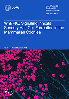
Cover Story (view full-size image): Sensory hair cells in the mammalian cochlea are essential for hearing and require precise developmental patterning. In this study, we demonstrate that Wnt4 and the Wnt antagonist sFRP2 jointly regulate inner hair cell boundary formation and differentiation through a non-canonical Wnt/Calcium/PKC pathway. Genetic and pharmacological analyses reveal that Wnt4 pathway activation inhibits proper hair cell formation, while its inhibition induces the formation of ectopic inner hair cells. We further identify Atoh1 as a downstream effector, as a mutation in one of its putative PKC target sites diminishes hair cell induction. This work reveals a novel Wnt4/Calcium/PKC pathway that controls cochlear boundary formation and hair cell specification. View this paper
  • Issues are regarded as officially published after their release is announced to the table of contents alert mailing list.
  • You may sign up for e-mail alerts to receive table of contents of newly released issues.
  • PDF is the official format for papers published in both, html and pdf forms. To view the papers in pdf format, click on the "PDF Full-text" link, and use the free Adobe Reader to open them.
Order results
Result details
Section
Select all
Export citation of selected articles as:
16 pages, 3056 KB  
Article
Muscle Spatial Transcriptomic Reveals Heterogeneous Profiles in Juvenile Dermatomyositis and Persistence of Abnormal Signature After Remission
by Margot Tragin, Séverine A. Degrelle, Baptiste Periou, Brigitte Bader-Meunier, Christine Barnerias, Christine Bodemer, Isabelle Desguerre, Mathieu Paul Rodero, François Jérôme Authier and Cyril Gitiaux
Cells 2025, 14(12), 939; https://doi.org/10.3390/cells14120939 - 19 Jun 2025
Viewed by 883
Abstract
This study aimed to investigate the spatial heterogeneity of molecular signature in the muscle of juvenile dermatomyositis (JDM) patients before and after treatment. Unsupervised reference-free deconvolution of spatial transcriptomics and standardized morphometry were performed in two JDM muscle biopsies with different clinical severity [...] Read more.
This study aimed to investigate the spatial heterogeneity of molecular signature in the muscle of juvenile dermatomyositis (JDM) patients before and after treatment. Unsupervised reference-free deconvolution of spatial transcriptomics and standardized morphometry were performed in two JDM muscle biopsies with different clinical severity at disease onset and compared to healthy muscle. Identified signatures were scored in two additional JDM muscle biopsies from the same patient before and after remission. Disappearance of the normal muscle signature mostly corresponding to mitochondrial biology was observed in JDM. Three pathological transcriptomic signatures were isolated, related to “myofibrillar stress”, “muscle remodeling” and “interferon signaling” signatures. The “myofibrillar stress signature” was prominent in the most severe biopsy while the “muscle remodeling” signature was mostly present in the biopsy from the patient with good outcome. These signatures unveiled genes not previously associated with JDM including ANKRD1 and FSLT1 for “myofibrillar stress” and “muscle remodeling” signatures, respectively. Post-treatment analysis of muscle after two years remission showed a persistence of pathological signatures. This pilot study of JDM muscle identified spatially distributed pathological signatures that persist after remission. This work paves the way for a better understanding of the pathophysiology in affected muscle and the identification of biomarkers that predict relapse. Full article
Show Figures

Figure 1

22 pages, 1349 KB  
Article
Cannabidiol Effects on Depressive-like Behavior and Neuroinflammation in Female Rats Exposed to High-Fat Diet and Unpredictable Chronic Mild Stress
by Tal Sabbag, Milly Kritman and Irit Akirav
Cells 2025, 14(12), 938; https://doi.org/10.3390/cells14120938 - 19 Jun 2025
Viewed by 719
Abstract
Depression and obesity are comorbid conditions linked through shared neuroinflammatory and immune mechanisms. This study examined the effects of chronic cannabidiol (CBD) treatment on behavior and neuroinflammatory gene expression in female rats exposed to a combined model of high-fat diet (HFD) and unpredictable [...] Read more.
Depression and obesity are comorbid conditions linked through shared neuroinflammatory and immune mechanisms. This study examined the effects of chronic cannabidiol (CBD) treatment on behavior and neuroinflammatory gene expression in female rats exposed to a combined model of high-fat diet (HFD) and unpredictable chronic mild stress (UCMS). Rats were subjected to an acute HFD for 2 weeks, followed by 4 weeks of UCMS. CBD (10 mg/kg, i.p.) or vehicle was administered during the final 2 weeks of UCMS. Specifically, mRNA levels of nuclear factor kappa B1 (NF-κB1), tumor necrosis factor alpha (TNF-α), interleukin-1 beta (IL-1β), and IL-6 were measured in the ventromedial prefrontal cortex (vmPFC) and CA1. CBD’s effects varied depending on the type of stressor. It promoted coping behavior, increased locomotion, reduced freezing, and restored UCMS-induced depressive-like behavior in a splash test. In the vmPFC, CBD normalized the HFD- and UCMS-induced increase in il1β, and downregulated nfkb1 and tnfa expression. In the CA1, it normalized stress-induced downregulation in nfkb1 expression. These findings suggest that the efficacy of CBD in modulating both behavior and neuroinflammation is contingent upon the nature of the stress exposure, highlighting its potential as a targeted treatment for stress-related neuropsychiatric disorders in females. Full article
Show Figures

Figure 1

17 pages, 2436 KB  
Review
Implications of Fatty Acids for Age-Related Macular Degeneration: Evidence and Recommendations
by Shivantika Bisen and Nikhlesh K. Singh
Cells 2025, 14(12), 937; https://doi.org/10.3390/cells14120937 - 19 Jun 2025
Viewed by 1006
Abstract
Age-related macular degeneration (AMD) is an ocular pathology in humans characterized by the buildup of lipid-rich extracellular deposits, which leads to retinal degeneration. In recent years, considerable effort has been made to observe the effect of dietary fatty acids on oxidative stress and [...] Read more.
Age-related macular degeneration (AMD) is an ocular pathology in humans characterized by the buildup of lipid-rich extracellular deposits, which leads to retinal degeneration. In recent years, considerable effort has been made to observe the effect of dietary fatty acids on oxidative stress and inflammation. In continuation of this, much effort has been made to study the effect of dietary fatty acids on the pathogenesis of AMD. Although studies have shown that dietary fatty acids are effective against few forms of AMD, particularly wet AMD or neovascular AMD, no dietary lipids have shown any conclusive results for dry AMD or geographic AMD. It is therefore important to look for new lipids and lipoproteins that can be helpful in treating various stages of AMD. This article reviews the impact of saturated fatty acids (SFAs), monounsaturated fatty acids (MUFAs), and polyunsaturated fatty acids (PUFAs) on retinal health and the progression of AMD. Furthermore, this manuscript discusses all studies investigating the implications of fatty acids on AMD, which may be beneficial for future treatment strategies and dietary guidelines related to it. In conclusion, studies suggest that omega-3 PUFAs, particularly docosahexaenoic acid (DHA) and eicosapentaenoic acid (EPA), might provide protection against AMD, highlighting the necessity for additional clinical trials to evaluate their efficacy in the prevention and treatment of AMD. Full article
(This article belongs to the Special Issue Retinal Disorders: Cellular Mechanisms and Targeted Therapies)
Show Figures

Graphical abstract

16 pages, 509 KB  
Review
Spatial Transcriptomics in Thyroid Cancer: Applications, Limitations, and Future Perspectives
by Chaerim Song, Hye-Ji Park and Man S. Kim
Cells 2025, 14(12), 936; https://doi.org/10.3390/cells14120936 - 19 Jun 2025
Viewed by 911
Abstract
Spatial transcriptomics (ST) is emerging as a powerful technology that transforms our understanding of thyroid cancer by offering a spatial context of gene expression within the tumor tissue. In this review, we synthesize the recent applications of ST in thyroid cancer research, with [...] Read more.
Spatial transcriptomics (ST) is emerging as a powerful technology that transforms our understanding of thyroid cancer by offering a spatial context of gene expression within the tumor tissue. In this review, we synthesize the recent applications of ST in thyroid cancer research, with a particular focus on the heterogeneity of the tumor microenvironment, tumor evolution, and cellular interactions. Studies have leveraged the spatial information provided by ST to map distinct cell types and expression patterns of genes and pathways across the different regions of thyroid cancer samples. The spatial context also allows a closer examination of invasion and metastasis, especially through the dysregulation at the tumor leading edge. Additionally, signaling pathways are inferred at a more accurate level through the spatial proximity of ligands and receptors. We also discuss the limitations that need to be overcome, including technical limitations like low resolution and sequencing depth, the need for high-quality samples, and complex data handling processes, and suggest future directions for a wider and more efficient application of ST in advancing personalized treatment of thyroid cancer. Full article
(This article belongs to the Special Issue Spatial Proteomics and Transcriptomics in Cells)
Show Figures

Figure 1

17 pages, 2220 KB  
Article
SOS3 from Avicennia marina Enhances Salt Stress Tolerance of Arabidopsis thaliana
by Mariam Alzaabi, John Orpilla, Khaled Michel Hazzouri, Ling Li and Khaled Amiri
Cells 2025, 14(12), 935; https://doi.org/10.3390/cells14120935 - 19 Jun 2025
Viewed by 527
Abstract
Abiotic stress poses a serious challenge in agriculture. Salinity inhibits crop growth and yields by disrupting ionic homeostasis and osmotic balance. One critical mechanism of salt tolerance is the activation of the Salt Overly Sensitive (SOS) signaling pathway. Investigating this pathway in halophytic [...] Read more.
Abiotic stress poses a serious challenge in agriculture. Salinity inhibits crop growth and yields by disrupting ionic homeostasis and osmotic balance. One critical mechanism of salt tolerance is the activation of the Salt Overly Sensitive (SOS) signaling pathway. Investigating this pathway in halophytic plants offers valuable insights into the molecular mechanisms underlying salt stress tolerance. This study explores the structure and function of SOS3/CBL4 from the gray mangrove, Avicennia marina (AmSOS3). Sequence analysis revealed that AmSOS3 shares significant similarities with orthologs of SOS3/CBL4, including Arabidopsis thaliana (AtSOS3). All essential functional domains of SOS3, including the four EF-hands, as well as the N-myristoylation and S-acylation motif, were conserved in AmSOS3. Structural modeling, using Modeller, predicted that AmSOS3 forms a homodimer stabilized by a hydrogen bond at the serine 140 position. Functional characterization further demonstrated that AmSOS3 complements the sos3-1 mutation in A. thaliana, thus confirming that AmSOS3 is an ortholog of AtSOS3. Overexpression of AmSOS3 in wild-type A. thaliana enhanced tolerance under salinity stress. The transgenic lines displayed reduced reactive oxygen species (ROS) accumulation and increased ROS-scavenging enzyme activity. These findings indicate that AmSOS3 plays a critical role in improving salt stress tolerance and maintaining cellular homeostasis. Full article
Show Figures

Figure 1

16 pages, 3399 KB  
Article
Investigating the Synergistic Neuroprotective Effects of Plant-Derived Antioxidants and the Psychedelic N,N-Dimethyltryptamine in Alzheimer’s Disease Therapy
by Júlia Jarne-Ferrer, Mercè Pallàs, Christian Griñán-Ferré and Aina Bellver-Sanchis
Cells 2025, 14(12), 934; https://doi.org/10.3390/cells14120934 - 19 Jun 2025
Viewed by 869
Abstract
Alzheimer’s disease (AD) is a chronic and complex neurodegenerative disorder characterized by progressive cognitive decline, memory loss, and irreversible impairment of brain functions. The etiology of AD is multifactorial, involving a complex interplay of genetic, environmental, and physiological factors, including the aggregation of [...] Read more.
Alzheimer’s disease (AD) is a chronic and complex neurodegenerative disorder characterized by progressive cognitive decline, memory loss, and irreversible impairment of brain functions. The etiology of AD is multifactorial, involving a complex interplay of genetic, environmental, and physiological factors, including the aggregation of amyloid-β (Aβ) and oxidative stress (OS). The role of OS in AD pathogenesis is of particular significance, given that an imbalance between oxidants and antioxidants promotes cellular damage, exacerbates Aβ deposition, and leads to cognitive deterioration. Despite extensive research, current therapeutic strategies have largely failed, likely due to the use of single-target drugs unable to halt the multifactorial progression of the disease. In this study, we investigated the synergistic therapeutic effect of plant-derived bioactive compounds Withanone, Apigenin, Bacoside A, Baicalin, and Thymoquinone in combination with N,N-Dimethyltryptamine (NN-DMT), a psychedelic molecule. We used a transgenic Caenorhabditis elegans model to assess the behavioral and molecular outcomes following compound exposure. Motility assays, thioflavin S staining, and survival assays under oxidative stress were employed to evaluate the treatment efficacy. The results of the behavioral and molecular analyses indicated that the combination therapy exhibited a higher efficacy than the monotherapies, leading to a significant reduction in age-related motility defects in the AD model. Furthermore, the combination treatment substantially reduced Aβ plaque burden, enhanced survival following OS insult, and demonstrated a synergistic effect in mitigating AD-related hallmarks. Taken together, these findings support the potential of combining NN-DMT with specific bioactive compounds as a promising multi-target therapeutic approach for AD. Full article
Show Figures

Figure 1

28 pages, 753 KB  
Review
Toll-like Receptors in Immuno-Metabolic Regulation of Emotion and Memory
by Carla Crespo-Quiles and Teresa Femenía
Cells 2025, 14(12), 933; https://doi.org/10.3390/cells14120933 - 19 Jun 2025
Viewed by 774
Abstract
Toll-like receptors (TLRs) comprise an evolutionarily conserved family of pattern recognition receptors that detect microbial-associated molecular patterns and endogenous danger signals to orchestrate innate immune responses. While traditionally positioned at the frontline of host defense, accumulating evidence suggests that TLRs are at the [...] Read more.
Toll-like receptors (TLRs) comprise an evolutionarily conserved family of pattern recognition receptors that detect microbial-associated molecular patterns and endogenous danger signals to orchestrate innate immune responses. While traditionally positioned at the frontline of host defense, accumulating evidence suggests that TLRs are at the nexus of immuno-metabolic regulation and central nervous system (CNS) homeostasis. They regulate a wide range of immune and non-immune functions, such as cytokine and chemokine signaling, and play key roles in modulating synaptic plasticity, neurogenesis, and neuronal survival. However, alterations in TLR signaling can drive a sustained pro-inflammatory state, mitochondrial dysfunction, and oxidative stress, which are highly associated with the disruption of emotional and cognitive functions and the pathogenesis of psychiatric disorders. In this review, we integrate findings from molecular to organismal levels to illustrate the diverse roles of TLRs in regulating emotion, cognition, metabolic balance, and gut–brain interactions. We also explore emerging molecular targets with the potential to guide the development of more effective therapeutic interventions. Full article
(This article belongs to the Special Issue Inflammatory Pathways in Psychiatric Disorders)
Show Figures

Figure 1

16 pages, 76646 KB  
Article
Cytokinesis in Suspension: A Distinctive Trait of Mesenchymal Stem Cells
by Bhavna Rani, Hong Qian and Staffan Johansson
Cells 2025, 14(12), 932; https://doi.org/10.3390/cells14120932 - 19 Jun 2025
Viewed by 576
Abstract
Mesenchymal stem cells (MSCs) have a broad clinical potential, but their selection and expansion on plastic cause unknown purity and phenotypic alterations, reducing therapy efficiency. Furthermore, their behavior in non-adherent conditions during systemic transplantation remains poorly understood. The sphere formation from single cells [...] Read more.
Mesenchymal stem cells (MSCs) have a broad clinical potential, but their selection and expansion on plastic cause unknown purity and phenotypic alterations, reducing therapy efficiency. Furthermore, their behavior in non-adherent conditions during systemic transplantation remains poorly understood. The sphere formation from single cells is commonly used to assess stemness, but MSCs lack this ability, raising questions about their anchorage dependence for proliferation. We investigated whether bone marrow-derived MSCs can complete cytokinesis in non-adherent environments. Primary human and mouse bone marrow-derived MSCs were synchronized in early mitosis using nocodazole and were cultured on soft, rigid, or non-adherent surfaces. Both human and mouse MSCs displayed an ALIX (abscission licensor) recruitment to the midbody 40–90 min post-nocodazole release, regardless of the substrate adherence. Cells maintained for 4hr in the suspension remained viable, and daughter cells rapidly migrated apart upon the re-adhesion to fibronectin-coated surfaces, demonstrating cytokinesis completion in suspension. These findings distinguish MSCs from fibroblasts (which require adhesion for division), provide a more general stemness feature, and suggest that adhesion-independent cytokinesis is a trait relevant to the post-transplantation survival and tissue homing. This property may offer strategies to expand MSCs with an improved purity and functionality and to enhance engraftment by leveraging cell cycle manipulation to promote an early extracellular matrix deposition at target sites. Full article
(This article belongs to the Section Stem Cells)
Show Figures

Figure 1

21 pages, 1693 KB  
Article
Free Methylglyoxal and Lactate Produced and Released by Cultured Cancer and Non-Cancer Cells: Implications for Tumor Growth and Development
by Dominique Belpomme, Philippe Irigaray, Jean-Marc Alberto, Clément Poletti, Charlotte Hinault-Boyer and Stéphanie Lacomme
Cells 2025, 14(12), 931; https://doi.org/10.3390/cells14120931 - 19 Jun 2025
Viewed by 701
Abstract
We have previously shown that in cancer patients, free methylglyoxal (MG), a side-product of glycolysis, is recovered from tumors at significantly higher levels than from their corresponding non-cancerous tissues. We also recently confirmed our initial experimental finding that in these patients, free MG [...] Read more.
We have previously shown that in cancer patients, free methylglyoxal (MG), a side-product of glycolysis, is recovered from tumors at significantly higher levels than from their corresponding non-cancerous tissues. We also recently confirmed our initial experimental finding that in these patients, free MG peripheral blood levels correlate positively with tumor growth, making free MG levels a new metabolic biomarker of tumor growth of interest to detect cancer and clinically follow cancer patients with no available biomarkers. Now we measure free MG and lactate produced by different cancer and normal cells cultured at low or high glucose concentration and in normoxic or hypoxic conditions to question whether cancer cells and non-cancer cells in tumors produce and release free MG and lactate. Surprisingly, we found that normal fibroblastic and endothelial cell lines grown in normoxic conditions produce and release high free MG levels, which we confirmed for non-transformed normal fibroblasts, albeit at significantly lower levels. Cancer cells generally significantly increased their free MG production and release when cultured in high glucose concentration, while normal cells generally did not. Furthermore, in normoxic conditions, normal fibroblastic cells, in addition to free MG, may produce and release lactate. From this data, we propose that in malignant tumors, both cancer and fibroblastic stromal cells may contribute to tumor growth and development by producing via glycolysis both free MG and D-lactate, which, in addition to L-lactate, may be part of the core hallmark of cell metabolic reprogramming in cancer. Full article
Show Figures

Figure 1

25 pages, 1580 KB  
Review
Inflammasomes and Signaling Pathways: Key Mechanisms in the Pathophysiology of Sepsis
by Jhan S. Saavedra-Torres, María Virginia Pinzón-Fernández, Martin Ocampo-Posada, H. A. Nati-Castillo, Laura Alejandra Jiménez Hincapie, Eder J. Cadrazo-Gil, Marlon Arias-Intriago, Marlon Rojas-Cadena, Andrea Tello-De-la-Torre, Walter Osejos and Juan S. Izquierdo-Condoy
Cells 2025, 14(12), 930; https://doi.org/10.3390/cells14120930 - 19 Jun 2025
Cited by 1 | Viewed by 2149
Abstract
Sepsis is a life-threatening syndrome characterized by a dysregulated immune response to infection, frequently leading to multiorgan failure and high mortality. Inflammasomes—cytosolic multiprotein complexes of the innate immune system—serve as critical platforms for sensing pathogen- and damage-associated molecular patterns (PAMPs and DAMPs). Key [...] Read more.
Sepsis is a life-threatening syndrome characterized by a dysregulated immune response to infection, frequently leading to multiorgan failure and high mortality. Inflammasomes—cytosolic multiprotein complexes of the innate immune system—serve as critical platforms for sensing pathogen- and damage-associated molecular patterns (PAMPs and DAMPs). Key sensors such as NLRP3, AIM2, and IFI16 initiate caspase-1 activation, IL-1β and IL-18 maturation, and gasdermin D–mediated pyroptosis. In sepsis, excessive inflammasome activation drives oxidative stress, endothelial dysfunction, immunothrombosis, and immune exhaustion. This maladaptive cascade is further aggravated by the release of DAMPs and procoagulant factors, compromising vascular integrity and immune homeostasis. Prolonged activation contributes to immunoparalysis, lymphopenia, and increased susceptibility to secondary infections. Inflammasome signaling also intersects with necroptosis and ferroptosis, amplifying systemic inflammation and tissue injury. Additionally, various pathogens exploit immune evasion strategies to modulate inflammasome responses and enhance virulence. Therapeutic interventions under investigation include selective NLRP3 inhibitors, IL-1 blockers, gasdermin D antagonists, and extracorporeal cytokine hemoadsorption. Emerging approaches emphasize biomarker-guided immunomodulation to achieve personalized therapy. While preclinical studies have shown promising results, clinical translation remains limited. Targeting inflammasomes may offer a path toward precision immunotherapy in sepsis, with potential to reduce organ dysfunction and improve survival. Full article
Show Figures

Figure 1

17 pages, 2444 KB  
Article
A Novel Modulator of Resistance for Oxaliplatin-Based Therapy for Colorectal Cancer: The ESCRT Family Member VPS4A
by Noha M. Abdelrazik, Anjana Patel, Andrew Conn, Christopher W. Sutton, Sriharsha Kantamneni and Steven D. Shnyder
Cells 2025, 14(12), 929; https://doi.org/10.3390/cells14120929 - 19 Jun 2025
Viewed by 706
Abstract
Drug resistance is still one of the main challenges for the treatment of colorectal cancer (CRC). Whilst some resistance mechanisms are well known, from the static therapy success rate, clearly, still much is undiscovered. Intracellular transport mechanisms have attracted attention as having a [...] Read more.
Drug resistance is still one of the main challenges for the treatment of colorectal cancer (CRC). Whilst some resistance mechanisms are well known, from the static therapy success rate, clearly, still much is undiscovered. Intracellular transport mechanisms have attracted attention as having a possible role in drug resistance, and here, the Endosomal Sorting Complex Required for Transport (ESCRT) protein family is studied as a source of drug resistance modulation using human CRC cell lines and clinical material. From an initial screening of ESCRT proteins in a panel of 10 CRC wild-type cell lines using immunoblotting, Vacuolar Protein Sorting-Associated Protein A4 (VPS4A) was identified as being consistently highly expressed, and it was selected for further investigation. Immunohistopathological evaluation in a small panel of CRC patient samples demonstrated high expression in the tumor epithelium compared to normal intestinal epithelium. The knockdown of VPS4A resulted in enhanced sensitivity of cells to oxaliplatin, and it was subsequently seen that oxaliplatin-resistant sublines had significantly higher VPS4A expression than their wild-type variants. In addition, it was demonstrated that a small molecule inhibitor of VPS4A, aloperine, could interact synergistically with oxaliplatin to enhance its sensitivity in an oxaliplatin-resistant cell line. We hypothesize from initial RNA sequencing analysis that the mechanism of action of VPS4A modulation is through depleting levels of the drug efflux transporter MRP2 in the cell, preventing oxaliplatin egress and increasing cell exposure to the drug. The evidence presented here thus indicates that ESCRT machinery, specifically VPS4A, may act as a modulator of oxaliplatin resistance in CRC. Full article
Show Figures

Graphical abstract

22 pages, 13702 KB  
Article
MicroRNA miR-193b-3p Regulates Esophageal Cancer Progression Through Targeting RSF1
by Yao Lin, Xudong Zhao, Zhenhua Du, Zhili Jia, Siyu Zhou, Gengsheng Cao and Hengbin Wang
Cells 2025, 14(12), 928; https://doi.org/10.3390/cells14120928 - 19 Jun 2025
Viewed by 611
Abstract
Esophageal cancer (ESCA) is the sixth leading cause of cancer-related mortality worldwide. Despite the significant impact, the molecular mechanisms underlying its initiation and progression remain poorly understood. In this study, we identified mircoRNA miR-193b-3p as a critical regulator of ESCA progression and the [...] Read more.
Esophageal cancer (ESCA) is the sixth leading cause of cancer-related mortality worldwide. Despite the significant impact, the molecular mechanisms underlying its initiation and progression remain poorly understood. In this study, we identified mircoRNA miR-193b-3p as a critical regulator of ESCA progression and the Remodeling and Spacing Factor 1 (RSF1) as an essential target of miR-193b-3p. Analysis of the TCGA_ESCA dataset and RT-qPCR experiments revealed that RSF1 levels are significantly elevated in ESCA and inversely correlated with miR-193b-3p levels. Using a dual-luciferase reporter assay, as well as transfection of miR-193-3p mimics or inhibitors, we confirmed RSF1 as a direct target of miR-193b-3p in ESCA cells. Transfection of miR-193b-3p suppresses ESCA cell proliferation, migration, and invasion. These effects were partially reversed by exogenous RSF1 expression. Injection of AgomiR-193b-3p into mice bearing ESCA xenografts impeded tumor growth. These findings underscore the critical role of the miR-193b-3p/RSF1 axis in esophageal cancer progression. Full article
(This article belongs to the Special Issue Epigenetic Mechanisms of Tumorigenesis)
Show Figures

Figure 1

23 pages, 3181 KB  
Article
Side-by-Side Comparison of Culture Media Uncovers Phenotypic and Functional Differences in Primary Mouse Aortic Mural Cells
by Iman Ghasemi, Rajinikanth Gogiraju, Sana’a Khraisat, Sven Pagel, Claudine Graf, Moritz Brandt, Thati Madhusudhan, Philip Wenzel, Guillermo Luxán, Philipp Lurz, Magdalena L. Bochenek and Katrin Schäfer
Cells 2025, 14(12), 927; https://doi.org/10.3390/cells14120927 - 19 Jun 2025
Viewed by 553
Abstract
(1) Background: Vascular mural cells reside in the media and outer layers of the vessel wall. Their ability to proliferate and migrate or to change phenotype in response to external cues is a central feature of the vascular response to injury. Genetically engineered [...] Read more.
(1) Background: Vascular mural cells reside in the media and outer layers of the vessel wall. Their ability to proliferate and migrate or to change phenotype in response to external cues is a central feature of the vascular response to injury. Genetically engineered mice are used for loss- or gain-of-function analyses or lineage tracing in vivo, their primary cells for mechanistic studies in vitro. Whether and how cultivation conditions affect their phenotype and function is often overlooked. (2) Methods: Here, we systematically studied how the cultivation of primary mural cells isolated from the aorta of adult wild-type mice in either basal medium (DMEM) or special media formulated for the cultivation of fibroblasts or pericytes affects their phenotype and function. (3) Results: Medium composition did not alter cell viability, but the mRNA levels of differentiated smooth muscle cell markers were highest in vascular mural cells expanded in DMEM. Conversely, significantly higher numbers of proliferating and migrating cells were observed in cells expanded in Pericyte medium, and cytoskeletal rearrangements supported increased migratory capacities. Significantly reduced telomere lengths and metabolic reprogramming was observed in aortic mural cells cultured in Fibroblast medium. (4) Conclusions: Our findings underline the plasticity of primary aortic mural cells and highlight the importance of the culture media composition during their expansion, which could be exploited to interrogate their responsiveness to external stimuli or conditions observed in vivo or in patients. Full article
Show Figures

Figure 1

21 pages, 6303 KB  
Article
Tight Spaces, Tighter Signals: Spatial Constraints as Drivers of Peripheral Myelination
by Luca Bartesaghi, Basilio Giangreco, Vanessa Chiappini, Maria Fernanda Veloz Castillo, Martina Monaco, Jean-Jaques Médard, Giovanna Gambarotta, Marco Agus and Corrado Calì
Cells 2025, 14(12), 926; https://doi.org/10.3390/cells14120926 - 18 Jun 2025
Viewed by 1612
Abstract
Peripheral myelination is driven by the intricate interplay between Schwann cells and axons, coordinated through molecular signaling and the structural organization of their shared environment. While the biochemical regulation of this process has been extensively studied, the influence of spatial architecture and mechanical [...] Read more.
Peripheral myelination is driven by the intricate interplay between Schwann cells and axons, coordinated through molecular signaling and the structural organization of their shared environment. While the biochemical regulation of this process has been extensively studied, the influence of spatial architecture and mechanical cues remains poorly understood. Here, we use in vitro co-culture models—featuring microfluidic devices and hydrogel-based scaffolds—to explore how extracellular organization, cellular density, and spatial constraints shape Schwann cell behavior. Our results show that (i) pro-myelinating effects triggered by ascorbic acid administration is distally propagated along axons in Schwann cell-DRG co-cultures, (ii) ascorbic acid modulates Neuregulin-1 expression, (iii) a critical threshold of cellular density is required to support proper Schwann cell differentiation and myelin formation, and (iv) spatial confinement promotes myelination in the absence of ascorbic acid. Together, these findings highlight how spatial and structural parameters regulate the cellular and molecular events underlying peripheral myelination, offering new physiologically relevant models of myelination and opening new avenues for peripheral nerve repair strategies. Full article
(This article belongs to the Special Issue Remyelination: From Basic Science to Therapies)
Show Figures

Graphical abstract

19 pages, 6474 KB  
Article
Transcriptomic Profiling of iPS Cell-Derived Hepatocyte-like Cells Reveals Their Close Similarity to Primary Liver Hepatocytes
by Saqlain Suleman, Sharmin Alhaque, Andrew Guo, Aaron Zhang, Serena Fawaz, Stefany Perera, Mohammad S. Khalifa, Hassan Rashidi, David C. Hay and Michael Themis
Cells 2025, 14(12), 925; https://doi.org/10.3390/cells14120925 - 18 Jun 2025
Viewed by 629
Abstract
Human-induced pluripotent stem cell (iPSC)-derived hepatocyte-like cells (HLCs) have been shown to be useful for the development of cell-based regenerative strategies and for modelling drug discovery. However, stem cell-derived HLCs are not identical in nature to primary human hepatocytes (PHHs), which could affect [...] Read more.
Human-induced pluripotent stem cell (iPSC)-derived hepatocyte-like cells (HLCs) have been shown to be useful for the development of cell-based regenerative strategies and for modelling drug discovery. However, stem cell-derived HLCs are not identical in nature to primary human hepatocytes (PHHs), which could affect the cell phenotype and, potentially, model reliability. Therefore, we employed the in-depth gene expression profiling of HLCs and other important and relevant cell types, which led to the identification of clear similarities and differences between them at the transcriptional level. Through gene set enrichment analysis, we identified that genes that are critical for immune signalling pathways become downregulated upon HLC differentiation. Our analysis also found that TAV.HLCs exhibit a mild gene signature characteristic of acute lymphoblastic leukaemia, but not other selected cancers. Importantly, HLCs present significant similarity to PHHs, making them genuinely valuable for modelling human liver biology in vitro and for the development of prototype cell-based therapies for pre-clinical testing. Full article
(This article belongs to the Section Tissues and Organs)
Show Figures

Figure 1

18 pages, 2478 KB  
Article
Concurrent Physical Activity Protects Against C26 Adenocarcinoma Tumor-Mediated Cardiac and Skeletal Muscle Dysfunction and Wasting in Males
by Louisa Tichy, Kimberly F. Allred, Erika T. Rezeli, Michael F. Coleman, Clinton D. Allred, Stephen D. Hursting and Traci L. Parry
Cells 2025, 14(12), 924; https://doi.org/10.3390/cells14120924 - 18 Jun 2025
Viewed by 466
Abstract
Muscle loss unresponsive to nutritional supplementation affects up to 80% of cancer patients and severely reduces survival and treatment response. Exercise may help preserve muscle mass and function, yet the translatability of preclinical methods remains questionable. This study aimed to assess how voluntary [...] Read more.
Muscle loss unresponsive to nutritional supplementation affects up to 80% of cancer patients and severely reduces survival and treatment response. Exercise may help preserve muscle mass and function, yet the translatability of preclinical methods remains questionable. This study aimed to assess how voluntary wheel running, a clinically relevant physical activity, protects skeletal and cardiac muscle against cancer-mediated dysfunction and identify underlying molecular mechanisms. Methods: BALB/c mice were assigned to sedentary nontumor-bearing (SED+NT), sedentary tumor-bearing (SED+T), wheel run nontumor-bearing (WR+NT), and wheel run tumor-bearing (WR+T). Tumor-bearing groups received 5 × 10⁵ C26 cells; WR mice had wheel access for 4 weeks. Muscle function and tissue were analyzed for protective mechanisms. Results: SED+T mice exhibited significant fat and lean mass loss, indicating cachexia, which was prevented in WR+T mice. SED+T also showed 15% reduced grip strength and cardiac dysfunction, while WR+T preserved function. WR+T mice had lower expression of muscle wasting markers (Atrogin1, MuRF1, GDF15, GDF8/11). Physical activity also reduced tumor mass by 57% and volume by 37%. Conclusion: Voluntary wheel running confers tumor-suppressive, myoprotective, and cardioprotective effects. These findings support physical activity as a non-pharmacological strategy to combat cancer-related muscle wasting and dysfunction. Full article
(This article belongs to the Special Issue Emerging Topics in Cachexia)
Show Figures

Figure 1

31 pages, 12256 KB  
Article
Inter-Relationship Between Melanoma Vemurafenib Tolerance Thresholds and Metabolic Pathway Choice
by Pratima Nangia-Makker, Madison Ahrens, Neeraja Purandare, Siddhesh Aras, Jing Li, Katherine Gurdziel, Hyejeong Jang, Seongho Kim and Malathy P Shekhar
Cells 2025, 14(12), 923; https://doi.org/10.3390/cells14120923 - 18 Jun 2025
Viewed by 922
Abstract
Melanomas quickly acquire resistance to vemurafenib, an important therapeutic for BRAFV600 mutant melanomas. Although combating vemurafenib resistance (VemR) to counter mitochondrial metabolic shift using mitochondria-targeting therapies has promise, no studies have analyzed the relationship between vemurafenib tolerance levels and metabolic plasticity. To determine [...] Read more.
Melanomas quickly acquire resistance to vemurafenib, an important therapeutic for BRAFV600 mutant melanomas. Although combating vemurafenib resistance (VemR) to counter mitochondrial metabolic shift using mitochondria-targeting therapies has promise, no studies have analyzed the relationship between vemurafenib tolerance levels and metabolic plasticity. To determine how vemurafenib endurance levels drive metabolic plasticity, we developed isogenic BRAFV600E VemR melanoma models with variant vemurafenib tolerances and performed an integrative analysis of metabolomic and transcriptome alterations using metabolome, Mitoplate-S1, Seahorse, and RNA-seq assays. Regardless of drug tolerance differences, both VemR models display resistance to MEK inhibitor and sensitivity to Wnt/β-catenin inhibitor, ICG-001. β-catenin, MITF, and ABCB5 levels are upregulated in both VemR models, and ICG-001 treatment restored vemurafenib sensitivity with reductions in MITF, ABCB5, phospho-ERK1/2, and mitochondrial respiration. Whereas β-catenin signaling induced TCA cycle and OXPHOS in highly drug tolerant A2058VemR cells, it activated pentose phosphate pathway in M14VemR cells with low vemurafenib tolerance, both of which are inhibited by ICG-001. These data implicate an important role for Wnt/β-catenin signaling in VemR-induced metabolic plasticity. Our data demonstrate that drug tolerance thresholds play a direct role in driving metabolic shifts towards specific routes, thus providing a new basis for delineating VemR melanomas for metabolism-targeting therapies. Full article
(This article belongs to the Collection Pathometabolism: Understanding Disease through Metabolism)
Show Figures

Graphical abstract

11 pages, 2471 KB  
Article
Lower Zinc but Higher Calcium Content in Rodent Spinal Cord Compared to Brain
by Alma I. Santos-Díaz, Brandon Bizup, Ana Karen Pantaleón-Gómez, Beatriz Osorio, Olivier Christophe Barbier, Thanos Tzounopoulos and Fanis Missirlis
Cells 2025, 14(12), 922; https://doi.org/10.3390/cells14120922 - 18 Jun 2025
Viewed by 542
Abstract
Metal ion measurements using inductively coupled plasma optical emission spectroscopy revealed twofold-higher zinc content in rat brain compared to spinal cord. One hypothesis to explain this difference is the high prevalence of synapses that corelease glutamate and zinc in the brain, marked by [...] Read more.
Metal ion measurements using inductively coupled plasma optical emission spectroscopy revealed twofold-higher zinc content in rat brain compared to spinal cord. One hypothesis to explain this difference is the high prevalence of synapses that corelease glutamate and zinc in the brain, marked by the vesicular Zinc Transporter-3 (ZnT3). In contrast, spinal cord tissue showed significantly higher calcium content, reflecting calcifications in the arachnoid. The above observations were made in 60-day-old adult male and female rats fed ad libitum or a restricted diet. In this study, we asked if the calcium and zinc content of the brain and spinal cord was species-specific or evolutionarily conserved, and whether the distinct concentration of zinc in the brain and spinal cord resulted from a different expression pattern of ZnT3, the primary transporter in synaptic vesicles. To address these questions, we examined 8-week-old wild-type male and female mice raised under conventional laboratory conditions and used a knock-in mouse that expresses a human influenza hemagglutinin epitope tag at the C terminus of the endogenous ZnT3 gene to assess the transporter’s abundance in spinal cord sections. Our results show conserved inverse differences in zinc and calcium content in mouse brain and spinal cord, but detectable ZnT3 signal in spinal cord. Whereas vesicular zinc modulates glutamatergic and GABAergic signaling and sensory processing, the functional significance of calcium aggregates in the arachnoid remains unknown. Full article
(This article belongs to the Special Issue Role of Zinc in Brain Homeostasis and Neurological Disorders)
Show Figures

Graphical abstract

31 pages, 1741 KB  
Review
Spotlight on Proteases: Roles in Ovarian Health and Disease
by Bhawna Kushawaha and Emanuele Pelosi
Cells 2025, 14(12), 921; https://doi.org/10.3390/cells14120921 - 18 Jun 2025
Viewed by 767
Abstract
Proteases play crucial roles in ovarian folliculogenesis, regulating several processes from primordial follicle activation to ovulation and corpus luteum formation. This review synthesizes the current knowledge on the diverse functions of proteases in ovarian physiology and pathology. We discuss the classification and regulation [...] Read more.
Proteases play crucial roles in ovarian folliculogenesis, regulating several processes from primordial follicle activation to ovulation and corpus luteum formation. This review synthesizes the current knowledge on the diverse functions of proteases in ovarian physiology and pathology. We discuss the classification and regulation of proteases, highlighting their importance in extracellular matrix remodeling, cell signaling, and apoptosis during ovarian follicular development. We explore the roles of several proteases including matrix metalloproteinases, tissue inhibitors of metalloproteinases, the plasminogen activator system, and cathepsins, and their roles in the critical functions of ovarian biology including follicle dynamics and senescence. Furthermore, we address the involvement of proteases in ovarian pathologies, including cancer, polycystic ovary syndrome, and primary ovarian insufficiency. By integrating recent findings from clinical genomics and animal models, this review provides a comprehensive overview of protease functions in the ovary, emphasizing their potential use for therapeutic interventions in reproductive medicine. Full article
(This article belongs to the Special Issue Cellular and Molecular Mechanisms in Gynecological Disorders)
Show Figures

Figure 1

18 pages, 3502 KB  
Review
Roles of Bile Acid-Activated Receptors in Monocytes-Macrophages and Dendritic Cells
by Huilin Jia, Xingli He, Tengfei Jiang and Fanzhi Kong
Cells 2025, 14(12), 920; https://doi.org/10.3390/cells14120920 - 18 Jun 2025
Viewed by 925
Abstract
Bile acids (BAs), essential for lipid metabolism and fat-soluble vitamin absorption, also act as signaling molecules that regulate immune homeostasis. This review focuses on the roles of four key BA-activated receptors, farnesoid X receptor (FXR), G protein-coupled bile acid receptor 1 (GPBAR1), liver [...] Read more.
Bile acids (BAs), essential for lipid metabolism and fat-soluble vitamin absorption, also act as signaling molecules that regulate immune homeostasis. This review focuses on the roles of four key BA-activated receptors, farnesoid X receptor (FXR), G protein-coupled bile acid receptor 1 (GPBAR1), liver X receptors (LXRs), and vitamin D receptor (VDR), in modulating the functions of monocytes-macrophages, and dendritic cells (DCs). The biological synthesis, transport, and metabolism of BAs were discussed and highlighted the feedback mechanisms regulating the synthesis and enterohepatic circulation of BAs. Each receptor’s role in shaping immune responses is detailed, including their function in inflammation, apoptosis, phagocytosis, and pathogen clearance. FXR and GPBAR1 activation generally exhibits anti-inflammatory effects, while LXR and VDR modulate a more nuanced interplay between immune responses and lipid homeostasis. We also explored the cross-talk between BA-activated receptors and Toll-like receptors, providing a comprehensive understanding of the complex interplay between BA signaling and innate immunity. This review culminates by highlighting the therapeutic potential of targeting these receptors for the treatment of inflammatory and autoimmune diseases. Full article
(This article belongs to the Section Cellular Immunology)
Show Figures

Graphical abstract

25 pages, 1926 KB  
Review
Impact of SGLT-2 Inhibitors on Biomarkers of Heart Failure
by Dana Darwish, Pooja Kumar, Khushi Urs and Siddharth Dave
Cells 2025, 14(12), 919; https://doi.org/10.3390/cells14120919 - 18 Jun 2025
Viewed by 984
Abstract
Type 2 diabetes mellitus affects nearly 7% of the world’s population and is a significant contributor to the development of cardiovascular disease and heart failure. Historically, the pharmacologic therapy of cardiovascular disease has centered around blood pressure control, insulin and cholesterol management, the [...] Read more.
Type 2 diabetes mellitus affects nearly 7% of the world’s population and is a significant contributor to the development of cardiovascular disease and heart failure. Historically, the pharmacologic therapy of cardiovascular disease has centered around blood pressure control, insulin and cholesterol management, the inhibition of the renin–angiotensin system, and catecholamine blockade. Recent evidence suggests that sodium–glucose cotransporter 2 (SGLT-2) inhibitors provide significant cardiovascular protection to patients with and without diabetes. The use of SGLT-2 inhibitors is associated with significant changes to serum biomarkers of cardiac function. In this narrative review, we summarize how biomarkers reflect physiologic aspects of cardiovascular function and how these are affected by the use of SGLT-2 inhibitors. Full article
(This article belongs to the Special Issue New Insights into Therapeutic Targets for Cardiovascular Diseases)
Show Figures

Figure 1

29 pages, 2689 KB  
Review
Cellular and Molecular Interactions in CNS Injury: The Role of Immune Cells and Inflammatory Responses in Damage and Repair
by Jai Chand Patel, Meenakshi Shukla and Manish Shukla
Cells 2025, 14(12), 918; https://doi.org/10.3390/cells14120918 - 18 Jun 2025
Cited by 1 | Viewed by 1159
Abstract
The central nervous system (CNS) is highly susceptible to damage due to its limited ability to regenerate. Injuries to the CNS, whether from trauma, ischemia, or neurodegenerative diseases, disrupt both cellular and vascular structures, leading to immediate (primary) and subsequent (secondary) damage. Primary [...] Read more.
The central nervous system (CNS) is highly susceptible to damage due to its limited ability to regenerate. Injuries to the CNS, whether from trauma, ischemia, or neurodegenerative diseases, disrupt both cellular and vascular structures, leading to immediate (primary) and subsequent (secondary) damage. Primary damage involves the physical disruption of cells and blood vessels, weakening the blood–brain barrier (BBB) and triggering excitotoxicity and calcium overload. Secondary damage develops over hours to days and is marked by ionic imbalance, mitochondrial dysfunction, oxidative stress, and chronic inflammation, which further aggravates tissue damage. Inflammation plays a dual role: acute inflammation helps in repair, while chronic inflammation accelerates neurodegeneration. Microglia and astrocytes play key roles in this inflammatory response, with M1-like microglia promoting pro-inflammatory responses and M2-like microglia supporting anti-inflammatory and repair processes. Neurodegenerative diseases are characterized by the accumulation of misfolded proteins such as Tau, amyloid-beta, TDP-43, and α-synuclein, which impair cellular function and lead to neuronal loss. Neurodegenerative diseases are characterized by the accumulation of misfolded proteins and influenced by genetic risk factors (e.g., APOE4, TARDBP). Despite the CNS’s limited regenerative abilities, processes like synaptogenesis, neurogenesis, axonal regeneration, and remyelination offer potential for recovery. Therapeutic approaches aim to target inflammatory pathways, enhance repair mechanisms, and develop neuroprotective treatments to counter excitotoxicity, oxidative stress, and apoptosis. Advances in stem cell therapy, gene therapy, and personalized medicine hold promise for improving outcomes. Future research should focus on combining strategies, utilizing advanced technologies, and conducting translational studies to bridge the gap between preclinical research and clinical application. By better understanding and leveraging the complex processes of CNS injury and repair, researchers hope to develop effective therapies to restore function and enhance the quality of life for individuals with CNS disorders. Full article
(This article belongs to the Collection Advances in Neurodegenerative Disease)
Show Figures

Figure 1

17 pages, 2124 KB  
Article
Reduced mtDNA Copy Number Links to Vascular Calcification and Restores After Transplantation
by Angelina Schwarz, Abdul Rashid Qureshi, Leah Hernandez, Lars Wennberg, Annika Wernerson, Karolina Kublickiene, Paul G. Shiels, Roberta Filograna, Peter Stenvinkel and Anna Witasp
Cells 2025, 14(12), 917; https://doi.org/10.3390/cells14120917 - 18 Jun 2025
Viewed by 978
Abstract
Patients with chronic kidney disease (CKD) face an increased risk of early vascular aging, progressive vascular calcification, and premature death. With increasing age, mitochondrial function and mitochondrial DNA copy number (mtDNA-cn) decline. This has been identified as an independent predictor of frailty and [...] Read more.
Patients with chronic kidney disease (CKD) face an increased risk of early vascular aging, progressive vascular calcification, and premature death. With increasing age, mitochondrial function and mitochondrial DNA copy number (mtDNA-cn) decline. This has been identified as an independent predictor of frailty and mortality in cardiovascular diseases (CVDs) and cancer. However, the relationship between mtDNA-cn and vascular calcification in the context of a uremic milieu remains ambiguous. We hypothesize that a lower mtDNA-cn is associated with medial calcification, as both are linked to impaired vascular health and accelerated aging. mtDNA-cn was analyzed in 211 CKD5 patients undergoing renal transplantation (RTx) and 196 healthy controls using quantitative PCR (qPCR) for three mtDNA genes (mtND1, mtND4, and mtCOX1) and single-locus nuclear gene hemoglobin beta (HbB). In 32 patients, mtDNA-cn was also quantified one year after RTx. The association between mtDNA-cn and vascular calcification scores, circulatory cell-free (ccf) mtDNA in plasma, and the surrogate marker of biological aging (skin autofluorescence) and CVD risk was assessed. mtDNA-cn was significantly lower in CKD5 patients than in controls and correlated with biological age, vascular calcification, and CVD risk. One year after RTx there was a significant recovery of mtDNA-cn in male patients compared to baseline levels. mtDNA-cn and ccf-mtDNA were inversely correlated. This prospective study provides novel insights into the link between low mtDNA-cn and vascular aging. It demonstrates that RTx restores mtDNA levels and may improve oxidative phosphorylation capacity in CKD. Further investigation is warranted to evaluate mtDNA as a biologically relevant biomarker and a potential therapeutic target for early vascular aging in the uremic environment. Full article
Show Figures

Graphical abstract

18 pages, 4053 KB  
Article
Molecular Insights into Outer Dynein Arm Defects in Primary Ciliary Dyskinesia: Involvement of ZMYND10 and GRP78
by İlker Levent Erdem, Zeynep Bengisu Kaya, Pergin Atilla, Nagehan Emiralioğlu, Cemil Can Eylem, Emirhan Nemutlu, Uğur Özçelik, Halime Nayır Büyükşahin, Ayşenur Daniş and Elif Karakoç
Cells 2025, 14(12), 916; https://doi.org/10.3390/cells14120916 - 17 Jun 2025
Viewed by 745
Abstract
Background: Primary ciliary dyskinesia (PCD) is a rare genetic disorder characterized by recurrent sinopulmonary infections due to motile cilia defects. The disease is genetically heterogeneous, with abnormalities in structural ciliary proteins. Zinc finger MYND-type containing 10 (ZMYND10) is essential for the assembly of [...] Read more.
Background: Primary ciliary dyskinesia (PCD) is a rare genetic disorder characterized by recurrent sinopulmonary infections due to motile cilia defects. The disease is genetically heterogeneous, with abnormalities in structural ciliary proteins. Zinc finger MYND-type containing 10 (ZMYND10) is essential for the assembly of outer dynein arms (ODA), with chaperones like Glucose-regulated protein 78 (GRP78) facilitating protein folding. This study investigates ZMYND10 and Dynein axonemal heavy chain 5 (DNAH5) mutations in individuals with PCD. Methods: Eight individuals aged 14–22 with clinical PCD symptoms and confirmed DNAH5 mutations were included. We analyzed the correlation between DNAH5 abnormalities and preassembly/chaperone proteins using immunofluorescence labeling. Nasal swabs were double-labeled (DNAH5–β-tubulin, β-tubulin–ZMYND10, β-tubulin–GRP78) and examined via fluorescence microscopy. Serum metabolomics and proteomics were also assessed. Results: The corrected total cell fluorescence (CTCF) levels of DNAH5, ZMYND10, and GRP78 were significantly different between PCD individuals and controls. Metabolomic analysis showed reduced valine, leucine, and isoleucine biosynthesis, with increased malate and triacylglycerol biosynthesis, malate-aspartate and glycerol phosphate shuttles, and arginine/proline metabolism, suggesting mitochondrial and ER stress. Conclusions: The altered expression of DNAH5, ZMYND10, and GRP78, along with metabolic shifts, points to a complex link between ciliary dysfunction and cellular stress in PCD. Further studies are needed to clarify the underlying mechanisms. Full article
(This article belongs to the Special Issue The Role of Cilia in Health and Diseases—2nd Edition)
Show Figures

Graphical abstract

18 pages, 696 KB  
Article
Exome Study of Single Nucleotide Variations in Patients with Syndromic and Non-Syndromic Autism Reveals Potential Candidate Genes for Diagnostics and Novel Single Nucleotide Variants
by Lyudmila Belenska-Todorova, Milen Zamfirov, Tihomir Todorov, Slavena Atemin, Mila Sleptsova, Zornitsa Pavlova, Tanya Kadiyska, Ales Maver, Borut Peterlin and Albena Todorova
Cells 2025, 14(12), 915; https://doi.org/10.3390/cells14120915 - 17 Jun 2025
Viewed by 2924
Abstract
Autism spectrum disorder (ASD) is a neurodevelopmental impairment that occurs due to mutations related to the formation of the nervous system, combined with the impact of various epigenetic and environmental factors. This necessitates the identification of the genetic variations involved in ASD pathogenesis. [...] Read more.
Autism spectrum disorder (ASD) is a neurodevelopmental impairment that occurs due to mutations related to the formation of the nervous system, combined with the impact of various epigenetic and environmental factors. This necessitates the identification of the genetic variations involved in ASD pathogenesis. We performed whole exome sequencing (WES) in a cohort of 22 Bulgarian male and female individuals showing ASD features alongside segregation analyses of their families. A targeted panel of genes was chosen and analyzed for each case, based on a detailed examination of clinical data. Gene analyses revealed that specific variants concern key neurobiological processes involving neuronal architecture, development, and function. These variants occur in a number of genes, including SHANK3, DLG3, NALCN, and PACS2 which are critical for synaptic signaling imbalance, CEP120 and BBS5 for ciliopathies, SPTAN1 for spectrins structure, SPATA5, TRAK1, and VPS13B for neuronal organelles trafficking and integrity, TAF6, SMARCB1, DDX3X, MECP2, and SETD1A for gene expression, CDK13 for cell cycle control, ALDH5A1, DPYD, FH, and PDHX for mitochondrial function, and PQBP1, HUWE1, and WDR45 for neuron homeostasis. Novel single nucleotide variants in the SPATA5, CEP120, BBS5, SETD1A, TRAK1, VPS13B, and DDX3X genes have been identified and proposed for use in ASD diagnostics. Our data contribute to a better understanding of the complex neurobiological features of autism and are applicable in the diagnosis and development of personalized therapeutic approaches. Full article
(This article belongs to the Special Issue Molecular Mechanisms of Autism Spectrum Disorder)
Show Figures

Figure 1

21 pages, 4231 KB  
Article
Microfungus Podosphaera fusca and the Fungus-like Organism Peronospora ficariae as Potential Inhalant Allergens in a Mouse Model of Asthma
by Piotr Wlaź, Katarzyna Socała, Marta Palusińska-Szysz, Urszula Świderska, Dominika Szczypior, Magdalena Krasowska and Agnieszka Szuster-Ciesielska
Cells 2025, 14(12), 914; https://doi.org/10.3390/cells14120914 - 17 Jun 2025
Viewed by 567
Abstract
Allergic conditions have surged to unprecedented levels globally, affecting approximately 30% of the population. Fungi are among the major sources of allergens, accounting for about 6% of respiratory issues. Identifying the causes of respiratory allergies is not always possible. Our study assessed the [...] Read more.
Allergic conditions have surged to unprecedented levels globally, affecting approximately 30% of the population. Fungi are among the major sources of allergens, accounting for about 6% of respiratory issues. Identifying the causes of respiratory allergies is not always possible. Our study assessed the capacity of two plant parasites, Podosphaera fusca and Peronospora ficariae, which infect Cucurbita pepo and Ficaria verna, to provoke inflammatory and asthmatic reactions in mouse models of acute and chronic asthma. We performed experiments by sensitizing mice through intranasal challenges with extracts from P. fusca and P. ficariae. Subsequently, we used ELISA tests to measure pro-inflammatory cytokines, including IL-4, IL-5, IL-13, TNF-α, and TGF-β. We evaluated specific IgE production through ELISA and examined histological changes in mouse lungs using hematoxylin-eosin staining. Our research revealed that P. fusca and P. ficariae induced significant production of all tested cytokines, increased specific IgE levels, and caused histological changes characteristic of acute and chronic asthma progression. Although weaker than the reference allergen ovalbumin, P. fusca and P. ficariae possess proinflammatory and asthma-inducing capabilities, indicating the potential to expand the current list of fungal allergens. Full article
Show Figures

Figure 1

20 pages, 2299 KB  
Article
Downregulated ALDH2 Contributes to Tumor Progression and Targeted Therapy Resistance in Human Metastatic Melanoma Cells
by Zili Zhai, Takeshi Yamauchi, Karenna Sandoval, Kira Villarreal, Man Wai Charlotte Kwong, Emily J. Swanson, Aik Choon Tan and Mayumi Fujita
Cells 2025, 14(12), 913; https://doi.org/10.3390/cells14120913 - 17 Jun 2025
Viewed by 840
Abstract
Aldehyde dehydrogenase 2 (ALDH2) is a crucial detoxifying enzyme that eliminates toxic aldehydes. ALDH2 deficiency has been linked to various human diseases, including certain cancers. We have previously reported ALDH2 downregulation in human melanoma tissues. Here, we further investigated the biological significance of [...] Read more.
Aldehyde dehydrogenase 2 (ALDH2) is a crucial detoxifying enzyme that eliminates toxic aldehydes. ALDH2 deficiency has been linked to various human diseases, including certain cancers. We have previously reported ALDH2 downregulation in human melanoma tissues. Here, we further investigated the biological significance of ALDH2 downregulation in this malignancy. Analysis of TCGA dataset revealed that low ALDH2 expression correlates with poorer survival in metastatic melanoma. Examination of human metastatic melanoma cell lines confirmed that most had ALDH2 downregulation (ALDH2-low) compared to primary melanocytes. In contrast, a small subset of metastatic melanoma cell lines exhibited normal ALDH2 levels (ALDH2-normal). CRISPR/Cas9-mediated ALDH2 knockout in ALDH2-normal A375 cells promoted tumor growth and MAPK/ERK activation. Given the pivotal role of MAPK/ERK signaling in melanoma and cellular response to acetaldehyde, we compared A375 with ALDH2-low SK-MEL-28 and 1205Lu cells. ALDH2-low cells were intrinsically resistant to BRAF and MEK inhibitors, whereas A375 cells were not. However, A375 cells acquired resistance upon ALDH2 knockout. Furthermore, melanoma cells with acquired resistance to these inhibitors displayed further ALDH2 downregulation. Our findings indicate that ALDH2 downregulation contributes to melanoma progression and therapy resistance in BRAF-mutated human metastatic melanoma cells, highlighting ALDH2 as a potential prognostic marker and therapeutic target in metastatic melanoma. Full article
Show Figures

Figure 1

19 pages, 8155 KB  
Article
Long-Term Treatment with Low-Level Arsenite Induces Aberrant Proliferation and Migration via Redox Rebalance in Human Urothelial Cells
by Xiangli Yan, Qing Zhou, Shuhua Xi and Peiyu Jin
Cells 2025, 14(12), 912; https://doi.org/10.3390/cells14120912 - 16 Jun 2025
Viewed by 512
Abstract
Chronic exposure to arsenic via drinking water can induce bladder cancer in humans. Nevertheless, there is little knowledge about the precise mechanisms of this. Abnormal elevations in cell proliferation and migration have repeatedly been identified as the first cellular traits of carcinogenesis. The [...] Read more.
Chronic exposure to arsenic via drinking water can induce bladder cancer in humans. Nevertheless, there is little knowledge about the precise mechanisms of this. Abnormal elevations in cell proliferation and migration have repeatedly been identified as the first cellular traits of carcinogenesis. The aims of this study are to uncover the molecular mechanisms underlying arsenic-induced aberrant proliferation and migration of uroepithelium cells by exploring the role of cellular redox modulation. Our results show significant elevations in the levels of ROS and GSH, Trx1, components of the Nrf2 system, and NLRP3 inflammasome activity in the cells chronically treated with arsenite, which also experienced markedly enhanced proliferation and migration capacities. Additionally, ROS inhibitors, NLRP3, and the above antioxidant system could suppress this enhancement of the proliferation and migration capacities and reverse overexpression in these cells. However, only the AKT and ERK inhibitors were capable of reversing EGF, TGFα, and HSP90 overexpression. In conclusion, our findings indicate that the cellular redox status in the uroepithelium following chronic treatment with low-level arsenite was rebalanced due to ROS overproduction and compensatory upregulation of the redox control systems, which may allow ROS and Trx1 to be maintained at higher levels to facilitate cell proliferation and migration via overstimulation of the related signaling pathways. Full article
Show Figures

Graphical abstract

26 pages, 959 KB  
Review
Autophagy and Alzheimer’s Disease: Mechanisms and Impact Beyond the Brain
by Zaw Myo Hein, Thirupathirao Vishnumukkala, Barani Karikalan, Aisyah Alkatiri, Farida Hussan, Saravanan Jagadeesan, Mohd Amir Kamaruzzaman, Muhammad Danial Che Ramli, Che Mohd Nasril Che Mohd Nassir and Prarthana Kalerammana Gopalakrishna
Cells 2025, 14(12), 911; https://doi.org/10.3390/cells14120911 - 16 Jun 2025
Viewed by 1670
Abstract
Alzheimer’s disease (AD) is a progressive neurodegenerative disorder marked by neuronal loss, cognitive decline, and pathological hallmarks such as amyloid-beta (Aβ) plaques and tau neurofibrillary tangles. Recent evidence highlights autophagy as a pivotal mechanism in cellular homeostasis, mediating the clearance of misfolded proteins [...] Read more.
Alzheimer’s disease (AD) is a progressive neurodegenerative disorder marked by neuronal loss, cognitive decline, and pathological hallmarks such as amyloid-beta (Aβ) plaques and tau neurofibrillary tangles. Recent evidence highlights autophagy as a pivotal mechanism in cellular homeostasis, mediating the clearance of misfolded proteins and damaged organelles. However, impaired autophagy contributes significantly to AD pathogenesis by disrupting proteostasis, exacerbating neuroinflammation, and promoting synaptic dysfunction. This review aims to scrutinize the intricate relationship between autophagy dysfunction and AD progression, explaining key pathways including macroautophagy, chaperone-mediated autophagy (CMA), and selective autophagy processes such as mitophagy and aggrephagy. This further extends the discussion beyond the central nervous system, evaluating the role of hepatic autophagy in Aβ clearance and systemic metabolic regulation. An understanding of autophagy’s involvement in AD pathology via various mechanisms could give rise to a novel therapeutic strategy targeting autophagic modulation to mitigate disease progression in the future. Full article
(This article belongs to the Special Issue Biological Mechanisms in the Treatment of Neuropsychiatric Diseases)
Show Figures

Figure 1

42 pages, 2491 KB  
Review
A Comprehensive Review of the Pathophysiology of Neonatal Stroke and a Critique of Current and Future Therapeutic Strategies
by Victor Mondal, Emily Ross-Munro, Gayathri K. Balasuriya, Ritu Kumari, Md. Munnaf Hossen, Mohammed Ageeli, Kate Firipis, David R. Nisbet, Glenn F. King, Richard J. Williams, Pierre Gressens, Jeanie L. Y. Cheong, Flora Y. Wong, David W. Walker, Mary Tolcos and Bobbi Fleiss
Cells 2025, 14(12), 910; https://doi.org/10.3390/cells14120910 - 16 Jun 2025
Viewed by 1679
Abstract
Within the first 28 days after birth, more than 1 in every 2500 newborns will suffer a stroke. The weekly-adjusted risk of stroke for a term-born infant is threefold higher than for a male smoker aged 50 to 59 years with hypertension and [...] Read more.
Within the first 28 days after birth, more than 1 in every 2500 newborns will suffer a stroke. The weekly-adjusted risk of stroke for a term-born infant is threefold higher than for a male smoker aged 50 to 59 years with hypertension and diabetes. Neonatal stroke has significant clinical and socio-economic consequences, leading to cerebral palsy, epilepsy, and a range of motor, sensory, and cognitive impairments. Currently, there is no treatment for the brain damage caused by neonatal stroke. In this review, we outline the differences in the complex interplay of inflammation, excitotoxicity, oxidative stress, and cell death after stroke between adults and neonates, which limits the direct transfer of knowledge between studies for understanding injury. We comprehensively document what is known about the pathophysiology of neonatal stroke and critically evaluate current therapeutic strategies, emphasising the urgent need for innovative treatments tailored to suit the neonatal brain. This analysis reveals that treatment with an injectable hydrogel scaffold, a three-dimensional, water-swollen polymer network, may be an innovative, viable approach to improve outcomes for infants suffering from the most severe forms of brain injury arising from neonatal stroke. Full article
(This article belongs to the Section Cells of the Nervous System)
Show Figures

Figure 1

Previous Issue
Next Issue
Back to TopTop