Next Issue
Volume 20, January-1
Previous Issue
Volume 19, November
 
 
ijms-logo

Journal Browser

Journal Browser

Int. J. Mol. Sci., Volume 19, Issue 12 (December 2018) – 433 articles

Cover Story (view full-size image): 
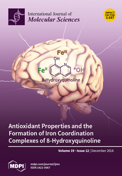
Various neurodegenerative diseases such as Alzheimer’s or Parkinson’s disease negatively affect the life quality of many patients. These diseases are associated with oxidative stress that increases the rate of brain tissue destruction. So far, the application of antioxidants represents a treatment possibility. Amongst antioxidants, 8-hydroxyquinoline and its derivatives showed promising results. In an attempt to obtain better insights into 8-hydroxyquinoline’s possible mode of action, this paper explores its redox chemistry with iron as a ligand in the iron coordination complex, and as a free molecule. View this paper.
  • Issues are regarded as officially published after their release is announced to the table of contents alert mailing list.
  • You may sign up for e-mail alerts to receive table of contents of newly released issues.
  • PDF is the official format for papers published in both, html and pdf forms. To view the papers in pdf format, click on the "PDF Full-text" link, and use the free Adobe Reader to open them.
Order results
Result details
Section
Select all
Export citation of selected articles as:
16 pages, 1284 KiB  
Review
Brain-Derived Neurotrophic Factor in Central Nervous System Myelination: A New Mechanism to Promote Myelin Plasticity and Repair
by Jessica L. Fletcher, Simon S. Murray and Junhua Xiao
Int. J. Mol. Sci. 2018, 19(12), 4131; https://doi.org/10.3390/ijms19124131 - 19 Dec 2018
Cited by 76 | Viewed by 14033
Abstract
Brain-derived neurotrophic factor (BDNF) plays vitally important roles in neural development and plasticity in both health and disease. Recent studies using mutant mice to selectively manipulate BDNF signalling in desired cell types, in combination with animal models of demyelinating disease, have demonstrated that [...] Read more.
Brain-derived neurotrophic factor (BDNF) plays vitally important roles in neural development and plasticity in both health and disease. Recent studies using mutant mice to selectively manipulate BDNF signalling in desired cell types, in combination with animal models of demyelinating disease, have demonstrated that BDNF not only potentiates normal central nervous system myelination in development but enhances recovery after myelin injury. However, the precise mechanisms by which BDNF enhances myelination in development and repair are unclear. Here, we review some of the recent progress made in understanding the influence BDNF exerts upon the myelinating process during development and after injury, and discuss the cellular and molecular mechanisms underlying its effects. In doing so, we raise new questions for future research. Full article
(This article belongs to the Special Issue Brain-Derived Neurotrophic Factor 2018)
Show Figures

Figure 1

19 pages, 2543 KiB  
Article
Progression of Repair and Injury in Human Liver Slices
by Alison E. M. Vickers, Anatoly V. Ulyanov and Robyn L. Fisher
Int. J. Mol. Sci. 2018, 19(12), 4130; https://doi.org/10.3390/ijms19124130 - 19 Dec 2018
Cited by 1 | Viewed by 4051
Abstract
Human liver slice function was stressed by daily dosing of acetaminophen (APAP) or diclofenac (DCF) to investigate injury and repair. Initially, untreated human liver and kidney slices were evaluated with the global human U133A array to assess the extended culture conditions. Then, drug [...] Read more.
Human liver slice function was stressed by daily dosing of acetaminophen (APAP) or diclofenac (DCF) to investigate injury and repair. Initially, untreated human liver and kidney slices were evaluated with the global human U133A array to assess the extended culture conditions. Then, drug induced injury and signals of repair in human liver slices exposed to APAP or DCF (1 mM) were evaluated via specific gene expression arrays. In culture, the untreated human liver and kidney slices remained differentiated and gene expression indicated that repair pathways were activated in both tissues. Morphologically the human liver slices exhibited evidence of repair and regeneration, while kidney slices did not. APAP and DCF exposure caused a direct multi-factorial response. APAP and DCF induced gene expression changes in transporters, oxidative stress and mitochondria energy. DCF caused a greater effect on heat shock and endoplasmic reticulum (ER) stress gene expression. Concerning wound repair, APAP caused a mild repression of gene expression; DCF suppressed the expression of matrix collagen genes, the remodeling metalloproteases, cell adhesion integrins, indicating a greater hinderance to wound repair than APAP. Thus, human liver slices are a relevant model to investigate the mechanisms of drug-induced injury and repair. Full article
(This article belongs to the Special Issue Liver Damage and Repair)
Show Figures

Figure 1

20 pages, 2025 KiB  
Review
Extracellular Interactions of Alpha-Synuclein in Multiple System Atrophy
by Dario Valdinocci, Rowan A. W. Radford, Michael Goulding, Junna Hayashi, Roger S. Chung and Dean L. Pountney
Int. J. Mol. Sci. 2018, 19(12), 4129; https://doi.org/10.3390/ijms19124129 - 19 Dec 2018
Cited by 21 | Viewed by 7679
Abstract
Multiple system atrophy, characterized by atypical Parkinsonism, results from central nervous system (CNS) cell loss and dysfunction linked to aggregates of the normally pre-synaptic α-synuclein protein. Mostly cytoplasmic pathological α-synuclein inclusion bodies occur predominantly in oligodendrocytes in affected brain regions and there is [...] Read more.
Multiple system atrophy, characterized by atypical Parkinsonism, results from central nervous system (CNS) cell loss and dysfunction linked to aggregates of the normally pre-synaptic α-synuclein protein. Mostly cytoplasmic pathological α-synuclein inclusion bodies occur predominantly in oligodendrocytes in affected brain regions and there is evidence that α-synuclein released by neurons is taken up preferentially by oligodendrocytes. However, extracellular α-synuclein has also been shown to interact with other neural cell types, including astrocytes and microglia, as well as extracellular factors, mediating neuroinflammation, cell-to-cell spread and other aspects of pathogenesis. Here, we review the current evidence for how α-synuclein present in the extracellular milieu may act at the cell surface to drive components of disease progression. A more detailed understanding of the important extracellular interactions of α-synuclein with neuronal and non-neuronal cell types both in the brain and periphery may provide new therapeutic targets to modulate the disease process. Full article
(This article belongs to the Special Issue Extracellular Matrix in Development and Disease 2.0)
Show Figures

Graphical abstract

21 pages, 4776 KiB  
Article
The Activity of Matrix Metalloproteinases (MMP-2, MMP-9) and Their Tissue Inhibitors (TIMP-1, TIMP-3) in the Cerebral Cortex and Hippocampus in Experimental Acanthamoebiasis
by Natalia Łanocha-Arendarczyk, Irena Baranowska-Bosiacka, Izabela Gutowska, Agnieszka Kolasa-Wołosiuk, Karolina Kot, Aleksandra Łanocha, Emilia Metryka, Barbara Wiszniewska, Dariusz Chlubek and Danuta Kosik-Bogacka
Int. J. Mol. Sci. 2018, 19(12), 4128; https://doi.org/10.3390/ijms19124128 - 19 Dec 2018
Cited by 11 | Viewed by 5902
Abstract
The pathological process occurring within the central nervous system (CNS) as a result of the infection by Acanthamoeba spp. is not fully understood. Therefore, the aim of this study was to determine whether Acanthamoeba spp. may affect the levels of matrix metalloproteinases (MMP-2,-9), [...] Read more.
The pathological process occurring within the central nervous system (CNS) as a result of the infection by Acanthamoeba spp. is not fully understood. Therefore, the aim of this study was to determine whether Acanthamoeba spp. may affect the levels of matrix metalloproteinases (MMP-2,-9), their tissue inhibitors (TIMP-1,-3) and MMP-9/TIMP-1, MMP-2/TIMP-3 ratios in the cerebral cortex and hippocampus, in relation to the host’s immunological status. Our results showed that Acanthamoeba spp. infection can change the levels of MMP and TIMP in the CNS and may be amenable targets for limiting amoebic encephalitis. The increase in the activity of matrix metalloproteinases during acanthamoebiasis may be primarily the result of inflammation process, probably an increased activity of proteolytic processes, but also (to a lesser extent) a defense mechanism preventing the processes of neurodegeneration. Full article
(This article belongs to the Section Biochemistry)
Show Figures

Graphical abstract

17 pages, 3345 KiB  
Article
Functional Assessment of Patient-Derived Retinal Pigment Epithelial Cells Edited by CRISPR/Cas9
by Leah P. Foltz, Sara E. Howden, James A. Thomson and Dennis O. Clegg
Int. J. Mol. Sci. 2018, 19(12), 4127; https://doi.org/10.3390/ijms19124127 - 19 Dec 2018
Cited by 17 | Viewed by 5149
Abstract
Retinitis pigmentosa is the most common form of inherited blindness and can be caused by a multitude of different genetic mutations that lead to similar phenotypes. Specifically, mutations in ubiquitously expressed splicing factor proteins are known to cause an autosomal dominant form of [...] Read more.
Retinitis pigmentosa is the most common form of inherited blindness and can be caused by a multitude of different genetic mutations that lead to similar phenotypes. Specifically, mutations in ubiquitously expressed splicing factor proteins are known to cause an autosomal dominant form of the disease, but the retina-specific pathology of these mutations is not well understood. Fibroblasts from a patient with splicing factor retinitis pigmentosa caused by a missense mutation in the PRPF8 splicing factor were used to produce three diseased and three CRISPR/Cas9-corrected induced pluripotent stem cell (iPSC) clones. We differentiated each of these clones into retinal pigment epithelial (RPE) cells via directed differentiation and analyzed the RPE cells in terms of gene and protein expression, apicobasal polarity, and phagocytic ability. We demonstrate that RPE cells can be produced from patient-derived and corrected cells and they exhibit morphology and functionality similar but not identical to wild-type RPE cells in vitro. Functionally, the RPE cells were able to establish apicobasal polarity and phagocytose photoreceptor outer segments at the same capacity as wild-type cells. These data suggest that patient-derived iPSCs, both diseased and corrected, are able to differentiate into RPE cells with a near normal phenotype and without differences in phagocytosis, a result that differs from previous mouse models. These RPE cells can now be studied to establish a disease-in-a-dish system relevant to retinitis pigmentosa. Full article
(This article belongs to the Special Issue Cell Reprogramming, II)
Show Figures

Figure 1

15 pages, 8676 KiB  
Article
Isoquercetin Improves Hepatic Lipid Accumulation by Activating AMPK Pathway and Suppressing TGF-β Signaling on an HFD-Induced Nonalcoholic Fatty Liver Disease Rat Model
by Guohong Qin, Ji Ma, Qiongshan Huang, Hongli Yin, Jichun Han, Maoru Li, Yang Deng, Bing Wang, Waseem Hassan and Jing Shang
Int. J. Mol. Sci. 2018, 19(12), 4126; https://doi.org/10.3390/ijms19124126 - 19 Dec 2018
Cited by 46 | Viewed by 7299
Abstract
Isoquercetin (IQ), a glucoside derivative of quercetin, has been reported to have beneficial effects in nonalcoholic fatty liver disease (NAFLD). In this study, we investigated the potential improvement of IQ in liver lipid accumulation, inflammation, oxidative condition, and activation in Kupffer cells (KCs) [...] Read more.
Isoquercetin (IQ), a glucoside derivative of quercetin, has been reported to have beneficial effects in nonalcoholic fatty liver disease (NAFLD). In this study, we investigated the potential improvement of IQ in liver lipid accumulation, inflammation, oxidative condition, and activation in Kupffer cells (KCs) on a high-fat diet (HFD) induced NAFLD models. Male Sprague-Dawley (SD) rats were induced by HFD, lipopolysaccharides/free fatty acids (LPS/FFA) induced co-culture cells model between primary hepatocytes and Kupffer cells was used to test the effects and the underlying mechanism of IQ. Molecular docking was performed to predict the potential target of IQ. Significant effects of IQ were found on reduced lipid accumulation, inflammation, and oxidative stress. In addition, AMP-activated protein kinase (AMPK) pathway was activated by IQ, and is plays an important role in lipid regulation. Meanwhile, IQ reversed the increase of activated KCs which caused by lipid overload, and also suppression of Transforming growth factor beta (TGF-β) signaling by TGF-β Recptor-1 and SMAD2/3 signaling. Finally, TGF-βR1 and TGF-βR2 were both found may involve in the mechanism of IQ. IQ can improve hepatic lipid accumulation and decrease inflammation and oxidative stress by its activating AMPK pathway and suppressing TGF-β signaling to alleviate NAFLD. Full article
(This article belongs to the Special Issue Liver Damage and Repair)
Show Figures

Graphical abstract

12 pages, 2358 KiB  
Article
Muller’s Ratchet and Ribosome Degeneration in the Obligate Intracellular Parasites Microsporidia
by Sergey V. Melnikov, Kasidet Manakongtreecheep, Keith D. Rivera, Arthur Makarenko, Darryl J. Pappin and Dieter Söll
Int. J. Mol. Sci. 2018, 19(12), 4125; https://doi.org/10.3390/ijms19124125 - 19 Dec 2018
Cited by 18 | Viewed by 5799
Abstract
Microsporidia are fungi-like parasites that have the smallest known eukaryotic genome, and for that reason they are used as a model to study the phenomenon of genome decay in parasitic forms of life. Similar to other intracellular parasites that reproduce asexually in an [...] Read more.
Microsporidia are fungi-like parasites that have the smallest known eukaryotic genome, and for that reason they are used as a model to study the phenomenon of genome decay in parasitic forms of life. Similar to other intracellular parasites that reproduce asexually in an environment with alleviated natural selection, Microsporidia experience continuous genome decay that is driven by Muller’s ratchet—an evolutionary process of irreversible accumulation of deleterious mutations that lead to gene loss and the miniaturization of cellular components. Particularly, Microsporidia have remarkably small ribosomes in which the rRNA is reduced to the minimal enzymatic core. In this study, we analyzed microsporidian ribosomes to study an apparent impact of Muller’s ratchet on structure of RNA and protein molecules in parasitic forms of life. Through mass spectrometry of microsporidian proteome and analysis of microsporidian genomes, we found that massive rRNA reduction in microsporidian ribosomes appears to annihilate the binding sites for ribosomal proteins eL8, eL27, and eS31, suggesting that these proteins are no longer bound to the ribosome in microsporidian species. We then provided an evidence that protein eS31 is retained in Microsporidia due to its non-ribosomal function in ubiquitin biogenesis. Our study illustrates that, while Microsporidia carry the same set of ribosomal proteins as non-parasitic eukaryotes, some ribosomal proteins are no longer participating in protein synthesis in Microsporidia and they are preserved from genome decay by having extra-ribosomal functions. More generally, our study shows that many components of parasitic cells, which are identified by automated annotation of pathogenic genomes, may lack part of their biological functions due to continuous genome decay. Full article
(This article belongs to the Special Issue Structure, Function and Evolution of the Ribosome)
Show Figures

Figure 1

20 pages, 6308 KiB  
Article
Medicinal Leech CNS as a Model for Exosome Studies in the Crosstalk between Microglia and Neurons
by Antonella Raffo-Romero, Tanina Arab, Issa S. Al-Amri, Francoise Le Marrec-Croq, Christelle Van Camp, Quentin Lemaire, Michel Salzet, Jacopo Vizioli, Pierre-Eric Sautiere and Christophe Lefebvre
Int. J. Mol. Sci. 2018, 19(12), 4124; https://doi.org/10.3390/ijms19124124 - 19 Dec 2018
Cited by 27 | Viewed by 5987
Abstract
In healthy or pathological brains, the neuroinflammatory state is supported by a strong communication involving microglia and neurons. Recent studies indicate that extracellular vesicles (EVs), including exosomes and microvesicles, play a key role in the physiological interactions between cells allowing central nervous system [...] Read more.
In healthy or pathological brains, the neuroinflammatory state is supported by a strong communication involving microglia and neurons. Recent studies indicate that extracellular vesicles (EVs), including exosomes and microvesicles, play a key role in the physiological interactions between cells allowing central nervous system (CNS) development and/or integrity. The present report used medicinal leech CNS to investigate microglia/neuron crosstalk from ex vivo approaches as well as primary cultures. The results demonstrated a large production of exosomes from microglia. Their incubation to primary neuronal cultures showed a strong interaction with neurites. In addition, neurite outgrowth assays demonstrated microglia exosomes to exhibit significant neurotrophic activities using at least a Transforming Growth Factor beta (TGF-β) family member, called nGDF (nervous Growth/Differentiation Factor). Of interest, the results also showed an EV-mediated dialog between leech microglia and rat cells highlighting this communication to be more a matter of molecules than of species. Taken together, the present report brings a new insight into the microglia/neuron crosstalk in CNS and would help deciphering the molecular evolution of such a cell communication in brain. Full article
(This article belongs to the Special Issue Neuroimmunology)
Show Figures

Figure 1

12 pages, 2915 KiB  
Article
High Throughput Chemical Screening Reveals Multiple Regulatory Proteins on FOXA1 in Breast Cancer Cell Lines
by Shixiong Wang, Sachin Kumar Singh, Madhumohan R. Katika, Sandra Lopez-Aviles and Antoni Hurtado
Int. J. Mol. Sci. 2018, 19(12), 4123; https://doi.org/10.3390/ijms19124123 - 19 Dec 2018
Cited by 9 | Viewed by 5613
Abstract
Forkhead box A1 (FOXA1) belongs to the forkhead class transcription factor family, playing pioneering function for hormone receptors in breast and prostate cancers, and mediating activation of linage specific enhancers. Interplay between FOXA1 and breast cancer specific signaling pathways has been reported previously, [...] Read more.
Forkhead box A1 (FOXA1) belongs to the forkhead class transcription factor family, playing pioneering function for hormone receptors in breast and prostate cancers, and mediating activation of linage specific enhancers. Interplay between FOXA1 and breast cancer specific signaling pathways has been reported previously, indicating a regulation network on FOXA1 in breast cancer cells. Here in this study, we aimed to identify which are the proteins that could potentially control FOXA1 function in breast cancer cell lines expressing different molecular markers. We first established a luciferase reporter system reflecting FOXA1 binding to DNA. Then, we applied high throughput chemical screening of multiple protein targets and mass spectrometry in breast cancer cell lines expressing different molecular markers: ER positive/HER2 negative (MCF-7), ER positive/HER2 positive (BT474), and ER negative/HER2 positive (MDA-MB-453). Regardless of estrogen receptor status, HER2 (human epidermal growth factor receptor 2) enriched cell lines showed similar response to kinase inhibitors, indicating the control of FOXA1 by cell signaling kinases. Among these kinases, we identified additional receptor tyrosine kinases and cyclin-dependent kinases as regulators of FOXA1. Furthermore, we performed proteomics experiments from FOXA1 inmunoprecipitated protein complex to identify that FOXA1 interacts with several proteins. Among all the targets, we identified cyclin-dependent kinase 1 (CDK1) as a positive factor to interact with FOXA1 in BT474 cell line. In silico analyses confirmed that cyclin-dependent kinases might be the kinases responsible for FOXA1 phosphorylation at the Forkhead domain and the transactivation domain. These results reveal that FOXA1 is potentially regulated by multiple kinases. The cell cycle control kinase CDK1 might control directly FOXA1 by phosphorylation and other kinases indirectly by means of regulating other proteins. Full article
(This article belongs to the Special Issue Sex Hormone Receptor Signals in Human Malignancies)
Show Figures

Graphical abstract

16 pages, 1974 KiB  
Perspective
S100A10 and Cancer Hallmarks: Structure, Functions, and its Emerging Role in Ovarian Cancer
by Tannith M. Noye, Noor A. Lokman, Martin K. Oehler and Carmela Ricciardelli
Int. J. Mol. Sci. 2018, 19(12), 4122; https://doi.org/10.3390/ijms19124122 - 19 Dec 2018
Cited by 30 | Viewed by 5806
Abstract
S100A10, which is also known as p11, is located in the plasma membrane and forms a heterotetramer with annexin A2. The heterotetramer, comprising of two subunits of annexin A2 and S100A10, activates the plasminogen activation pathway, which is involved in cellular repair of [...] Read more.
S100A10, which is also known as p11, is located in the plasma membrane and forms a heterotetramer with annexin A2. The heterotetramer, comprising of two subunits of annexin A2 and S100A10, activates the plasminogen activation pathway, which is involved in cellular repair of normal tissues. Increased expression of annexin A2 and S100A10 in cancer cells leads to increased levels of plasmin—which promotes the degradation of the extracellular matrix—increased angiogenesis, and the invasion of the surrounding organs. Although many studies have investigated the functional role of annexin A2 in cancer cells, including ovarian cancer, S100A10 has been less studied. We recently demonstrated that high stromal annexin A2 and high cytoplasmic S100A10 expression is associated with a 3.4-fold increased risk of progression and 7.9-fold risk of death in ovarian cancer patients. Other studies have linked S100A10 with multidrug resistance in ovarian cancer; however, no functional studies to date have been performed in ovarian cancer cells. This article reviews the current understanding of S100A10 function in cancer with a particular focus on ovarian cancer. Full article
Show Figures

Figure 1

21 pages, 2259 KiB  
Review
Hydrogen Sulfide: A Therapeutic Option in Systemic Sclerosis
by Amaal Eman Abdulle, Harry Van Goor and Douwe J. Mulder
Int. J. Mol. Sci. 2018, 19(12), 4121; https://doi.org/10.3390/ijms19124121 - 19 Dec 2018
Cited by 15 | Viewed by 5293 | Correction
Abstract
Systemic sclerosis (SSc) is a lethal disease that is characterized by auto-immunity, vascular injury, and progressive fibrosis of multiple organ systems. Despite the fact that the exact etiology of SSc remains unknown, oxidative stress has been associated with a large range of SSc-related [...] Read more.
Systemic sclerosis (SSc) is a lethal disease that is characterized by auto-immunity, vascular injury, and progressive fibrosis of multiple organ systems. Despite the fact that the exact etiology of SSc remains unknown, oxidative stress has been associated with a large range of SSc-related complications. In addition to the well-known detrimental properties of reactive oxygen species (ROS), gasotransmitters (e.g., nitric oxide (NO), carbon monoxide (CO), and hydrogen sulfide (H2S)) are also thought to play an important role in SSc. Accordingly, the diverse physiologic actions of NO and CO and their role in SSc have been previously studied. Recently, multiple studies have also shown the importance of the third gasotransmitter H2S in both vascular physiology and pathophysiology. Interestingly, homocysteine (which is converted into H2S through the transsulfuration pathway) is often found to be elevated in SSc patients; suggesting defects in the transsulfuration pathway. Hydrogen sulfide, which is known to have several effects, including a strong antioxidant and vasodilator effect, could potentially play a prominent role in the initiation and progression of vasculopathy. A better understanding of the actions of gasotransmitters, like H2S, in the development of SSc-related vasculopathy, could help to create early interventions to attenuate the disease course. This paper will review the role of H2S in vascular (patho-)physiology and potential disturbances in SSc. Moreover, current data from experimental animal studies will be reviewed. Lastly, we will evaluate potential interventional strategies. Full article
Show Figures

Graphical abstract

13 pages, 4290 KiB  
Article
Providing Antibacterial Activity to Poly(2-Hydroxy Ethyl Methacrylate) by Copolymerization with a Methacrylic Thiazolium Derivative
by Alexandra Muñoz-Bonilla, Daniel López and Marta Fernández-García
Int. J. Mol. Sci. 2018, 19(12), 4120; https://doi.org/10.3390/ijms19124120 - 19 Dec 2018
Cited by 15 | Viewed by 4218
Abstract
Antimicrobial polymers and coatings are potent types of materials for fighting microbial infections, and as such, they have attracted increased attention in many fields. Here, a series of antimicrobial copolymers were prepared by radical copolymerization of 2-hydroxyethyl methacrylate (HEMA), which is widely employed [...] Read more.
Antimicrobial polymers and coatings are potent types of materials for fighting microbial infections, and as such, they have attracted increased attention in many fields. Here, a series of antimicrobial copolymers were prepared by radical copolymerization of 2-hydroxyethyl methacrylate (HEMA), which is widely employed in the manufacturing of biomedical devices, and the monomer 2-(4-methylthiazol-5-yl)ethyl methacrylate (MTA), which bears thiazole side groups susceptible to quaternization, to provide a positive charge. The copolymers were further quantitatively quaternized with either methyl or butyl iodide, as demonstrated by nuclear magnetic resonance (NMR) and attenuated total reflection Fourier-transform infrared spectroscopy (ATR-FTIR). Then, the polycations were characterized by zeta potential measurements to evaluate their effective charge and by differential scanning calorimetry (DSC) and thermogravimetric analysis (TGA) to evaluate their thermal properties. The ζ-potential study revealed that the quaternized copolymers with intermediate compositions present higher charges than the corresponding homopolymers. The cationic copolymers showed greater glass transition temperatures than poly(2-hydroxyethyl methacrylate) (PHEMA), with values higher than 100 °C, in particular those quaternized with methyl iodide. The TGA studies showed that the thermal stability of polycations varies with the composition, improving as the content of HEMA in the copolymer increases. Microbial assays targeting Gram-positive and Gram-negative bacteria confirmed that the incorporation of a low number of cationic units into PHEMA provides antimicrobial character with a minimum inhibitory concentration (MIC) of 128 µg mL−1. Remarkably, copolymers with MTA molar fractions higher than 0.50 exhibited MIC values as low as 8 µg mL−1. Full article
(This article belongs to the Special Issue Polymeric Systems as Antimicrobial or Antifouling Agents)
Show Figures

Figure 1

13 pages, 5016 KiB  
Article
Hyaluronic Acid/Bone Substitute Complex Implanted on Chick Embryo Chorioallantoic Membrane Induces Osteoblastic Differentiation and Angiogenesis, but not Inflammation
by Laura Cirligeriu, Anca Maria Cimpean, Horia Calniceanu, Mircea Vladau, Simona Sarb, Marius Raica and Luminita Nica
Int. J. Mol. Sci. 2018, 19(12), 4119; https://doi.org/10.3390/ijms19124119 - 19 Dec 2018
Cited by 14 | Viewed by 4234
Abstract
Microscopic and molecular events related to alveolar ridge augmentation are less known because of the lack of experimental models and limited molecular markers used to evaluate this process. We propose here the chick embryo chorioallantoic membrane (CAM) as an in vivo model to [...] Read more.
Microscopic and molecular events related to alveolar ridge augmentation are less known because of the lack of experimental models and limited molecular markers used to evaluate this process. We propose here the chick embryo chorioallantoic membrane (CAM) as an in vivo model to study the interaction between CAM and bone substitutes (B) combined with hyaluronic acid (BH), saline solution (BHS and BS, respectively), or both, aiming to point out the microscopic and molecular events assessed by Runt-related transcription factor 2 (RUNX 2), osteonectin (SPARC), and Bone Morphogenic Protein 4 (BMP4). The BH complex induced osteoprogenitor and osteoblastic differentiation of CAM mesenchymal cells, certified by the RUNX2 +, BMP4 +, and SPARC + phenotypes capable of bone matrix synthesis and mineralization. A strong angiogenic response without inflammation was detected on microscopic specimens of the BH combination compared with an inflammatory induced angiogenesis for the BS and BHS combinations. A multilayered organization of the BH complex grafted on CAM was detected with a differential expression of RUNX2, BMP4, and SPARC. The BH complex induced CAM mesenchymal cells differentiation through osteoblastic lineage with a sustained angiogenic response not related with inflammation. Thus, bone granules resuspended in hyaluronic acid seem to be the best combination for a proper non-inflammatory response in alveolar ridge augmentation. The CAM model allows us to assess the early events of the bone substitutes–mesenchymal cells interaction related to osteoblastic differentiation, an important step in alveolar ridge augmentation. Full article
(This article belongs to the Section Molecular Pathology, Diagnostics, and Therapeutics)
Show Figures

Figure 1

11 pages, 2222 KiB  
Case Report
Oligoclonal T Cells Transiently Expand and Express Tim-3 and PD-1 Following Anti-CD19 CAR T Cell Therapy: A Case Report
by Christopher Ronald Funk, Christopher T. Petersen, Neera Jagirdar, Sruthi Ravindranathan, David L. Jaye, Christopher R. Flowers, Amelia Langston and Edmund K. Waller
Int. J. Mol. Sci. 2018, 19(12), 4118; https://doi.org/10.3390/ijms19124118 - 19 Dec 2018
Cited by 7 | Viewed by 4774
Abstract
Clinical trials of chimeric antigen receptor (CAR) T cells in hematologic malignancy associate remissions with two profiles of CAR T cell proliferation kinetics, which differ based upon costimulatory domain. Additional T cell intrinsic factors that influence or predict clinical response remain unclear. To [...] Read more.
Clinical trials of chimeric antigen receptor (CAR) T cells in hematologic malignancy associate remissions with two profiles of CAR T cell proliferation kinetics, which differ based upon costimulatory domain. Additional T cell intrinsic factors that influence or predict clinical response remain unclear. To address this gap, we report the case of a 68-year-old woman with refractory/relapsed diffuse large B cell lymphoma (DLBCL), treated with tisagenlecleucel (anti-CD19), with a CD137 costimulatory domain (4-1BB) on an investigational new drug application (#16944). For two months post-infusion, the patient experienced dramatic regression of subcutaneous nodules of DLBCL. Unfortunately, her CAR T exhibited kinetics unassociated with remission, and she died of DLBCL-related sequelae. Serial phenotypic analysis of peripheral blood alongside sequencing of the β-peptide variable region of the T cell receptor (TCRβ) revealed distinct waves of oligoclonal T cell expansion with dynamic expression of immune checkpoint molecules. One week prior to CAR T cell contraction, T cell immunoglobulin mucin domain 3 (Tim-3) and programmed cell death protein 1 (PD-1) exhibited peak expressions on both the CD8 T cell (Tim-3 ≈ 50%; PD-1 ≈ 17%) and CAR T cell subsets (Tim-3 ≈ 78%; PD-1 ≈ 40%). These correlative observations draw attention to Tim-3 and PD-1 signaling pathways in context of CAR T cell exhaustion. Full article
(This article belongs to the Special Issue CAR-T Cell Therapy)
Show Figures

Figure 1

79 pages, 3140 KiB  
Review
Tissue-Engineered Grafts from Human Decellularized Extracellular Matrices: A Systematic Review and Future Perspectives
by Andrea Porzionato, Elena Stocco, Silvia Barbon, Francesca Grandi, Veronica Macchi and Raffaele De Caro
Int. J. Mol. Sci. 2018, 19(12), 4117; https://doi.org/10.3390/ijms19124117 - 18 Dec 2018
Cited by 221 | Viewed by 14411
Abstract
Tissue engineering and regenerative medicine involve many different artificial and biologic materials, frequently integrated in composite scaffolds, which can be repopulated with various cell types. One of the most promising scaffolds is decellularized allogeneic extracellular matrix (ECM) then recellularized by autologous or stem [...] Read more.
Tissue engineering and regenerative medicine involve many different artificial and biologic materials, frequently integrated in composite scaffolds, which can be repopulated with various cell types. One of the most promising scaffolds is decellularized allogeneic extracellular matrix (ECM) then recellularized by autologous or stem cells, in order to develop fully personalized clinical approaches. Decellularization protocols have to efficiently remove immunogenic cellular materials, maintaining the nonimmunogenic ECM, which is endowed with specific inductive/differentiating actions due to its architecture and bioactive factors. In the present paper, we review the available literature about the development of grafts from decellularized human tissues/organs. Human tissues may be obtained not only from surgery but also from cadavers, suggesting possible development of Human Tissue BioBanks from body donation programs. Many human tissues/organs have been decellularized for tissue engineering purposes, such as cartilage, bone, skeletal muscle, tendons, adipose tissue, heart, vessels, lung, dental pulp, intestine, liver, pancreas, kidney, gonads, uterus, childbirth products, cornea, and peripheral nerves. In vitro recellularizations have been reported with various cell types and procedures (seeding, injection, and perfusion). Conversely, studies about in vivo behaviour are poorly represented. Actually, the future challenge will be the development of human grafts to be implanted fully restored in all their structural/functional aspects. Full article
(This article belongs to the Special Issue Cell Colonization in Scaffolds)
Show Figures

Figure 1

12 pages, 1336 KiB  
Article
Association of HbA1C Variability and Renal Progression in Patients with Type 2 Diabetes with Chronic Kidney Disease Stages 3–4
by Mei-Yueh Lee, Jiun-Chi Huang, Szu-Chia Chen, Hsin-Ying Clair Chiou and Pei-Yu Wu
Int. J. Mol. Sci. 2018, 19(12), 4116; https://doi.org/10.3390/ijms19124116 - 18 Dec 2018
Cited by 28 | Viewed by 4874
Abstract
Little is known about the predictive value of glycosylated hemoglobin (HbA1C) variability in patients with advanced chronic kidney disease (CKD). The aim of this study was to investigate whether HbA1C variability is associated with progression to end-stage renal disease in [...] Read more.
Little is known about the predictive value of glycosylated hemoglobin (HbA1C) variability in patients with advanced chronic kidney disease (CKD). The aim of this study was to investigate whether HbA1C variability is associated with progression to end-stage renal disease in diabetic patients with stages 3–5 CKD, and whether different stages of CKD affect these associations. Three hundred and eighty-eight patients with diabetes and stages 3–5 CKD were enrolled in this longitudinal study. Intra-individual HbA1C variability was defined as the standard deviation (SD) of HbA1C, and the renal endpoint was defined as commencing dialysis. The results indicated that, during a median follow-up period of 3.5 years, 108 patients started dialysis. Adjusted Cox analysis showed an association between the highest tertile of HbA1C SD (tertile 3 vs. tertile 1) and a lower risk of the renal endpoint (hazard ratio = 0.175; 95% confidence interval = 0.059–0.518; p = 0.002) in the patients with an HbA1C level ≥ 7% and stages 3–4 CKD, but not in stage 5 CKD. Further subgroup analysis showed that the highest two tertiles of HbA1C SD were associated with a lower risk of the renal endpoint in the group with a decreasing trend of HbA1C. Our results demonstrated that greater HbA1C variability and a decreasing trend of HbA1C, which may be related to intensive diabetes control, was associated with a lower risk of progression to dialysis in the patients with stages 3–4 CKD and poor glycemic control (HbA1c ≥ 7%). Full article
(This article belongs to the Special Issue Kidney Injury: From Molecular Basis to Therapies)
Show Figures

Figure 1

18 pages, 1037 KiB  
Review
Liver Regeneration: Different Sub-Populations of Parenchymal Cells at Play Choreographed by an Injury-Specific Microenvironment
by Rita Manco, Isabelle A. Leclercq and Laure-Alix Clerbaux
Int. J. Mol. Sci. 2018, 19(12), 4115; https://doi.org/10.3390/ijms19124115 - 18 Dec 2018
Cited by 20 | Viewed by 10562
Abstract
Liver regeneration is crucial for the maintenance of liver functional mass during homeostasis and diseases. In a disease context-dependent manner, liver regeneration is contributed to by hepatocytes or progenitor cells. As long as they are replicatively competent, hepatocytes are the main cell type [...] Read more.
Liver regeneration is crucial for the maintenance of liver functional mass during homeostasis and diseases. In a disease context-dependent manner, liver regeneration is contributed to by hepatocytes or progenitor cells. As long as they are replicatively competent, hepatocytes are the main cell type responsible for supporting liver size homeostasisand regeneration. The concept that all hepatocytes within the lobule have the same proliferative capacity but are differentially recruited according to the localization of the wound, or whether a yet to be defined sub-population of hepatocytes supports regeneration is still debated. In a chronically or severely injured liver, hepatocytes may enter a state of replicative senescence. In such conditions, small biliary cells activate and expand, a process called ductular reaction (DR). Work in the last few decades has demonstrated that DR cells can differentiate into hepatocytes and thereby contribute to parenchymal reconstitution. In this study we will review the molecular mechanisms supporting these two processes to determine potential targets that would be amenable for therapeutic manipulation to enhance liver regeneration. Full article
(This article belongs to the Special Issue Liver Damage and Repair)
Show Figures

Graphical abstract

13 pages, 3532 KiB  
Article
Tunicamycin-Induced ER Stress is Accompanied with Oxidative Stress via Abrogation of Sulfur Amino Acids Metabolism in the Liver
by Sou Hyun Kim, Do-young Kwon, Jae-Hwan Kwak, Seunghyun Lee, Yun-Hee Lee, Jieun Yun, Tae Gen Son and Young-Suk Jung
Int. J. Mol. Sci. 2018, 19(12), 4114; https://doi.org/10.3390/ijms19124114 - 18 Dec 2018
Cited by 37 | Viewed by 6579
Abstract
Endoplasmic reticulum (ER) stress is involved in non-alcoholic fatty liver disease (NAFLD), but the relationship between oxidative stress, another well-known risk factor of NAFLD, and ER stress has yet to be elucidated. In this study, we treated mice with tunicamycin (TM) (2 mg/kg [...] Read more.
Endoplasmic reticulum (ER) stress is involved in non-alcoholic fatty liver disease (NAFLD), but the relationship between oxidative stress, another well-known risk factor of NAFLD, and ER stress has yet to be elucidated. In this study, we treated mice with tunicamycin (TM) (2 mg/kg body weight) for 48 h to induce ER stress in the liver and examined the metabolic pathway that synthesizes the endogenous antioxidant, glutathione (GSH). Tunicamycin (TM) treatment significantly increased mRNA levels of CHOP and GRP78, and induced lipid accumulation in the liver. Lipid peroxidation in the liver tissue also increased from TM treatment (CON vs. TM; 3.0 ± 1.8 vs. 11.1 ± 0.8 nmol MDA/g liver, p < 0.001), which reflects an imbalance between the generation of reactive substances and antioxidant capacity. To examine the involvement of GSH synthetic pathway, we determined the metabolomic changes of sulfur amino acids in the liver. TM significantly decreased hepatic S-adenosylmethionine concentration in the methionine cycle. The levels of cysteine in the liver were increased, while taurine concentration was maintained and GSH levels profoundly decreased (CON vs. TM; 8.7 ± 1.5 vs. 5.4 ± 0.9 µmol GSH/g liver, p < 0.001). These results suggest that abnormal cysteine metabolism by TM treatment resulted in a decrease in GSH, followed by an increase in oxidative stress in the liver. In HepG2 cells, decreased GSH levels were examined by TM treatment in a dose dependent manner. Furthermore, pretreatment with TM in HepG2 cells potentiated oxidative cell death, by exacerbating the effects of tert-butyl hydroperoxide. In conclusion, TM-induced ER stress was accompanied by oxidative stress by reducing the GSH synthesis, which made the liver more susceptible to oxidative stress. Full article
(This article belongs to the Section Molecular Pathology, Diagnostics, and Therapeutics)
Show Figures

Figure 1

18 pages, 4279 KiB  
Article
Activation of TRPV1 and TRPM8 Channels in the Larynx and Associated Laryngopharyngeal Regions Facilitates the Swallowing Reflex
by Mohammad Zakir Hossain, Hiroshi Ando, Shumpei Unno, Yuji Masuda and Junichi Kitagawa
Int. J. Mol. Sci. 2018, 19(12), 4113; https://doi.org/10.3390/ijms19124113 - 18 Dec 2018
Cited by 28 | Viewed by 5842
Abstract
The larynx and associated laryngopharyngeal regions are innervated by the superior laryngeal nerve (SLN) and are highly reflexogenic. Transient receptor potential (TRP) channels have recently been detected in SLN innervated regions; however, their involvement in the swallowing reflex has not been fully elucidated. [...] Read more.
The larynx and associated laryngopharyngeal regions are innervated by the superior laryngeal nerve (SLN) and are highly reflexogenic. Transient receptor potential (TRP) channels have recently been detected in SLN innervated regions; however, their involvement in the swallowing reflex has not been fully elucidated. Here, we explore the contribution of two TRP channels, TRPV1 and TRPM8, located in SLN-innervated regions to the swallowing reflex. Immunohistochemistry identified TRPV1 and TRPM8 on cell bodies of SLN afferents located in the nodose-petrosal-jugular ganglionic complex. The majority of TRPV1 and TRPM8 immunoreactivity was located on unmyelinated neurons. Topical application of different concentrations of TRPV1 and TRPM8 agonists modulated SLN activity. Application of the agonists evoked a significantly greater number of swallowing reflexes compared with the number evoked by distilled water. The interval between the reflexes evoked by the agonists was shorter than that produced by distilled water. Prior topical application of respective TRPV1 or TRPM8 antagonists significantly reduced the number of agonist-evoked reflexes. The findings suggest that the activation of TRPV1 and TRPM8 channels present in the swallowing-related regions can facilitate the evoking of swallowing reflex. Targeting the TRP channels could be a potential therapeutic strategy for the management of dysphagia. Full article
(This article belongs to the Section Molecular Neurobiology)
Show Figures

Figure 1

24 pages, 3045 KiB  
Article
Synthesis and Evaluation of the 4-Substituted 2-Hydroxy-5-Iodochalcones and Their 7-Substituted 6-Iodoflavonol Derivatives for Inhibitory Effect on Cholinesterases and β-Secretase
by Malose J. Mphahlele, Emmanuel N. Agbo and Samantha Gildenhuys
Int. J. Mol. Sci. 2018, 19(12), 4112; https://doi.org/10.3390/ijms19124112 - 18 Dec 2018
Cited by 16 | Viewed by 3454
Abstract
A series of 2-aryl-3-hydroxy-6-iodo-4H-chromen-4-ones substituted at the 7-position with a halogen atom (X = F, Cl and Br) or methoxy group and their corresponding 4-substituted 2-hydroxy-5-iodochalcone precursors were evaluated in vitro for inhibitory effect against acetylcholinesterase (AChE), butyrylcholinesterase (BChE) and [...] Read more.
A series of 2-aryl-3-hydroxy-6-iodo-4H-chromen-4-ones substituted at the 7-position with a halogen atom (X = F, Cl and Br) or methoxy group and their corresponding 4-substituted 2-hydroxy-5-iodochalcone precursors were evaluated in vitro for inhibitory effect against acetylcholinesterase (AChE), butyrylcholinesterase (BChE) and β-secretase (BACE1) activities. Although moderate inhibitory effect was observed for the chalcones against AChE, derivatives 2h, 2j and 2n exhibited significant inhibitory effect against BChE and BACE-1. The 2-aryl-7-fluoro-8-iodoflavonols 3b and 3c, on the other hand, exhibited increased activity and selectivity against AChE and reduced effect on BACE-1. The flavonols 3h, 3i, 3k, 3l and 3p exhibited moderate inhibitory effect against AChE, but significant inhibition against BChE. Compounds 2j and 3l exhibited non-competitive mode of inhibition against BACE-1. Molecular docking predicted strong interactions with the protein residues in the active site of BACE-1 implying these compounds bind with the substrate. Similarly docking studies predicted interaction of the most active compounds with both CAS and PAS of either AChE or BChE with mixed type of enzyme inhibition confirmed by kinetic studies. Full article
(This article belongs to the Section Molecular Biophysics)
Show Figures

Graphical abstract

18 pages, 1414 KiB  
Review
Dynamics of Axl Receptor Shedding in Hepatocellular Carcinoma and Its Implication for Theranostics
by Elisa Holstein, Mathias Binder and Wolfgang Mikulits
Int. J. Mol. Sci. 2018, 19(12), 4111; https://doi.org/10.3390/ijms19124111 - 18 Dec 2018
Cited by 21 | Viewed by 6862
Abstract
Signaling of the receptor tyrosine kinase Axl and its ligand Gas6 is crucially involved in the development of liver fibrosis and hepatocellular carcinoma (HCC) by activation of hepatic stellate cells and modulation of hepatocyte differentiation. Shedding of Axl’s ectodomain leads to the release [...] Read more.
Signaling of the receptor tyrosine kinase Axl and its ligand Gas6 is crucially involved in the development of liver fibrosis and hepatocellular carcinoma (HCC) by activation of hepatic stellate cells and modulation of hepatocyte differentiation. Shedding of Axl’s ectodomain leads to the release of soluble Axl (sAxl), which is increased in advanced fibrosis and in early-to-late stage HCC in the presence and absence of cirrhosis. Here, we focus on the dynamics of Axl receptor shedding and delineate possible scenarios how Axl signaling might act as driver of fibrosis progression and HCC development. Based on experimental and clinical data, we discuss the consequences of modifying Axl signaling by sAxl cleavage, as well as cellular strategies to escape from antagonizing effects of Axl shedding by the involvement of the hepatic microenvironment. We emphasize a correlation between free Gas6 and free sAxl levels favoring abundant Gas6/Axl signaling in advanced fibrosis and HCC. The raised scenario provides a solid basis for theranostics allowing the use of sAxl as an accurate diagnostic biomarker of liver cirrhosis and HCC, as well as Axl receptor signaling for therapeutic intervention in stratified HCC patients. Full article
Show Figures

Figure 1

16 pages, 1964 KiB  
Review
Using Mouse and Drosophila Models to Investigate the Mechanistic Links between Diet, Obesity, Type II Diabetes, and Cancer
by Coral G. Warr, Katherine H. Shaw, Arani Azim, Matthew D. W. Piper and Linda M. Parsons
Int. J. Mol. Sci. 2018, 19(12), 4110; https://doi.org/10.3390/ijms19124110 - 18 Dec 2018
Cited by 21 | Viewed by 6487
Abstract
Many of the links between diet and cancer are controversial and over simplified. To date, human epidemiological studies consistently reveal that patients who suffer diet-related obesity and/or type II diabetes have an increased risk of cancer, suffer more aggressive cancers, and respond poorly [...] Read more.
Many of the links between diet and cancer are controversial and over simplified. To date, human epidemiological studies consistently reveal that patients who suffer diet-related obesity and/or type II diabetes have an increased risk of cancer, suffer more aggressive cancers, and respond poorly to current therapies. However, the underlying molecular mechanisms that increase cancer risk and decrease the response to cancer therapies in these patients remain largely unknown. Here, we review studies in mouse cancer models in which either dietary or genetic manipulation has been used to model obesity and/or type II diabetes. These studies demonstrate an emerging role for the conserved insulin and insulin-like growth factor signaling pathways as links between diet and cancer progression. However, these models are time consuming to develop and expensive to maintain. As the world faces an epidemic of obesity and type II diabetes we argue that the development of novel animal models is urgently required. We make the case for Drosophila as providing an unparalleled opportunity to combine dietary manipulation with models of human metabolic disease and cancer. Thus, combining diet and cancer models in Drosophila can rapidly and significantly advance our understanding of the conserved molecular mechanisms that link diet and diet-related metabolic disorders to poor cancer patient prognosis. Full article
(This article belongs to the Special Issue Drosophila Model and Human Disease)
Show Figures

Graphical abstract

12 pages, 809 KiB  
Review
Types of Gastric Carcinomas
by Helge L. Waldum and Reidar Fossmark
Int. J. Mol. Sci. 2018, 19(12), 4109; https://doi.org/10.3390/ijms19124109 - 18 Dec 2018
Cited by 65 | Viewed by 13948
Abstract
Gastric cancer has reduced prevalence, but poor prognoses. To improve treatment, better knowledge of carcinogenesis and cells of origin should be sought. Stomach cancers are typically localized to one of the three mucosae; cardial, oxyntic and antral. Moreover, not only the stem cell, [...] Read more.
Gastric cancer has reduced prevalence, but poor prognoses. To improve treatment, better knowledge of carcinogenesis and cells of origin should be sought. Stomach cancers are typically localized to one of the three mucosae; cardial, oxyntic and antral. Moreover, not only the stem cell, but the ECL cell may proliferate and give rise to tumours. According to Laurén, the classification of gastric carcinomas seems to reflect biological important differences and possible different cell of origin since the two subtypes, intestinal and diffuse, do not transform into the other and show different epidemiology. The stem cell probably gives rise to the intestinal type, whereas the ECL cell may be important in the diffuse type. Elevation of gastrin may be the carcinogenic factor for Helicobacter pylori as well as the recently described increased risk of gastric cancer due to proton pump inhibitor treatment. Therefore, it is essential to determine the role of the gastrin target cell, the ECL cell, in gastric carcinogenesis. Clinical trials with gastrin antagonists could improve prognoses in those with gastrin receptor positive tumours. However, further studies on gastric carcinomas applying relative available methods and with the highest sensitivity are warranted to improve our knowledge of gastric carcinogenesis. Full article
(This article belongs to the Special Issue Gastric Cancers: Molecular Pathways and Candidate Biomarkers)
Show Figures

Figure 1

15 pages, 897 KiB  
Review
Metabolic Signaling into Chromatin Modifications in the Regulation of Gene Expression
by Tian Gao, Zyanya Díaz-Hirashi and Francisco Verdeguer
Int. J. Mol. Sci. 2018, 19(12), 4108; https://doi.org/10.3390/ijms19124108 - 18 Dec 2018
Cited by 20 | Viewed by 8048
Abstract
The regulation of cellular metabolism is coordinated through a tissue cross-talk by hormonal control. This leads to the establishment of specific transcriptional gene programs which adapt to environmental stimuli. On the other hand, recent advances suggest that metabolic pathways could directly signal into [...] Read more.
The regulation of cellular metabolism is coordinated through a tissue cross-talk by hormonal control. This leads to the establishment of specific transcriptional gene programs which adapt to environmental stimuli. On the other hand, recent advances suggest that metabolic pathways could directly signal into chromatin modifications and impact on specific gene programs. The key metabolites acetyl-CoA or S-adenosyl-methionine (SAM) are examples of important metabolic hubs which play in addition a role in chromatin acetylation and methylation. In this review, we will discuss how intermediary metabolism impacts on transcription regulation and the epigenome with a particular focus in metabolic disorders. Full article
(This article belongs to the Special Issue Nutrition, Epigenetics, and Diseases)
Show Figures

Graphical abstract

14 pages, 452 KiB  
Review
Effective Food Ingredients for Fatty Liver: Soy Protein β-Conglycinin and Fish Oil
by Tomomi Yamazaki, Dongyang Li and Reina Ikaga
Int. J. Mol. Sci. 2018, 19(12), 4107; https://doi.org/10.3390/ijms19124107 - 18 Dec 2018
Cited by 13 | Viewed by 9119
Abstract
Obesity is prevalent in modern society because of a lifestyle consisting of high dietary fat and sucrose consumption combined with little exercise. Among the consequences of obesity are the emerging epidemics of hepatic steatosis and nonalcoholic fatty liver disease (NAFLD). Sterol regulatory element-binding [...] Read more.
Obesity is prevalent in modern society because of a lifestyle consisting of high dietary fat and sucrose consumption combined with little exercise. Among the consequences of obesity are the emerging epidemics of hepatic steatosis and nonalcoholic fatty liver disease (NAFLD). Sterol regulatory element-binding protein-1c (SREBP-1c) is a transcription factor that stimulates gene expression related to de novo lipogenesis in the liver. In response to a high-fat diet, the expression of peroxisome proliferator-activated receptor (PPAR) γ2, another nuclear receptor, is increased, which leads to the development of NAFLD. β-Conglycinin, a soy protein, prevents NAFLD induced by diets high in sucrose/fructose or fat by decreasing the expression and function of these nuclear receptors. β-Conglycinin also improves NAFLD via the same mechanism as for prevention. Fish oil contains n-3 polyunsaturated fatty acids such as eicosapentaenoic acid and docosahexaenoic acid. Fish oil is more effective at preventing NAFLD induced by sucrose/fructose because SREBP-1c activity is inhibited. However, the effect of fish oil on NAFLD induced by fat is controversial because fish oil further increases PPARγ2 expression, depending upon the experimental conditions. Alcohol intake also causes an alcoholic fatty liver, which is induced by increased SREBP-1c and PPARγ2 expression and decreased PPARα expression. β-Conglycinin and fish oil are effective at preventing alcoholic fatty liver because β-conglycinin decreases the function of SREBP-1c and PPARγ2, and fish oil decreases the function of SREBP-1c and increases that of PPARα. Full article
(This article belongs to the Special Issue Transcriptional Regulation in Lipid Metabolism)
Show Figures

Graphical abstract

12 pages, 1619 KiB  
Article
Effects of Space Flight on Mouse Liver versus Kidney: Gene Pathway Analyses
by Timothy G. Hammond, Patricia L. Allen and Holly H. Birdsall
Int. J. Mol. Sci. 2018, 19(12), 4106; https://doi.org/10.3390/ijms19124106 - 18 Dec 2018
Cited by 18 | Viewed by 4343
Abstract
Understanding genome wide, tissue-specific, and spaceflight-induced changes in gene expression is critical to develop effective countermeasures. Transcriptome analysis has been performed on diverse tissues harvested from animals flown in space, but not the kidney. We determined the genome wide gene expression using a [...] Read more.
Understanding genome wide, tissue-specific, and spaceflight-induced changes in gene expression is critical to develop effective countermeasures. Transcriptome analysis has been performed on diverse tissues harvested from animals flown in space, but not the kidney. We determined the genome wide gene expression using a gene array analysis of kidney and liver tissue from mice flown in space for 12 days versus ground based control animals. By comparing the transcriptome of liver and kidney from animals flown in space versus ground control animals, we tested a unique hypothesis: Are there common gene expression pathways activated in multiple tissue types in response to spaceflight stimuli? Although there were tissue-specific changes, both liver and kidney overexpressed genes in the same four areas: (a) cellular responses to peptides, hormones, and nitrogen/organonitrogen compounds; (b) apoptosis and cell death; (c) fat cell differentiation and (d) negative regulation of protein kinase. Full article
(This article belongs to the Special Issue Adaptation of Living Organisms in Space: From Mammals to Plants)
Show Figures

Figure 1

14 pages, 4918 KiB  
Article
Macrophage Migration Inhibitory Factor (MIF) Inhibition in a Murine Model of Bleomycin-Induced Pulmonary Fibrosis
by Sven Günther, Jennifer Bordenave, Thông Hua-Huy, Carole Nicco, Amélie Cumont, Raphaël Thuillet, Ly Tu, Timothée Quatremarre, Thomas Guilbert, Gaël Jalce, Frédéric Batteux, Marc Humbert, Laurent Savale, Christophe Guignabert and Anh-Tuan Dinh-Xuan
Int. J. Mol. Sci. 2018, 19(12), 4105; https://doi.org/10.3390/ijms19124105 - 18 Dec 2018
Cited by 22 | Viewed by 6362
Abstract
Background: Pulmonary hypertension (PH) is a common complication of idiopathic pulmonary fibrosis (IPF) that significantly contributes to morbidity and mortality. Macrophage migration inhibitory factor (MIF) is a critical factor in vascular remodeling of the pulmonary circulation. Objectives: We tested the effects of two [...] Read more.
Background: Pulmonary hypertension (PH) is a common complication of idiopathic pulmonary fibrosis (IPF) that significantly contributes to morbidity and mortality. Macrophage migration inhibitory factor (MIF) is a critical factor in vascular remodeling of the pulmonary circulation. Objectives: We tested the effects of two small molecules targeting MIF on bleomycin (BLM)-induced collagen deposition, PH, and vascular remodeling in mouse lungs. Methods: We examined the distribution pattern of MIF, CD74, and CXCR4 in the lungs of patients with IPF-PH and the lungs of BLM-injected mice. Then, treatments were realized with (S,R)-3-(4-hydroxyphenyl)-4,5-dihydro-5-isoxazole acetic acid methyl ester (ISO-1) and N-(3-hydroxy-4-fluorobenzyl)-5 trifluoromethylbenzoxazol-2-thione 31 (20 mg/kg/day per os for 3 weeks) started 24 h after an intratracheal BLM administration. Results: More intense immunoreactivity was noted for MIF, CD74, and CXCR4 in lungs from IPF-PH patients and BLM-injected mice. Furthermore, we found that treatments of BLM-injected mice with ISO-1 or compound 31 attenuated lung collagen deposition and right ventricular systolic pressure increase. Additionally, reduced pulmonary inflammatory infiltration and pulmonary arterial muscularization were observed in the lungs of BLM-injected mice treated with ISO-1 or compound 31. Conclusions: Treatments with ISO-1 or compound 31 attenuates BLM-induced inflammation and fibrosis in lung, and prevents PH development in mice, suggesting that MIF is an important factor for IPF-PH development. Full article
(This article belongs to the Special Issue Molecular Research on Pulmonary Hypertension)
Show Figures

Graphical abstract

14 pages, 751 KiB  
Review
Dissecting Pathogenetic Mechanisms and Therapeutic Strategies in Drosophila Models of Myotonic Dystrophy Type 1
by Anissa Souidi, Monika Zmojdzian and Krzysztof Jagla
Int. J. Mol. Sci. 2018, 19(12), 4104; https://doi.org/10.3390/ijms19124104 - 18 Dec 2018
Cited by 14 | Viewed by 4343
Abstract
Myotonic dystrophy type 1 (DM1), the most common cause of adult-onset muscular dystrophy, is autosomal dominant, multisystemic disease with characteristic symptoms including myotonia, heart defects, cataracts and testicular atrophy. DM1 disease is being successfully modelled in Drosophila allowing to identify and validate new [...] Read more.
Myotonic dystrophy type 1 (DM1), the most common cause of adult-onset muscular dystrophy, is autosomal dominant, multisystemic disease with characteristic symptoms including myotonia, heart defects, cataracts and testicular atrophy. DM1 disease is being successfully modelled in Drosophila allowing to identify and validate new pathogenic mechanisms and potential therapeutic strategies. Here we provide an overview of insights gained from fruit fly DM1 models, either: (i) fundamental with particular focus on newly identified gene deregulations and their link with DM1 symptoms; or (ii) applied via genetic modifiers and drug screens to identify promising therapeutic targets. Full article
(This article belongs to the Special Issue Drosophila Model and Human Disease)
Show Figures

Figure 1

4 pages, 151 KiB  
Editorial
Molecular Pharmacology and Pathology of Strokes
by Joen-Rong Sheu
Int. J. Mol. Sci. 2018, 19(12), 4103; https://doi.org/10.3390/ijms19124103 - 18 Dec 2018
Viewed by 2855
Abstract
Stroke, an important neurological disease, is becoming an increasingly non-communicable ailment and is the second leading cause of death after coronary heart disease in developed countries [...] Full article
(This article belongs to the Special Issue Molecular Pharmacology and Pathology of Strokes)
10 pages, 2980 KiB  
Review
Pathological Process of Prompt Connection between Host and Donor Tissue Vasculature Causing Rapid Perfusion of the Engineered Donor Tissue after Transplantation
by Sachiko Sekiya, Shunichi Morikawa, Taichi Ezaki and Tatsuya Shimizu
Int. J. Mol. Sci. 2018, 19(12), 4102; https://doi.org/10.3390/ijms19124102 - 18 Dec 2018
Cited by 3 | Viewed by 4190
Abstract
The shortage of donors for transplantation therapy is a serious issue worldwide. Tissue engineering is considered a potential solution to this problem. Connection and perfusion in engineered tissues after transplantation is vital for the survival of the transplanted tissue, especially for tissues requiring [...] Read more.
The shortage of donors for transplantation therapy is a serious issue worldwide. Tissue engineering is considered a potential solution to this problem. Connection and perfusion in engineered tissues after transplantation is vital for the survival of the transplanted tissue, especially for tissues requiring blood perfusion to receive nutrients, such as the heart. A myocardial cell sheet containing an endothelial cell network structure was fabricated in vitro using cell sheet technology. Transplantation of the three-dimensional (3D) tissue by layering myocardial sheets could ameliorate ischemic heart disease in a rat model. The endothelial cell network in the 3D tissue was able to rapidly connect to host vasculature and begin perfusion within 24 h after transplantation. In this review, we compare and discuss the engineered tissue–host vasculature connection process between tissue engineered constructs with hydrogels and cell sheets by histological analysis. This review provides information that may be useful for further improvements of in vivo engineered tissue vascularization techniques. Full article
(This article belongs to the Special Issue Vascular Endothelial Cells)
Show Figures

Figure 1

Previous Issue
Back to TopTop