Next Issue
Volume 17, July
Previous Issue
Volume 17, May
 
 
ijms-logo

Journal Browser

Journal Browser

Int. J. Mol. Sci., Volume 17, Issue 6 (June 2016) – 200 articles

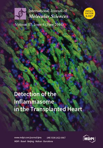
Cover Story (view full-size image): Detection of the Inflammasome in the Transplanted Heart.
Bioptic samples are used to monitor the type and severity of organ rejection. The fluorescent staining of a bioptic sample is depicted in this picture, to detect the presence of the inflammasome scaffold protein, ASC (in red). The inflammasome is part of the innate immune response. The co-staining of the cardiac alpha-actin (in green) is used to mark the cardiac myocytes. Myocytes are surrounded by infiltrating cells (leukocytes), visible through the nuclear staining (blue). In this particular picture, the myocardial structure is disrupted and ASC positivity is detected in both cardiomyocytes and non-cardiomyocyte cells.
By Adolfo Gabriele Mauro. View this article.
  • Issues are regarded as officially published after their release is announced to the table of contents alert mailing list.
  • You may sign up for e-mail alerts to receive table of contents of newly released issues.
  • PDF is the official format for papers published in both, html and pdf forms. To view the papers in pdf format, click on the "PDF Full-text" link, and use the free Adobe Reader to open them.
Order results
Result details
Section
Select all
Export citation of selected articles as:
399 KiB  
Article
Molecular Characteristics of Methicillin-Resistant Staphylococcus epidermidis on the Abdominal Skin of Females before Laparotomy
by Pin-Jia Wang, Cheng-Bin Xie, Feng-Hui Sun, Li-Juan Guo, Min Dai, Xi Cheng and Yong-Xin Ma
Int. J. Mol. Sci. 2016, 17(6), 992; https://doi.org/10.3390/ijms17060992 - 22 Jun 2016
Cited by 15 | Viewed by 6489
Abstract
Staphylococcus epidermidis, especially methicillin-resistant strains, may be the source of surgical site infections and may be a reservoir of staphylococcal cassette chromosome mec (SCCmec) for S. aureus. The aim of this study was to investigate the prevalence of methicillin-resistant [...] Read more.
Staphylococcus epidermidis, especially methicillin-resistant strains, may be the source of surgical site infections and may be a reservoir of staphylococcal cassette chromosome mec (SCCmec) for S. aureus. The aim of this study was to investigate the prevalence of methicillin-resistant S. epidermidis (MRSE) on the abdominal skin of females before laparotomy and determine the molecular characteristics and antimicrobial susceptibility patterns of these isolates. MRSE was found in 54 of 157 isolates based on mecA gene detection, and there was no difference in icaA gene carriage rate between MRSE and methicillin-susceptible S. epidermidis (MSSE) isolates. Antimicrobial susceptibility profiles were determined by broth microdilution antimicrobial susceptibility testing according to the latest CLSI manuals. All MRSE isolates had unfavorable antimicrobial susceptibility patterns. Twenty-three MRSE strains (42.6%) were multi-drug resistant. SCCmec typing and pulsed field gel electrophoresis (PFGE) typing was performed. Thirty-nine (72.2%) had a single SCCmec type, whereas 1.9% had two types. Fourteen strains (25.9%) were non-typeable (NT). The most frequent MRSE genotype was SCCmec type IVa. High diversity with PFGE patterns was obtained for MRSE, and there were no isolates exhibiting identical pulsotype. The results confirm that methicillin-resistant strains are frequently present among S. epidermidis on the abdominal skin of females before laparotomy. Moreover, resistance profiles seem to have no association with the SCCmec types or PFGE types for most common antibiotics. Full article
(This article belongs to the Special Issue Inflammatory Skin Conditions)
Show Figures

Graphical abstract

742 KiB  
Communication
Serum Levels of Substance P and Mortality in Patients with a Severe Acute Ischemic Stroke
by Leonardo Lorente, María M. Martín, Teresa Almeida, Antonia Pérez-Cejas, Luis Ramos, Mónica Argueso, Marta Riaño-Ruiz, Jordi Solé-Violán and Mariano Hernández
Int. J. Mol. Sci. 2016, 17(6), 991; https://doi.org/10.3390/ijms17060991 - 22 Jun 2016
Cited by 25 | Viewed by 5177
Abstract
Substance P (SP), a member of tachykinin family, is involved in the inflammation of the central nervous system and in the appearance of cerebral edema. Higher serum levels of SP have been found in 18 patients with cerebral ischemia compared with healthy controls. [...] Read more.
Substance P (SP), a member of tachykinin family, is involved in the inflammation of the central nervous system and in the appearance of cerebral edema. Higher serum levels of SP have been found in 18 patients with cerebral ischemia compared with healthy controls. The aim of our multi-center study was to analyze the possible association between serum levels of SP and mortality in ischemic stroke patients. We included patients with malignant middle cerebral artery infarction (MMCAI) and a Glasgow Coma Scale (GCS) lower than 9. Non-surviving patients at 30 days (n = 31) had higher serum concentrations of SP levels at diagnosis of severe MMCAI than survivors (n = 30) (p < 0.001). We found in multiple regression an association between serum concentrations of SP higher than 362 pg/mL and mortality at 30 days (Odds Ratio = 5.33; 95% confidence interval = 1.541–18.470; p = 0.008) after controlling for age and GCS. Thus, the major novel finding of our study was the association between serum levels of SP and mortality in patients suffering from severe acute ischemic stroke. Full article
(This article belongs to the Special Issue Neurological Injuries’ Monitoring, Tracking and Treatment 2016)
Show Figures

Graphical abstract

773 KiB  
Review
DNA Damage and Pulmonary Hypertension
by Benoît Ranchoux, Jolyane Meloche, Roxane Paulin, Olivier Boucherat, Steeve Provencher and Sébastien Bonnet
Int. J. Mol. Sci. 2016, 17(6), 990; https://doi.org/10.3390/ijms17060990 - 22 Jun 2016
Cited by 89 | Viewed by 10150
Abstract
Pulmonary hypertension (PH) is defined by a mean pulmonary arterial pressure over 25 mmHg at rest and is diagnosed by right heart catheterization. Among the different groups of PH, pulmonary arterial hypertension (PAH) is characterized by a progressive obstruction of distal pulmonary arteries, [...] Read more.
Pulmonary hypertension (PH) is defined by a mean pulmonary arterial pressure over 25 mmHg at rest and is diagnosed by right heart catheterization. Among the different groups of PH, pulmonary arterial hypertension (PAH) is characterized by a progressive obstruction of distal pulmonary arteries, related to endothelial cell dysfunction and vascular cell proliferation, which leads to an increased pulmonary vascular resistance, right ventricular hypertrophy, and right heart failure. Although the primary trigger of PAH remains unknown, oxidative stress and inflammation have been shown to play a key role in the development and progression of vascular remodeling. These factors are known to increase DNA damage that might favor the emergence of the proliferative and apoptosis-resistant phenotype observed in PAH vascular cells. High levels of DNA damage were reported to occur in PAH lungs and remodeled arteries as well as in animal models of PH. Moreover, recent studies have demonstrated that impaired DNA-response mechanisms may lead to an increased mutagen sensitivity in PAH patients. Finally, PAH was linked with decreased breast cancer 1 protein (BRCA1) and DNA topoisomerase 2-binding protein 1 (TopBP1) expression, both involved in maintaining genome integrity. This review aims to provide an overview of recent evidence of DNA damage and DNA repair deficiency and their implication in PAH pathogenesis. Full article
(This article belongs to the Special Issue DNA Damage and Repair in Degenerative Diseases 2016)
Show Figures

Figure 1

265 KiB  
Review
The Obesity-Breast Cancer Conundrum: An Analysis of the Issues
by Shawna B. Matthews and Henry J. Thompson
Int. J. Mol. Sci. 2016, 17(6), 989; https://doi.org/10.3390/ijms17060989 - 22 Jun 2016
Cited by 46 | Viewed by 11211
Abstract
Breast cancer develops over a timeframe of 2–3 decades prior to clinical detection. Given this prolonged latency, it is somewhat unexpected from a biological perspective that obesity has no effect or reduces the risk for breast cancer in premenopausal women yet increases the [...] Read more.
Breast cancer develops over a timeframe of 2–3 decades prior to clinical detection. Given this prolonged latency, it is somewhat unexpected from a biological perspective that obesity has no effect or reduces the risk for breast cancer in premenopausal women yet increases the risk for breast cancer in postmenopausal women. This conundrum is particularly striking in light of the generally negative effects of obesity on breast cancer outcomes, including larger tumor size at diagnosis and poorer prognosis in both pre- and postmenopausal women. This review and analysis identifies factors that may contribute to this apparent conundrum, issues that merit further investigation, and characteristics of preclinical models for breast cancer and obesity that should be considered if animal models are used to deconstruct the conundrum. Full article
(This article belongs to the Special Issue Advances in Nutritional Epidemiology)
Show Figures

Graphical abstract

545 KiB  
Review
Bone Metastasis from Renal Cell Carcinoma
by Szu-Chia Chen and Po-Lin Kuo
Int. J. Mol. Sci. 2016, 17(6), 987; https://doi.org/10.3390/ijms17060987 - 22 Jun 2016
Cited by 65 | Viewed by 9324
Abstract
About one-third of patients with advanced renal cell carcinoma (RCC) have bone metastasis that are often osteolytic and cause substantial morbidity, such as pain, pathologic fracture, spinal cord compression and hypercalcemia. The presence of bone metastasis in RCC is also associated with poor [...] Read more.
About one-third of patients with advanced renal cell carcinoma (RCC) have bone metastasis that are often osteolytic and cause substantial morbidity, such as pain, pathologic fracture, spinal cord compression and hypercalcemia. The presence of bone metastasis in RCC is also associated with poor prognosis. Bone-targeted treatment using bisphosphonate and denosumab can reduce skeletal complications in RCC, but does not cure the disease or improve survival. Elucidating the molecular mechanisms of tumor-induced changes in the bone microenvironment is needed to develop effective treatment. The “vicious cycle” hypothesis has been used to describe how tumor cells interact with the bone microenvironment to drive bone destruction and tumor growth. Tumor cells secrete factors like parathyroid hormone-related peptide, transforming growth factor-β and vascular endothelial growth factor, which stimulate osteoblasts and increase the production of the receptor activator of nuclear factor κB ligand (RANKL). In turn, the overexpression of RANKL leads to increased osteoclast formation, activation and survival, thereby enhancing bone resorption. This review presents a general survey on bone metastasis in RCC by natural history, interaction among the immune system, bone and tumor, molecular mechanisms, bone turnover markers, therapies and healthcare burden. Full article
(This article belongs to the Special Issue Cellular and Molecular Mechanisms of Bone Metastasis)
Show Figures

Figure 1

6761 KiB  
Article
Characteristics and Antitumor Activity of Morchella esculenta Polysaccharide Extracted by Pulsed Electric Field
by Chao Liu, Yonghai Sun, Qian Mao, Xiaolei Guo, Peng Li, Yang Liu and Na Xu
Int. J. Mol. Sci. 2016, 17(6), 986; https://doi.org/10.3390/ijms17060986 - 22 Jun 2016
Cited by 94 | Viewed by 7367
Abstract
Polysaccharides from Morchella esculenta have been proven to be functional and helpful for humans. The purpose of this study was to investigate the chemical structure and anti-proliferating and antitumor activities of a Morchella esculenta polysaccharide (MEP) extracted by pulsed electric field (PEF) in [...] Read more.
Polysaccharides from Morchella esculenta have been proven to be functional and helpful for humans. The purpose of this study was to investigate the chemical structure and anti-proliferating and antitumor activities of a Morchella esculenta polysaccharide (MEP) extracted by pulsed electric field (PEF) in submerged fermentation. The endo-polysaccharide was separated and purified by column chromatography and Gel permeation chromatography, and analyzed by gas chromatography. The MEP with an average molecular weight of 81,835 Da consisted of xylose, glucose, mannose, rhamnose and galactose at the ratio of 5.4:5.0:6.5:7.8:72.3. Structure of MEP was further analyzed by Fourier-transform infrared spectroscopy and 1H and 13C liquid-state nuclear magnetic resonance spectroscopy. Apoptosis tests proved that MEP could inhibit the proliferation and growth of human colon cancer HT-29 cells in a time- and dose-dependent manner within 48 h. This study provides more information on chemical structure of anti-proliferating polysaccharides isolated from Morchella esculenta. Full article
(This article belongs to the Section Biochemistry)
Show Figures

Figure 1

1602 KiB  
Review
RNA Secondary Structure Modulates FMRP’s Bi-Functional Role in the MicroRNA Pathway
by Phillip Kenny and Stephanie Ceman
Int. J. Mol. Sci. 2016, 17(6), 985; https://doi.org/10.3390/ijms17060985 - 22 Jun 2016
Cited by 23 | Viewed by 9121
Abstract
MicroRNAs act by post-transcriptionally regulating the gene expression of 30%–60% of mammalian genomes. MicroRNAs are key regulators in all cellular processes, though the mechanism by which the cell activates or represses microRNA-mediated translational regulation is poorly understood. In this review, we discuss the [...] Read more.
MicroRNAs act by post-transcriptionally regulating the gene expression of 30%–60% of mammalian genomes. MicroRNAs are key regulators in all cellular processes, though the mechanism by which the cell activates or represses microRNA-mediated translational regulation is poorly understood. In this review, we discuss the RNA binding protein Fragile X Mental Retardation Protein (FMRP) and its role in microRNA-mediated translational regulation. Historically, FMRP is known to function as a translational suppressor. However, emerging data suggests that FMRP has both an agonistic and antagonistic role in regulating microRNA-mediated translational suppression. This bi-functional role is dependent on FMRP’s interaction with the RNA helicase Moloney leukemia virus 10 (MOV10), which modifies the structural landscape of bound mRNA, therefore facilitating or inhibiting its association with the RNA-Induced Silencing Complex. Full article
(This article belongs to the Special Issue MicroRNA Regulation)
Show Figures

Graphical abstract

639 KiB  
Review
ROS and Brain Gliomas: An Overview of Potential and Innovative Therapeutic Strategies
by Mariagrazia Rinaldi, Maria Caffo, Letteria Minutoli, Herbert Marini, Rosaria Viola Abbritti, Francesco Squadrito, Vincenzo Trichilo, Andrea Valenti, Valeria Barresi, Domenica Altavilla, Marcello Passalacqua and Gerardo Caruso
Int. J. Mol. Sci. 2016, 17(6), 984; https://doi.org/10.3390/ijms17060984 - 22 Jun 2016
Cited by 98 | Viewed by 9075
Abstract
Reactive oxygen species (ROS) represent reactive products belonging to the partial reduction of oxygen. It has been reported that ROS are involved in different signaling pathways to control cellular stability. Under normal conditions, the correct function of redox systems leads to the prevention [...] Read more.
Reactive oxygen species (ROS) represent reactive products belonging to the partial reduction of oxygen. It has been reported that ROS are involved in different signaling pathways to control cellular stability. Under normal conditions, the correct function of redox systems leads to the prevention of cell oxidative damage. When ROS exceed the antioxidant defense system, cellular stress occurs. The cellular redox impairment is strictly related to tumorigenesis. Tumor cells, through the generation of hydrogen peroxide, tend to the alteration of cell cycle phases and, finally to cancer progression. In adults, the most common form of primary malignant brain tumors is represented by gliomas. The gliomagenesis is characterized by numerous molecular processes all characterized by an altered production of growth factor receptors. The difficulty to treat brain cancer depends on several biological mechanisms such as failure of drug delivery through the blood-brain barrier, tumor response to chemotherapy, and intrinsic resistance of tumor cells. Understanding the mechanisms of ROS action could allow the formulation of new therapeutic protocols to treat brain gliomas. Full article
(This article belongs to the Section Molecular Pathology, Diagnostics, and Therapeutics)
Show Figures

Graphical abstract

1852 KiB  
Article
Selection of Suitable Reference Genes for Quantitative Real-Time PCR Normalization in Three Types of Rat Adipose Tissue
by Wan-Xia Zhang, Jie Fan, Jing Ma, Yi-Song Rao, Li Zhang and You-E Yan
Int. J. Mol. Sci. 2016, 17(6), 968; https://doi.org/10.3390/ijms17060968 - 22 Jun 2016
Cited by 40 | Viewed by 9691
Abstract
Quantitative real-time PCR (qRT-PCR) is the most classical technique in the field of gene expression study. This method requires an appropriate reference gene to normalize mRNA levels. In this study, the expression stability of four frequently-used reference genes in epididymal white adipose tissue [...] Read more.
Quantitative real-time PCR (qRT-PCR) is the most classical technique in the field of gene expression study. This method requires an appropriate reference gene to normalize mRNA levels. In this study, the expression stability of four frequently-used reference genes in epididymal white adipose tissue (eWAT), inguinal beige adipose tissue (iBeAT) and brown adipose tissue (BAT) from obese and lean rats were evaluated by geNorm, NormFinder and BestKeeper. Based on the Minimum Information for Publication of Quantitative Real-Time PCR Experiments (MIQE) guidelines, the two most stable reference genes were recommended in each type of adipose tissue. Two target genes were applied to test the stability of the reference genes. The geNorm and NormFinder results revealed that GAPDH and 36B4 exhibited the highest expression stabilities in eWAT, while 36B4 and β-actin had the highest expression stabilities in iBeAT and BAT. According to the results of the BestKeeper analysis, 36B4 was the most stable gene in eWAT, iBeAT and BAT, in terms of the coefficient of variance. In terms of the coefficient of correlation, GAPDH, 36B4 and β-actin were the most stable genes in eWAT, iBeAT and BAT, respectively. Additionally, expected results and statistical significance were obtained using a combination of two suitable reference genes for data normalization. In conclusion, 36B4 and GAPDH, in combination, are the best reference genes for eWAT, while 36B4 and β-actin are two most suitable reference genes for both iBeAT and BAT. We recommend using these reference genes accordingly. Full article
(This article belongs to the Section Biochemistry)
Show Figures

Graphical abstract

8870 KiB  
Article
Expression of Stipa purpurea SpCIPK26 in Arabidopsis thaliana Enhances Salt and Drought Tolerance and Regulates Abscisic Acid Signaling
by Yanli Zhou, Xudong Sun, Yunqiang Yang, Xiong Li, Ying Cheng and Yongping Yang
Int. J. Mol. Sci. 2016, 17(6), 966; https://doi.org/10.3390/ijms17060966 - 22 Jun 2016
Cited by 15 | Viewed by 6963
Abstract
Stipa purpurea (S. purpurea) is the dominant plant species in the alpine steppe of the Qinghai-Tibet Plateau, China. It is highly resistant to cold and drought conditions. However, the underlying mechanisms regulating the stress tolerance are unknown. In this study, a [...] Read more.
Stipa purpurea (S. purpurea) is the dominant plant species in the alpine steppe of the Qinghai-Tibet Plateau, China. It is highly resistant to cold and drought conditions. However, the underlying mechanisms regulating the stress tolerance are unknown. In this study, a CIPK gene from S. purpurea (SpCIPK26) was isolated. The SpCIPK26 coding region consisted of 1392 bp that encoded 464 amino acids. The protein has a highly conserved catalytic structure and regulatory domain. The expression of SpCIPK26 was induced by drought and salt stress. SpCIPK26 overexpression in Arabidopsis thaliana (A. thaliana) plants provided increased tolerance to drought and salt stress in an abscisic acid (ABA)-dependent manner. Compared with wild-type A. thaliana plants, SpCIPK26-overexpressing plants had higher survival rates, water potentials, and photosynthetic efficiency (Fv/Fm), as well as lower levels of reactive oxygen species (ROS) following exposure to drought and salt stress. Gene expression analyses indicated stress-inducible genes (RD29A, RD29B, and ABF2) and a ROS-scavenger gene (CAT1) were upregulated in SpCIPK26-overexpressing plants after stress treatments. All of these marker genes are associated with ABA-responsive cis-acting elements. Additionally, the similarities in the gene expression patterns following ABA, mannitol, and NaCl treatments suggest SpCIPK26 has an important role during plant responses to drought and salt stress and in regulating ABA signaling. Full article
(This article belongs to the Special Issue Kinase Signal Transduction)
Show Figures

Graphical abstract

3459 KiB  
Article
Tumor Necrosis Factor-Related Apoptosis-Inducing Ligand-Induced Apoptosis in Prostate Cancer Cells after Treatment with Xanthohumol—A Natural Compound Present in Humulus lupulus L.
by Małgorzata Kłósek, Anna Mertas, Wojciech Król, Dagmara Jaworska, Jan Szymszal and Ewelina Szliszka
Int. J. Mol. Sci. 2016, 17(6), 837; https://doi.org/10.3390/ijms17060837 - 22 Jun 2016
Cited by 46 | Viewed by 6696
Abstract
TRAIL (tumor necrosis factor-related apoptosis-inducing ligand) is an endogenous ligand, which plays role in immune surveillance and anti-tumor immunity. It has ability to selectively kill tumor cells showing no toxicity to normal cells. We tested the apoptotic and cytotoxic activities of xanthohumol, a [...] Read more.
TRAIL (tumor necrosis factor-related apoptosis-inducing ligand) is an endogenous ligand, which plays role in immune surveillance and anti-tumor immunity. It has ability to selectively kill tumor cells showing no toxicity to normal cells. We tested the apoptotic and cytotoxic activities of xanthohumol, a prenylated chalcone found in Humulus lupulus on androgen-sensitive human prostate adenocarcinoma cells (LNCaP) in combination with TRAIL. Cytotoxicity was measured by 3-(4,5-dimethylthiazol-2-yl)-2,5-diphenyltetrazolium bromide tetrazolium reduction assay (MTT) and lactate dehydrogenase assay (LDH). The expression of death receptors (DR4/TRAIL-R1 and DR5/TRAIL-R2) and apoptosis were detected using flow cytometry. We examined mitochondrial membrane potential (ΔΨm) by DePsipher reagent using fluorescence microscopy. The intracellular expression of proteins was evaluated by Western blotting. Our study showed that xanthohumol enhanced cytotoxic and apoptotic effects of TRAIL. The tested compounds activated caspases-3, -8, -9, Bid, and increased the expression of Bax. They also decreased expression of Bcl-xL and decreased mitochondrial membrane potential, while the expression of death receptors was not changed. The findings suggest that xanthohumol is a compound of potential use in chemoprevention of prostate cancer due to its sensitization of cancer cells to TRAIL-mediated apoptosis. Full article
(This article belongs to the Special Issue The Mechanism of Action of Food Components in Disease Prevention)
Show Figures

Graphical abstract

260 KiB  
Review
Strategies to Optimize Adult Stem Cell Therapy for Tissue Regeneration
by Shan Liu, Jingli Zhou, Xuan Zhang, Yang Liu, Jin Chen, Bo Hu, Jinlin Song and Yuanyuan Zhang
Int. J. Mol. Sci. 2016, 17(6), 982; https://doi.org/10.3390/ijms17060982 - 21 Jun 2016
Cited by 120 | Viewed by 13219
Abstract
Stem cell therapy aims to replace damaged or aged cells with healthy functioning cells in congenital defects, tissue injuries, autoimmune disorders, and neurogenic degenerative diseases. Among various types of stem cells, adult stem cells (i.e., tissue-specific stem cells) commit to becoming [...] Read more.
Stem cell therapy aims to replace damaged or aged cells with healthy functioning cells in congenital defects, tissue injuries, autoimmune disorders, and neurogenic degenerative diseases. Among various types of stem cells, adult stem cells (i.e., tissue-specific stem cells) commit to becoming the functional cells from their tissue of origin. These cells are the most commonly used in cell-based therapy since they do not confer risk of teratomas, do not require fetal stem cell maneuvers and thus are free of ethical concerns, and they confer low immunogenicity (even if allogenous). The goal of this review is to summarize the current state of the art and advances in using stem cell therapy for tissue repair in solid organs. Here we address key factors in cell preparation, such as the source of adult stem cells, optimal cell types for implantation (universal mesenchymal stem cells vs. tissue-specific stem cells, or induced vs. non-induced stem cells), early or late passages of stem cells, stem cells with endogenous or exogenous growth factors, preconditioning of stem cells (hypoxia, growth factors, or conditioned medium), using various controlled release systems to deliver growth factors with hydrogels or microspheres to provide apposite interactions of stem cells and their niche. We also review several approaches of cell delivery that affect the outcomes of cell therapy, including the appropriate routes of cell administration (systemic, intravenous, or intraperitoneal vs. local administration), timing for cell therapy (immediate vs. a few days after injury), single injection of a large number of cells vs. multiple smaller injections, a single site for injection vs. multiple sites and use of rodents vs. larger animal models. Future directions of stem cell-based therapies are also discussed to guide potential clinical applications. Full article
Show Figures

Graphical abstract

2232 KiB  
Article
Melanoma-Derived BRAFV600E Mutation in Peritumoral Stromal Cells: Implications for in Vivo Cell Fusion
by Zsuzsanna Kurgyis, Lajos V. Kemény, Tünde Buknicz, Gergely Groma, Judit Oláh, Ádám Jakab, Hilda Polyánka, Kurt Zänker, Thomas Dittmar, Lajos Kemény and István B. Németh
Int. J. Mol. Sci. 2016, 17(6), 980; https://doi.org/10.3390/ijms17060980 - 21 Jun 2016
Cited by 13 | Viewed by 5317
Abstract
Melanoma often recurs in patients after the removal of the primary tumor, suggesting the presence of recurrent tumor-initiating cells that are undetectable using standard diagnostic methods. As cell fusion has been implicated to facilitate the alteration of a cell’s phenotype, we hypothesized that [...] Read more.
Melanoma often recurs in patients after the removal of the primary tumor, suggesting the presence of recurrent tumor-initiating cells that are undetectable using standard diagnostic methods. As cell fusion has been implicated to facilitate the alteration of a cell’s phenotype, we hypothesized that cells in the peritumoral stroma having a stromal phenotype that initiate recurrent tumors might originate from the fusion of tumor and stromal cells. Here, we show that in patients with BRAFV600E melanoma, melanoma antigen recognized by T-cells (MART1)-negative peritumoral stromal cells express BRAFV600E protein. To confirm the presence of the oncogene at the genetic level, peritumoral stromal cells were microdissected and screened for the presence of BRAFV600E with a mutation-specific polymerase chain reaction. Interestingly, cells carrying the BRAFV600E mutation were not only found among cells surrounding the primary tumor but were also present in the stroma of melanoma metastases as well as in a histologically tumor-free re-excision sample from a patient who subsequently developed a local recurrence. We did not detect any BRAFV600E mutation or protein in the peritumoral stroma of BRAFWT melanoma. Therefore, our results suggest that peritumoral stromal cells contain melanoma-derived oncogenic information, potentially as a result of cell fusion. These hybrid cells display the phenotype of stromal cells and are therefore undetectable using routine histological assessments. Our results highlight the importance of genetic analyses and the application of mutation-specific antibodies in the identification of potentially recurrent-tumor-initiating cells, which may help better predict patient survival and disease outcome. Full article
(This article belongs to the Special Issue Cell Fusion in Cancer)
Show Figures

Graphical abstract

1417 KiB  
Letter
The Chlorophyll a Fluorescence Modulated by All-Trans-β-Carotene in the Process of Photosystem II
by Tianyu Li, Ye Zhang, Nan Gong, Zuowei Li, Chenglin Sun and Zhiwei Men
Int. J. Mol. Sci. 2016, 17(6), 978; https://doi.org/10.3390/ijms17060978 - 21 Jun 2016
Cited by 8 | Viewed by 7481
Abstract
Modulating the chlorophyll a (Chl-a) fluorescence by all-trans-β-Carotene (β-Car) in the polarity and non-polarity solutions was investigated. The fluorescence intensity of Chl-a decreased as the concentration of β-Car increased. The excited electronic levels of Chl-a and β-Car became much closer owing to the [...] Read more.
Modulating the chlorophyll a (Chl-a) fluorescence by all-trans-β-Carotene (β-Car) in the polarity and non-polarity solutions was investigated. The fluorescence intensity of Chl-a decreased as the concentration of β-Car increased. The excited electronic levels of Chl-a and β-Car became much closer owing to the solvent effect, which led to the electron transfer between both two molecules. A electron-separated pair Chl·Chl+ that is not luminous was formed due to electron transfer. The solution of Chl-a and β-car in C3H6O was similar to the internal environment of chloroplast. We conclude that the polar solvent is good for the fluorescent modulation in photosystem II. Full article
(This article belongs to the Special Issue Chemical Bond and Bonding 2016)
Show Figures

Graphical abstract

3318 KiB  
Article
Multi-Elemental Profiling of Tibial and Maxillary Trabecular Bone in Ovariectomised Rats
by Pingping Han, Shifeier Lu, Yinghong Zhou, Karine Moromizato, Zhibin Du, Thor Friis and Yin Xiao
Int. J. Mol. Sci. 2016, 17(6), 977; https://doi.org/10.3390/ijms17060977 - 21 Jun 2016
Cited by 3 | Viewed by 6248
Abstract
Atomic minerals are the smallest components of bone and the content of Ca, being the most abundant mineral in bone, correlates strongly with the risk of osteoporosis. Postmenopausal women have a far greater risk of suffering from OP due to low Ca concentrations [...] Read more.
Atomic minerals are the smallest components of bone and the content of Ca, being the most abundant mineral in bone, correlates strongly with the risk of osteoporosis. Postmenopausal women have a far greater risk of suffering from OP due to low Ca concentrations in their bones and this is associated with low bone mass and higher bone fracture rates. However, bone strength is determined not only by Ca level, but also a number of metallic and non-metallic elements in bone. Thus, in this study, the difference of metallic and non-metallic elements in ovariectomy-induced osteoporosis tibial and maxillary trabecular bone was investigated in comparison with sham operated normal bone by laser ablation inductively-coupled plasma mass spectrometry using a rat model. The results demonstrated that the average concentrations of 25Mg, 28Si, 39K, 47Ti, 56Fe, 59Co, 77Se, 88Sr, 137Ba, and 208Pb were generally higher in tibia than those in maxilla. Compared with the sham group, Ovariectomy induced more significant changes of these elements in tibia than maxilla, indicating tibial trabecular bones are more sensitive to changes of circulating estrogen. In addition, the concentrations of 28Si, 77Se, 208Pb, and Ca/P ratios were higher in tibia and maxilla in ovariectomised rats than those in normal bone at all time-points. The present study indicates that ovariectomy could significantly impact the element distribution and concentrations between tibia and maxilla. Full article
(This article belongs to the Special Issue Molecular Research on Dental Materials and Biomaterials)
Show Figures

Graphical abstract

3837 KiB  
Article
Rhizobacterial Strain Bacillus megaterium BOFC15 Induces Cellular Polyamine Changes that Improve Plant Growth and Drought Resistance
by Cheng Zhou, Zhongyou Ma, Lin Zhu, Xin Xiao, Yue Xie, Jian Zhu and Jianfei Wang
Int. J. Mol. Sci. 2016, 17(6), 976; https://doi.org/10.3390/ijms17060976 - 21 Jun 2016
Cited by 168 | Viewed by 9366
Abstract
Plant-growth-promoting rhizobacteria can improve plant growth, development, and stress adaptation. However, the underlying mechanisms are still largely unclear. We investigated the effects of Bacillus megaterium BOFC15 on Arabidopsis plants. BOFC15 produced and secreted spermidine (Spd), a type of polyamine (PA) that plays an [...] Read more.
Plant-growth-promoting rhizobacteria can improve plant growth, development, and stress adaptation. However, the underlying mechanisms are still largely unclear. We investigated the effects of Bacillus megaterium BOFC15 on Arabidopsis plants. BOFC15 produced and secreted spermidine (Spd), a type of polyamine (PA) that plays an important role in plant growth. Moreover, BOFC15 induced changes in the cellular PAs of plants that promoted an increase of free Spd and spermine levels. However, these effects were remarkably abolished by the addition of dicyclohexylamine (DCHA), a Spd biosynthetic inhibitor. Additionally, the inoculation with BOFC15 remarkably increased plant biomass, improved root system architecture, and augmented photosynthetic capacity. Inoculated plants also displayed stronger ability to tolerate drought stress than non-inoculated (control) plants. Abscisic acid (ABA) content was notably higher in the inoculated plants than in the control plants under drought stress and polyethylene glycol (PEG)-induced stress conditions. However, the BOFC15-induced ABA synthesis was markedly inhibited by DCHA. Thus, microbial Spd participated in the modulation of the ABA levels. The Spd-producing BOFC15 improved plant drought tolerance, which was associated with altered cellular ABA levels and activated adaptive responses. Full article
(This article belongs to the Section Molecular Plant Sciences)
Show Figures

Graphical abstract

1371 KiB  
Review
Role of mTOR Inhibitors in Kidney Disease
by Moto Kajiwara and Satohiro Masuda
Int. J. Mol. Sci. 2016, 17(6), 975; https://doi.org/10.3390/ijms17060975 - 21 Jun 2016
Cited by 48 | Viewed by 9643
Abstract
The first compound that inhibited the mammalian target of rapamycin (mTOR), sirolimus (rapamycin) was discovered in the 1970s as a soil bacterium metabolite collected on Easter Island (Rapa Nui). Because sirolimus showed antiproliferative activity, researchers investigated its molecular target and identified the TOR1 [...] Read more.
The first compound that inhibited the mammalian target of rapamycin (mTOR), sirolimus (rapamycin) was discovered in the 1970s as a soil bacterium metabolite collected on Easter Island (Rapa Nui). Because sirolimus showed antiproliferative activity, researchers investigated its molecular target and identified the TOR1 and TOR2. The mTOR consists of mTOR complex 1 (mTORC1) and mTORC2. Rapalogues including sirolimus, everolimus, and temsirolimus exert their effect mainly on mTORC1, whereas their inhibitory effect on mTORC2 is mild. To obtain compounds with more potent antiproliferative effects, ATP-competitive inhibitors of mTOR targeting both mTORC1 and mTORC2 have been developed and tested in clinical trials as anticancer drugs. Currently, mTOR inhibitors are used as anticancer drugs against several solid tumors, and immunosuppressive agents for transplantation of various organs. This review discusses the role of mTOR inhibitors in renal disease with a particular focus on renal cancer, diabetic nephropathy, and kidney transplantation. Full article
(This article belongs to the Special Issue Advances in Chronic Kidney Disease)
Show Figures

Graphical abstract

10352 KiB  
Article
Toxicological Effects of Caco-2 Cells Following Short-Term and Long-Term Exposure to Ag Nanoparticles
by Ni Chen, Zheng-Mei Song, Huan Tang, Wen-Song Xi, Aoneng Cao, Yuanfang Liu and Haifang Wang
Int. J. Mol. Sci. 2016, 17(6), 974; https://doi.org/10.3390/ijms17060974 - 21 Jun 2016
Cited by 50 | Viewed by 9093
Abstract
Extensive utilization increases the exposure of humans to Ag nanoparticles (NPs) via the oral pathway. To comprehensively address the action of Ag NPs to the gastrointestinal systems in real situations, i.e., the long-term low-dose exposure, we evaluated and compared the toxicity of [...] Read more.
Extensive utilization increases the exposure of humans to Ag nanoparticles (NPs) via the oral pathway. To comprehensively address the action of Ag NPs to the gastrointestinal systems in real situations, i.e., the long-term low-dose exposure, we evaluated and compared the toxicity of three Ag NPs (20–30 nm with different surface coatings) to the human intestine cell Caco-2 after 1-day and 21-day exposures, using various biological assays. In both the short- and long-term exposures, the variety of surface coating predominated the toxicity of Ag NPs in a descending order of citrate-coated Ag NP (Ag-CIT), bare Ag NP (Ag-B), and poly (N-vinyl-2-pyrrolidone)-coated Ag NP (Ag-PVP). The short-term exposure induced cell growth inhibition and death. The cell viability loss appeared after cells were exposed to 0.7 μg/mL Ag-CIT, 0.9 μg/mL Ag-B or >1.0 μg/mL Ag-PVP for 24 h. The short-term and higher-dose exposure also induced reactive oxygen species (ROS) generation, mitochondrial damage, cell membrane leakage, apoptosis, and inflammation (IL-8 level). The long-term exposure only inhibited the cell proliferation. After 21-day exposure to 0.4 μg/mL Ag-CIT, the cell viability dropped to less than 50%, while cells exposed to 0.5 μg/mL Ag-PVP remained normal as the control. Generally, 0.3 μg/mL is the non-toxic dose for the long-term exposure of Caco-2 cells to Ag NPs in this study. However, cells presented inflammation after exposure to Ag NPs with the non-toxic dose in the long-term exposure. Full article
(This article belongs to the Special Issue Cellular Toxicity of Nanoparticles)
Show Figures

Graphical abstract

2117 KiB  
Article
Cell Type-Specific Modulation of Respiratory Chain Supercomplex Organization
by Dayan Sun, Bin Li, Ruyi Qiu, Hezhi Fang and Jianxin Lyu
Int. J. Mol. Sci. 2016, 17(6), 926; https://doi.org/10.3390/ijms17060926 - 21 Jun 2016
Cited by 30 | Viewed by 8156
Abstract
Respiratory chain complexes are organized into large supercomplexes among which supercomplex In + IIIn + IVn is the only one that can directly transfer electrons from NADH to oxygen. Recently, it was reported that the formation of supercomplex In + IIIn + IVn [...] Read more.
Respiratory chain complexes are organized into large supercomplexes among which supercomplex In + IIIn + IVn is the only one that can directly transfer electrons from NADH to oxygen. Recently, it was reported that the formation of supercomplex In + IIIn + IVn in mice largely depends on their genetic background. However, in this study, we showed that the composition of supercomplex In + IIIn + IVn is well conserved in various mouse and human cell lines. Strikingly, we found that a minimal supercomplex In + IIIn, termed “lowest supercomplex” (LSC) in this study because of its migration at the lowest position close to complex V dimers in blue native polyacrylamide gel electrophoresis, was associated with complex IV to form a supercomplex In + IIIn + IVn in some, but not all of the human and mouse cells. In addition, we observed that the 3697G>A mutation in mitochondrial-encoded NADH dehydrogenase 1 (ND1) in one patient with Leigh’s disease specifically affected the assembly of supercomplex In + IIIn + IVn containing LSC, leading to decreased cellular respiration and ATP generation. In conclusion, we showed the existence of LSC In + IIIn + IVn and impairment of this supercomplex causes disease. Full article
(This article belongs to the Section Biochemistry)
Show Figures

Graphical abstract

2652 KiB  
Article
Mobilization of Intracellular Copper by Gossypol and Apogossypolone Leads to Reactive Oxygen Species-Mediated Cell Death: Putative Anticancer Mechanism
by Haseeb Zubair, Shafquat Azim, Husain Yar Khan, Mohammad Fahad Ullah, Daocheng Wu, Ajay Pratap Singh, Sheikh Mumtaz Hadi and Aamir Ahmad
Int. J. Mol. Sci. 2016, 17(6), 973; https://doi.org/10.3390/ijms17060973 - 20 Jun 2016
Cited by 18 | Viewed by 7383
Abstract
There is compelling evidence that serum, tissue and intracellular levels of copper are elevated in all types of cancer. Copper has been suggested as an important co-factor for angiogenesis. It is also a major metal ion present inside the nucleus, bound to DNA [...] Read more.
There is compelling evidence that serum, tissue and intracellular levels of copper are elevated in all types of cancer. Copper has been suggested as an important co-factor for angiogenesis. It is also a major metal ion present inside the nucleus, bound to DNA bases, particularly guanine. We have earlier proposed that the interaction of phenolic-antioxidants with intracellular copper leads to the generation of reactive oxygen species (ROS) that ultimately serve as DNA cleaving agents. To further validate our hypothesis we show here that the antioxidant gossypol and its semi-synthetic derivative apogossypolone induce copper-mediated apoptosis in breast MDA-MB-231, prostate PC3 and pancreatic BxPC-3 cancer cells, through the generation of ROS. MCF10A breast epithelial cells refractory to the cytotoxic property of these compounds become sensitized to treatment against gossypol, as well as apogossypolone, when pre-incubated with copper. Our present results confirm our earlier findings and strengthen our hypothesis that plant-derived antioxidants mobilize intracellular copper instigating ROS-mediated cellular DNA breakage. As cancer cells exist under significant oxidative stress, this increase in ROS-stress to cytotoxic levels could be a successful anticancer approach. Full article
(This article belongs to the Special Issue Recent Advances in Metal Based Drugs)
Show Figures

Graphical abstract

571 KiB  
Review
Role of Receptor Tyrosine Kinase Signaling in Renal Fibrosis
by Feng Liu and Shougang Zhuang
Int. J. Mol. Sci. 2016, 17(6), 972; https://doi.org/10.3390/ijms17060972 - 20 Jun 2016
Cited by 47 | Viewed by 12440
Abstract
Renal fibrosis can be induced in different renal diseases, but ultimately progresses to end stage renal disease. Although the pathophysiologic process of renal fibrosis have not been fully elucidated, it is characterized by glomerulosclerosis and/or tubular interstitial fibrosis, and is believed to be [...] Read more.
Renal fibrosis can be induced in different renal diseases, but ultimately progresses to end stage renal disease. Although the pathophysiologic process of renal fibrosis have not been fully elucidated, it is characterized by glomerulosclerosis and/or tubular interstitial fibrosis, and is believed to be caused by the proliferation of renal inherent cells, including glomerular epithelial cells, mesangial cells, and endothelial cells, along with defective kidney repair, renal interstitial fibroblasts activation, and extracellular matrix deposition. Receptor tyrosine kinases (RTKs) regulate a variety of cell physiological processes, including metabolism, growth, differentiation, and survival. Many studies from in vitro and animal models have provided evidence that RTKs play important roles in the pathogenic process of renal fibrosis. It is also showed that tyrosine kinases inhibitors (TKIs) have anti-fibrotic effects in basic research and clinical trials. In this review, we summarize the evidence for involvement of specific RTKs in renal fibrosis process and the employment of TKIs as a therapeutic approach for renal fibrosis. Full article
(This article belongs to the Special Issue Advances in Chronic Kidney Disease)
Show Figures

Figure 1

4322 KiB  
Article
New Insights into Mechanisms and Functions of Chemokine (C-X-C Motif) Receptor 4 Heteromerization in Vascular Smooth Muscle
by Ann E. Evans, Abhishek Tripathi, Heather M. LaPorte, Lioubov I. Brueggemann, Abhay Kumar Singh, Lauren J. Albee, Kenneth L. Byron, Nadya I. Tarasova, Brian F. Volkman, Thomas Yoonsang Cho, Vadim Gaponenko and Matthias Majetschak
Int. J. Mol. Sci. 2016, 17(6), 971; https://doi.org/10.3390/ijms17060971 - 20 Jun 2016
Cited by 31 | Viewed by 7459
Abstract
Recent evidence suggests that C-X-C chemokine receptor type 4 (CXCR4) heteromerizes with α1A/B-adrenoceptors (AR) and atypical chemokine receptor 3 (ACKR3) and that CXCR4:α1A/B-AR heteromers are important for α1-AR function in vascular smooth muscle cells (VSMC). Structural determinants [...] Read more.
Recent evidence suggests that C-X-C chemokine receptor type 4 (CXCR4) heteromerizes with α1A/B-adrenoceptors (AR) and atypical chemokine receptor 3 (ACKR3) and that CXCR4:α1A/B-AR heteromers are important for α1-AR function in vascular smooth muscle cells (VSMC). Structural determinants for CXCR4 heteromerization and functional consequences of CXCR4:α1A/B-AR heteromerization in intact arteries, however, remain unknown. Utilizing proximity ligation assays (PLA) to visualize receptor interactions in VSMC, we show that peptide analogs of transmembrane-domain (TM) 2 and TM4 of CXCR4 selectively reduce PLA signals for CXCR4:α1A-AR and CXCR4:ACKR3 interactions, respectively. While both peptides inhibit CXCL12-induced chemotaxis, only the TM2 peptide inhibits phenylephrine-induced Ca2+-fluxes, contraction of VSMC and reduces efficacy of phenylephrine to constrict isolated arteries. In a Cre-loxP mouse model to delete CXCR4 in VSMC, we observed 60% knockdown of CXCR4. PLA signals for CXCR4:α1A/B-AR and CXCR4:ACKR3 interactions in VSMC, however, remained constant. Our observations point towards TM2/4 of CXCR4 as possible contact sites for heteromerization and suggest that TM-derived peptide analogs permit selective targeting of CXCR4 heteromers. A molecular dynamics simulation of a receptor complex in which the CXCR4 homodimer interacts with α1A-AR via TM2 and with ACKR3 via TM4 is presented. Our findings further imply that CXCR4:α1A-AR heteromers are important for intrinsic α1-AR function in intact arteries and provide initial and unexpected insights into the regulation of CXCR4 heteromerization in VSMC. Full article
(This article belongs to the Collection G Protein-Coupled Receptor Signaling and Regulation)
Show Figures

Graphical abstract

5241 KiB  
Article
Differential Apoptosis Radiosensitivity of Neural Progenitors in Adult Mouse Hippocampus
by Yu-Qing Li, Zoey Cheng and Shun Wong
Int. J. Mol. Sci. 2016, 17(6), 970; https://doi.org/10.3390/ijms17060970 - 20 Jun 2016
Cited by 18 | Viewed by 8667
Abstract
Mammalian tissue-specific stem cells and progenitors demonstrate differential DNA damage response. Neural progenitors in dentate gyrus of the hippocampus are known to undergo apoptosis after irradiation. Using a mouse model of hippocampal neuronal development, we characterized the apoptosis sensitivity of the different neural [...] Read more.
Mammalian tissue-specific stem cells and progenitors demonstrate differential DNA damage response. Neural progenitors in dentate gyrus of the hippocampus are known to undergo apoptosis after irradiation. Using a mouse model of hippocampal neuronal development, we characterized the apoptosis sensitivity of the different neural progenitor subpopulations in adult mouse dentate gyrus after irradiation. Two different bromodeoxyuridine incorporation paradigms were used for cell fate mapping. We identified two apoptosis sensitive neural progenitor subpopulations after irradiation. The first represented non-proliferative and non-newborn neuroblasts and immature neurons that expressed doublecortin, calretinin or both. The second consisted of proliferative intermediate neural progenitors. The putative radial glia-like neural stem cells or type-1 cells, regardless of proliferation status, were apoptosis resistant after irradiation. There was no evidence of radiation-induced apoptosis in the absence of the Trp53 (p53) gene but absence of Cdkn1a (p21) did not alter the apoptotic response. Upregulation of nuclear p53 was observed in neuroblasts after irradiation. We conclude that adult hippocampal neural progenitors may demonstrate differential p53-dependent apoptosis sensitivity after irradiation. Full article
(This article belongs to the Collection Programmed Cell Death and Apoptosis)
Show Figures

Graphical abstract

7372 KiB  
Article
Enhancement of Anti-Inflammatory Activity of Curcumin Using Phosphatidylserine-Containing Nanoparticles in Cultured Macrophages
by Ji Wang, Yu-Xia Kang, Wen Pan, Wan Lei, Bin Feng and Xiao-Juan Wang
Int. J. Mol. Sci. 2016, 17(6), 969; https://doi.org/10.3390/ijms17060969 - 20 Jun 2016
Cited by 42 | Viewed by 9686
Abstract
Macrophages are one kind of innate immune cells, and produce a variety of inflammatory cytokines in response to various stimuli, such as oxidized low density lipoprotein found in the pathogenesis of atherosclerosis. In this study, the effect of phosphatidylserine on anti-inflammatory activity of [...] Read more.
Macrophages are one kind of innate immune cells, and produce a variety of inflammatory cytokines in response to various stimuli, such as oxidized low density lipoprotein found in the pathogenesis of atherosclerosis. In this study, the effect of phosphatidylserine on anti-inflammatory activity of curcumin-loaded nanostructured lipid carriers was investigated using macrophage cultures. Different amounts of phosphatidylserine were used in the preparation of curcumin nanoparticles, their physicochemical properties and biocompatibilities were then compared. Cellular uptake of the nanoparticles was investigated using a confocal laser scanning microscope and flow cytometry analysis in order to determine the optimal phosphatidylserine concentration. In vitro anti-inflammatory activities were evaluated in macrophages to test whether curcumin and phosphatidylserine have interactive effects on macrophage lipid uptake behavior and anti-inflammatory responses. Here, we showed that macrophage uptake of phosphatidylserine-containing nanostructured lipid carriers increased with increasing amount of phosphatidylserine in the range of 0%–8%, and decreased when the phosphatidylserine molar ratio reached over 12%. curcumin-loaded nanostructured lipid carriers significantly inhibited lipid accumulation and pro-inflammatory factor production in cultured macrophages, and evidently promoted release of anti-inflammatory cytokines, when compared with curcumin or phosphatidylserine alone. These results suggest that the delivery system using PS-based nanoparticles has great potential for efficient delivery of drugs such as curcumin, specifically targeting macrophages and modulation of their anti-inflammatory functions. Full article
(This article belongs to the Section Materials Science)
Show Figures

Graphical abstract

3236 KiB  
Article
Metabolic Responses of Poplar to Apripona germari (Hope) as Revealed by Metabolite Profiling
by Lijuan Wang, Liangjian Qu, Liwei Zhang, Jianjun Hu, Fang Tang and Mengzhu Lu
Int. J. Mol. Sci. 2016, 17(6), 923; https://doi.org/10.3390/ijms17060923 - 20 Jun 2016
Cited by 14 | Viewed by 6540
Abstract
Plants have developed biochemical responses to adapt to biotic stress. To characterize the resistance mechanisms in poplar tree against Apripona germari, comprehensive metabolomic changes of poplar bark and xylem in response to A. germari infection were examined by gas chromatography time-of-flight mass [...] Read more.
Plants have developed biochemical responses to adapt to biotic stress. To characterize the resistance mechanisms in poplar tree against Apripona germari, comprehensive metabolomic changes of poplar bark and xylem in response to A. germari infection were examined by gas chromatography time-of-flight mass spectrometry (GC–TOF/MS). It was found that, four days after feeding (stage I), A. germari infection brought about changes in various metabolites, such as phenolics, amino acids and sugars in both bark and xylem. Quinic acid, epicatechin, epigallocatechin and salicin might play a role in resistance response in bark, while coniferyl alcohol, ferulic acid and salicin contribute resistance in xylem. At feeding stages II when the larvae fed for more than one month, fewer defensive metabolites were induced, but levels of many intermediates of glycolysis and the tricarboxylic acid (TCA) cycle were reduced, especially in xylem. These results suggested that the defense strategies against A. germari might depend mainly on the early defense responses in poplar. In addition, it was found that bark and xylem in infected trees accumulated higher levels of salicylic acid and 4-aminobutyric acid, respectively, these tissues displaying a direct and systemic reaction against A. germari. However, the actual role of the two metabolites in A. germari-induced defense in poplar requires further investigation. Full article
(This article belongs to the Special Issue Metabolomics in the Plant Sciences)
Show Figures

Graphical abstract

735 KiB  
Article
TSPO Ligand-Methotrexate Prodrug Conjugates: Design, Synthesis, and Biological Evaluation
by Valentino Laquintana, Nunzio Denora, Annalisa Cutrignelli, Mara Perrone, Rosa Maria Iacobazzi, Cosimo Annese, Antonio Lopalco, Angela Assunta Lopedota and Massimo Franco
Int. J. Mol. Sci. 2016, 17(6), 967; https://doi.org/10.3390/ijms17060967 - 18 Jun 2016
Cited by 9 | Viewed by 6860
Abstract
The 18-kDa translocator protein (TSPO) is a potential mitochondrial target for drug delivery to tumors overexpressing TSPO, including brain cancers, and selective TSPO ligands have been successfully used to selectively deliver drugs into the target. Methotrexate (MTX) is an anticancer drug of choice [...] Read more.
The 18-kDa translocator protein (TSPO) is a potential mitochondrial target for drug delivery to tumors overexpressing TSPO, including brain cancers, and selective TSPO ligands have been successfully used to selectively deliver drugs into the target. Methotrexate (MTX) is an anticancer drug of choice for the treatment of several cancers, but its permeability through the blood brain barrier (BBB) is poor, making it unsuitable for the treatment of brain tumors. Therefore, in this study, MTX was selected to achieve two TSPO ligand-MTX conjugates (TSPO ligand α-MTX and TSPO ligand γ-MTX), potentially useful for the treatment of TSPO-rich cancers, including brain tumors. In this work, we have presented the synthesis, the physicochemical characterizations, as well as the in vitro stabilities of the new TSPO ligand-MTX conjugates. The binding affinity for TSPO and the selectivity versus central-type benzodiazepine receptor (CBR) was also investigated. The cytotoxicity of prepared conjugates was evaluated on MTX-sensitive human and rat glioma cell lines overexpressing TSPO. The estimated coefficients of lipophilicity and the stability studies of the conjugates confirm that the synthesized molecules are stable enough in buffer solution at pH 7.4, as well in physiological medium, and show an increased lipophilicity compared to the MTX, compatible with a likely ability to cross the blood brain barrier. The latter feature of two TSPO ligand-MTX conjugates was also confirmed by in vitro permeability studies conducted on Madin-Darby canine kidney cells transfected with the human MDR1 gene (MDCK-MDR1) monolayers. TSPO ligand-MTX conjugates have shown to possess a high binding affinity for TSPO, with IC50 values ranging from 7.2 to 40.3 nM, and exhibited marked toxicity against glioma cells overexpressing TSPO, in comparison with the parent drug MTX. Full article
(This article belongs to the Special Issue Translocator Protein (TSPO))
Show Figures

Graphical abstract

663 KiB  
Article
Antibacterial Activity of Juglone against Staphylococcus aureus: From Apparent to Proteomic
by Jiayi Wang, Yuhuan Cheng, Rina Wu, Donghua Jiang, Bing Bai, Dehong Tan, Tingcai Yan, Xiyun Sun, Qi Zhang and Zhaoxia Wu
Int. J. Mol. Sci. 2016, 17(6), 965; https://doi.org/10.3390/ijms17060965 - 18 Jun 2016
Cited by 53 | Viewed by 7453
Abstract
The proportion of foodborne disease caused by pathogenic microorganisms is rising worldwide, with staphylococcal food poisoning being one of the main causes of this increase. Juglone is a plant-derived 1,4-naphthoquinone with confirmed antibacterial and antitumor activities. However, the specific mechanism underlying its antibacterial [...] Read more.
The proportion of foodborne disease caused by pathogenic microorganisms is rising worldwide, with staphylococcal food poisoning being one of the main causes of this increase. Juglone is a plant-derived 1,4-naphthoquinone with confirmed antibacterial and antitumor activities. However, the specific mechanism underlying its antibacterial activity against Staphylococcus aureus remains unclear. To elucidate the mechanism underlying its antibacterial activity, isobaric tags for relative and absolute quantitation methods of quantitative proteomics were applied for analysis of the 53 proteins that were differentially expressed after treatment with juglone. Combined with verification experiments, such as detection of changes in DNA and RNA content and quantification of oxidative damage, our results suggested that juglone effectively increased the protein expression of oxidoreductase and created a peroxidative environment within the cell, significantly reducing cell wall formation and increasing membrane permeability. We hypothesize that juglone binds to DNA and reduces DNA transcription and replication directly. This is the first study to adopt a proteomic approach to investigate the antibacterial mechanism of juglone. Full article
(This article belongs to the Section Bioactives and Nutraceuticals)
Show Figures

Graphical abstract

2163 KiB  
Article
Benzbromarone, Quercetin, and Folic Acid Inhibit Amylin Aggregation
by Laura C. López, Olga Varea, Susanna Navarro, José A. Carrodeguas, Natalia Sanchez de Groot, Salvador Ventura and Javier Sancho
Int. J. Mol. Sci. 2016, 17(6), 964; https://doi.org/10.3390/ijms17060964 - 18 Jun 2016
Cited by 37 | Viewed by 8211
Abstract
Human Amylin, or islet amyloid polypeptide (hIAPP), is a small hormone secreted by pancreatic β-cells that forms aggregates under insulin deficiency metabolic conditions, and it constitutes a pathological hallmark of type II diabetes mellitus. In type II diabetes patients, amylin is abnormally increased, [...] Read more.
Human Amylin, or islet amyloid polypeptide (hIAPP), is a small hormone secreted by pancreatic β-cells that forms aggregates under insulin deficiency metabolic conditions, and it constitutes a pathological hallmark of type II diabetes mellitus. In type II diabetes patients, amylin is abnormally increased, self-assembled into amyloid aggregates, and ultimately contributes to the apoptotic death of β-cells by mechanisms that are not completely understood. We have screened a library of approved drugs in order to identify inhibitors of amylin aggregation that could be used as tools to investigate the role of amylin aggregation in type II diabetes or as therapeutics in order to reduce β-cell damage. Interestingly, three of the compounds analyzed—benzbromarone, quercetin, and folic acid—are able to slow down amylin fiber formation according to Thioflavin T binding, turbidimetry, and Transmission Electron Microscopy assays. In addition to the in vitro assays, we have tested the effect of these compounds in an amyloid toxicity cell culture model and we have found that one of them, quercetin, has the ability to partly protect cultured pancreatic insulinoma cells from the cytotoxic effect of amylin. Our data suggests that quercetin can contribute to reduce oxidative damage in pancreatic insulinoma β cells by modulating the aggregation propensity of amylin. Full article
(This article belongs to the Special Issue Protein Folding)
Show Figures

Figure 1

5627 KiB  
Article
Enhancement of Matrix Metalloproteinase-2 (MMP-2) as a Potential Chondrogenic Marker during Chondrogenic Differentiation of Human Adipose-Derived Stem Cells
by Yoshie Arai, Sunghyun Park, Bogyu Choi, Kyoung-Won Ko, Won Chul Choi, Joong-Myung Lee, Dong-Wook Han, Hun-Kuk Park, Inbo Han, Jong Hun Lee and Soo-Hong Lee
Int. J. Mol. Sci. 2016, 17(6), 963; https://doi.org/10.3390/ijms17060963 - 17 Jun 2016
Cited by 18 | Viewed by 6851
Abstract
Human adipose-derived stem cells (hASCs) have a capacity to undergo adipogenic, chondrogenic, and osteogenic differentiation. Recently, hASCs were applied to various fields including cell therapy for tissue regeneration. However, it is hard to predict the direction of differentiation of hASCs in real-time. Matrix [...] Read more.
Human adipose-derived stem cells (hASCs) have a capacity to undergo adipogenic, chondrogenic, and osteogenic differentiation. Recently, hASCs were applied to various fields including cell therapy for tissue regeneration. However, it is hard to predict the direction of differentiation of hASCs in real-time. Matrix metalloproteinases (MMPs) are one family of proteolytic enzymes that plays a pivotal role in regulating the biology of stem cells. MMPs secreted by hASCs are expected to show different expression patterns depending on the differentiation state of hASCs because biological functions exhibit different patterns during the differentiation of stem cells. Here, we investigated proteolytic enzyme activity, especially MMP-2 activity, in hASCs during their differentiation. The activities of proteolytic enzymes and MMP-2 were higher during chondrogenic differentiation than during adipogenic and osteogenic differentiation. During chondrogenic differentiation, mRNA expression of MMP-2 and the level of the active form of MMP-2 were increased, which also correlated with Col II. It is concluded that proteolytic enzyme activity and the level of the active form of MMP-2 were increased during chondrogenic differentiation, which was accelerated in the presence of Col II protein. According to our findings, MMP-2 could be a candidate maker for real-time detection of chondrogenic differentiation of hASCs. Full article
(This article belongs to the Special Issue Metalloproteins)
Show Figures

Graphical abstract

294 KiB  
Review
The Potential for Microalgae as Bioreactors to Produce Pharmaceuticals
by Na Yan, Chengming Fan, Yuhong Chen and Zanmin Hu
Int. J. Mol. Sci. 2016, 17(6), 962; https://doi.org/10.3390/ijms17060962 - 17 Jun 2016
Cited by 178 | Viewed by 15682
Abstract
As photosynthetic organisms, microalgae can efficiently convert solar energy into biomass. Microalgae are currently used as an important source of valuable natural biologically active molecules, such as carotenoids, chlorophyll, long-chain polyunsaturated fatty acids, phycobiliproteins, carotenoids and enzymes. Significant advances have been achieved in [...] Read more.
As photosynthetic organisms, microalgae can efficiently convert solar energy into biomass. Microalgae are currently used as an important source of valuable natural biologically active molecules, such as carotenoids, chlorophyll, long-chain polyunsaturated fatty acids, phycobiliproteins, carotenoids and enzymes. Significant advances have been achieved in microalgae biotechnology over the last decade, and the use of microalgae as bioreactors for expressing recombinant proteins is receiving increased interest. Compared with the bioreactor systems that are currently in use, microalgae may be an attractive alternative for the production of pharmaceuticals, recombinant proteins and other valuable products. Products synthesized via the genetic engineering of microalgae include vaccines, antibodies, enzymes, blood-clotting factors, immune regulators, growth factors, hormones, and other valuable products, such as the anticancer agent Taxol. In this paper, we briefly compare the currently used bioreactor systems, summarize the progress in genetic engineering of microalgae, and discuss the potential for microalgae as bioreactors to produce pharmaceuticals. Full article
(This article belongs to the Special Issue Plant-Derived Pharmaceuticals by Molecular Farming 2016)
Show Figures

Graphical abstract

Previous Issue
Next Issue
Back to TopTop