


Pathogens 2023, 12(6), 833; https://doi.org/10.3390/pathogens12060833 - 15 Jun 2023
Cited by 9 | Viewed by 3522
Abstract
►
Show Figures
The canine distemper virus (CDV), a paramyxovirus that is closely related to the human measles virus and rinderpest virus of cattle, is a highly contagious viral disease in dogs and wild carnivores worldwide. CDV represents a serious threat to domestic and wild animals,
[...] Read more.
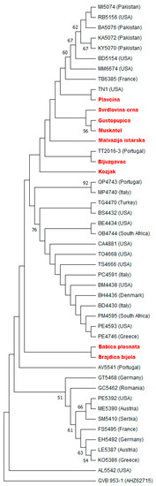
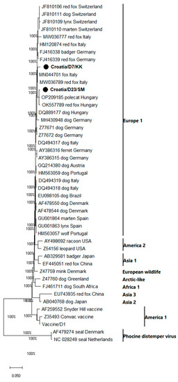
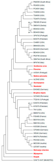
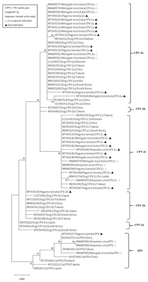
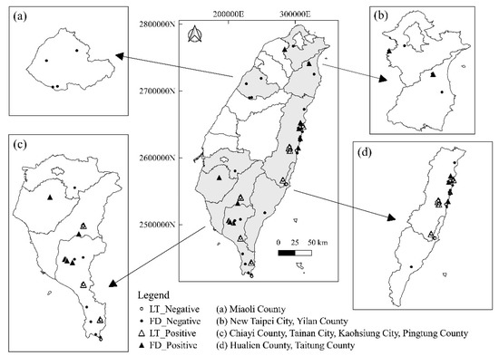
The canine distemper virus (CDV), a paramyxovirus that is closely related to the human measles virus and rinderpest virus of cattle, is a highly contagious viral disease in dogs and wild carnivores worldwide. CDV represents a serious threat to domestic and wild animals, especially to the conservation of endangered wild carnivores. Our study aims to investigate the occurrence of CDV in free-living wild canines in Croatia. For this purpose, 176 red foxes and 24 jackal brain samples collected in the frame of the active surveillance of rabies during winter 2021/2022 were tested. This study provided the first comprehensive overview of the prevalence and spatial distribution of CDV in the wildlife of Croatia, including the molecular phylogenetic analysis of the H gene sequence of field CDV strains circulating in red fox and jackal populations of Croatia. The molecular characterization of hemagglutinin gene genomic regions confirmed the phylogenetic clustering of obtained sequences into the Europa 1 genotype. The obtained CDV red fox sequences were mutually very similar (97.60%). This study indicates the high genetic similarity of Croatian CDV red fox sequences and CDV red fox sequences from Italy and Germany, badger sequences from Germany, polecat sequences from Hungary, and dog sequences from Hungary and Germany.
Full article
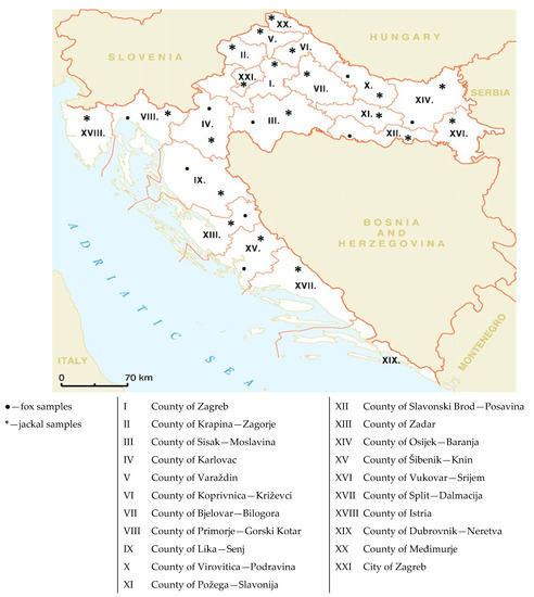
Figure 1


