
Investigation of Bufavirus and Parvovirus 4 in Patients with Gastro-Enteritis from the South-East of France

 ,
, 
Abstract
:1. Introduction
2. Results and Discussion
2.1. BuV and PARV4 Detection in Stool Samples from Patients with Diarrhea (Cohort 1)
2.1.1. Parvovirus 4 Detection
2.1.2. Bufavirus Detection
Demographic Data
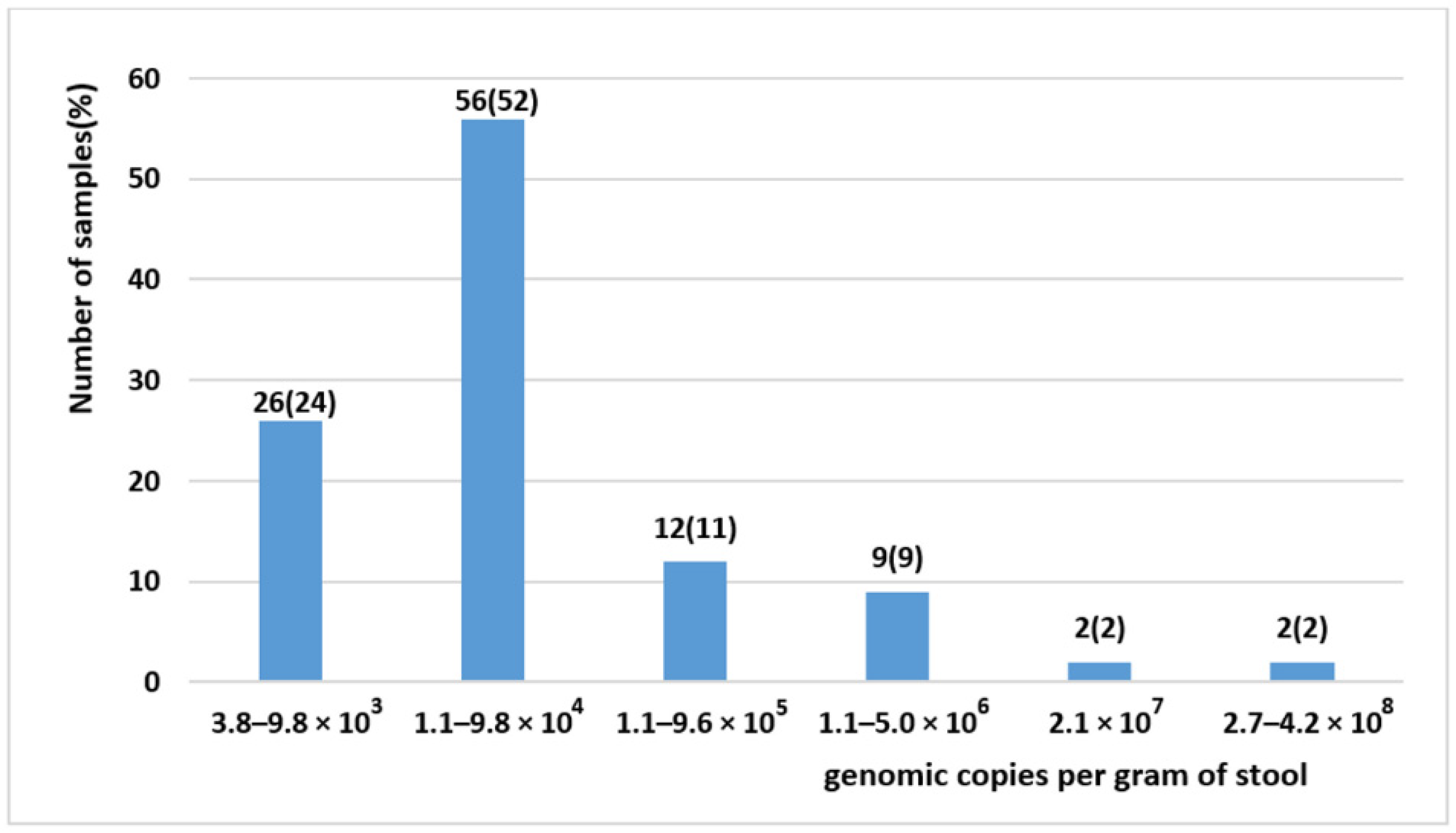
Characteristics of BuV-Positive Patients
BuV Co-Infection
2.2. BuV Testing in Blood and Respiratory Samples from Patients with Buv-Positive Stool Samples
2.3. BuV Detection Rate in Stool Samples from Patients with and without Diarrhea (Cohort 2)
2.4. Genotyping of BuV-Positive Samples
3. Materials and Methods
3.1. Study Population
3.2. Sample Preparation
3.3. Nucleic acid Extraction
3.4. Sample Testing
3.5. Positive Control
3.6. Taqman Real-Time PCR Assays
3.7. Statistical Analysis
Supplementary Materials
Author Contributions
Funding
Institutional Review Board Statement
Informed Consent Statement
Data Availability Statement
Acknowledgments
Conflicts of Interest
References
- Soderlund-Venermo, M. Emerging Human Parvoviruses: The Rocky Road to Fame. Annu. Rev. Virol. 2019, 6, 71–91. [Google Scholar] [CrossRef] [PubMed]
- Vaisanen, E.; Mohanraj, U.; Kinnunen, P.M.; Jokelainen, P.; Al-Hello, H.; Barakat, A.M.; Sadeghi, M.; Jalilian, F.A.; Majlesi, A.; Masika, M.; et al. Global Distribution of Human Protoparvoviruses. Emerg. Infect. Dis. 2018, 24, 1292–1299. [Google Scholar] [CrossRef] [Green Version]
- Vaisanen, E.; Fu, Y.; Hedman, K.; Soderlund-Venermo, M. Human Protoparvoviruses. Viruses 2017, 9, 354. [Google Scholar] [CrossRef] [Green Version]
- Phan, T.G.; Vo, N.P.; Bonkoungou, I.J.; Kapoor, A.; Barro, N.; O’Ryan, M.; Kapusinszky, B.; Wang, C.; Delwart, E. Acute diarrhea in West African children: Diverse enteric viruses and a novel parvovirus genus. J. Virol. 2012, 86, 11024–11030. [Google Scholar] [CrossRef] [PubMed] [Green Version]
- Cotmore, S.F.; Agbandje-McKenna, M.; Canuti, M.; Chiorini, J.A.; Eis-Hubinger, A.; Hughes, J.; Mietzsch, M.; Modha, S.; Ogliastro, M.; Pénzes, J.J.; et al. ICTV Virus Taxonomy Profile: Parvoviridae. J. Gen. Virol. 2019, 100, 367–368. [Google Scholar] [CrossRef] [PubMed]
- Mohanraj, U.; Jokinen, M.; Thapa, R.R.; Paloniemi, M.; Vesikari, T.; Lappalainen, M.; Tarkka, E.; Nora-Krukle, Z.; Vilmane, A.; Vettenranta, K.; et al. Human Protoparvovirus DNA and IgG in Children and Adults with and without Respiratory or Gastrointestinal Infections. Viruses 2021, 13, 483. [Google Scholar] [CrossRef]
- Dapra, V.; Galliano, I.; Montanari, P.; Zaniol, E.; Calvi, C.; Alliaudi, C.; Bergallo, M. Bufavirus, Cosavirus, and Salivirus in Diarrheal Italian Infants. Intervirology 2021, 1–4. [Google Scholar] [CrossRef]
- Altay Kocak, A.; Ocal, M.; Polat, M.; Kanik Yuksek, S.; Aktas Tapisiz, A.; Tezer, H.; Ozkul, A.; Ergunay, K.; Bozdayi, G.; Ahmed, K. Multicenter investigation of bufavirus in the etiology of viral central nervous system infections of adults and children. Mikrobiyol Bul. 2017, 51, 191–194. [Google Scholar] [CrossRef]
- Vaisanen, E.; Paloniemi, M.; Kuisma, I.; Lithovius, V.; Kumar, A.; Franssila, R.; Ahmed, K.; Delwart, E.; Vesikari, T.; Hedman, K.; et al. Epidemiology of two human protoparvoviruses, bufavirus and tusavirus. Sci. Rep. 2016, 6, 39267. [Google Scholar] [CrossRef] [Green Version]
- Chieochansin, T.; Vutithanachot, V.; Theamboonlers, A.; Poovorawan, Y. Bufavirus in fecal specimens of patients with and without diarrhea in Thailand. Arch. Virol. 2015, 160, 1781–1784. [Google Scholar] [CrossRef]
- Altay, A.; Yahiro, T.; Bozdayi, G.; Matsumoto, T.; Sahin, F.; Ozkan, S.; Nishizono, A.; Soderlund-Venermo, M.; Ahmed, K. Bufavirus genotype 3 in Turkish children with severe diarrhoea. Clin. Microbiol. Infect. 2015, 21, 965e1–965e4. [Google Scholar] [CrossRef] [PubMed] [Green Version]
- Okitsu, S.; Khamrin, P.; Takanashi, S.; Thongprachum, A.; Hoque, S.A.; Takeuchi, H.; Khan, M.A.; Hasan, S.M.T.; Iwata, T.; Shimizu, H.; et al. Molecular detection of enteric viruses in the stool samples of children without diarrhea in Bangladesh. Infect. Genet. Evol. 2020, 77, 104055. [Google Scholar] [CrossRef] [PubMed]
- Prakash, S.; Shukla, S.; Ramakrishna, V.; Mishra, H.; Bhagat, A.K.; Jain, A. Human Parvovirus 4: A harmless bystander or a pathogen of severe acute respiratory illness. Int. J. Infect. Dis. 2020, 90, 21–25. [Google Scholar] [CrossRef] [Green Version]
- Manning, A.; Willey, S.J.; Bell, J.E.; Simmonds, P. Comparison of tissue distribution, persistence, and molecular epidemiology of parvovirus B19 and novel human parvoviruses PARV4 and human bocavirus. J. Infect. Dis. 2007, 195, 1345–1352. [Google Scholar] [CrossRef] [PubMed] [Green Version]
- Matthews, P.C.; Sharp, C.; Simmonds, P.; Klenerman, P. Human parvovirus 4 ‘PARV4’ remains elusive despite a decade of study. F1000Research 2017, 6, 82. [Google Scholar] [CrossRef] [Green Version]
- Simmons, R.; Sharp, C.; McClure, C.P.; Rohrbach, J.; Kovari, H.; Frangou, E.; Simmonds, P.; Irving, W.; Rauch, A.; Bowness, P.; et al. Parvovirus 4 infection and clinical outcome in high-risk populations. J. Infect. Dis. 2012, 205, 1816–1820. [Google Scholar] [CrossRef]
- Drexler, J.F.; Reber, U.; Muth, D.; Herzog, P.; Annan, A.; Ebach, F.; Sarpong, N.; Acquah, S.; Adlkofer, J.; Adu-Sarkodie, Y.; et al. Human parvovirus 4 in nasal and fecal specimens from children, Ghana. Emerg. Infect. Dis. 2012, 18, 1650–1653. [Google Scholar] [CrossRef]
- Vaisanen, E.; Kuisma, I.; Phan, T.G.; Delwart, E.; Lappalainen, M.; Tarkka, E.; Hedman, K.; Soderlund-Venermo, M. Bufavirus in feces of patients with gastroenteritis, Finland. Emerg. Infect. Dis. 2014, 20, 1077–1079. [Google Scholar] [CrossRef]
- Smits, S.L.; Schapendonk, C.M.; van Beek, J.; Vennema, H.; Schurch, A.C.; Schipper, D.; Bodewes, R.; Haagmans, B.L.; Osterhaus, A.D.; Koopmans, M.P. New viruses in idiopathic human diarrhea cases, the Netherlands. Emerg. Infect. Dis. 2014, 20, 1218–1222. [Google Scholar] [CrossRef] [Green Version]
- Huang, D.D.; Wang, W.; Lu, Q.B.; Zhao, J.; Guo, C.T.; Wang, H.Y.; Zhang, X.A.; Tong, Y.G.; Liu, W.; Cao, W.C. Identification of Bufavirus-1 and Bufavirus-3 in Feces of Patients with Acute Diarrhea, China. Sci. Rep. 2015, 5, 13272. [Google Scholar] [CrossRef] [Green Version]
- CNRvge. Rapport Annuel D’activité 2019, Année D’exercice 2018. Dijon, France. 2019, pp. 31–32. Available online: http://www.cnr-ve.org/wp-content/uploads/documents/RAPPORT%20ACTIVITES%202019.pdf (accessed on 4 September 2021).
- Pratte-Santos, R.; Miagostovich, M.P.; Fumian, T.M.; Maciel, E.L.; Martins, S.A.; Cassini, S.T.; Keller, R. High prevalence of enteric viruses associated with acute gastroenteritis in pediatric patients in a low-income area in Vitoria, Southeastern Brazil. J. Med. Virol. 2019, 91, 744–750. [Google Scholar] [CrossRef] [PubMed]
- Chan, M.C.; Sung, J.J.; Lam, R.K.; Chan, P.K.; Lee, N.L.; Lai, R.W.; Leung, W.K. Fecal viral load and norovirus-associated gastroenteritis. Emerg. Infect. Dis. 2006, 12, 1278–1280. [Google Scholar] [CrossRef]
- Mollerup, S.; Fridholm, H.; Vinner, L.; Kjartansdottir, K.R.; Friis-Nielsen, J.; Asplund, M.; Herrera, J.A.; Steiniche, T.; Mourier, T.; Brunak, S.; et al. Cutavirus in Cutaneous Malignant Melanoma. Emerg. Infect. Dis. 2017, 23, 363–365. [Google Scholar] [CrossRef] [PubMed] [Green Version]
- Phan, T.G.; Dreno, B.; da Costa, A.C.; Li, L.; Orlandi, P.; Deng, X.; Kapusinszky, B.; Siqueira, J.; Knol, A.C.; Halary, F.; et al. A new protoparvovirus in human fecal samples and cutaneous T cell lymphomas (mycosis fungoides). Virology 2016, 496, 299–305. [Google Scholar] [CrossRef] [PubMed]
- Phan, T.; Nagaro, K. Cutavirus: A newly discovered parvovirus on the rise. Infect. Genet. Evol. 2020, 80, 104175. [Google Scholar] [CrossRef]
- Yahiro, T.; Wangchuk, S.; Tshering, K.; Bandhari, P.; Zangmo, S.; Dorji, T.; Tshering, K.; Matsumoto, T.; Nishizono, A.; Soderlund-Venermo, M.; et al. Novel human bufavirus genotype 3 in children with severe diarrhea, Bhutan. Emerg. Infect. Dis. 2014, 20, 1037–1039. [Google Scholar] [CrossRef]
- Ayouni, S.; Estienney, M.; Hammami, S.; Neji Guediche, M.; Pothier, P.; Aouni, M.; Belliot, G.; de Rougemont, A. Cosavirus, Salivirus and Bufavirus in Diarrheal Tunisian Infants. PLoS ONE 2016, 11, e0162255. [Google Scholar] [CrossRef]
- Zanella, M.C.; Cordey, S.; Laubscher, F.; Docquier, M.; Vieille, G.; Van Delden, C.; Braunersreuther, V.; Ta, M.K.; Lobrinus, J.A.; Masouridi-Levrat, S.; et al. Unmasking viral sequences by metagenomic next-generation sequencing in adult human blood samples during steroid-refractory/dependent graft-versus-host disease. Microbiome 2021, 9, 28. [Google Scholar] [CrossRef]
- Auffret, N.; de Rougemont, A. Détermination des Séroprévalences de Bufavirus et Tusavirus, Deux Nouveaux Protoparvovirus, en Bourgogne; Mémoire du Diplôme D’études Spécialisées de Biologie Médicale. 2020. Available online: https://nuxeo.u-bourgogne.fr/nuxeo/nxfile/default/f8bcd3ad-a451-4fd1-9736-907a7b832b0f/file:content/AUFFRET_THESEPHIE_2020-2.pdf (accessed on 4 September 2021).
- Fryer, J.F.; Delwart, E.; Hecht, F.M.; Bernardin, F.; Jones, M.S.; Shah, N.; Baylis, S.A. Frequent detection of the parvoviruses, PARV4 and PARV5, in plasma from blood donors and symptomatic individuals. Transfusion 2007, 47, 1054–1061. [Google Scholar] [CrossRef]
- Panning, M.; Kobbe, R.; Vollbach, S.; Drexler, J.F.; Adjei, S.; Adjei, O.; Drosten, C.; May, J.; Eis-Hubinger, A.M. Novel human parvovirus 4 genotype 3 in infants, Ghana. Emerg. Infect. Dis. 2010, 16, 1143–1146. [Google Scholar] [CrossRef]
- Kageyama, T.; Kojima, S.; Shinohara, M.; Uchida, K.; Fukushi, S.; Hoshino, F.B.; Takeda, N.; Katayama, K. Broadly reactive and highly sensitive assay for Norwalk-like viruses based on real-time quantitative reverse transcription-PCR. J. Clin. Microbiol. 2003, 41, 1548–1557. [Google Scholar] [CrossRef] [PubMed] [Green Version]
- Le Guyader, F.S.; Parnaudeau, S.; Schaeffer, J.; Bosch, A.; Loisy, F.; Pommepuy, M.; Atmar, R.L. Detection and quantification of noroviruses in shellfish. Appl. Environ. Microbiol. 2009, 75, 618–624. [Google Scholar] [CrossRef] [PubMed] [Green Version]
- Freeman, M.M.; Kerin, T.; Hull, J.; McCaustland, K.; Gentsch, J. Enhancement of detection and quantification of rotavirus in stool using a modified real-time RT-PCR assay. J. Med. Virol. 2008, 80, 1489–1496. [Google Scholar] [CrossRef] [PubMed]
- Logan, C.; O’Leary, J.J.; O’Sullivan, N. Real-time reverse transcription PCR detection of norovirus, sapovirus and astrovirus as causative agents of acute viral gastroenteritis. J. Virol. Methods. 2007, 146, 36–44. [Google Scholar] [CrossRef]
- Heim, A.; Ebnet, C.; Harste, G.; Pring-Akerblom, P. Rapid and quantitative detection of human adenovirus DNA by real-time PCR. J. Med. Virol. 2003, 70, 228–239. [Google Scholar] [CrossRef]


| BuV DNA + Patients (N = 92) | p-Value | Study Population (N = 2583) | |
|---|---|---|---|
| Residence, n (%) | |||
| Marseille | 83 (3.5) | 2375 | |
| Nice | 9 (4.3) | 208 | |
| Sex, n (%) | |||
| Men | 69 (5.3) | 1305 | |
| Women | 23 (1.8) | 1278 | |
| Sex Ratio (M/W) | 3.00 | <0.0001 | 1.02 |
| Age, Years | |||
| Median | 58 | 56 | |
| Range | 1–92 | 0–101 | |
| Children (≤15 years) | 4 (0.7) | 539 | |
| Adults (>15 years) | 88 (4.3) | 2044 | |
| <0.0001 | |||
| Age/Sex Groups, n (%) | |||
| Young (≤15-Year-old) Men | 4 (1.4) | 286 | |
| Young (≤15-Year-old) Women | 0 (0.0) | 253 | |
| Adult (>15-Year-old) Men | 65 (6.4) | 1019 | |
| Adult (>15-Year-old) Women | 23 (2.2) | 1025 | |
| <0.0001 | |||
| Seasonality; n (%) | |||
| Cumulated Winters | 36 (3.17) | 1136 | |
| Cumulated Springs | 26 (3.45) | 754 | |
| Cumulated Summers | 14 (3.33) | 420 | |
| Cumulated Autumns | 31 (3.70) | 838 | |
| 0.94 |
| N. | Sex | N of Days * | BuV Viral Load in Stool Samples ** | Others Viruses Found in Stool | Blood Samples *** (Days before Death) |
|---|---|---|---|---|---|
| 1 | M | 1 | − | − | Negative (64) |
| 9 | 4.2 × 10⁸ | ADV † and Noro ‡ GII | − | ||
| 57 | − | − | 2.0 × 10⁴ (8) | ||
| 58 | 2.7 × 10⁸ | ADV and Noro GII | − | ||
| 61 | − | − | 3.3 × 10³ (4) | ||
| 64 | − | − | 5.8 × 10³ (1) | ||
| 2 | M | − | 2.1 × 10⁷ | − | − |
| 3 | M | − | 2.1 × 10⁷ | − | − |
| 4 | M | 1 | 5.0 × 10⁶ | ADV | Negative |
| 5 | F | − | 4.6 × 10⁶ | − | − |
| 6 | F | − | 2.7 × 10⁶ | − | − |
| 7 | M | − | 2.4 × 10⁶ | − | − |
| 8 | M | − | 1.7 × 10⁶ | − | − |
| 9 | M | 1 | 1.7 × 10⁶ | − | − |
| 10 | 6.8 × 10⁵ | − | − | ||
| 14 | − | − | Negative | ||
| 10 | M | − | 1.6 × 10⁶ | − | − |
| 11 | F | − | 1.1 × 10⁶ | − | − |
| 12 | M | − | 9.6 × 10⁵ | − | − |
| 13 | M | 1 | 9.1 × 10⁵ | − | Negative |
| 14 | M | 1 | − | − | Negative (82) |
| 5 | 8.6 × 10⁵ | − | − | ||
| 22 | − | − | Negative (61) | ||
| 32 | 2.8 × 10⁴ | − | − | ||
| 15 | M | 1 | 7.9 × 10⁵ | − | − |
| 8 | − | − | Negative | ||
| 16 | M | 1 | − | − | Negative |
| 3 | 4.6 × 10⁵ | − | − | ||
| 17 | M | − | 2.7 × 10⁵ | − | − |
| 18 | F | − | 2.3 × 10⁵ | − | − |
| 19 | F | 1 | 1.7 × 10⁵ | − | − |
| 189 | 4.7 × 10⁴ | − | − | ||
| 189 | 3.0 × 10⁴ | − | − | ||
| 245 | 1.1 × 10⁶ | − | − | ||
| 20 | M | 1 | 1.6 × 10⁵ | − | − |
| 1 | 3.8 × 10⁴ | − | − | ||
| 21 | M | − | 1.3 × 10⁵ | − | − |
| 22 | F | − | 9.8 × 10⁴ | ADV | − |
| 23 | M | − | 8.6 × 10⁴ | − | − |
| 24 | M | 1 | 8.4 × 10⁴ | − | − |
| 6 | 3.0 × 10⁴ | − | − | ||
| 25 | F | − | 8.3 × 10⁴ | − | − |
| 26 | M | − | 6.3 × 10⁴ | − | − |
| 27 | M | − | 5.6 × 10⁴ | ADV | − |
| 28 | M | − | 5.4 × 10⁴ | − | − |
| 29 | M | − | 5.3 × 10⁴ | − | − |
| 30 | F | − | 5.3 × 10⁴ | − | − |
| 31 | M | − | 5.2 × 10⁴ | − | − |
| 32 | M | 1 | 5.0 × 10⁴ | − | − |
| 215 | − | − | Negative (16) | ||
| 33 | F | − | 4.9 × 10⁴ | − | − |
| 34 | M | − | 4.7 × 10⁴ | − | − |
| 35 | M | 1 | − | − | Negative |
| 5 | 4.6 × 10⁴ | − | − | ||
| 36 | M | − | 4.5 × 10⁴ | − | − |
| 37 | M | 1 | 4.1 × 10⁴ | − | − |
| 2 | − | − | Negative | ||
| 67 | Negative | − | |||
| 38 | M | 1 | 3.9 × 10⁴ | − | − |
| 26 | 7.1 × 10³ | − | − | ||
| 39 | M | 1 | 3.8 × 10⁴ | − | Negative |
| 40 | F | 1 | − | − | Negative |
| 2 | 3.3 × 10⁴ | − | − | ||
| 41 | F | − | 3.2 × 10⁴ | − | − |
| 42 | M | 1 | 3.1 × 10⁴ | − | Negative |
| 43 | F | − | 3.0 × 10⁴ | − | − |
| 44 | M | 1 | − | − | 1.2 × 10³ |
| 4 | 3.0 × 10⁴ | − | − | ||
| 9 | 2.3 × 10⁴ | − | |||
| 45 | F | − | 2.9 × 10⁴ | − | − |
| 46 | M | − | 2.8 × 10⁴ | − | − |
| 47 | M | − | 2.7 × 10⁴ | − | − |
| 48 | F | − | 2.6 × 10⁴ | − | − |
| 49 | M | − | 2.3 × 10⁴ | − | − |
| 50 | M | − | 2.2 × 10⁴ | ADV | − |
| 51 | M | 1 | 2.2 × 10⁴ | ADV | − |
| 7 | − | − | Negative | ||
| 52 | F | 1 | − | − | Negative |
| 5 | 2.0 × 10⁴ | − | − | ||
| 5 | 1.7 × 10⁴ | − | − | ||
| 53 | M | − | 1.9 × 10⁴ | − | − |
| 54 | M | 1 | 1.7 × 10⁴ | − | − |
| 7 | 1.8 × 10⁴ | − | − | ||
| 55 | M | 1 | − | − | Negative |
| 4 | 1.7 × 10⁴ | − | − | ||
| 56 | M | − | 1.5 × 10⁴ | − | − |
| 57 | M | − | 1.5 × 10⁴ | − | − |
| 58 | F | 1 | 1.4 × 10⁴ | − | − |
| 417 | Negative | − | − | ||
| 59 | M | 1 | 1.4 × 10⁴ | − | Negative |
| 60 | F | − | 1.4 × 10⁴ | − | − |
| 61 | F | − | 1.3 × 10⁴ | − | − |
| 62 | M | 1 | − | − | Negative |
| 2 | 1.3 × 10⁴ | − | − | ||
| 63 | M | − | 1.3 × 10⁴ | − | − |
| 64 | F | 1 | 1.2 × 10⁴ | − | − |
| 6 | 1.9 × 10⁴ | − | − | ||
| 65 | M | 1 | − | − | Negative |
| 16 | 1.2 × 10⁴ | − | − | ||
| 66 | M | − | 1.2 × 10⁴ | − | − |
| 67 | F | − | 1.1 × 10⁴ | − | − |
| 68 | M | − | 9.8 × 10³ | − | − |
| 69 | M | 1 | 9.8 × 10³ | Norovirus GII | Negative |
| 70 | F | − | 9.3 × 10³ | − | − |
| 71 | M | − | 9.2 × 10³ | − | − |
| 72 | M | 1 | 8.7 × 10³ | − | − |
| 3 | − | − | Negative | ||
| 73 | M | 1 | 8.2 × 10³ | Norovirus GI | Negative |
| 74 | M | − | 8.1 × 10³ | − | − |
| 75 | M | 1 | − | − | Negative |
| 12 | 8.1 × 10³ | − | − | ||
| 76 | M | − | 7.9 × 10³ | Astrovirus | − |
| 77 | M | 1 | − | − | Negative (41) |
| 5 | 7.7 × 10³ | − | Negative (37) | ||
| 9 | Negative | − | − | ||
| 38 | − | − | Negative (4) | ||
| 78 | M | 1 | − | − | Negative |
| 2 | Negative | − | − | ||
| 3 | 7.6 × 10³ | − | − | ||
| 79 | M | 1 | 7.1 × 10³ | − | − |
| 14 | 1.1 × 10⁴ | − | − | ||
| 80 | M | 1 | 6.5 × 10³ | − | Negative |
| 81 | M | − | 6.5 × 10³ | − | − |
| 82 | M | 1 | Negative | − | |
| 215 | 6.5 × 10³ | − | Negative (83) | ||
| 277 | 1.1 × 10⁵ | − | − | ||
| 284 | − | − | 2.8 × 10³ (15) | ||
| 83 | F | − | 6.4 × 10³ | − | − |
| 84 | F | − | 6.2 × 10³ | − | − |
| 85 | M | 1 | − | − | Negative (76) |
| 3 | − | − | Negative (74) | ||
| 77 | 6.1 × 10³ | − | − | ||
| 86 | M | 1 | 5.6 × 10³ | − | − |
| 4 | Negative | − | |||
| 87 | M | − | 5.6 × 10³ | − | − |
| 88 | M | − | 5.4 × 10³ | − | − |
| 89 | M | 1 | − | − | Negative |
| 3 | 5.1 × 10³ | − | − | ||
| 90 | M | − | 4.7 × 10³ | − | − |
| 91 | M | − | 4.1 × 10³ | − | − |
| 92 | M | − | 3.8 × 10³ | − | − |
| BuV + Patients | Study Population (30–75 Years of Age) | ||
|---|---|---|---|
| Men | 23 (10.50) | 219 | |
| Patients with Diarrhea; n (%) | Women | 5 (4.59) | 109 |
| N = 328 | Men/Women Ratio | 23/5 (4.60) | 219/109 (2.01) |
| Age, Range (Median), Years | 30–75 (60) | 30–75 (61) | |
| Men | 11 (10.00) | 110 | |
| Patients without Diarrhea; n (%) | Women | 6 (4.41) | 136 |
| N = 246 | Men/Women Ratio | 11/6 (1.83) | 110/136 (0.81) |
| Age, Range (Median), Years | 33–74 (61) | 30–75 (53) |
| BuV + Patients | Study Population (30–75 Years of Age) | ||
|---|---|---|---|
| Cohort 1, November 2017–April 2018; n (%) N = 578 | Men | 23 (7.4) | 311 |
| Women | 8 (3.0) | 267 | |
| Men/Women ratio | 23/8 (2.9) | 311/267 (1.2) | |
| Age, range (median), years | 30−75 (59) | 30−75 (60) | |
| Cohort 2, November 2020–April 2021; n (%) N = 328 | Men | 23 (10.5) | 219 |
| Women | 5 (4.6) | 109 | |
| Men/Women ratio | 23/5 (4.6) | 219/109 (2.0) | |
| Age, range (median), years | 30–75 (60) | 30–75 (61) |
Publisher’s Note: MDPI stays neutral with regard to jurisdictional claims in published maps and institutional affiliations. |
© 2021 by the authors. Licensee MDPI, Basel, Switzerland. This article is an open access article distributed under the terms and conditions of the Creative Commons Attribution (CC BY) license (https://creativecommons.org/licenses/by/4.0/).
Share and Cite
Simo-Fouda, F.; Thirion, L.; Nougairède, A.; Luciani, L.; Driouich, J.-S.; Petit, P.R.; Delaunay, P.; Charrel, R.N. Investigation of Bufavirus and Parvovirus 4 in Patients with Gastro-Enteritis from the South-East of France. Pathogens 2021, 10, 1151. https://doi.org/10.3390/pathogens10091151
Simo-Fouda F, Thirion L, Nougairède A, Luciani L, Driouich J-S, Petit PR, Delaunay P, Charrel RN. Investigation of Bufavirus and Parvovirus 4 in Patients with Gastro-Enteritis from the South-East of France. Pathogens. 2021; 10(9):1151. https://doi.org/10.3390/pathogens10091151
Chicago/Turabian StyleSimo-Fouda, Francis, Laurence Thirion, Antoine Nougairède, Léa Luciani, Jean-Sélim Driouich, Paul Rémi Petit, Pascal Delaunay, and Remi N. Charrel. 2021. "Investigation of Bufavirus and Parvovirus 4 in Patients with Gastro-Enteritis from the South-East of France" Pathogens 10, no. 9: 1151. https://doi.org/10.3390/pathogens10091151
APA StyleSimo-Fouda, F., Thirion, L., Nougairède, A., Luciani, L., Driouich, J.-S., Petit, P. R., Delaunay, P., & Charrel, R. N. (2021). Investigation of Bufavirus and Parvovirus 4 in Patients with Gastro-Enteritis from the South-East of France. Pathogens, 10(9), 1151. https://doi.org/10.3390/pathogens10091151