

Journal Description
Diagnostics
Diagnostics
is an international, peer-reviewed, open access journal on medical diagnosis published semimonthly online by MDPI. The British Neuro-Oncology Society (BNOS), the International Society for Infectious Diseases in Obstetrics and Gynaecology (ISIDOG) and the Swiss Union of Laboratory Medicine (SULM) are affiliated with Diagnostics and their members receive a discount on the article processing charges.
- Open Access— free for readers, with article processing charges (APC) paid by authors or their institutions.
- High Visibility: indexed within Scopus, SCIE (Web of Science), PubMed, PMC, Embase, Inspec, CAPlus / SciFinder, and other databases.
- Journal Rank: JCR - Q1 (Medicine, General and Internal) / CiteScore - Q2 (Internal Medicine)
- Rapid Publication: manuscripts are peer-reviewed and a first decision is provided to authors approximately 21.6 days after submission; acceptance to publication is undertaken in 2.7 days (median values for papers published in this journal in the second half of 2025).
- Recognition of Reviewers: reviewers who provide timely, thorough peer-review reports receive vouchers entitling them to a discount on the APC of their next publication in any MDPI journal, in appreciation of the work done.
- Companion journals for Diagnostics include: LabMed and AI in Medicine.
Impact Factor:
3.3 (2024);
5-Year Impact Factor:
3.3 (2024)
Latest Articles
Personalized Estimates of Brain Cortical Structural Similarity in Major Depressive Disorder: Evidence from a Multi-Site Neuroimaging Dataset
Diagnostics 2026, 16(11), 1632; https://doi.org/10.3390/diagnostics16111632 (registering DOI) - 26 May 2026
Abstract
Background: Major depressive disorder (MDD) is increasingly recognized as a highly heterogeneous disorder. Although the person-based similarity index (PBSI) provides a useful framework for characterizing individualized brain structural similarity, existing studies in MDD remain limited by either small samples or a lack
[...] Read more.
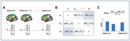
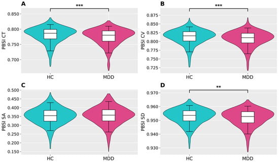
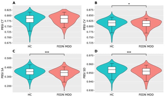
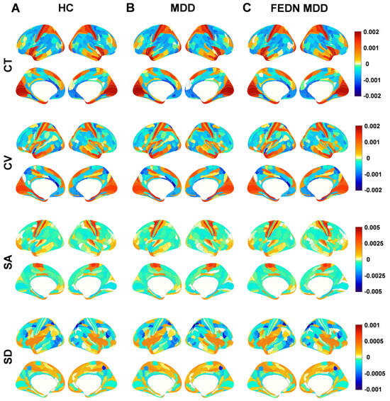
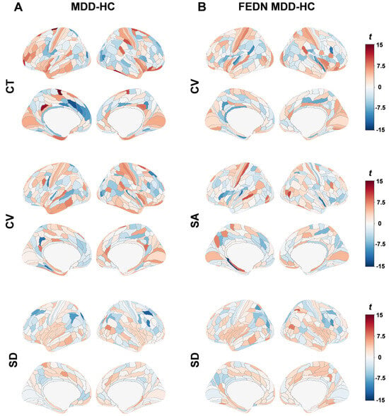
Background: Major depressive disorder (MDD) is increasingly recognized as a highly heterogeneous disorder. Although the person-based similarity index (PBSI) provides a useful framework for characterizing individualized brain structural similarity, existing studies in MDD remain limited by either small samples or a lack of integration across different morphological features. Methods: We used structural MRI data from 1442 patients with MDD and 1277 healthy controls to calculate PBSI scores of cortical morphology measures based on cortical thickness (CT), cortical volume (CV), cortical surface area (SA), and sulcal depth (SD). Group comparisons of whole-brain PBSI and regional contributions to PBSI scores were then performed, and a subgroup analysis in 243 first-episode, drug-naive (FEDN) patients with MDD was further conducted. Results: Patients with MDD showed significant alterations in PBSI. Specifically, PBSI scores were significantly reduced for CT, CV, and SD, whereas no significant group difference was observed for SA in the main analysis. Analyses of regional contributions to PBSI further revealed significant between-group differences across multiple cortical regions. These alterations were mainly distributed in the default mode, ventral attention, and visual networks for CT; in the default mode, ventral attention, sensorimotor, and visual networks for CV; and in the default mode, dorsal attention, frontoparietal, and sensorimotor networks for SD. Similar patterns were also observed in the FEDN MDD subgroup. Conclusions: These findings provide neurobiological evidence for the marked structural heterogeneity of MDD and highlight the potential of PBSI as an individualized neuroimaging marker for more precise diagnosis and personalized intervention.
Full article
(This article belongs to the Section Machine Learning and Artificial Intelligence in Diagnostics)
►
Show Figures
Open AccessArticle
Validation of a Novel Smartphone-Based Point-of-Care Semen Analysis System to Evaluate Male Reproductive Potential: A Concordance Study with Computer-Assisted Sperm Analysis
by
Byeong Jun Mun, Seung A Oh, Jin Young An, Seong Jung Kim, Yu Ha Shim, Ji Soo Ryu, Hyun Seung Lee, Tae Eun Shin, Ji Hoon Kim, Yu Jin Lee, Jun Ho Ji, Dae Keun Kim and Jae Ho Lee
Diagnostics 2026, 16(11), 1631; https://doi.org/10.3390/diagnostics16111631 (registering DOI) - 26 May 2026
Abstract
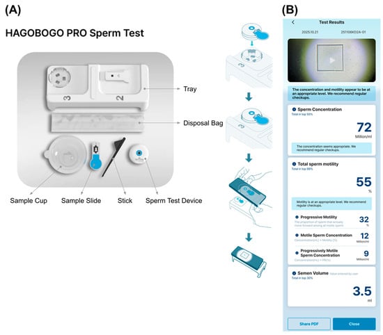
Background/Objectives: Male factor infertility contributes to approximately 40–50% of infertility cases globally, yet traditional laboratory-based semen analysis often imposes logistical and psychological barriers. This study aimed to evaluate the analytical performance and diagnostic concordance of a novel smartphone-based point-of-care testing (POCT) system, Hagobogo
[...] Read more.
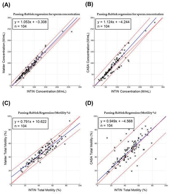
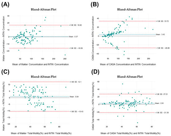
Background/Objectives: Male factor infertility contributes to approximately 40–50% of infertility cases globally, yet traditional laboratory-based semen analysis often imposes logistical and psychological barriers. This study aimed to evaluate the analytical performance and diagnostic concordance of a novel smartphone-based point-of-care testing (POCT) system, Hagobogo Pro, compared with a laboratory-based computer-assisted sperm analysis (CASA) reference system. Methods: This retrospective validation study analyzed 520 video microscopy clips obtained from 104 men undergoing infertility evaluation at a tertiary fertility center. Following World Health Organization (WHO) 2021 guidelines, sperm concentration and total motility were measured using the Hagobogo Pro smartphone device and the reference system. Analytical performance was assessed based on intra-assay precision, operational time, and method agreement using Passing–Bablok regression, Bland–Altman analysis, and Spearman correlation. Results: The smartphone-based system demonstrated strong analytical agreement with the CASA reference, with high correlations observed for sperm concentration (ρ = 0.943) and motility (ρ = 0.7335). Bland–Altman analysis indicated minimal systematic bias, and intra-assay precision showed coefficients of variation below 6%. There were no statistically significant differences in mean parameters between the smartphone device, CASA, and manual assessment. Conclusions: The Hagobogo Pro platform enables rapid, reliable, and standardized sperm concentration and motility quantification, and results showed good agreement with laboratory CASA. While not a replacement for holistic laboratory evaluations, this technology improves access to preliminary male fertility screening and may empower patients by mitigating barriers to initial testing.
Full article
(This article belongs to the Special Issue Revolutionizing Male Infertility: Novel Mechanistic Insights, Diagnostic Innovations, and Management Strategies)
►▼
Show Figures

Figure 1

Open AccessInteresting Images
Femoral Osteochondritis Dissecans and Tibial Osteochondral Defect in an Adult Revealed by Bone SPECT/CT
by
Tzyy-Ling Chuang, Keng-Chang Liu, Chih-Wen Lin, Chun-Hsi Huang and Yuh-Feng Wang
Diagnostics 2026, 16(11), 1630; https://doi.org/10.3390/diagnostics16111630 (registering DOI) - 26 May 2026
Abstract
A 46-year-old woman presented with persistent right knee pain and swelling six months after a fall. MRI initially showed a lateral meniscus tear, leading to meniscus repair and later meniscectomy, but symptoms persisted. Retrospective review of the MRI revealed edema in the tibial
[...] Read more.
A 46-year-old woman presented with persistent right knee pain and swelling six months after a fall. MRI initially showed a lateral meniscus tear, leading to meniscus repair and later meniscectomy, but symptoms persisted. Retrospective review of the MRI revealed edema in the tibial plateau and distal femoral condyle. Arthroscopic debridement demonstrated severe synovitis, marked cartilage loss of the lateral femoral condyle with a loose body, and tibial plateau cartilage damage. Bone SPECT/CT showed bony destruction, cleft formation, and focal tracer uptake in the distal femur and proximal tibia. Femoral osteochondritis dissecans and a tibial osteochondral defect were diagnosed based on arthroscopic and imaging findings.
Full article
(This article belongs to the Section Medical Imaging and Theranostics)
Open AccessArticle
Federated Privacy-Preserving Multi-Modal Deep Learning for Breast Cancer Diagnosis: A Physics-Aware Approach
by
Ahmed Lateef Salih Al-Karawi, Hayder Mohammedqasim and Rüya Yılmaz
Diagnostics 2026, 16(11), 1629; https://doi.org/10.3390/diagnostics16111629 - 26 May 2026
Abstract
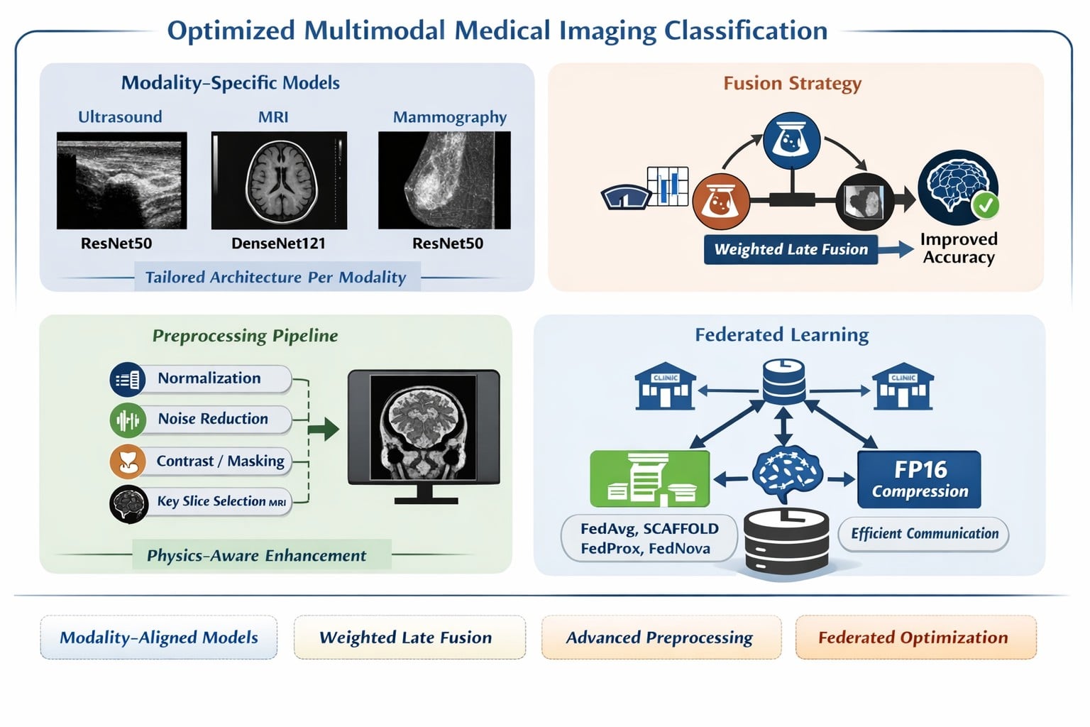
Background/Objectives: Breast cancer remains a leading cause of cancer-related mortality among women worldwide. This study presents a systematically justified multi-modal breast cancer classification pipeline that combines established, physically motivated preprocessing operations, modality-specific deep learning models, late-fusion inference, and a deployment-aware federated learning evaluation.
[...] Read more.
Background/Objectives: Breast cancer remains a leading cause of cancer-related mortality among women worldwide. This study presents a systematically justified multi-modal breast cancer classification pipeline that combines established, physically motivated preprocessing operations, modality-specific deep learning models, late-fusion inference, and a deployment-aware federated learning evaluation. Rather than introducing new image restoration or federated optimization algorithms, this work formalizes how standard preprocessing methods can be organized according to the dominant degradation characteristics of ultrasound, MRI, and mammography, and evaluates their contribution under centralized and simulated federated learning settings. Methods: Patient-wise stratified five-fold cross-validation was applied across ultrasound (BUSI, ), dynamic contrast-enhanced MRI (DUKE, ), and mammography (CBIS-DDSM, ). A five-algorithm federated learning comparison, including FedAvg, FedProx, SCAFFOLD, FedNova, and FP16-FedAvg, was conducted under IID and non-IID conditions using a Dirichlet distribution with . The evaluation reports diagnostic performance together with per-round training time, communication time, latency-related measurements, and cumulative bandwidth. Ablation experiments, McNemar’s test, Cohen’s h effect sizes, and confidence intervals were used to support the analysis. Results: Per-modality models achieved 92.50 ± 1.2%, 90.63 ± 1.5%, and 92.00 ± 1.3% accuracy for ultrasound, MRI, and mammography, respectively, with statistically significant improvements over the corresponding baselines according to McNemar’s test ( ). Weighted late fusion achieved 93.10 ± 1.1% accuracy and improved performance compared with the best individual modality ( ). FP16 transmission reduced cumulative bandwidth from 8.14 GB to 1.23 GB ( ) without a statistically significant performance difference compared with FP32 transmission ( ), while SCAFFOLD achieved the highest non-IID accuracy (90.50%). Conclusions: The findings demonstrate internal technical validity and deployment-relevant trade-offs, but they should be interpreted cautiously because the federated evaluation is simulation-based, key-slice extraction may require annotation-assisted assumptions, and external multi-center validation remains necessary before clinical deployment. Reported improvements are statistically significant in several comparisons, but corresponding Cohen’s h effect sizes are small, and clinical meaningfulness requires independent validation rather than inference from p-values alone.
Full article
(This article belongs to the Section Machine Learning and Artificial Intelligence in Diagnostics)
►▼
Show Figures

Graphical abstract
Open AccessInteresting Images
Cerebral Radionecrosis Following Stereotactic Irradiation of Skull Bone Metastases: A Diagnostic Pitfall to Be Aware of
by
Gianluca Ferini, Anna Viola, Valentina Zagardo, Antonio Pontoriero, Saveria Spadola, Gianluca Scalia and Giuseppe Emmanuele Umana
Diagnostics 2026, 16(11), 1628; https://doi.org/10.3390/diagnostics16111628 - 26 May 2026
Abstract
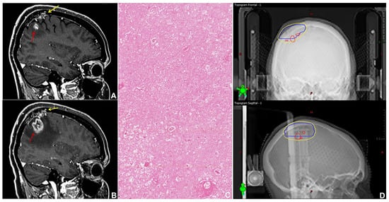
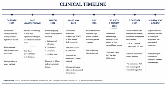
We report an unusual case of brain radionecrosis following fractionated stereotactic radiotherapy (FSRT) delivered to a calvarial metastasis in a patient with metastatic luminal A breast cancer. A 40-year-old woman developed three subcentimetric brain lesions during follow-up, initially interpreted as new metastases and
[...] Read more.
We report an unusual case of brain radionecrosis following fractionated stereotactic radiotherapy (FSRT) delivered to a calvarial metastasis in a patient with metastatic luminal A breast cancer. A 40-year-old woman developed three subcentimetric brain lesions during follow-up, initially interpreted as new metastases and treated with FSRT. Subsequent radiological progression and advanced imaging suggested radionecrosis, which was confirmed histologically after surgical resection. Retrospective dosimetric analysis revealed that all lesions arose within the high-dose isodose region of the previously irradiated skull metastasis, despite compliance with established brain dose constraints. This case highlights a previously unreported risk of brain radionecrosis after stereotactic irradiation of skull bone metastases and underscores the need for caution in treatment planning and imaging interpretation.
Full article
(This article belongs to the Section Medical Imaging and Theranostics)
►▼
Show Figures

Figure 1
Open AccessArticle
A Combined Serum Biomarker Panel for Early Prediction of Response to Anti-TNF Therapy in Rheumatoid Arthritis: Toward a Precision Medicine Approach
by
Bogdan Ion Gavrila, Claudia Ciofu and Marilena Stoian
Diagnostics 2026, 16(11), 1627; https://doi.org/10.3390/diagnostics16111627 - 26 May 2026
Abstract
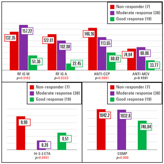
Background/Objectives: Response to TNF inhibitors in RA remains heterogeneous and reliable predictors of treatment response are still lacking. Biomarker-based stratification may improve therapeutic decision-making and aligns with the emerging paradigm of precision medicine. Methods: We conducted a prospective observational study including 64 biologic-naïve
[...] Read more.
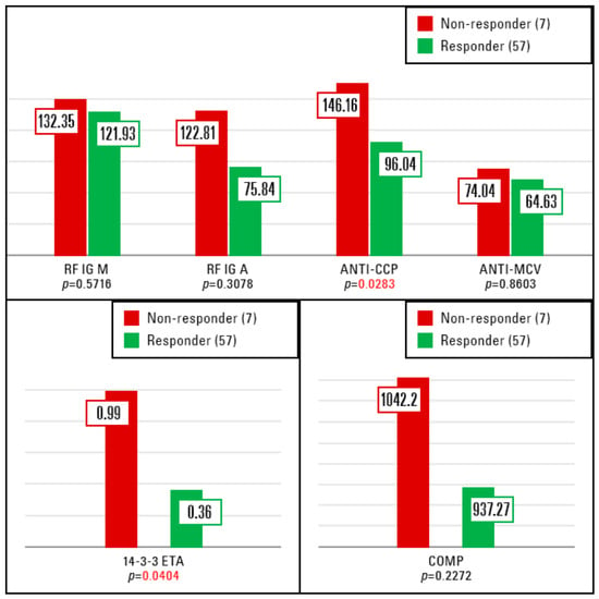
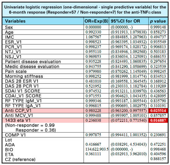
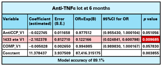
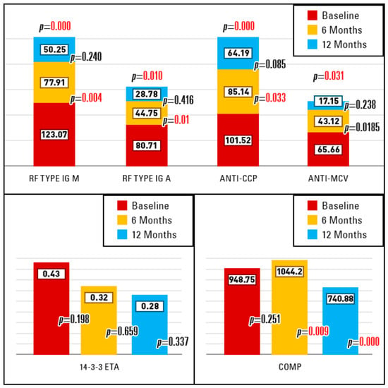
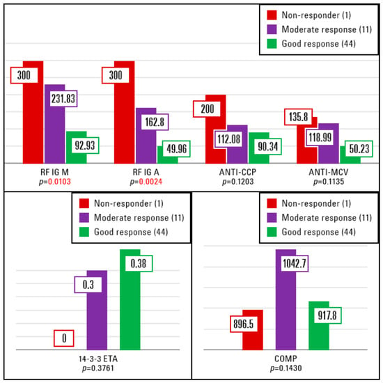
Background/Objectives: Response to TNF inhibitors in RA remains heterogeneous and reliable predictors of treatment response are still lacking. Biomarker-based stratification may improve therapeutic decision-making and aligns with the emerging paradigm of precision medicine. Methods: We conducted a prospective observational study including 64 biologic-naïve patients with active RA being inadequately controlled by csDMARDs. All patients initiated anti-TNF therapy and were followed for 12 months. Clinical response was assessed at 6 and 12 months using EULAR response criteria based on DAS28-CRP. Baseline serum levels of classical biomarkers (RF type IgM, RF type IgA, anti-CCP) and additional biomarkers (anti-MCV,14-3-3η protein, COMP) were evaluated. Logistic regression analyses were performed to identify predictors of treatment response. Results: At 6 months, 7 patients were classified as non-responders, 38 as moderate responders, and 19 as good responders Lower baseline levels of RF isotypes, anti-CCP antibodies, 14-3-3η protein, and COMP were associated with favorable clinical response at 6 months. Baseline anti-CCP and 14-3-3η protein levels emerged as significant predictors in univariate analysis. Multivariate logistic regression yielded a predictive model incorporating anti-CCP, 14-3-3η protein, and COMP, achieving an overall prediction accuracy of 89.1%. At 12 months, baseline RF isotypes remained associated with treatment response, whereas the predictive value of other biomarkers diminished. Longitudinal analysis demonstrated significant reductions mainly for classical autoantibody levels under anti-TNF α inhibitors. Conclusions: A combined serum biomarker panel may support early prediction of response to anti-TNF therapy in RA. These findings highlight the potential of integrated biomarker-based stratification to optimize therapeutic decisions and support the implementation of precision medicine approaches in RA.
Full article
(This article belongs to the Special Issue Current Perspectives and Gaps in the Diagnosis and Management of Rheumatic Diseases)
►▼
Show Figures

Figure 1

Open AccessArticle
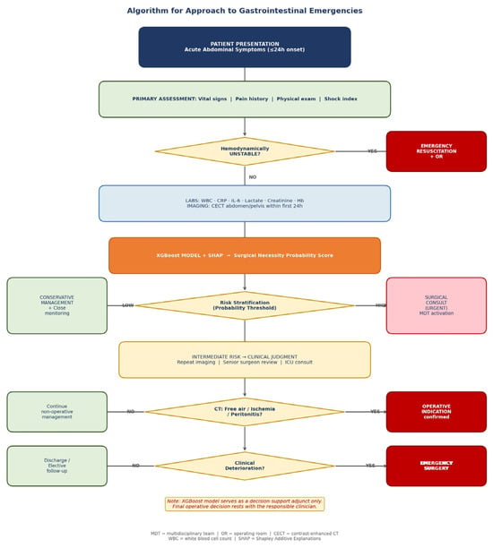
Explainable Machine Learning for Early Prediction of Surgical Necessity in Gastrointestinal Emergencies: A Multimodal Diagnostic Study
by
Oprescu Macovei Anca Monica, Dana Paula Venter, Stefan Mihai, Constantin Oprescu, Andrei Gabriel, Dumitriu Bogdan, Micle Bianca-Maria, Valcea Sebastian, Oana-Mihaela Plotogea and Ilie Madalina
Diagnostics 2026, 16(11), 1626; https://doi.org/10.3390/diagnostics16111626 - 26 May 2026
Abstract
►▼
Show Figures
Background/Objectives: Acute gastrointestinal (GI) emergencies require timely and accurate prediction of surgical necessity to avoid delayed intervention and improve patient outcomes. Traditional scoring systems offer limited accuracy and fail to integrate multimodal data. This study aimed to develop and validate an explainable machine
[...] Read more.
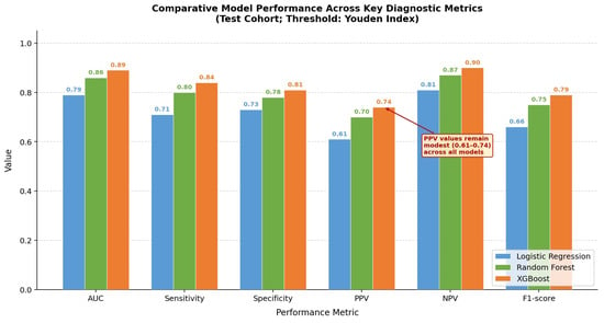
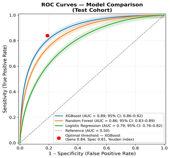
Background/Objectives: Acute gastrointestinal (GI) emergencies require timely and accurate prediction of surgical necessity to avoid delayed intervention and improve patient outcomes. Traditional scoring systems offer limited accuracy and fail to integrate multimodal data. This study aimed to develop and validate an explainable machine learning model for early prediction of surgical necessity in patients presenting with GI emergencies. Methods: A retrospective cohort of 1032 consecutive adult patients admitted with acute GI emergencies at a tertiary referral center (2019–2024) was analyzed. Three predictive models were developed: logistic regression, Random Forest, and XGBoost. Features included clinical, laboratory, and contrast-enhanced CT imaging variables available within the first 24 h. Model performance was evaluated using AUC, sensitivity, specificity, PPV, NPV, and F1-score. Shapley Additive Explanations (SHAP) were applied for global and individual-level interpretability. The study followed STROBE and TRIPOD+AI reporting guidelines. Results: Surgical intervention was required in 312 patients (30.2%). The XGBoost model achieved the highest AUC (0.89; 95% CI: 0.86–0.92), outperforming Random Forest (AUC 0.86) and logistic regression (AUC 0.79), with sensitivity 0.84, specificity 0.81, and NPV 0.90. The most influential predictors were serum lactate, CT findings (free intraperitoneal air, bowel ischemia), IL-6, and shock index. Decision curve analysis confirmed net clinical benefit across threshold probabilities of 10–70%. Subgroup performance remained robust across diagnostic categories (AUC 0.87–0.91). Conclusions: An explainable XGBoost model integrating early clinical, laboratory, and imaging data accurately predicts surgical necessity in GI emergencies and outperforms traditional scoring systems. SHAP-based explainability supports clinical adoption and transparency. Prospective multicenter validation is warranted. The positive predictive value of 0.74 indicates that approximately one in four patients flagged as requiring surgery may not need operative intervention. The model should be regarded as a decision-support adjunct, rather than a standalone surgical decision tool, that is most relevant in settings where immediate experienced surgical judgment is limited.
Full article

Figure 1
Open AccessArticle
Radiological Outcomes of PEEK Versus Rigid Rod Stabilization in Lumbar Spinal Stenosis Surgery: The Role of Preoperative and Postoperative Findings in Adjacent Segment Disease
by
Merih Can Yilmaz, Ozgur Ozaydin and Keramettin Aydin
Diagnostics 2026, 16(11), 1625; https://doi.org/10.3390/diagnostics16111625 - 26 May 2026
Abstract
Background/Objectives: Lumbar spondylosis is a degenerative disorder that may require decompression and stabilization surgery. Rigid titanium rods provide strong fixation, whereas polyetheretherketone (PEEK) rods have been proposed to offer a more flexible load distribution profile. This study compared the radiological outcomes after
[...] Read more.
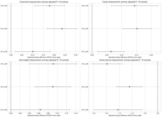
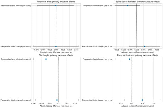
Background/Objectives: Lumbar spondylosis is a degenerative disorder that may require decompression and stabilization surgery. Rigid titanium rods provide strong fixation, whereas polyetheretherketone (PEEK) rods have been proposed to offer a more flexible load distribution profile. This study compared the radiological outcomes after PEEK versus rigid rod stabilization and evaluated whether the preoperative degenerative findings contributed independently to the postoperative adjacent segment radiological status. Methods: A retrospective cohort of 106 patients undergoing lumbar decompression and posterior stabilization (2020–2025) was analyzed. Rod allocation followed routine clinical practice rather than randomization. Radiological parameters (foraminal area, canal diameter, disc height, and facet volume) were measured preoperatively and at one year postoperatively. Baseline-adjusted ANCOVA models with HC3 robust inference compared PEEK and rigid rods across the two-, three-, and four-segment constructs. Additional models assessed the independent effects of preoperative facet effusion and Modic changes. Results: PEEK rods were associated with the statistically reliable preservation of spinal canal diameter, foraminal area, disc height (particularly in the three- and four-segment constructs), and reduced facet joint volume increase compared with rigid rods after multiple-comparison correction. The findings for the two-segment constructs were less consistent and partly influence-sensitive. Preoperative facet effusion and Modic changes showed no statistically reliable independent association with postoperative radiological outcomes after adjustment. Conclusions: PEEK rod systems were associated with favorable baseline-adjusted radiological preservation patterns, especially in long-segment stabilization. These findings should be interpreted as radiological associations rather than proof of clinical superiority or causal reduction in adjacent segment disease, because rod allocation was nonrandom and clinical, and fusion-related outcomes were not assessed.
Full article
(This article belongs to the Special Issue Advances in the Diagnosis and Management of Low-Back Pain)
►▼
Show Figures

Figure 1

Open AccessArticle
Differential Association of Visceral and Subcutaneous Adipose Tissue with Treatment Response to Neoadjuvant Chemoradiotherapy in Locally Advanced Rectal Cancer
by
Hye Jin Kang, Yong Kyun Won, Eun Seog Kim, Sang Mi Lee, Ik Dong Yoo, Jeong Won Lee, Sun-pyo Hong, Moo-Jun Baek, Dong Hyun Kang, Mee-Hye Oh, Ji-Hye Lee, Si-Hyong Jang, Nam Hun Heo, Ji An Seo, Jae Won Kim, Taesung Ahn and In Young Jo
Diagnostics 2026, 16(11), 1624; https://doi.org/10.3390/diagnostics16111624 - 26 May 2026
Abstract
Background: In locally advanced rectal cancer, neoadjuvant concurrent chemoradiotherapy (CCRT) followed by surgery is the standard treatment. Pathologic complete response (pCR) is strongly associated with favorable long-term outcomes; however, reliable pre-treatment biomarkers for predicting treatment response remain limited. This study aimed to investigate
[...] Read more.
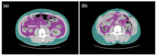
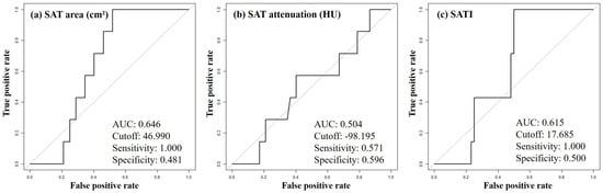
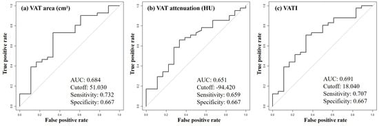
Background: In locally advanced rectal cancer, neoadjuvant concurrent chemoradiotherapy (CCRT) followed by surgery is the standard treatment. Pathologic complete response (pCR) is strongly associated with favorable long-term outcomes; however, reliable pre-treatment biomarkers for predicting treatment response remain limited. This study aimed to investigate the association between CT-derived adipose tissue parameters and pathologic response following neoadjuvant CCRT. Methods: A total of 61 patients with locally advanced rectal cancer who underwent neoadjuvant CCRT followed by surgery between 2020 and 2024 were retrospectively analyzed. Visceral adipose tissue (VAT) and subcutaneous adipose tissue (SAT) parameters, including area, index, and mean attenuation (Hounsfield unit, HU), were measured at the L3 level on pre-treatment CT. Patients were classified into favorable (complete or near-complete response) and unfavorable response groups, as well as response and no-response groups. Statistical analyses included independent t-tests, chi-square tests, and receiver operating characteristic (ROC) curve analysis. Results: In the favorable versus unfavorable response analysis, higher body mass index (BMI), larger VAT area, higher VAT index (VATI), and lower mean VAT attenuation were significantly associated with favorable response (all p < 0.05), whereas SAT-related parameters were not. In the response versus no-response analysis, SAT area and SAT index (SATI), but not mean SAT attenuation and VAT-related parameters, were significantly associated with treatment response (all p < 0.05). BMI was significantly associated with pathologic response only in the favorable versus unfavorable response group analysis, whereas no significant association was observed in the response versus no-response group analysis. Conclusions: CT-derived adipose tissue parameters were differentially associated with pathologic response to neoadjuvant CCRT in locally advanced rectal cancer. VAT parameters, including both quantity and attenuation, were associated with favorable response, whereas SAT parameters were associated with overall treatment response, suggesting compartment-specific roles of adipose tissue in modulating treatment outcomes. While BMI demonstrated a significant association in one subgroup analysis, CT-based body composition analysis may provide more comprehensive and compartment-specific information beyond conventional anthropometric measures, and may serve as a potential imaging biomarker for predicting treatment response.
Full article
(This article belongs to the Special Issue Advancements in Diagnosis of Colorectal Cancer)
►▼
Show Figures

Figure 1
Open AccessArticle
A Simple Admission-Based Score for Early Mortality Risk Stratification in Non-Shock Sepsis: A Pilot Study
by
Simona Maria Borta, Romana Olivia Popetiu, Larisa Alexandra Rus, Anamaria Vîlcea, Cristina Maghera, Renata Padurean, Dragoş Vasile Nica and Adrian Silviu Crişan
Diagnostics 2026, 16(11), 1623; https://doi.org/10.3390/diagnostics16111623 - 26 May 2026
Abstract
Background/Objectives: Early risk stratification in non-shock sepsis remains challenging, as patients may appear clinically stable despite ongoing deterioration. We used key variables across seven pathophysiological domains (cardiovascular, hematological, metabolic, hepatic, renal, immune, and comorbidity burden) to identify admission-based predictors of in-hospital mortality
[...] Read more.
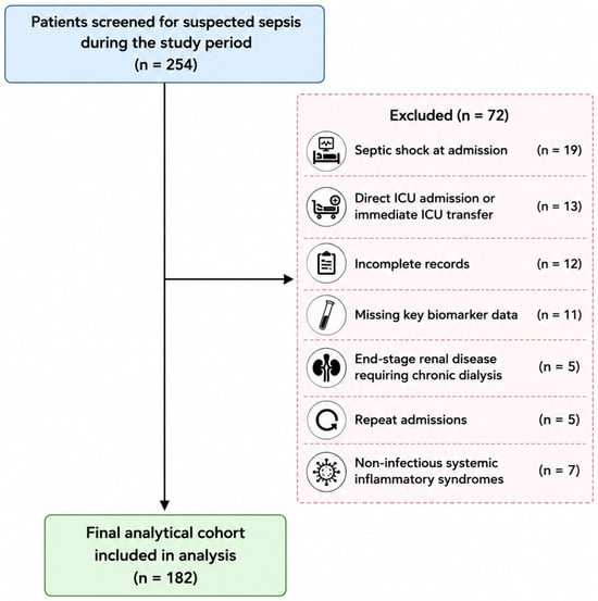
Background/Objectives: Early risk stratification in non-shock sepsis remains challenging, as patients may appear clinically stable despite ongoing deterioration. We used key variables across seven pathophysiological domains (cardiovascular, hematological, metabolic, hepatic, renal, immune, and comorbidity burden) to identify admission-based predictors of in-hospital mortality for these patients and derive a simple, clinically applicable triage score. Methods: This prospective pilot study included 182 adult non-shock sepsis patients transferred from the Emergency Department to the internal medicine ward in a tertiary hospital in Arad (Romania). Markers of cardiovascular (atrial fibrillation), renal (creatinine, urea), immune–inflammatory (IL-6, CRP, ESR, procalcitonin, presepsin), metabolic (FBG), hepatic (AST, ALT), and comorbidity burden (CCI) domains were analyzed using logistic and LASSO regression with bootstrap validation. Results: Non-survivors exhibited a significantly higher prevalence of atrial fibrillation (p = 0.012), as well as significantly higher creatinine, urea, IL-6, CRP, ESR, CCI, and presepsin (p ≤ 0.042). Although most variables, (except IL-6) were significant in univariate analysis (p ≤ 0.047), only atrial fibrillation (AOR = 2.31, 95% CI: 1.09–2.84, p = 0.021) and creatinine (AOR = 1.40, 95% CI: 1.00–1.95, p = 0.048) remained independent predictors in the multivariate model. LASSO regression (1000 bootstrap iterations, selection frequency ≥ 80%) confirmed their robustness. Three-parameter models combining atrial fibrillation, creatinine, and inflammatory biomarkers showed good discrimination, with the presepsin-based model achieving an AUC of 0.843, 80.0% sensitivity, 82.9% specificity, and NPV > 95% at the optimal cut-offs (creatinine > 1.49 mg/dL; presepsin > 1446 pg/mL). The Sepsis CORE score demonstrated progressive risk stratification, with mortality rising from 0% to 60.0% across score categories. Conclusions: The proposed score integrates cardiovascular, renal, and immune domains into a simple admission-based tool with high negative predictive value. It may support early triage and risk stratification in non-shock sepsis patients admitted to general wards, although external validation is required.
Full article
(This article belongs to the Section Clinical Diagnosis and Prognosis)
►▼
Show Figures

Figure 1
Open AccessCase Report
Concurrent HHV-8-Associated Multicentric Castleman Disease and Kaposi Sarcoma in an HIV-Negative Patient: A Case Report
by
Alin-Marian Vasile, Raluca-Maria Closca, Marina Rakitovan, Maria Iordache and Flavia Zara
Diagnostics 2026, 16(11), 1622; https://doi.org/10.3390/diagnostics16111622 - 26 May 2026
Abstract
Background: Human Herpesvirus 8-associated multicentric Castleman disease is a rare, lymphoproliferative disorder characterized by recurrent episodes of systemic inflammation. The disease is predominantly observed in human immunodeficiency virus-positive patients, but there is evidence of its occurrence in negative individuals. Its pathogenesis is driven
[...] Read more.
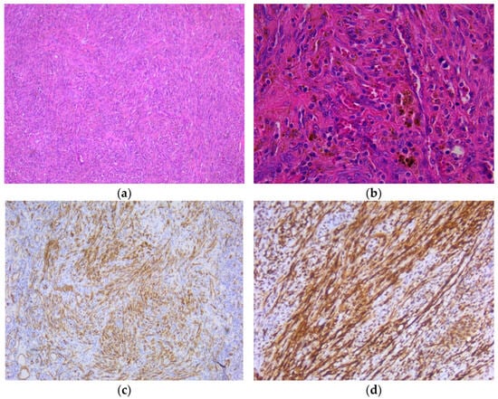
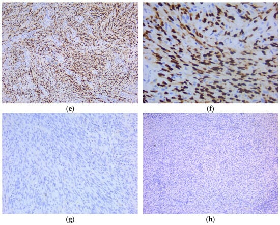
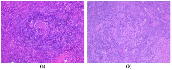
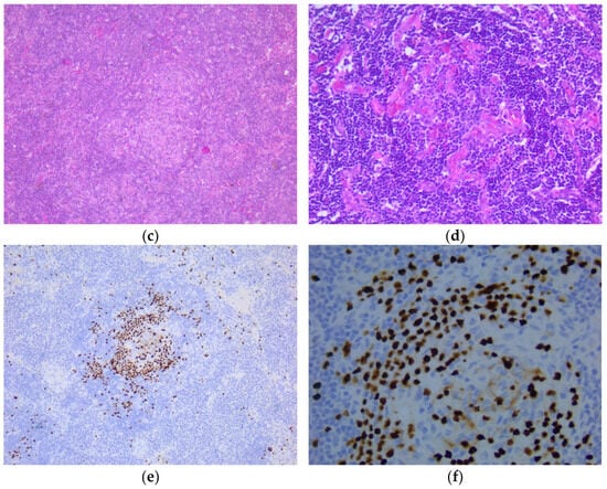
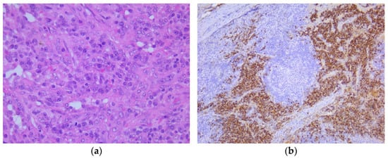
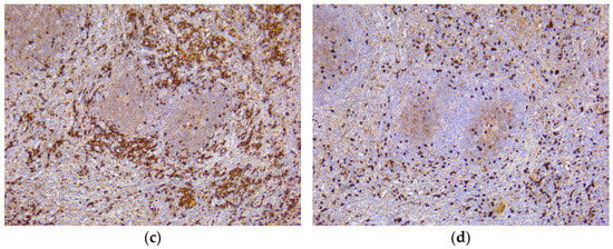
Background: Human Herpesvirus 8-associated multicentric Castleman disease is a rare, lymphoproliferative disorder characterized by recurrent episodes of systemic inflammation. The disease is predominantly observed in human immunodeficiency virus-positive patients, but there is evidence of its occurrence in negative individuals. Its pathogenesis is driven by dysregulated cytokine activity, particularly interleukin 6. Additionally, these individuals have an increased risk of developing Kaposi Sarcoma, which may present simultaneously. Case Presentation: The current paper presents a case of a 58-year-old male patient admitted to the Hematology Department of the Emergency City Hospital in Timisoara, Romania, in October 2024, accusing fever, night sweats, palpitations, weight loss and general deterioration, approximately three months prior, with gradual progression. Clinical examination revealed bilateral lymphadenopathy in the cervical and inguinal regions. No cutaneous lesions were observed initially. Laboratory tests showed elevated inflammatory markers, pancytopenia, hypergammaglobulinemia and hyponatremia. HIV serology had negative results. CT imaging revealed extensive lymphadenopathy and splenomegaly. Further excisional biopsy of the inguinal and cervical lymphadenopathies was performed. Following the microscopic examination, the final diagnosis of Human Herpesvirus 8-associated multicentric Castleman disease concurrent with Kaposi Sarcoma was established. Conclusions: Human Herpesvirus 8-associated multicentric Castleman disease is predominantly observed in HIV-positive patients, but there is evidence of its occurrence in human immunodeficiency virus-negative individuals, presenting distinct epidemiological and pathological characteristics. Early and precise diagnosis is essential, as the disease can progress rapidly and may lead to severe or fatal outcomes.
Full article
(This article belongs to the Special Issue Diagnosis and Management of Hematologic Malignancies)
►▼
Show Figures

Figure 1
Open AccessArticle
Lean Psoas Muscle Area Is Associated with Length of Stay After Lower Limb Revascularization for CLTI
by
Jagoda Bobula, Joanna Halman, Kamil Myszczyński, Jakub Dybcio, Nina Kimilu, Agnieszka Blacha, Grzegorz Owedyk and Mariusz Siemiński
Diagnostics 2026, 16(11), 1621; https://doi.org/10.3390/diagnostics16111621 - 26 May 2026
Abstract
Background: Chronic limb-threatening ischemia (CLTI) is associated with high morbidity and substantial healthcare utilization. Length of hospital stay (LOS) after lower limb revascularization is influenced by procedural complexity, but patient physiological reserve may also play a role. We evaluated whether CT-derived lean
[...] Read more.
Background: Chronic limb-threatening ischemia (CLTI) is associated with high morbidity and substantial healthcare utilization. Length of hospital stay (LOS) after lower limb revascularization is influenced by procedural complexity, but patient physiological reserve may also play a role. We evaluated whether CT-derived lean psoas muscle area (LPMA) is independently associated with LOS in patients undergoing revascularization for CLTI. Methods: We retrospectively analyzed 234 consecutive patients treated with endovascular, hybrid, or open revascularization for CLTI (Rutherford 4–5) between 2018 and 2021. Sarcopenia markers were derived from preoperative CT at the L3 level, including psoas muscle area (PMA), muscle density (PMD), and LPMA. Multivariable linear regression models with log-transformed LOS were used to estimate relative effects on hospitalization duration. Results: Median age was 68 years and 65.4% were male; 76.5% of admissions were urgent. Median LOS was 6 days (IQR 4–9). Procedure type was the strongest determinant of LOS: hybrid (β = 0.69, p < 0.001) and open surgery (β = 0.73, p < 0.001) were associated with approximately 99% and 108% longer LOS compared with endovascular treatment. Higher LPMA was independently associated with shorter LOS (β = −0.00049, p = 0.004). Smoking (β = −0.21, p = 0.003) and history of myocardial infarction (β = −0.19, p = 0.030) were associated with shorter LOS, whereas dialysis showed a non-significant trend toward longer hospitalization (β = 0.36, p = 0.056). Conclusions: In patients undergoing lower limb revascularization for CLTI, CT-derived LPMA demonstrated a modest but independent association with hospital stay duration after adjustment for procedural and clinical factors. Given the exploratory nature of this study, these hypothesis-generating findings support further evaluation of imaging-based muscle assessment as an adjunct marker of physiological reserve in this high-risk population.
Full article
(This article belongs to the Section Medical Imaging and Theranostics)
Open AccessBrief Report
Comparison Between Chemiluminescent Assay and Enzyme-Linked ImmunoSorbent Assay Techniques for the Detection of Anti-Cardiolipin and Anti-β2 Glycoprotein I Antibody Values
by
Fulvio Castelgrande, Sergio Bernardini and Marzia Nuccetelli
Diagnostics 2026, 16(11), 1620; https://doi.org/10.3390/diagnostics16111620 - 25 May 2026
Abstract
Background: Antiphospholipid antibodies (aPLs) are essential for antiphospholipid syndrome (APS) diagnosis, which is based on clinical and laboratory parameters, including the detection of lupus-anticoagulant (LAC), anti-cardiolipin (aCL) and anti-β2-glycoprotein-I (aβ2-GPI) antibodies. The enzyme-linked immunosorbent assay (ELISA) is the reference methodology for classification
[...] Read more.
Background: Antiphospholipid antibodies (aPLs) are essential for antiphospholipid syndrome (APS) diagnosis, which is based on clinical and laboratory parameters, including the detection of lupus-anticoagulant (LAC), anti-cardiolipin (aCL) and anti-β2-glycoprotein-I (aβ2-GPI) antibodies. The enzyme-linked immunosorbent assay (ELISA) is the reference methodology for classification criteria, although chemiluminescence immunoassays (CLIA) are more common in clinical practice. Methods: Since LAC reflects the functional activity of a large subset of antiphospholipids, through coagulation assays that enhance a phospholipid-dependent inhibitory effect, it has been used as a reference for assessing ELISA and CLIA reliability. Samples, separated into positive and negative LAC, were selected by CLIA detection in negative and positive IgG/IgM aCL and aβ2-GPI (cut-off > 20 U/mL). Results: The ELISA/CLIA comparison showed a 100% concordance in triple negative groups, highlighting an optimal analytical specificity; a higher concordance in the aβ2-GPI IgM-positive groups compared to positive aCL IgM (100% vs. 76% in LAC-positive groups; 82% vs. 71% in LAC-negative groups), as well as in aβ2-GPI IgM-negative groups compared to negative aCL IgM in LAC-positive groups (100% vs. 87.5%); and a massive concordance reduction in positive IgG aβ2-GPI and aCL groups (44% vs. 50% in LAC-positive groups; 4.8% vs. 4.5% in LAC-negative groups). Concordance increased in all groups with a higher CLIA cut-off (>50 U/mL). Conclusions: Although CLIA performances partly differed from ELISA, this does not preclude their use in APS diagnosis, which aims for higher sensitivity in detecting cases of disease. ELISA is confirmed to be more reliable for classification criteria that aim for high specificity to reduce false positives.
Full article
(This article belongs to the Special Issue Recent Advances in Clinical Biochemistry, 2nd Edition)
Open AccessSystematic Review
MRI-Based Brain Signatures of Chemotherapy-Induced Peripheral Neuropathy in Cancer Patients: A Systematic Review and Meta-Analysis
by
Ioana Creangă-Murariu, Eliza-Maria Armeanu, Vladimir Poroch, Bogdan-Ionel Tamba, Teodora Alexa-Stratulat, Bogdan Gafton, Mihai-Vasile Marinca, Vlad-Adrian Afrasanie, Diana Maria Puscasu, Matei Ioan Rusu and Iulian Prutianu
Diagnostics 2026, 16(11), 1619; https://doi.org/10.3390/diagnostics16111619 - 25 May 2026
Abstract
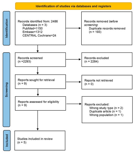
Background: Chemotherapy-induced peripheral neuropathy (CIPN) is a common, disabling toxicity with no validated biomarkers. MRI-based functional neuroimaging could offer insight into central pain processing and may reveal reproducible brain signatures of CIPN. Methods: Following PRISMA 2020 (PROSPERO: CRD420251132102), we systematically reviewed
[...] Read more.
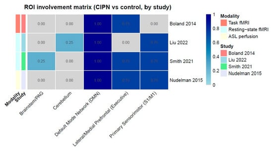
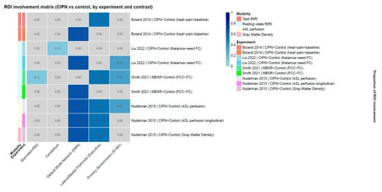
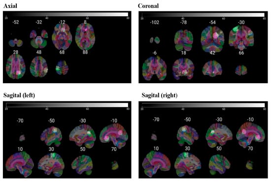
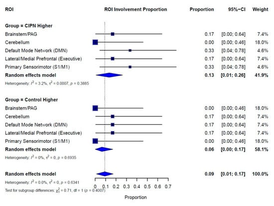
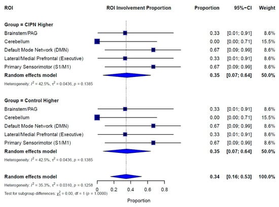
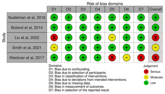
Background: Chemotherapy-induced peripheral neuropathy (CIPN) is a common, disabling toxicity with no validated biomarkers. MRI-based functional neuroimaging could offer insight into central pain processing and may reveal reproducible brain signatures of CIPN. Methods: Following PRISMA 2020 (PROSPERO: CRD420251132102), we systematically reviewed whole-brain MRI studies in adult cancer patients with CIPN. Eligible MRI techniques included task-based fMRI, resting-state fMRI, perfusion MRI, and structural MRI. Data were synthesized through voxelwise activation likelihood estimation (ALE), systems-level region-of-interest (ROI) mapping, and proportion meta-analysis of regional involvement. Results: Of 2488 screened records, five observational studies were included. The voxelwise ALE analysis did not identify clusters surviving correction, but dispersed foci appeared within the default mode network (DMN), prefrontal executive cortex, and primary sensorimotor regions, suggesting the engagement of these pain-processing networks. ROI synthesis confirmed consistent alterations in the DMN and executive prefrontal and sensorimotor cortices in CIPN patients compared with controls, while the brainstem/periaqueductal gray and cerebellum were rarely implicated. Proportion meta-analysis further quantified these differences: CIPN patients showed altered involvement in 30% (95% CI 0.16–0.48) of contrasts, with the highest frequencies in the DMN (50%), sensorimotor (33%), and executive prefrontal regions (33%). By contrast, control-higher contrasts were less frequent (10%, 95% CI 0.03–0.27), highlighting CIPN-related increases particularly in self-referential and somatosensory networks. Conclusions: Across analytic approaches, CIPN is characterized by reproducible alterations in the DMN and executive prefrontal and sensorimotor networks. These central pain signatures represent promising MRI-based biomarkers for identifying and monitoring CIPN in oncology.
Full article
(This article belongs to the Special Issue Diagnostic Tools and Monitoring Strategies for Chronic Diseases: Integrating Patient-Reported Outcomes to Enhance Clinical Decision-Making)
►▼
Show Figures

Figure 1
Open AccessCase Report
When Lymph Nodes Don’t Lie: Report of Three Unusual Presentations of Thoracic Tumors
by
Stefano Lucà, Francesco Barbato, Amedeo Di Maio, Liliana Montella, Stefano Farese, Gaetano Di Guida, Beatrice Leonardi, Rosa Giannatiempo, Rosario Salvi, Marco Montella, Carminia Maria Della Corte, Morena Fasano, Michele Orditura, Alfonso Fiorelli, Floriana Morgillo and Renato Franco
Diagnostics 2026, 16(11), 1618; https://doi.org/10.3390/diagnostics16111618 - 25 May 2026
Abstract
Background and Clinical Significance: Lymph node metastases from carcinoma of unknown primary origin (CUP) are a rare and diagnostically challenging entity, particularly when arising from thoracic malignancies with atypical clinical presentations. This study aims to illustrate the essential nature of multidisciplinary integration, with
[...] Read more.
Background and Clinical Significance: Lymph node metastases from carcinoma of unknown primary origin (CUP) are a rare and diagnostically challenging entity, particularly when arising from thoracic malignancies with atypical clinical presentations. This study aims to illustrate the essential nature of multidisciplinary integration, with a particular emphasis on the role of the pathologist in identifying occult thoracic tumors. Case Presentation: We report three cases of patients presenting with cervical or systemic lymphadenopathy as the initial clinical manifestation. Comprehensive diagnostic workups included advanced imaging (CT, MRI, and PET), comprehensive histopathological analysis, and next-generation sequencing of circulating tumor DNA. Case one and case two were diagnosed as occult primary non-mucinous lung adenocarcinomas, based on the integration of morphological features and immunohistochemical co-expression of TTF-1 and Napsin A, despite the absence of identifiable lung lesions. One case harbored an ALK rearrangement, guiding effective targeted therapy with alectinib. Case three involved metastatic pleural epithelioid mesothelioma, which presented with systemic lymphadenopathy and was initially misclassified as metastatic adenocarcinoma. Diagnosis was confirmed by the loss of BAP1 expression by immunohistochemistry and the detection of a BAP1 S160fs*1 mutation, emphasizing the role of molecular pathology. Conclusions: Lymphadenopathy as the first manifestation of thoracic malignancy is a rare but clinically significant occurrence. In such atypical presentations, pathologists play a pivotal role in diagnosis, often leading the process when clinical or radiological clues are minimal or absent. Accurate histopathological assessment is essential to establish a correct diagnosis and guide appropriate therapy. A multidisciplinary approach remains the cornerstone of diagnostic precision in CUP cases.
Full article
(This article belongs to the Section Clinical Diagnosis and Prognosis)
Open AccessArticle
Radiomics-Based Machine Learning for Sarcopenia Detection in Abdominal and Low-Dose CT
by
Soo-Been Kim, Young Jae Kim and Kwang Gi Kim
Diagnostics 2026, 16(11), 1617; https://doi.org/10.3390/diagnostics16111617 - 25 May 2026
Abstract
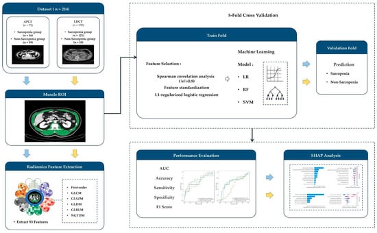
Background: Sarcopenia, characterized by progressive loss of skeletal muscle mass and function, is becoming increasingly prevalent with the global population aging. Computed tomography (CT) is widely used for muscle assessment; however, concerns regarding radiation exposure have prompted interest in lower-dose imaging protocols.
[...] Read more.
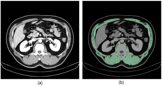
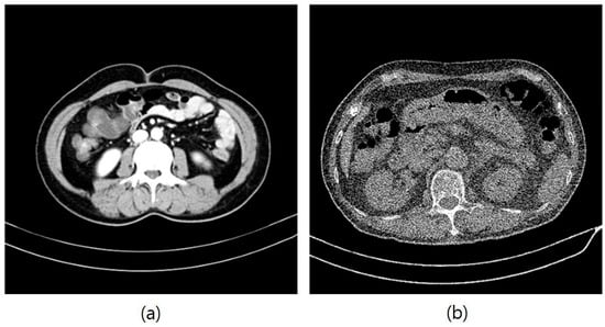
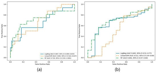
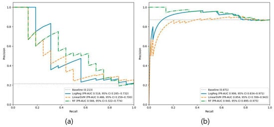
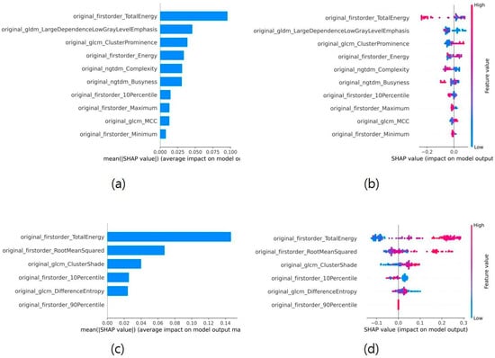
Background: Sarcopenia, characterized by progressive loss of skeletal muscle mass and function, is becoming increasingly prevalent with the global population aging. Computed tomography (CT) is widely used for muscle assessment; however, concerns regarding radiation exposure have prompted interest in lower-dose imaging protocols. This study investigated the performance of radiomics-based machine learning (ML) models for sarcopenia detection using abdominal CT (APCT) and low-dose CT (LDCT). Methods: Radiomics features were extracted from CT images following skeletal muscle segmentation, and ML models were developed using logistic regression, support vector machine, and random forest. Model performance was evaluated using fivefold cross-validation with out-of-fold predictions. Results: The random forest model demonstrated the best performance among the evaluated models, achieving an area under the receiver operating characteristic curve of 0.720 (95% CI: 0.532–0.881) for APCT and 0.692 (95% CI: 0.573–0.801) for LDCT. Model interpretation using SHapley Additive exPlanations analysis identified several intensity-based radiomics features, including TotalEnergy, as important contributors to sarcopenia prediction. Conclusions: These findings suggest that radiomics features derived from LDCT images may provide useful information for sarcopenia detection. Because LDCT is widely used in clinical settings such as lung cancer screening, radiomics analysis of LDCT images may offer an additional opportunity for opportunistic sarcopenia assessment.
Full article
(This article belongs to the Special Issue AI-Powered Medical Image Diagnosis: From Algorithm Development to Clinical Validation)
►▼
Show Figures

Figure 1
Open AccessTutorial
Establishing a Cone Beam CT-Guided Bronchoscopy Program: A Stepwise Guide for Interventional Pulmonologists
by
Sammy Onyancha, Naveed Mohamoud Merali, Nishma Elesh Gajjar, Peter Waweru Munyu, Angelique Holland and Gernot Rohde
Diagnostics 2026, 16(11), 1616; https://doi.org/10.3390/diagnostics16111616 - 25 May 2026
Abstract
Cone beam computed tomography (CBCT)-guided bronchoscopy has emerged as a powerful tool in the diagnosis of peripheral lung lesions, offering real-time, image-confirmed biopsy capabilities that enhance precision and diagnostic yield. However, implementation of a CBCT program presents significant logistical and technical challenges. This
[...] Read more.
Cone beam computed tomography (CBCT)-guided bronchoscopy has emerged as a powerful tool in the diagnosis of peripheral lung lesions, offering real-time, image-confirmed biopsy capabilities that enhance precision and diagnostic yield. However, implementation of a CBCT program presents significant logistical and technical challenges. This article presents an experience-based implementation tutorial outlining a stepwise approach to establishing a CBCT-guided bronchoscopy program. The framework is derived from iterative workflow development across more than 300 procedures at our institution, St. Elisabethen Hospital in Frankfurt, Germany, as well as our implementation efforts at Avenue Hospital Parklands in Nairobi, Kenya. Key domains addressed include infrastructure assessment, access strategy, training, procedural logistics, ventilation protocols, case selection, workflow optimization, safety considerations, and business case development. This roadmap aims to support interventional pulmonologists in integrating CBCT into clinical practice, while emphasising the need for local adaptation based on institutional resources and multidisciplinary collaboration.
Full article
(This article belongs to the Special Issue Advances in Interventional Pulmonology)
Open AccessCase Report
When Palpitations Unmask Crista Terminalis Hypertrophy: A Case Report and Review of Current Literature
by
Antonia Racz, Alexandra Dădârlat-Pop, Adela Șerban, Raluca Tomoaia, Alexandru Oprea and Horia Rosianu
Diagnostics 2026, 16(11), 1615; https://doi.org/10.3390/diagnostics16111615 - 25 May 2026
Abstract
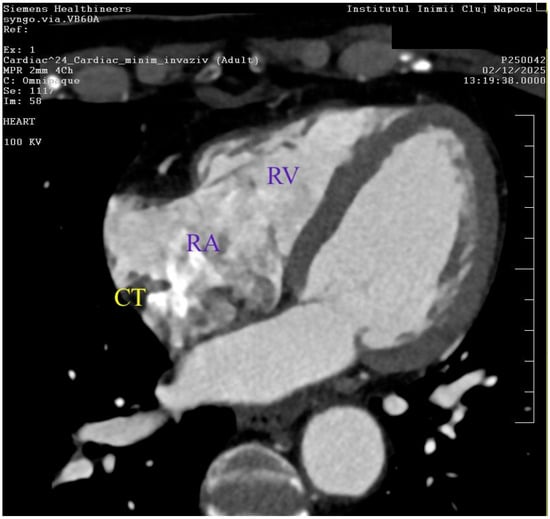
Background and Clinical Significance: The crista terminalis (CT) is a physiological fibromuscular ridge in the right atrium. While benign, rare cases of CT hypertrophy present a diagnostic challenge, as it can mimic a pathological right atrial mass on cardiac imaging. The CT also
[...] Read more.
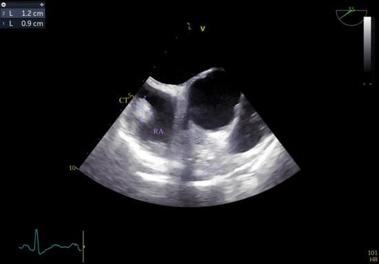
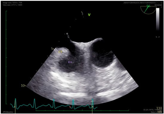
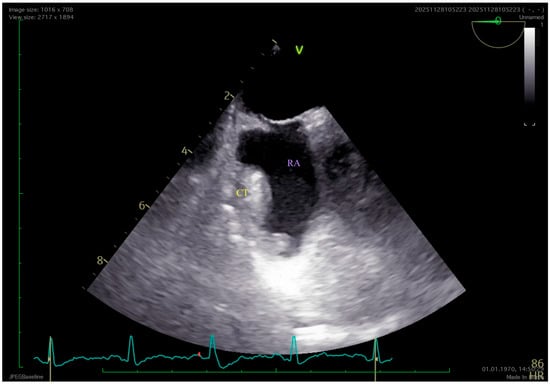
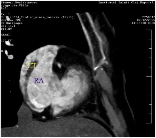
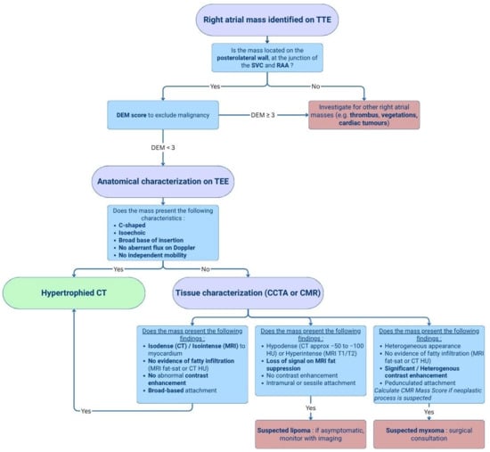
Background and Clinical Significance: The crista terminalis (CT) is a physiological fibromuscular ridge in the right atrium. While benign, rare cases of CT hypertrophy present a diagnostic challenge, as it can mimic a pathological right atrial mass on cardiac imaging. The CT also presents arrhythmogenic potential and is known to be associated with right atrial tachyarrhythmias. Case Presentation: We present the case of a 58-year-old female that presented with rapid, irregular palpitations, accompanied by hypertension. Holter electrocardiography (ECG) confirmed self-limiting episodes of atrial tachycardia (max heart rate 170 bpm). Initial transthoracic echocardiography (TTE) identified an echogenic, non-mobile mass on the posterolateral right atrial wall. Transesophageal echocardiography (TEE) confirmed a 12 × 9 mm homogenous structure with a broad base of implantation and no intrinsic mobility, initially raising the suspicion of an atrial lipoma. Subsequent cardiac computed tomography angiography (CCTA) provided high-resolution tissue characterization, identifying the mass as a hypertrophied CT due to its precise anatomical orientation and its lack of contrast enhancement, also ruling out neoplastic and thrombotic aetiologies. Conclusions: CT hypertrophy is a key differential diagnosis for right atrial masses, particularly in females in their sixth decade. A multimodal imaging approach, transitioning from TTE to TEE and finally CCTA or Cardiac Magnetic Resonance Imaging (CMR), is advantageous in preventing unnecessary invasive interventions or anticoagulation.
Full article
(This article belongs to the Special Issue Clinical Anatomy and Diagnosis in 2026)
►▼
Show Figures

Figure 1
Open AccessCase Report
Chronic Chylothorax in Childhood: Clinical Complexity and Management Challenges—A Case Report and Literature Overview
by
Teofana-Otilia Bizerea-Moga, Cristina-Loredana Pantea, Giorgiana Flavia Brad, Otilia Mărginean and Tudor-Voicu Moga
Diagnostics 2026, 16(11), 1614; https://doi.org/10.3390/diagnostics16111614 - 25 May 2026
Abstract
Background and Clinical Significance: Pleural effusion results from an imbalance between fluid production and lymphatic drainage within the pleural space and may arise from a wide range of conditions. Chylothorax, characterized by the accumulation of chyle in the pleural cavity, is an uncommon
[...] Read more.
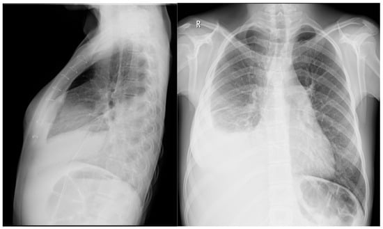
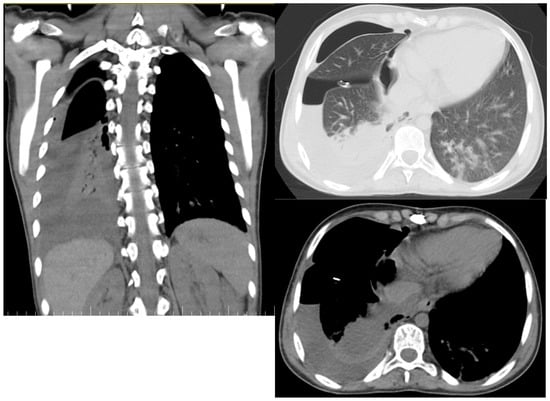
Background and Clinical Significance: Pleural effusion results from an imbalance between fluid production and lymphatic drainage within the pleural space and may arise from a wide range of conditions. Chylothorax, characterized by the accumulation of chyle in the pleural cavity, is an uncommon cause of pleural effusion in the pediatric population. Although octreotide has been widely used in neonatal postoperative chylothorax, evidence supporting its role in acquired chylothorax beyond infancy remains limited, and dosing strategies are not standardized. This report describes the management of massive chylothorax in an adolescent patient. Case Presentation: A 13-year-old girl presented with fatigue, decreased exercise tolerance, vertigo, and diaphoresis and was diagnosed with massive right-sided chylothorax based on imaging and pleural fluid analysis. Treatment consisted of pleural drainage, dietary measures, and continuous intravenous octreotide administered within a multidisciplinary care framework. Chylous drainage declined progressively, with complete resolution of the effusion achieved without surgical intervention. Conclusions: Chylothorax in older children and adolescents may present with nonspecific symptoms and requires a high index of clinical suspicion. Early diagnosis, appropriate drainage, and individualized therapy are essential for favorable outcomes. Continuous octreotide infusion may represent an effective adjunctive treatment in selected pediatric cases, but further studies are needed to define optimal dosing and treatment duration.
Full article
(This article belongs to the Special Issue Advances in the Diagnosis and Management of Chronic Disease)
►▼
Show Figures

Figure 1
Open AccessArticle
An Attention-Enhanced RegNetY Framework for Detection and Classification of Vertical Misfit in Implant-Supported Restorations: A Retrospective Study
by
Tuba Talo Yildirim, Aybike Cengiz Dagtekin, Nurullah Düger, Ayşe Rençber Kizilkaya, Furkan Talo, Emre Arslan, Mucahit Karaduman and Muhammed Yildirim
Diagnostics 2026, 16(11), 1613; https://doi.org/10.3390/diagnostics16111613 - 25 May 2026
Abstract
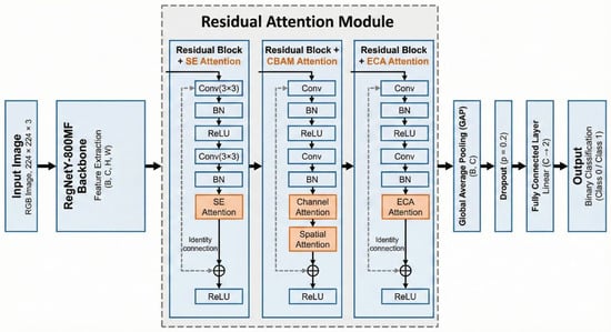
Background/Objectives: The aim of this study is to test different convolutional neural network (CNN) and Transformer-based models to detect and classify vertical misfit at the abutment-prosthesis interface on panoramic radiographs, and to develop a hybrid deep learning model enhanced with attention mechanisms.
[...] Read more.
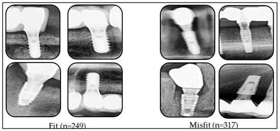
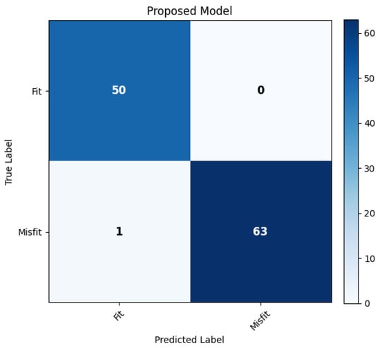
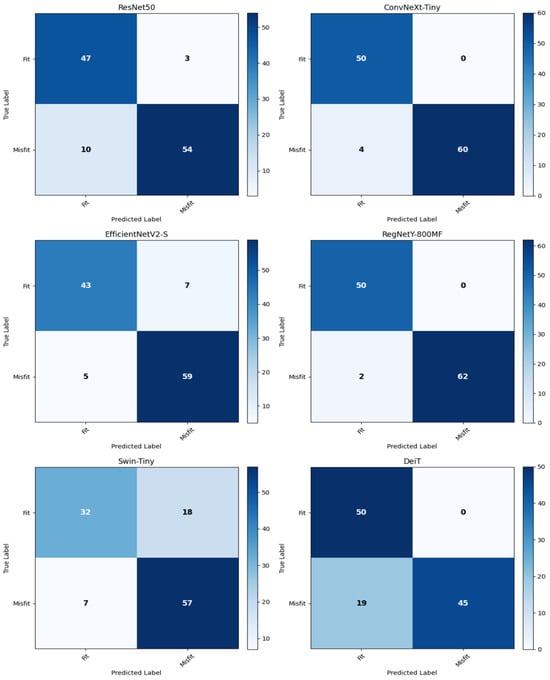
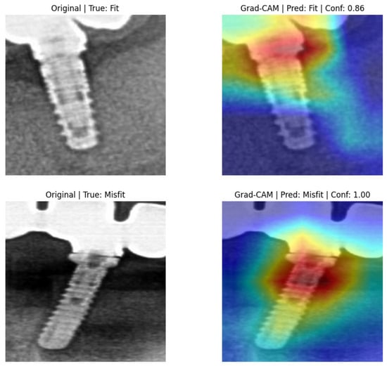
Background/Objectives: The aim of this study is to test different convolutional neural network (CNN) and Transformer-based models to detect and classify vertical misfit at the abutment-prosthesis interface on panoramic radiographs, and to develop a hybrid deep learning model enhanced with attention mechanisms. Methods: A dataset consisting of a total of 566 images, manually classified as 249 ‘fit’ and 317 ‘misfit’ cases by two experts, was created. Images were resized to 224 × 224 and divided into training, validation, and test groups. The deep learning model yielding the most successful results was determined as the backbone; a hybrid model was developed by integrating three different attention modules (SE, CBAM, and ECA) into this structure. Model performance was evaluated using accuracy, precision, sensitivity, and F1 score metrics. Results: CNN-based models (RegNetY-800MF, ConvNeXt-Tiny, EfficientNetV2-S, ResNet50) performed better than Transformer-based models (DeiT, Swin-Tiny) in all metrics. The proposed hybrid model exhibited the highest success among all tested models with a 99.12% accuracy rate. This model reached a 100% precision value in the misfit group and yielded no false positive results. The F1 scores of the hybrid model were recorded as 99.01% for the fit group and 99.21% for the misfit group. Conclusions: The findings of this study demonstrate that attention-enhancing deep learning frameworks have the potential to significantly improve the diagnostic utility of routine panoramic radiographs. It shows that panoramic imaging, when supported by advanced artificial intelligence, can provide valuable diagnostic support in detecting vertical misfit. The developed model has the potential to become a reliable clinical decision support system.
Full article
(This article belongs to the Special Issue Advances in Dental Diagnostics)
►▼
Show Figures

Figure 1
Journal Menu
► ▼ Journal Menu-
- Diagnostics Home
- Aims & Scope
- Editorial Board
- Reviewer Board
- Topical Advisory Panel
- Instructions for Authors
- Special Issues
- Topics
- Sections & Collections
- Article Processing Charge
- Indexing & Archiving
- Editor’s Choice Articles
- Most Cited & Viewed
- Journal Statistics
- Journal History
- Journal Awards
- Society Collaborations
- Conferences
- Editorial Office
Journal Browser
► ▼ Journal BrowserHighly Accessed Articles
Latest Books
E-Mail Alert
News
Topics
Topic in
Biology, JCM, Diagnostics, Dentistry Journal
Assessment of Craniofacial Morphology: Traditional Methods and Innovative Approaches
Topic Editors: Nikolaos Gkantidis, Carlalberta VernaDeadline: 1 June 2026
Topic in
Diagnostics, Electronics, Eng, Micro, Micromachines
Micro-Nanoelectronic Systems for Diagnosis and Therapies
Topic Editors: Wubin Bai, Jiho ShinDeadline: 30 June 2026
Topic in
Applied Sciences, Diagnostics, JCM, Clinics and Practice, Therapeutics
Advances in Diagnosis and Treatment of Dental Diseases and Temporomandibular Joints
Topic Editors: Rafał Obuchowicz, Małgorzata Pihut, Karolina Nurzynska, Andrzej UrbanikDeadline: 31 July 2026
Topic in
Cancers, Diagnostics, Gastrointestinal Disorders, JCM, Current Oncology
Metastatic Colorectal Cancer: From Laboratory to Clinical Studies, 2nd Edition
Topic Editors: Ioannis Ntanasis-Stathopoulos, Diamantis I. TsilimigrasDeadline: 20 August 2026
Conferences
Special Issues
Special Issue in
Diagnostics
Cardiovascular Imaging, 2nd Edition
Guest Editor: Riemer H.J.A. SlartDeadline: 26 May 2026
Special Issue in
Diagnostics
Advances in Nephrology
Guest Editor: Marijn M. SpeeckaertDeadline: 30 May 2026
Special Issue in
Diagnostics
Trends and Novelties in Bone Imaging
Guest Editor: Kamyar MoradiDeadline: 31 May 2026
Special Issue in
Diagnostics
Advances in the Diagnosis and Management of Idiopathic Normal Pressure Hydrocephalus
Guest Editors: Elisabeth Kapaki, Efstratios-Stylianos PyrgelisDeadline: 31 May 2026
Topical Collections
Topical Collection in
Diagnostics
Clinical Guidelines/Expert Consensus on Diagnostics
Collection Editor: Zhongheng Zhang
Topical Collection in
Diagnostics
Diagnosis of Endometriosis: Biomarkers and Clinical Methods
Collection Editor: Antonio Simone Laganà
Topical Collection in
Diagnostics
Nuclear Medicine and Molecular Imaging Technology
Collection Editor: Andreas Kjaer