Topic Editors

Department of Orthodontics and Dentofacial Orthopedics, School of Dental Medicine, University of Bern, CH-3010 Bern, Switzerland
Prof. Dr. Carlalberta Verna
Department of Pediatric Oral Health and Orthodontics, University Center for Dental Medicine Basel UZB, University of Basel, Hebelstrasse 3, CH-4056 Basel, Switzerland

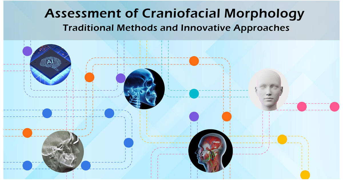
Assessment of Craniofacial Morphology: Traditional Methods and Innovative Approaches

Abstract submission deadline
20 March 2025
Manuscript submission deadline
20 June 2025
Viewed by
1502

Topic Information

Dear Colleagues,

This Topic is a continuation of the previous successful Topic “Diagnosis of Craniofacial Changes: Conventional Approaches and Novel Methodologies”.

The accurate assessment of craniofacial morphology is essential across multiple disciplines to ensure proper diagnosis, treatment planning, and outcome evaluation. Variations and structural changes within and between individuals can be attributed to genetic effects, physiological processes like growth, development, and aging, as well as pathological conditions and medical interventions. In addition to facilitating correct diagnosis and documentation, which serve both medical and legal purposes, accurate patient representations at different time points also impact the ability to track morphological changes. It's important to recognize that even sequential craniofacial images, acquired within a brief timeframe and under identical conditions, may exhibit variations related to the imaging technique or the subject itself. The challenge of reliably assessing craniofacial morphology and associated changes over extended periods is even more daunting.

The craniofacial region is of utmost importance for human life, highly impacting both function and aesthetics. As a result, the accurate assessment of craniofacial morphology is fundamental, and various approaches have been developed to address this need. Traditional methods primarily revolve around two-dimensional imaging, while three-dimensional (and four-dimensional) imaging is increasingly becoming an indispensable component of both daily clinical practice and cutting-edge research. The upcoming article collection will feature literature reviews and original studies focusing on the development and application of imaging modalities and analysis methodologies for the assessment of craniofacial morphology.

Dr. Nikolaos Gkantidis
Prof. Dr. Carlalberta Verna
Topic Editors

Keywords

  • craniofacial imaging
  • radiography
  • facial photography
  • anthropometry
  • craniofacial morphology
  • cephalometrics
  • computed tomography
  • stereophotogrammetry
  • superimposition
  • geometric morphometrics
  • finite element analysis
  • machine learning
  • artificial intelligence

Participating Journals

Journal Name Impact Factor CiteScore Launched Year First Decision (median) APC
Biology
biology
3.6 5.7 2012 16.1 Days CHF 2700 Submit
Journal of Clinical Medicine
jcm
3.0 5.7 2012 17.3 Days CHF 2600 Submit
Diagnostics
diagnostics
3.0 4.7 2011 20.5 Days CHF 2600 Submit
Dentistry Journal
dentistry
2.5 3.7 2013 26 Days CHF 2000 Submit

Preprints.org is a multidiscipline platform providing preprint service that is dedicated to sharing your research from the start and empowering your research journey.

MDPI Topics is cooperating with Preprints.org and has built a direct connection between MDPI journals and Preprints.org. Authors are encouraged to enjoy the benefits by posting a preprint at Preprints.org prior to publication:

  1. Immediately share your ideas ahead of publication and establish your research priority;
  2. Protect your idea from being stolen with this time-stamped preprint article;
  3. Enhance the exposure and impact of your research;
  4. Receive feedback from your peers in advance;
  5. Have it indexed in Web of Science (Preprint Citation Index), Google Scholar, Crossref, SHARE, PrePubMed, Scilit and Europe PMC.

Published Papers (2 papers)

Order results
Result details
Journals
Select all
Export citation of selected articles as:
15 pages, 853 KiB  
Review
The Evolution of Custom Subperiosteal Implants for Treatment of Partial or Complete Edentulism in Patients with Severe Alveolar Ridge Atrophy
by Jan Łoginoff, Agata Majos and Marcin Elgalal
J. Clin. Med. 2024, 13(12), 3582; https://doi.org/10.3390/jcm13123582 - 19 Jun 2024
Viewed by 467
Abstract
Dental implants have always played an important role in dentistry and have been used to replace missing teeth since around 600 AD. They can be classified into three groups: endosteal, subperiosteal, and transosteal. Over time, different materials have been used to manufacture dental [...] Read more.
Dental implants have always played an important role in dentistry and have been used to replace missing teeth since around 600 AD. They can be classified into three groups: endosteal, subperiosteal, and transosteal. Over time, different materials have been used to manufacture dental implants and these, in turn, can be divided into three groups: metals, ceramics, and polymers. Today, the most commonly used treatment for edentulism is the use of endosteal implants. However, such an approach cannot be used in patients with severe alveolar ridge atrophy and, in such cases, custom subperiosteal implants are an alternative. This review article focuses on historical developments and improvements that have been made over recent years in treatment options for patients suffering from edentulism and significant resorption of the alveolar ridge. These treatment options involve the utilization of custom subperiosteal implants. This paper looks at the historical evolution of these implants, the significance of diagnostic imaging, and the application of the contemporary methods of production, such as CAD-CAM and additive manufacturing. The research emphasizes the importance of accuracy and personalization provided by these emerging technologies that have rendered subperiosteal implants a more feasible and less intrusive alternative for patients suffering from significant bone loss. Full article
Show Figures

Figure 1

14 pages, 2486 KiB  
Article
Effects of Occlusal Contact on Maxillary Alveolar Bone Morphology in Patients with and without Anterior Open Bite: A Cross-Sectional Study
by Chiyo Shimizu-Tomoda, Yuji Ishida, Aiko Ishizaki-Terauchi, Yukari Mizoguchi, Shuji Oishi and Takashi Ono
J. Clin. Med. 2024, 13(11), 3061; https://doi.org/10.3390/jcm13113061 - 23 May 2024
Viewed by 448
Abstract
Background/Objectives: Anterior open bite (AOB) is characterized by the absence of occlusal contact between the maxillary and mandibular anterior teeth, while the posterior teeth are in contact when occluded. Here, we aimed to clarify the difference in maxillary alveolar bone morphology in adult [...] Read more.
Background/Objectives: Anterior open bite (AOB) is characterized by the absence of occlusal contact between the maxillary and mandibular anterior teeth, while the posterior teeth are in contact when occluded. Here, we aimed to clarify the difference in maxillary alveolar bone morphology in adult patients with and without AOB. Methods: This cross-sectional study was conducted on 50 adults aged 18–39 years: 25 patients without AOB (control group; 13 males and 12 females; age: mean ± standard deviation [SD], 22.2 ± 4.5 years) and 25 patients with AOB (9 males and 16 females; age: 24.2 ± 6.4 years). Using cone-beam computed tomography images, the height of the maxillary alveolar bone crest in the anterior and posterior teeth and thickness of the alveolar cortical bone on the labial and palatal sides were measured and compared between the two groups. An independent t-test and Pearson’s correlation analysis were used to examine statistical significance (p < 0.05). Results: The AOB group showed a significantly longer (p = 0.016) posterior alveolar crest and thinner cortical bone on the buccal (p < 0.001) and lingual (p = 0.009) sides of the anterior region and the buccal (p = 0.006) sides of the posterior region than the control group did. Moreover, a significant negative correlation (p = 0.046; r = −0.403) was observed between bite force and cortical bone thickness on the buccal side of the posterior region in the AOB group. Conclusions: It is suggested that the absence of occlusal contact in the anterior area influences the alveolar bone morphology of the maxilla. Full article
Show Figures

Figure 1

Back to TopTop