When Palpitations Unmask Crista Terminalis Hypertrophy: A Case Report and Review of Current Literature
 , and
, and
Abstract
1. Introduction
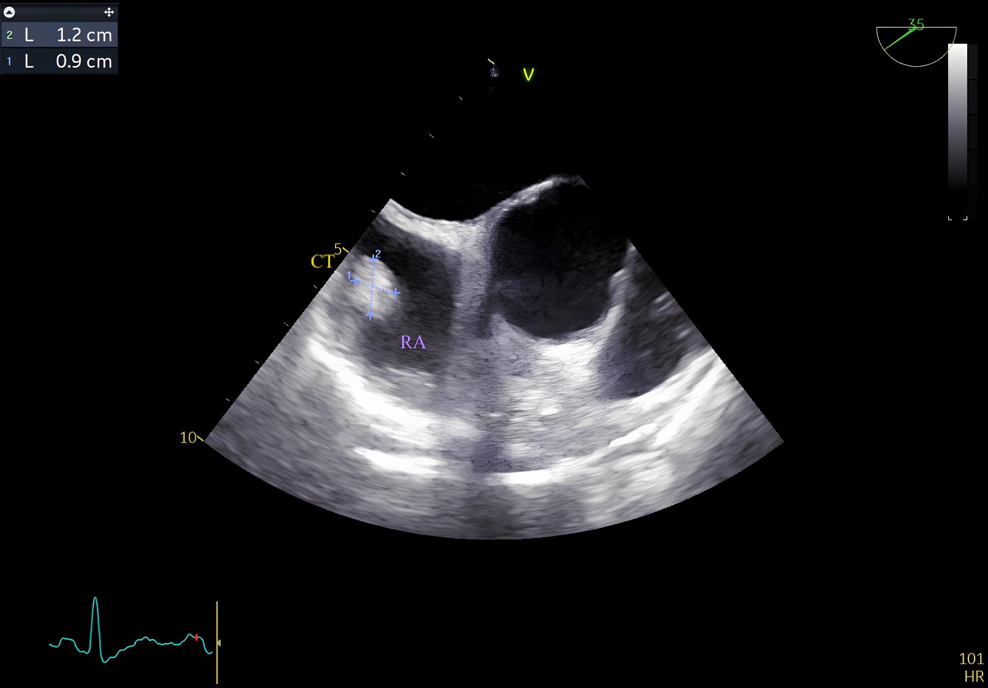
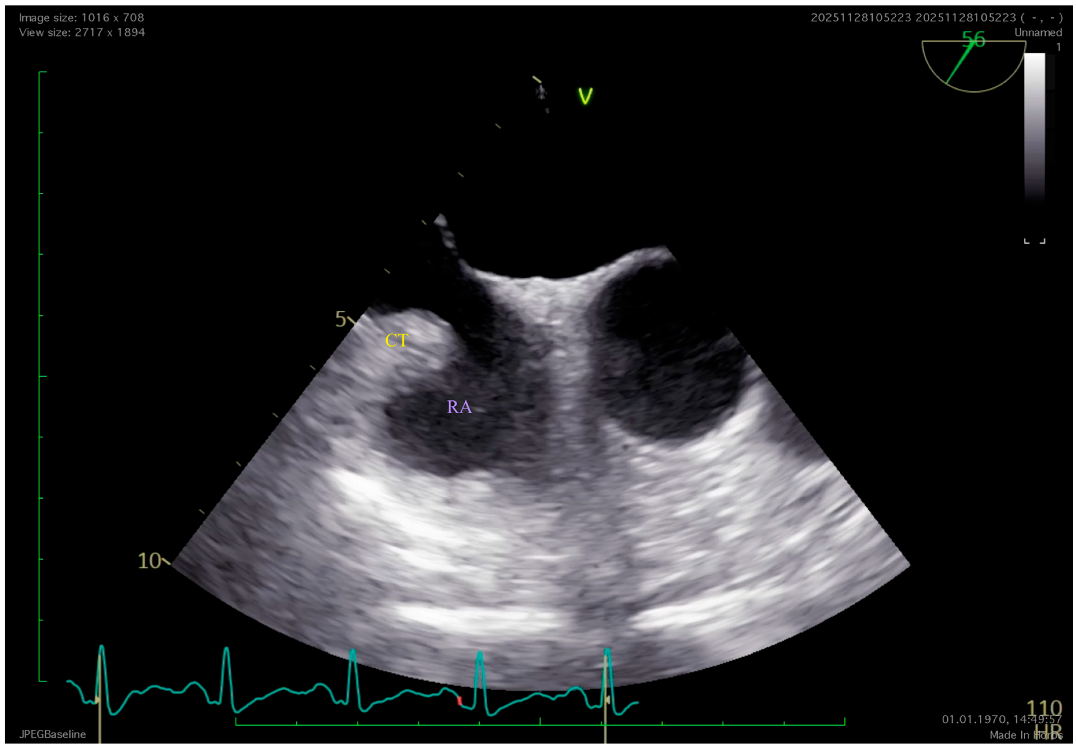
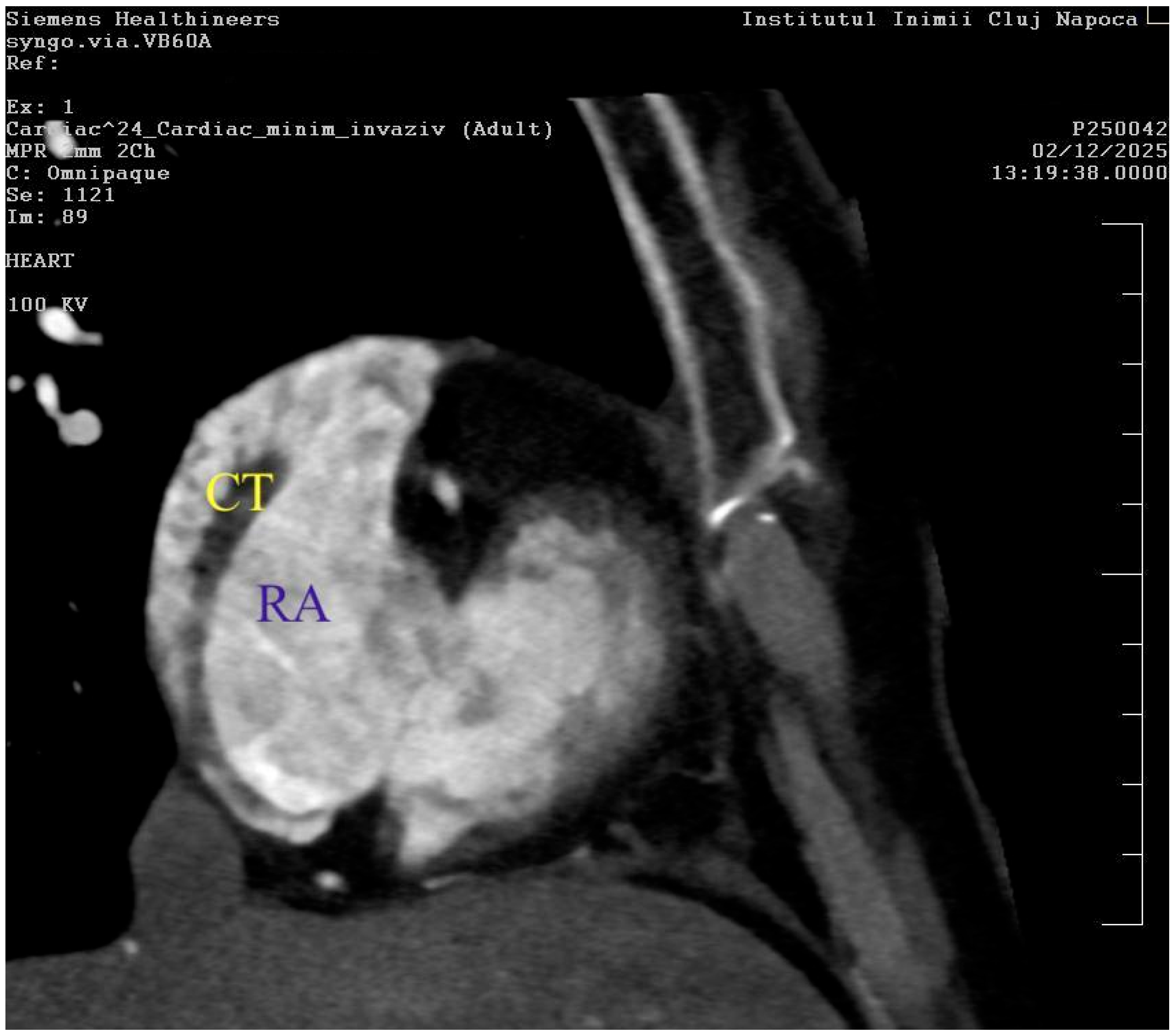
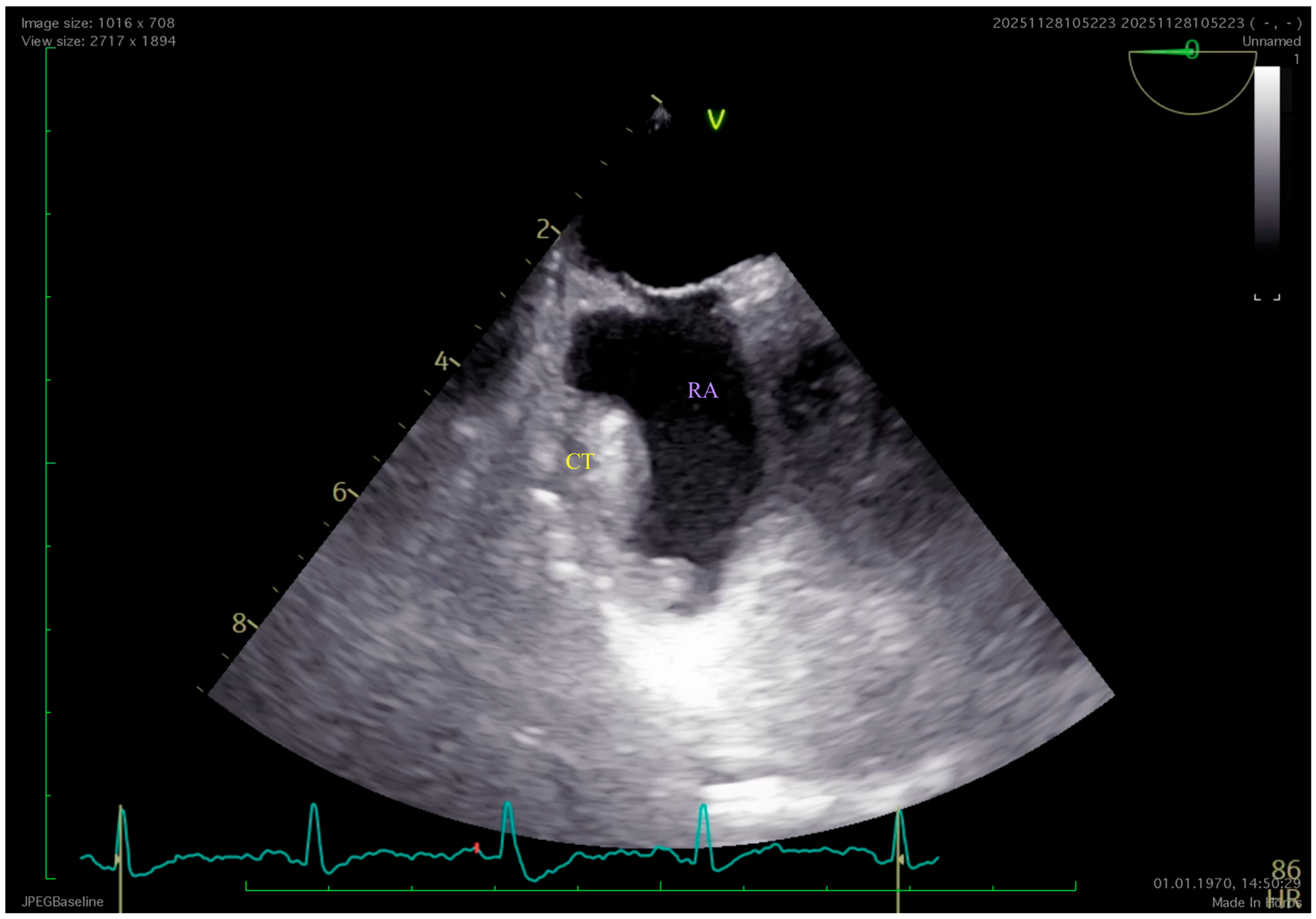
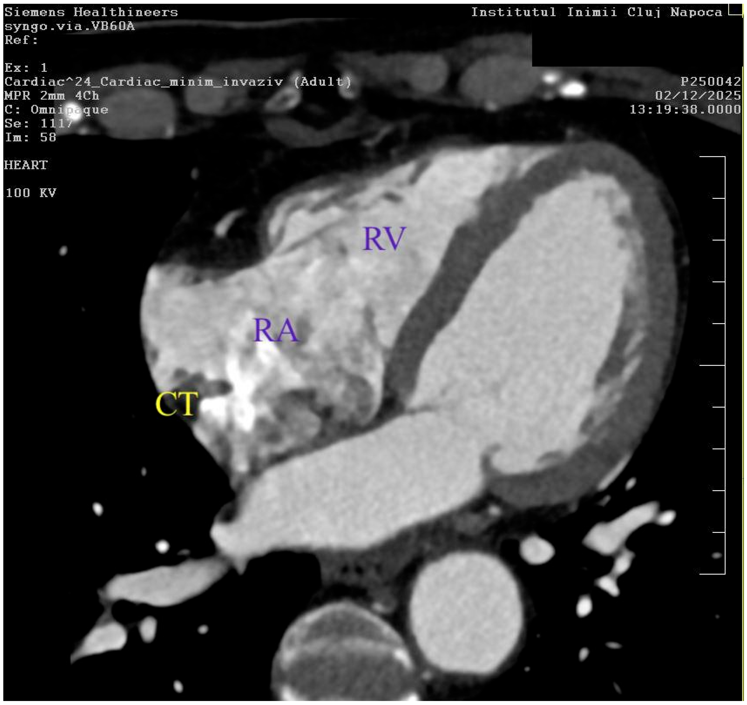
2. Detailed Case Description



3. Discussion
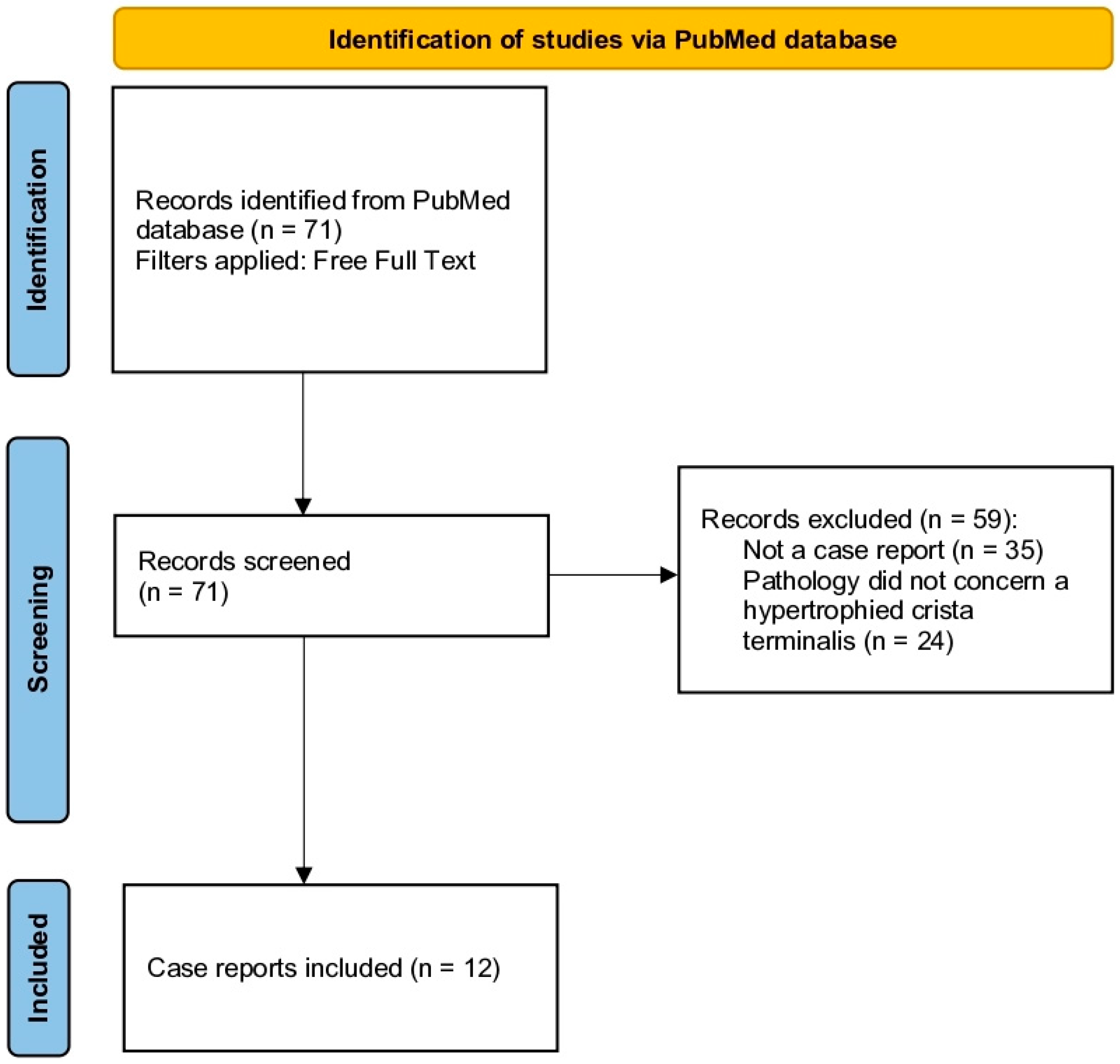
3.1. Crista Terminalis Hypertrophy in the Current Literature
3.1.1. Association with Atrial Tachyarrhythmia
3.1.2. Diagnostic Imaging and Differential Diagnosis
First-Line Assessment and Risk Stratification
Second-Level Imaging
Tertiary Modalities and Further Risk Stratification
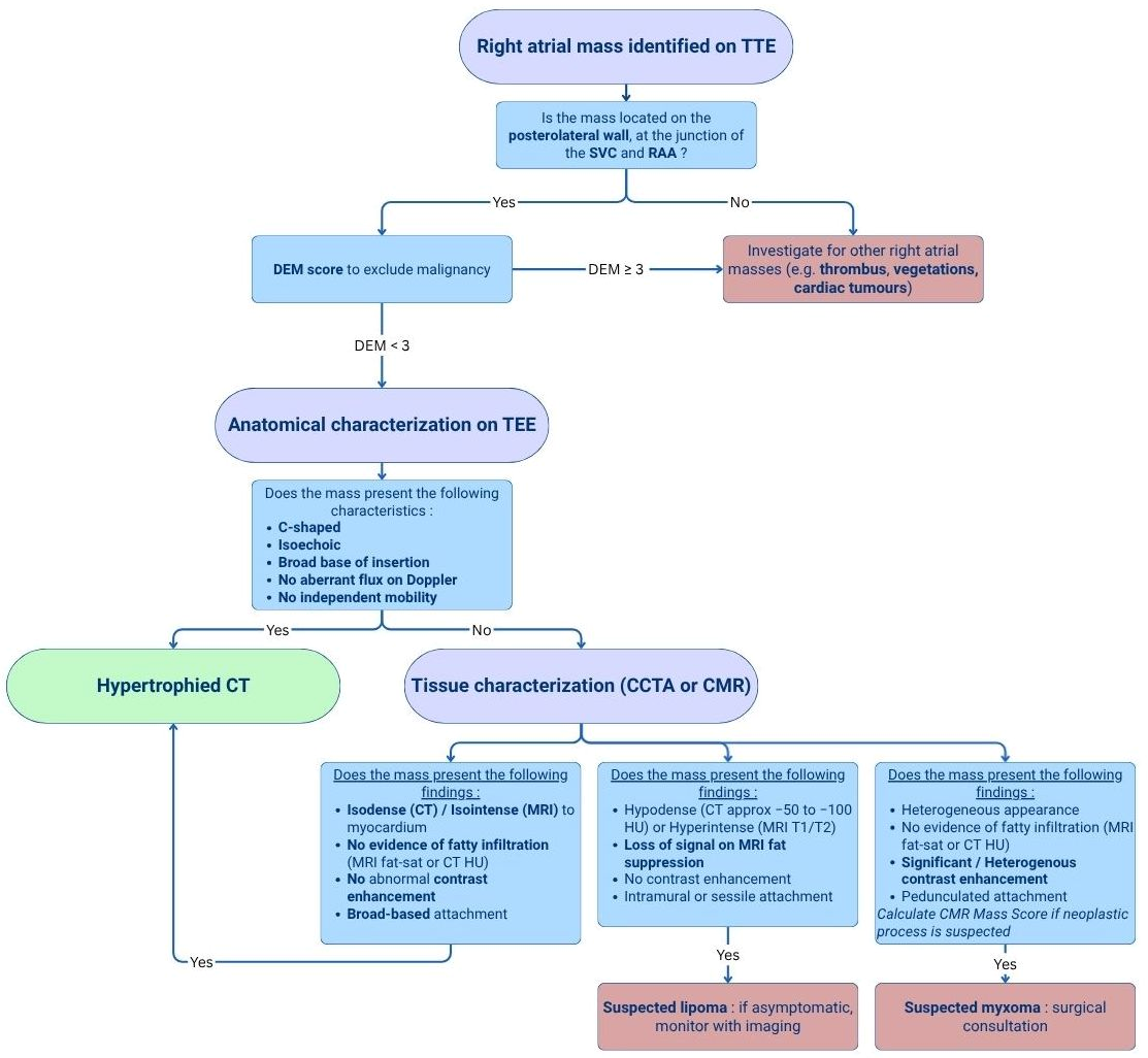
3.1.3. Proposed Diagnostic Algorithm for Prominent Crista Terminalis
3.2. Significant Contributions
3.3. Limitations
4. Conclusions
Author Contributions
Funding
Institutional Review Board Statement
Informed Consent Statement
Data Availability Statement
Conflicts of Interest
Abbreviations
| CT | Crista terminalis |
| TTE | Transthoracic echocardiography |
| TEE | Transesophageal echocardiography |
| CCTA | Cardiac computed tomography angiography |
| CMR | Cardiac magnetic resonance imagery |
| ECG | Electrocardiography |
| LVEF | Left ventricular systolic function |
| SA | Sino-atrial |
| CAD | Coronary artery disease |
| CAD-RADS | Coronary Artery Disease Reporting and Data System |
| LAD | Left anterior descending artery |
| CTI | Cavotricuspid isthmus |
| HCN | Hyperpolarization-activated Cyclic Nucleotide-gated |
| CTD | Cor triatriatum dexter |
| SVC | Superior vena cava |
| IVC | Inferior vena cava |
| DEM | Diagnostic echocardiographic mass |
| MDCT | Multi-detector computer tomography |
| RAA | Right atrial appendage |
| 18F-FDG PET-CT | Fluorine-18 Fluorodeoxyglucose Positron Emission Tomography/Computed Tomography |
References
- Salim, H.; Palit, A.; Maher, A. When Is a Mass Not a Mass? An Unusual Presentation of Prominent Crista Terminalis. BMJ Case Rep. 2016, 2016, bcr2015211532. [Google Scholar] [CrossRef]
- Salustri, A.; Bakir, S.; Sana, A.; Lange, P.; Al Mahmeed, W.A. Prominent Crista Terminalis Mimicking a Right Atrial Mass: Case Report. Cardiovasc. Ultrasound 2010, 8, 47. [Google Scholar] [CrossRef] [PubMed]
- Evong, Y.; Warren, A.; Mohsin, H. Prominent Crista Terminalis in a Fetus. Images Paediatr. Cardiol. 2018, 20, 1–4. [Google Scholar]
- Morris, G.M.; Segan, L.; Wong, G.; Wynn, G.; Watts, T.; Heck, P.; Walters, T.E.; Nisbet, A.; Sparks, P.; Morton, J.B.; et al. Atrial Tachycardia Arising From the Crista Terminalis, Detailed Electrophysiological Features and Long-Term Ablation Outcomes. JACC Clin. Electrophysiol. 2019, 5, 448–458. [Google Scholar] [CrossRef]
- Ahmed, A.S. Hypertrophied Crista Terminalis—The Great Masquerader and Saviour. Anatol. J. Cardiol. 2019, 23, 5011–5012. [Google Scholar] [CrossRef]
- Nalawade, D.D.; Wadhokar, P.S.; Jadhav, A.K.; Manade, V.V. Prominent Crista Terminalis Mimicking Right Atrial Thrombus in a Case of Permanent Pacemaker Implantation—Role of Two- and Three-Dimensional Transesophageal Echocardiography. J. Cardiovasc. Echogr. 2024, 34, 149–151. [Google Scholar] [CrossRef] [PubMed]
- Lakhani, D.A.; Balar, A.B.; Kim, C. Prominent Crista Terminalis Mimicking a Right Atrial Mass: A Case Report and Brief Review of the Literature. Radiol. Case Rep. 2022, 17, 434–438. [Google Scholar] [CrossRef]
- Ghesani, A.; Cangut, B.; Stempler, L. Not Everything That Shines Is Gold, Normal Uptake in Crista Terminalis on FDG PET/CT Masquerading as a Tumor Thrombus Approaching Right Heart. Radiol. Case Rep. 2024, 19, 513–518. [Google Scholar] [CrossRef]
- Wang, J.; Wang, G.; Bi, X.; Zhang, R.; Liu, C. An Unusual Presentation of Prominent Crista Terminalis Mimicking a Right Atrial Mass: A Case Report. BMC Cardiovasc. Disord. 2018, 18, 210. [Google Scholar] [CrossRef] [PubMed]
- Na, J.O.; Kim, E.J.; Mun, S.J.; Choi, E.H.; Mun, J.H.; Lee, H.R.; Kim, Y.K.; Yong, H.S. Prominent Crista Terminalis in Patients with Embolic Events. J. Cardiovasc. Ultrasound 2011, 19, 156. [Google Scholar] [CrossRef]
- Bhatia, S.; Qasim, A.; Jiwani, A.K.; Aly, A.M. Benign Structures Mimicking Right Atrial Masses on Prenatal Ultrasound. Case Rep. Pediatr. 2021, 2021, 8889941. [Google Scholar] [CrossRef] [PubMed]
- Pati, M.; Bonasoni, M.P.; Musarò, A.; Petrachi, B.; Di Dio, F.; Chesi, E.; Leo, F.; Barbato, G.; Ricò, S.; Gargano, G.; et al. Prenatal Detection of a Right Atrial Echogenic Mass: A Case Report of Hypertrophic Crista Terminalis. Diagnostics 2025, 16, 17. [Google Scholar] [CrossRef] [PubMed]
- Shoji, Y.; Saito, Y.; Okumura, Y. Prominent Crista Terminalis Mimicking a Right Atrial Mass. Intern. Med. 2024, 63, 3259–3260. [Google Scholar] [CrossRef]
- Zhao, J.; Kennelly, J.; Nalar, A.; Kulathilaka, A.; Sharma, R.; Bai, J.; Li, N.; Fedorov, V.V. Chamber-Specific Wall Thickness Features in Human Atrial Fibrillation. Interface Focus 2023, 13, 20230044. [Google Scholar] [CrossRef]
- Diamant, M.J.; Andrade, J.G.; Virani, S.A.; Jhund, P.S.; Petrie, M.C.; Hawkins, N.M. Heart Failure and Atrial Flutter: A Systematic Review of Current Knowledge and Practices. ESC Heart Fail. 2021, 8, 4484–4496. [Google Scholar] [CrossRef]
- Meyer, C.; Kahle, A.-K.; Dagres, N.; Derval, N.; Dewland, T.A.; Drago, F.; Eckardt, L.; Frontera, A.; Gerstenfeld, E.P.; Hardy, C.; et al. Management of Patients with Atrial Tachycardia: A Clinical Consensus Statement of the European Heart Rhythm Association (EHRA) of the ESC, Endorsed by the Heart Rhythm Society (HRS), the Asia Pacific Heart Rhythm Society (APHRS), the Latin American Heart Rhythm Society (LAHRS), and the Association for European Paediatric and Congenital Cardiology (AEPC). Europace 2025, 27, euaf324. [Google Scholar] [CrossRef] [PubMed]
- Esmel-Vilomara, R.; Castellote, A.; Santana, L.; Albert, D.C. Neonatal Supraventricular Extrasystole as Early Clinical Debut of Cardiac Rhabdomyoma. Ann. Pediatr. Cardiol. 2018, 11, 318–321. [Google Scholar] [CrossRef]
- Kalyanasundaram, A.; Li, N.; Augostini, R.S.; Weiss, R.; Hummel, J.D.; Fedorov, V.V. Three-Dimensional Functional Anatomy of the Human Sinoatrial Node for Epicardial and Endocardial Mapping and Ablation. Heart Rhythm 2023, 20, 122–133. [Google Scholar] [CrossRef]
- Sánchez-Quintana, D.; Anderson, R.H.; Cabrera, J.A.; Climent, V.; Martin, R.; Farré, J.; Ho, S.Y. The Terminal Crest: Morphological Features Relevant to Electrophysiology. Heart 2002, 88, 406–411. [Google Scholar] [CrossRef]
- Pambrun, T.; Derval, N.; Duchateau, J.; Ramirez, F.D.; Chauvel, R.; Tixier, R.; Marchand, H.; Bouyer, B.; Welte, N.; André, C.; et al. Sinus Node Exit, Crista Terminalis Conduction, Interatrial Connection, and Wavefront Collision: Key Features of Human Atrial Activation in Sinus Rhythm. Heart Rhythm 2022, 19, 701–709. [Google Scholar] [CrossRef]
- Amsaleg, A.; Sánchez, J.; Mikut, R.; Loewe, A. Characterization of the Pace-and-Drive Capacity of the Human Sinoatrial Node: A 3D in Silico Study. Biophys. J. 2022, 121, 4247–4259. [Google Scholar] [CrossRef]
- Bhargav, A.; Velayutham, R.; Selvaraj, R.J. Lower Loop Re-entrant Flutter. J. Arrhythmia 2025, 41, e70081. [Google Scholar] [CrossRef] [PubMed]
- Hu, X.; Jiang, W.; Liu, X. Catheter Ablation of Baffle-Crista Suture–Related Macro-Re-Entrant Flutter in a Lateral Tunnel Fontan Patient. JACC Case Rep. 2025, 30, 105899. [Google Scholar] [CrossRef]
- Liwanag, M.; Willoughby, C. Atrial Tachycardia. In StatPearls; StatPearls Publishing: Treasure Island, FL, USA, 2026. [Google Scholar]
- Mishra, P.; Narayanan, R. The Enigmatic HCN Channels: A Cellular Neurophysiology Perspective. Proteins Struct. Funct. Bioinform. 2025, 93, 72–92. [Google Scholar] [CrossRef] [PubMed]
- Pan, T.; Liu, Y.; Yu, Y.; Zhang, D.; Sun, Y.-H.; Zhang, H.-W.; Li, C.-Y. Association of Quantitative Computed Tomography-Based Right Atrial Appendage and Right Atrium Parameters with Postradiofrequency Ablation Recurrence of Atrial Fibrillation. Quant. Imaging Med. Surg. 2023, 13, 3802–3815. [Google Scholar] [CrossRef] [PubMed]
- Pan, T.; Tian, X.; Liu, Y.; Yang, H.-Q.; Ma, G.-J.; Han, X.-N.; Li, C.-Y. The Role of the Right Atrial Appendage and Right Atrium in Post-Radiofrequency Ablation Recurrence in Different Types of Atrial Fibrillation. Clin. Radiol. 2024, 79, e1312–e1320. [Google Scholar] [CrossRef]
- Kanda, T.; Masuda, M.; Kurata, N.; Matsuda, Y.; Osamu, I.; Asai, M.; Mano, T. An Unusual Propagation Pattern along the Tricuspid Annulus after Cavo-Tricuspid Isthmus Ablation: Insights into Posterior Transverse Conduction Revealed by an Ultra-High-Resolution 3-Dimensional Mapping System. J. Cardiol. Cases 2019, 19, 101–105. [Google Scholar] [CrossRef]
- Diaz Delgado, P.J.; Batlle, J.C.; Saenger, E.; Mellado, S.; Rodriguez Lopez, J.; Nayak, H.M.; Anderson, A.S.; Velasco, A. Radiofrequency Ablation of Crista Terminalis Focal Atrial Tachycardia Using End Inspiration to Prevent Phrenic Nerve Paralysis. JACC Case Rep. 2025, 30, 103559. [Google Scholar] [CrossRef]
- Paolisso, P.; Foà, A.; Magnani, I.; Bergamaschi, L.; Graziosi, M.; Angeli, F.; Chiti, C.; Fabrizio, M.; Rinaldi, A.; Stefanizzi, A.; et al. Development and Validation of a Diagnostic Echocardiographic Mass Score in the Approach to Cardiac Masses. JACC Cardiovasc. Imaging 2022, 15, 2010–2012. [Google Scholar] [CrossRef]
- Paolisso, P.; Foà, A.; Bergamaschi, L.; Graziosi, M.; Rinaldi, A.; Magnani, I.; Angeli, F.; Stefanizzi, A.; Armillotta, M.; Sansonetti, A.; et al. Echocardiographic Markers in the Diagnosis of Cardiac Masses. J. Am. Soc. Echocardiogr. Off. Publ. Am. Soc. Echocardiogr. 2023, 36, 464–473.e2. [Google Scholar] [CrossRef]
- Turton, E.W.; Ender, J. Role of 3D Echocardiography in Cardiac Surgery: Strengths and Limitations. Curr. Anesthesiol. Rep. 2017, 7, 291–298. [Google Scholar] [CrossRef]
- Hassan, M.; Abdrabou, M.M.; Wahba, W.A.; Samaan, A.A.; Baghdady, Y.; Elamragy, A.A. Agreement between 4D Transesophageal Echocardiography and Multi-Detector Computed Tomography in Measuring Aortic Root Dimensions and Coronary Ostia Heights. Int. J. Cardiovasc. Imaging 2023, 39, 1561–1569. [Google Scholar] [CrossRef] [PubMed]
- Lang, R.M.; Cameli, M.; Sade, L.E.; Faletra, F.F.; Fortuni, F.; Rossi, A.; Soulat-Dufour, L. Imaging Assessment of the Right Atrium: Anatomy and Function. Eur. Heart J.—Cardiovasc. Imaging 2022, 23, 867–884. [Google Scholar] [CrossRef]
- Hrabak-Paar, M.; Muršić, M.; Balaško-Josipović, T.; Dilber, D.; Bulj, N. Multimodality Imaging of Cardiac Myxomas. Rev. Cardiovasc. Med. 2024, 25, 204. [Google Scholar] [CrossRef]
- Arisha, M.J.; Hsiung, M.C.; Nanda, N.C.; Gupta, A.; George, D.C.; Elkaryoni, A.; Barssoum, K.; Mohamed, A.H.; Srialluri, S. Two- and Three-dimensional Transthoracic Echocardiographic Assessment of Superior Vena Cava, Crista Terminalis, and Right Atrial Appendage Using the Right Parasternal Approach. Echocardiography 2017, 34, 1919–1929. [Google Scholar] [CrossRef] [PubMed]
- Shah, B.; Uppal, A.; Kunal, S.; Prajapati, S.; Gupta, A. Triatrial Appearance in a Patient With Atrial Septal Defect: A Case Report. Cureus 2025, 17, e84011. [Google Scholar] [CrossRef]
- Phuyal, N.; Pandey, N.; Mishra, A.K.; Shrestha, A. Chiari Network: A Case Report. BMC Cardiovasc. Disord. 2025, 25, 543. [Google Scholar] [CrossRef]
- Nguyen, M.; Gross, C.; Buttafuoco, J.; Ansari, S.; Nagi, H.; Maki, D. Persistent Eustachian Valve Causing Abdominal and Pelvic Venous System Dilation in a 44-Year-Old Male: A Case Report. Cureus 2024, 16, e60994. [Google Scholar] [CrossRef] [PubMed]
- Sławek-Szmyt, S.; Szmyt, K.; Żaba, C.; Grygier, M.; Lesiak, M.; Araszkiewicz, A. Peculiarities in Coronary Sinus Anatomy: Implications for Successful Cannulation from an Autoptic Study. EP Eur. 2021, 23, 1787–1794. [Google Scholar] [CrossRef]
- Teo, T.W.; Yeo, T.C. Different Faces of Atrial Septal Aneurysm. Ann. Acad. Med. Singap. 2025, 54, 786. [Google Scholar] [CrossRef]
- Clemente, A.; Seitun, S.; Mantini, C.; Gentile, G.; Federici, D.; Barison, A.; Rossi, A.; Cuman, M.; Pizzuto, A.; Ait-Ali, L.; et al. Cardiac CT Angiography: Normal and Pathological Anatomical Features—A Narrative Review. Cardiovasc. Diagn. Ther. 2020, 10, 1918–1945. [Google Scholar] [CrossRef] [PubMed]
- Matusik, P.S.; Mikrut, K.; Bryll, A.; Popiela, T.J.; Matusik, P.T. Cardiac Magnetic Resonance Imaging in Diagnostics and Cardiovascular Risk Assessment. Diagnostics 2025, 15, 178. [Google Scholar] [CrossRef]
- Paolisso, P.; Bergamaschi, L.; Angeli, F.; Belmonte, M.; Foà, A.; Canton, L.; Fedele, D.; Armillotta, M.; Sansonetti, A.; Bodega, F.; et al. Cardiac Magnetic Resonance to Predict Cardiac Mass Malignancy: The CMR Mass Score. Circ. Cardiovasc. Imaging 2024, 17, e016115. [Google Scholar] [CrossRef]
- Iwamiya, S.; Ihara, K.; Nitta, G.; Sasano, T. Atrial Fibrillation and Underlying Structural and Electrophysiological Heterogeneity. Int. J. Mol. Sci. 2024, 25, 10193. [Google Scholar] [CrossRef]

| References | Sex (F/M) | Age | Concomitant Arrhythmia | Key Imaging |
|---|---|---|---|---|
| Pati et al. (2025) [12] | NR 1 | Fetus (32 w) | NR | Prenatal ultrasound |
| Nalawade et al. (2024) [6] | F | 65 | NR | Real-time 3D TEE 2 |
| Shoji et al. (2024) [13] | M | 57 | NR | Real-time 3D TEE |
| Ghesani et al. (2023) [8] | F | 79 | NR | 18F-FDG PET-CT 3, TEE |
| Bhatia et al. (2021) [11] | NR | Fetus (23 w) | NR | Prenatal ultrasound |
| Lakhani et al. (2021) [7] | M | 78 | NR | CMR 4 |
| Ahmed et al. (2020) [5] | F | 59 | Atrial fibrillation | CCTA 5 |
| Evong et al. (2018) [3] | NR | Fetus (20 w) | NR | Prenatal ultrasound |
| Wang et al. (2018) [9] | M | 54 | NR | Cardiac PET/MRI 6 |
| Salim et al. (2016) [1] | F | 32 | Paroxysmal atrial fibrillation | CMR |
| Na et al. (2011) [10] | F | 73 | NR | TEE |
| Salustri et al. (2010) [2] | F | 26 | NR | TEE |
Disclaimer/Publisher’s Note: The statements, opinions and data contained in all publications are solely those of the individual author(s) and contributor(s) and not of MDPI and/or the editor(s). MDPI and/or the editor(s) disclaim responsibility for any injury to people or property resulting from any ideas, methods, instructions or products referred to in the content. |
© 2026 by the authors. Licensee MDPI, Basel, Switzerland. This article is an open access article distributed under the terms and conditions of the Creative Commons Attribution (CC BY) license.
Share and Cite
Racz, A.; Dădârlat-Pop, A.; Șerban, A.; Tomoaia, R.; Oprea, A.; Rosianu, H. When Palpitations Unmask Crista Terminalis Hypertrophy: A Case Report and Review of Current Literature. Diagnostics 2026, 16, 1615. https://doi.org/10.3390/diagnostics16111615
Racz A, Dădârlat-Pop A, Șerban A, Tomoaia R, Oprea A, Rosianu H. When Palpitations Unmask Crista Terminalis Hypertrophy: A Case Report and Review of Current Literature. Diagnostics. 2026; 16(11):1615. https://doi.org/10.3390/diagnostics16111615
Chicago/Turabian StyleRacz, Antonia, Alexandra Dădârlat-Pop, Adela Șerban, Raluca Tomoaia, Alexandru Oprea, and Horia Rosianu. 2026. "When Palpitations Unmask Crista Terminalis Hypertrophy: A Case Report and Review of Current Literature" Diagnostics 16, no. 11: 1615. https://doi.org/10.3390/diagnostics16111615
APA StyleRacz, A., Dădârlat-Pop, A., Șerban, A., Tomoaia, R., Oprea, A., & Rosianu, H. (2026). When Palpitations Unmask Crista Terminalis Hypertrophy: A Case Report and Review of Current Literature. Diagnostics, 16(11), 1615. https://doi.org/10.3390/diagnostics16111615