Case Report

When Lymph Nodes Don’t Lie: Report of Three Unusual Presentations of Thoracic Tumors

1
Pathology Unit, Department of Mental and Physical Health and Preventive Medicine, Università degli Studi della Campania “Luigi Vanvitelli”, Via Luciano Armanni 5, 80138 Naples, Italy
2
Stroke Unit, Department of Emergency and Urgent Medicine, Santa Maria Delle Grazie Hospital, 80078 Pozzuoli, Italy
3
Oncology Operative Unit, “Santa Maria delle Grazie” Hospital, ASL NA2 NORD, 80078 Pozzuoli, Italy
4
Department of Precision Medicine, Medical Oncology, Università degli Studi della Campania “Luigi Vanvitelli”, 80131 Naples, Italy
5
Division of Thoracic Surgery, Università degli Studi della Campania “Luigi Vanvitelli”, Piazza Miraglia, 2, 80138 Naples, Italy
6
Pathology Unit, Ospedale Evangelico Villa Betania, 80147 Naples, Italy
7
Division of Thoracic Surgery, S.G. Moscati Hospital, 83100 Avellino, Italy
*
Author to whom correspondence should be addressed.
Diagnostics 2026, 16(11), 1618; https://doi.org/10.3390/diagnostics16111618
Submission received: 26 February 2026 / Revised: 21 April 2026 / Accepted: 6 May 2026 / Published: 25 May 2026
(This article belongs to the Section Clinical Diagnosis and Prognosis)

Abstract

Background and Clinical Significance: Lymph node metastases from carcinoma of unknown primary origin (CUP) are a rare and diagnostically challenging entity, particularly when arising from thoracic malignancies with atypical clinical presentations. This study aims to illustrate the essential nature of multidisciplinary integration, with a particular emphasis on the role of the pathologist in identifying occult thoracic tumors. Case Presentation: We report three cases of patients presenting with cervical or systemic lymphadenopathy as the initial clinical manifestation. Comprehensive diagnostic workups included advanced imaging (CT, MRI, and PET), comprehensive histopathological analysis, and next-generation sequencing of circulating tumor DNA. Case one and case two were diagnosed as occult primary non-mucinous lung adenocarcinomas, based on the integration of morphological features and immunohistochemical co-expression of TTF-1 and Napsin A, despite the absence of identifiable lung lesions. One case harbored an ALK rearrangement, guiding effective targeted therapy with alectinib. Case three involved metastatic pleural epithelioid mesothelioma, which presented with systemic lymphadenopathy and was initially misclassified as metastatic adenocarcinoma. Diagnosis was confirmed by the loss of BAP1 expression by immunohistochemistry and the detection of a BAP1 S160fs*1 mutation, emphasizing the role of molecular pathology. Conclusions: Lymphadenopathy as the first manifestation of thoracic malignancy is a rare but clinically significant occurrence. In such atypical presentations, pathologists play a pivotal role in diagnosis, often leading the process when clinical or radiological clues are minimal or absent. Accurate histopathological assessment is essential to establish a correct diagnosis and guide appropriate therapy. A multidisciplinary approach remains the cornerstone of diagnostic precision in CUP cases.

1. Introduction

Lymph node metastases from carcinoma of unknown primary (CUP) comprise a heterogeneous group of tumors with unidentified primary sites. These rare cases typically show rapid, aggressive progression and poor prognosis, with a median survival of under one year. Metastases may display various histological subtypes, most commonly adenocarcinoma [1]. The diagnosis of metastatic lesions requires a multidisciplinary approach, with radiologists and pathologists playing key roles in identifying the primary tumor. Radiological evaluation, particularly through computed tomography (CT), and histopathological assessment—including morphological analysis and immunohistochemical profiling—are essential components of the diagnostic process [2]. Histology often reveals poorly differentiated tumors, making classification based on morphology alone challenging. Therefore, immunohistochemistry (IHC) plays a crucial role in characterizing the neoplastic cell population and guiding the identification of the tumor’s site of origin [3]. In 1995, Wang et al. first described the diagnostic value of differential cytokeratin-7 (CK7) and cytokeratin-20 (CK20) expression patterns in predicting the site of tumor origin in pathological analysis [4]. Several IHC-based algorithms have been proposed for the evaluation of CUP, and they always included the coordinated expression of CK7 and CK20 as fundamental biomarkers in the initial diagnostic strategy [5,6,7,8]. The integration of clinical data, oncological history, and analysis of excised lymph nodes, together with increasingly available lineage-specific biomarkers and molecular techniques, has markedly improved diagnostic accuracy in carcinoma of unknown primary (CUP) while preserving tissue for subsequent predictive analyses [9]. We report a small series of thoracic tumors with highly atypical clinical presentations. These cases highlight the critical role of a multidisciplinary diagnostic approach, in which the pathologist either led the diagnostic process or contributed substantially within a collaborative framework.

2. Report of Cases

2.1. Case One

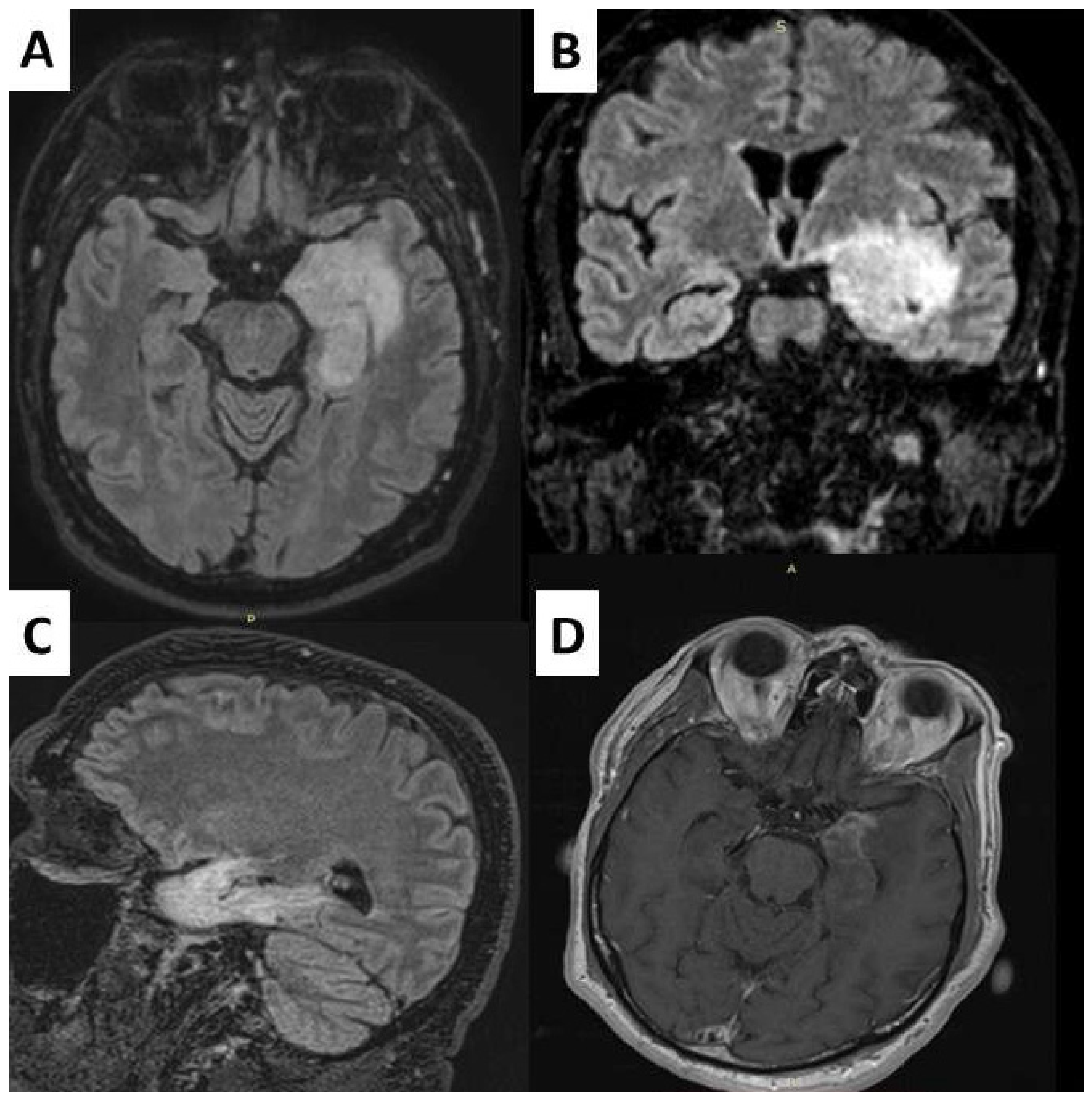
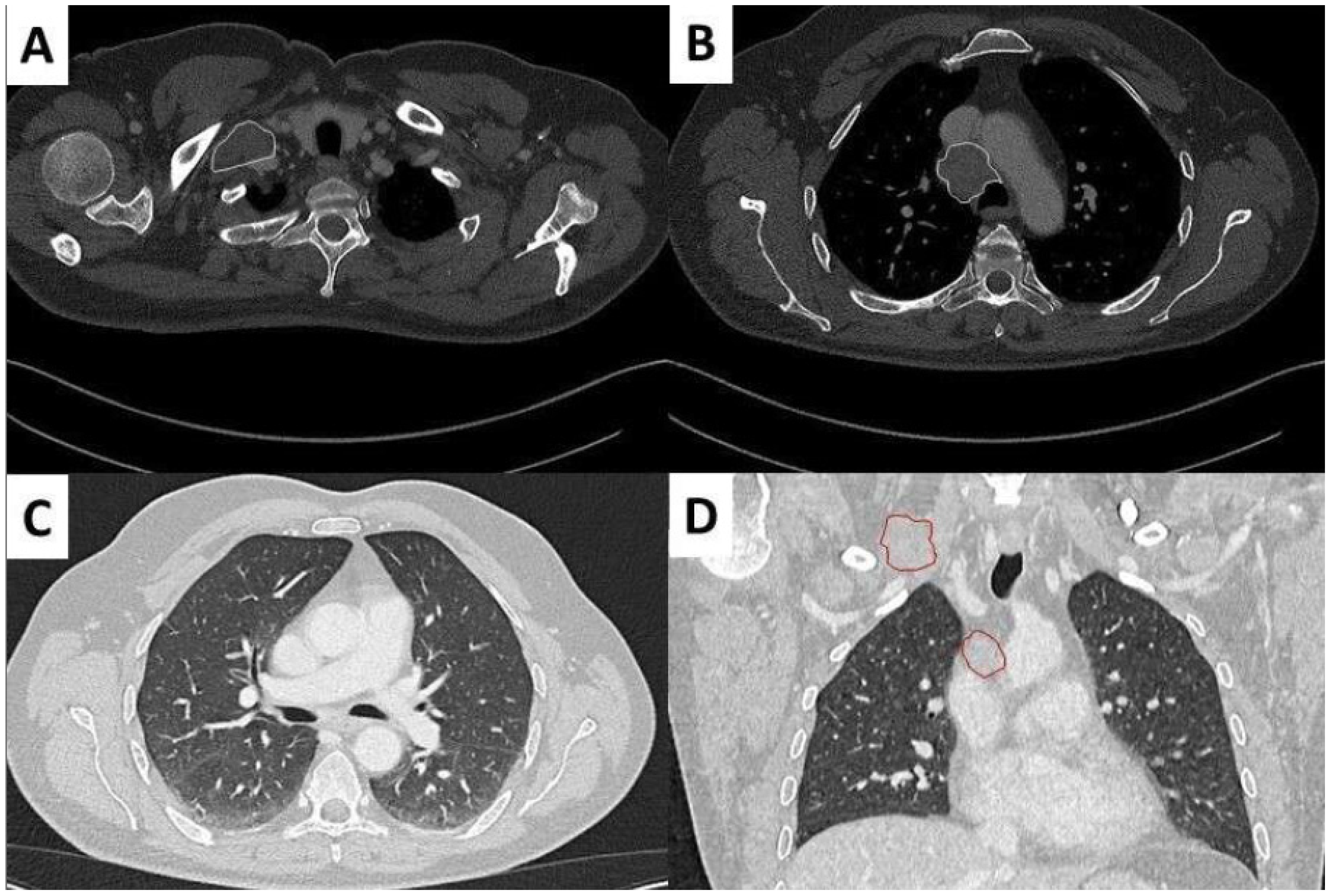
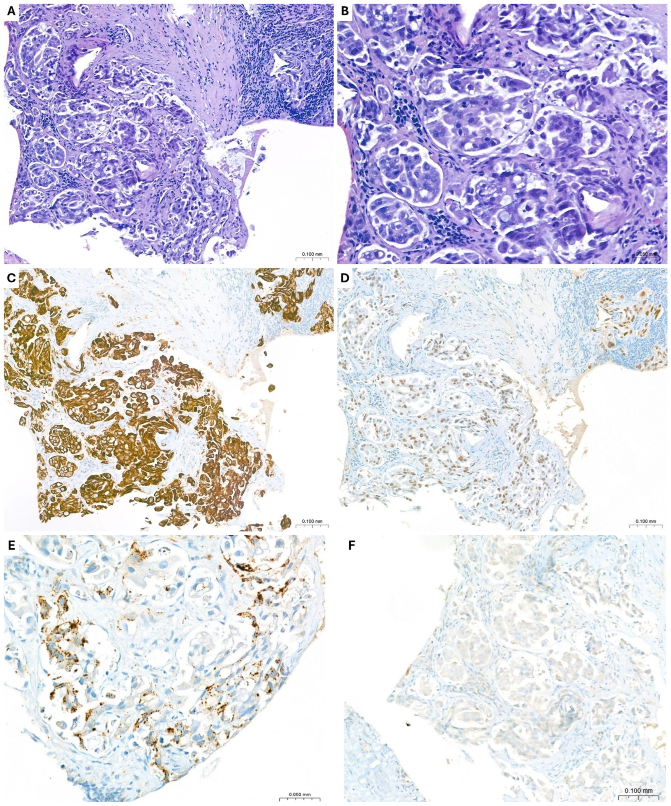
A 56-year-old man with a medical history of hypertension and HBV-related liver disease was admitted after a fall preceded by two days of confusion and language disturbance. On admission, a neurological examination revealed fluent aphasia and mild right upper limb pronation. A brain CT initially suggested a left temporo-insular ischemic lesion, but MRI demonstrated bilateral T2/FLAIR hyperintensities involving the left temporo-mesial cortex, thalami, and right medial frontal gyrus, with focal contrast enhancement (Figure 1). CSF analysis revealed mild pleocytosis (33 cells/µL) and elevated protein (86 mg/dL), while serum and CSF testing showed positivity for anti-Ma2 antibodies, consistent with autoimmune encephalitis. EEG demonstrated diffuse slowing without epileptiform activity. Whole-body imaging revealed mediastinal and right supraclavicular lymphadenopathies without a clearly identifiable lung lesion (Figure 2). Thus, a supraclavicular lymph node biopsy was performed. Gross examination showed fragments measuring 7 × 5.5 mm, while microscopic evaluation revealed lymph node parenchyma infiltrated by epithelial neoplastic cells arranged in aggregates, occasionally forming micropapillary structures. The neoplastic cells demonstrated marked cytological atypia and pleomorphism, ranging from medium to large in size. The cytoplasm was mildly eosinophilic and frequently vacuolated, with occasional signet ring cell-like features. Nuclei were enlarged, irregular in contour, hyperchromatic, and sometimes eccentrically located (Figure 3).
During hospitalization, the patient developed right popliteal deep vein thrombosis in the context of heterozygous factor II G20210A mutation and a urinary tract infection due to Escherichia coli. He was treated with intravenous high-dose steroids followed by an oral taper, as well as IGEV for the autoimmune encephalitis, with neurological and radiological improvement. At discharge, the patient was clinically improved, continued on oral steroids, and referred to hematology–oncology follow-up.
Multiple neoplastic emboli were observed in the lymphatic sinuses. Immunohistochemistry demonstrated diffuse expression of CK7, TTF-1 (clone 8G7G3/1), and Napsin A, while CK20, CDX2, and ALK (D5F3) were negative (Figure 3). Considering the morphological and immunophenotypic features, a diagnosis of lymph node metastasis from primary non-mucinous adenocarcinoma of the lung (NM-LUAD) was established. Molecular analysis was performed using next-generation sequencing (NGS) on DNA and RNA extracted from formalin-fixed paraffin-embedded (FFPE) tissue to detect recurrent mutations in the EGFR, KRAS, ERBB2, KEAP1, STK11, and BRAF genes, as well as gene fusions involving ALK, ROS1, RET, NTRK1, NTRK2, NTRK3, and MET exon 14 skipping. No pathogenic alterations were identified; however, a rare variant of unknown significance (VUS) in EGFR (c.379G>A, p.Ala127Thr, located in codon 127 of exon 3) was detected with a variant allele frequency (VAF) of 33.2%.

2.2. Case Two

A 49-year-old man, with no significant comorbidities and who had never smoked, presented with right laterocervical lymphadenopathy as his sole initial clinical manifestation. A neck ultrasound revealed multiple suspicious lymph nodes and a thyroid nodule. Fine-needle aspiration (FNA) of the thyroid was not malignant (TIR 1c). Subsequently, an excisional biopsy demonstrated a 1.5 cm lymph node with a solid, whitish cut surface.
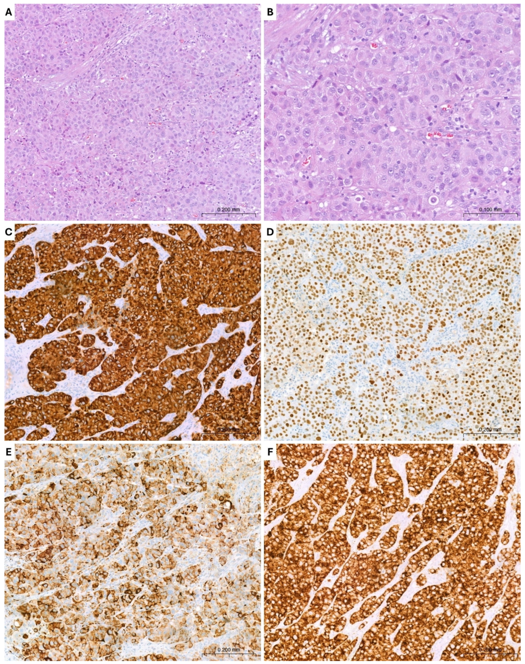
Microscopic examination revealed lymph node involvement by a malignant neoplasm with a solid growth pattern. The neoplastic cells are medium to large in size, featuring eosinophilic cytoplasm and round nuclei with irregular nuclear membranes and often prominent nucleoli (Figure 4). The immunohistochemistry revealed a positivity for CK7, TTF1 (clone 8G7G3/1), and Napsin A while CK20, PAX8, S100, SOX10 were negative (Figure 4). Based on morphological and immunophenotypic findings, lymph node metastasis from primary NM-LUAD was diagnosed.
Immunostaining for ALK (D5F3) showed positivity in the neoplastic cells (Figure 4), a finding subsequently confirmed by FISH analysis using a break-apart probe (ALK-2p23 probe), which demonstrated the presence of an ALK gene rearrangement. RET and ROS1 gene rearrangements were not detected by FISH analysis. Furthermore, molecular analysis, performed using NGS on DNA extracted from FFPE tissue, to assess for recurrent mutations in the EGFR, KRAS, and BRAF genes, showed no pathogenic molecular alterations.
Staging with PET/CT and brain MRI revealed cervical and mediastinal lymphadenopathy, along with three sub-centimetric brain metastases, consistent with stage IV NM-LUAD (cN3, cM1c). Notably, no distinct primary lung lesion was identified, highlighting the atypical nature of this presentation. The case was discussed in a multidisciplinary tumor board, and the diagnosis of ALK-positive stage IV NM-LUAD was made. Given the limited number and the small size of brain metastases, the absence of neurological symptoms, and the well-documented intracranial activity of alectinib in ALK-rearranged non-small-cell lung cancer, stereotactic brain radiotherapy was deferred in favor of upfront systemic therapy.
The patient initiated first-line targeted therapy with alectinib. Throughout treatment, he maintained an excellent clinical condition (ECOG 0), experiencing only mild adverse events including fatigue, asymptomatic bradycardia, and peripheral edema. Early imaging assessments demonstrated a marked intracranial response, with reduction and eventual complete resolution of brain metastases. Serial chest–abdomen CT scans confirmed sustained systemic disease stability; notably, pulmonary micronodules, absent at initial staging and subsequently interpreted as intrapulmonary metastatic lesions, remained stable with no evidence of progression.
The patient continued on alectinib with no significant toxicities and preserved performance status. Repeated imaging confirmed ongoing complete intracranial remission and systemic stability, highlighting the durable efficacy and favorable tolerability of alectinib in the management of ALK-rearranged NM-LUAD.

2.3. Case Three

A 66-year-old male with no significant past medical history presented with progressive enlargement of a right supraclavicular lymph node, adjacent to the thyroid lodge, measuring up to 3 × 2.2 cm.
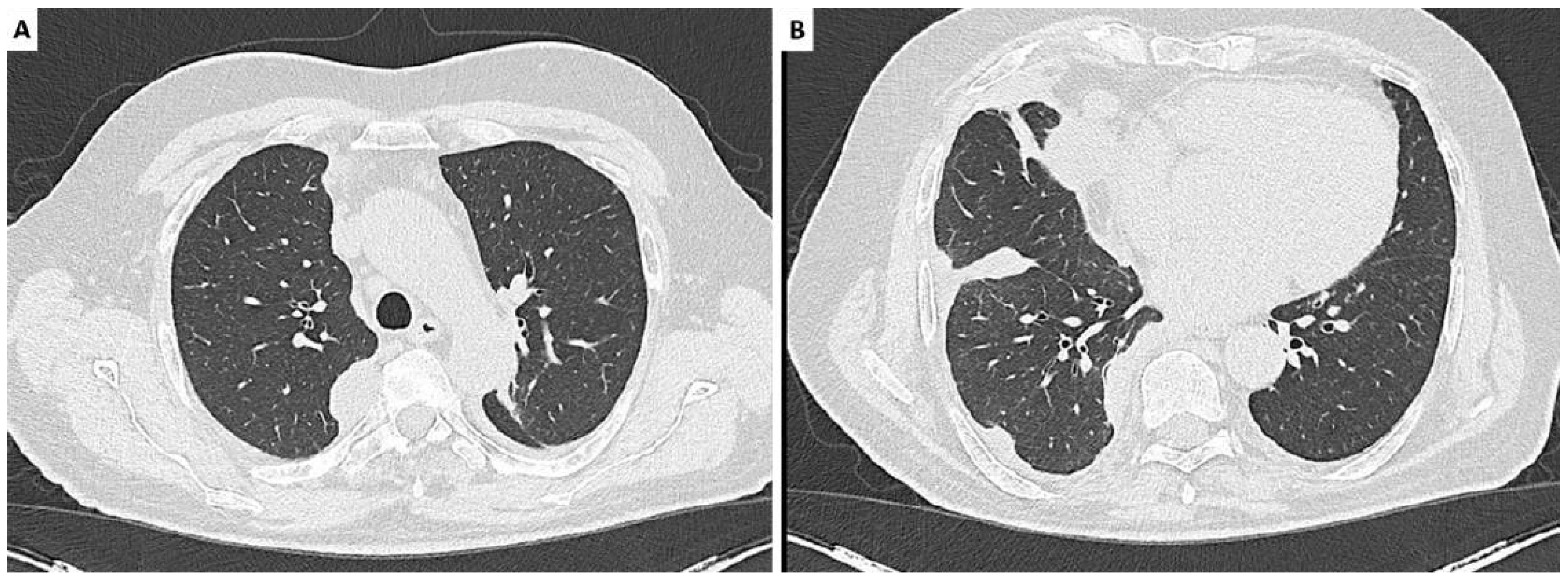
A total-body CT scan revealed subcentimetric cervical lymphadenopathy and enlarged lymph nodes in the Barety space, anterior mediastinum, subcarinal region extending to the right paravertebral area and cardiophrenic angle, along with a right-sided parieto-basal pleural effusion and basal and posterior pleural thickening. Focal thickenings were particularly notable in the upper costovertebral groove between the fourth and sixth costovertebral joints (Figure 5). The patient, however, denied exposure to asbestos or other relevant environmental or occupational risk factors, and his professional history was not associated with known exposure risks.
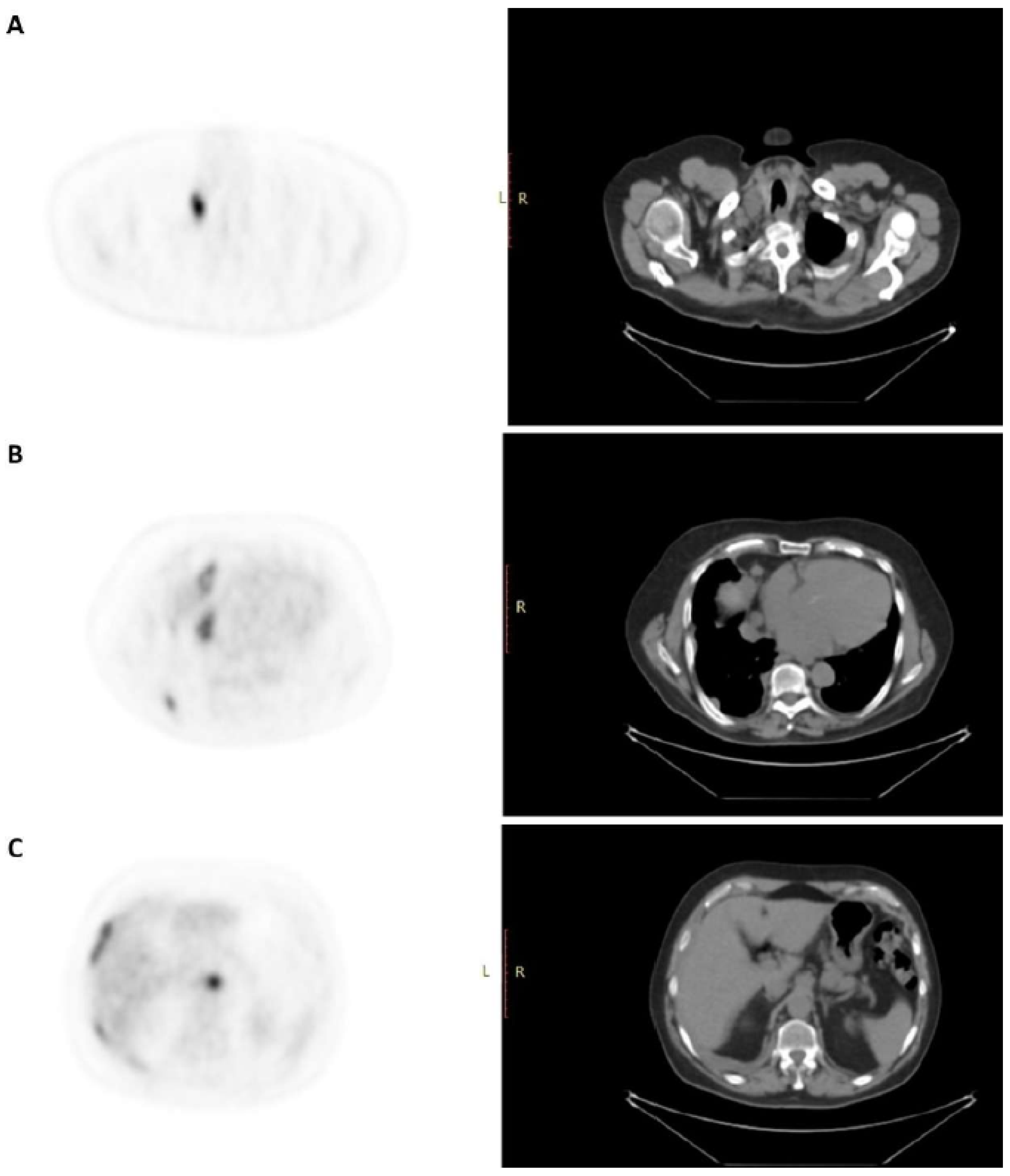
A PET scan with 18F-FDG demonstrated high metabolic uptake in multiple lymph node stations, including the right supraclavicular region (SUV max 13.8), Barety space (SUV max 9.9), anterior mediastinum (SUV max 11.8), subcarinal region extending to the right paravertebral area (SUV max 4.8), celiac–pancreatic (SUV max 11.5), intercavo-paraaortic (SUV max 6.4), and lumboaortic (SUV max 11.8) regions. Elevated FDG uptake was also noted in the right pleural space and adjacent structures, particularly the right cardiophrenic angle (SUV max 8.5), anterior (SUV max 7.2) and posterior (SUV max 7.3) costodiaphragmatic recesses, basal pleural region (SUV max 10.2), and posterior pleura, with the highest intensity in the upper right costovertebral groove (SUV max 10.8) (Figure 6). These radiological findings were consistent with metabolically active lymphadenopathy and pleural involvement, suggestive of a metastatic process, most likely of pulmonary origin. Therefore, excisional biopsy of the right supraclavicular lymph node was performed, revealing an enlarged 2.2 × 1 cm lymph node with a fleshy, translucent white cut surface.
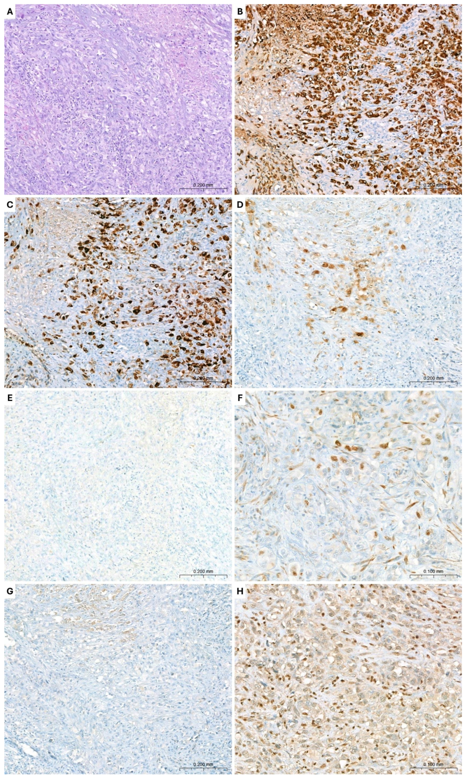
Microscopic examination revealed lymph node parenchyma involved by neoplastic proliferation with a diffuse growth pattern, composed of large, polygonal, epithelioid cells arranged in sheets. The neoplastic cells exhibited marked cyto-morphological atypia and pronounced nuclear pleomorphism, with well-defined membranes, abundant eosinophilic or clear cytoplasm, and large nuclei exhibiting round to irregular contours, dispersed chromatin, and prominent nucleoli. Extensive areas of tumor necrosis were also observed (Figure 7).
Immunohistochemistry demonstrated diffuse positivity for CK AE1/AE3 and CK7. Scattered tumor cells showed focal expression of calretinin, and very rare cells displayed weak positivity for WT1. The neoplastic cells were negative for Claudin-4, Ber-EP4, TTF1 (clone 8G7G3/1), Napsin A, CK5/6, Podoplanin (D2-40), CK20, PSA, SOX10, and Synaptophysin (Figure 7).
Molecular analysis was performed using NGS on DNA and RNA extracted from an FFPE tissue to assess for recurrent mutations in the EGFR, KRAS, and BRAF genes, as well as gene fusions involving ALK, ERBB2, KEAP1, STK11, ROS1, RET, NTRK1, NTRK2, NTRK3, and MET exon 14 skipping. No pathogenic molecular alterations were identified.
Given the clinico-radiological and morpho-immunophenotypic features, additional immunohistochemical staining for BAP1 and MTAP was performed. The results showed a loss of nuclear BAP1 expression (Figure 7) and retained cytoplasmic and nuclear MTAP expression. Concurrently, the NGS assay performed on circulating cell-free DNA identified a clinically relevant genomic alteration, namely the BAP1 S160fs*1 mutation, with a VAF of 12.4%. The BAP1 S160fs*1 mutation is a frameshift (fs) mutation caused by an insertion or deletion at codon 160, resulting in a premature stop codon after one (*1) altered amino acid (S160—serine at position 160 of the protein). This leads to a truncated, likely nonfunctional protein and explains the observed loss of BAP1 immunohistochemical expression. Therefore, based on clinico-radiological, histopathological, immunophenotypic, and molecular findings, a diagnosis of metastatic pleural epithelioid mesothelioma (PEM) was rendered. The patient started first-line chemotherapy with Carboplatin plus Pemetrexed administered every 3 weeks, in accordance with international standards for unresectable malignant pleural mesothelioma (MPM). Vitamin B12 and folic acid supplementation were also administered. At the time of reporting, the patient had completed three cycles of chemotherapy, with three additional cycles planned. He maintained a good performance status without significant high-grade toxicities. Clinical and laboratory follow-up is ongoing, with close monitoring of hematologic parameters and treatment tolerability.

3. Discussion

3.1. Diagnoses Challenges

Correlation of pathological findings with clinical history, data from prior pathology specimens, and imaging studies is essential in the routine diagnostic pathology, particularly in this type of clinical scenario. In such cases, a systematic approach is especially valuable. Key factors to consider include the patient’s age, sex, anatomical site of the metastasis, radiological findings, as well as the macroscopic and, most importantly, histopathological features of the lesion, prior to selecting an appropriate panel of immunohistochemical stains and/or molecular tests [10]. Nonetheless, cases with rare and unusual clinical presentations can occasionally pose diagnostic challenges that the pathologist must address and resolve independently, often without substantial external support.

3.2. Non-Mucinous Lung Adenocarcinoma

The first two cases exemplify these complexities, illustrating instances of NM-LUAD involving cervical lymph nodes without an identifiable primary tumor, consistent with occult primary NM-LUAD (OP-NM-LUAD). OP-NM-LUAD is a rare diagnosis and an exceptionally uncommon presentation, particularly in non-mediastinal lymph nodes, with only 11 cases reported in the literature to date (Table 1).
Differentiating NM-LUAD from other adenocarcinomas, particularly poorly differentiated tumors, remains a significant diagnostic challenge in the era of targeted therapy [14]. Accordingly, reliable LUAD-specific markers are required for accurate differential diagnosis. The identification of such markers relies in part on the retention of features of putative precursor cells, namely type II pneumocytes and Clara cells, by primary LUADs. In the present case, TTF-1 and Napsin A, expressed in differentiated type II pneumocytes and Clara cells, were employed to support the diagnosis of primary NM-LUAD [15]. TTF-1 is a highly conserved transcription factor essential for the early development of the lung, thyroid, and parts of the forebrain. In adult lungs, it is expressed in type II pneumocytes and distal bronchiolar epithelial cells, regulating several lung-specific proteins, including surfactant proteins, Napsin A, ABCA3, and Clara cell secretory protein, thereby directing lineage-specific lung development.
However, it is important to note that data on the rate of TTF-1 expression in various tumors are heterogeneous. The literature reports show TTF-1 expression in 42–100% of LUADs, 15–93% of small-cell lung cancers (SCLCs), 0–50% of squamous cell carcinomas of the lung (LUSCs), 0–42% of malignant mesotheliomas, 88–100% of follicular thyroid carcinomas, 66–100% of papillary thyroid carcinomas, 50–100% of medullary thyroid carcinomas, 5–100% of anaplastic thyroid carcinomas, 23–75% of ovarian endometrioid carcinomas, 5–80% of serous endometrial carcinomas, 0–80% of Merkel cell carcinomas and 7–25% of endometrial clear cell carcinomas. Among the various commercially available TTF-1 clones, clone 8G7G3/1 is by far characterized by the highest diagnostic specificity, being associated with a very low number of false positives and therefore representing the clone of choice for defining pulmonary lineage [16,17,18]. These findings indicate that, although TTF-1 is a highly sensitive diagnostic marker, its specificity is not optimal for absolutely reliable differentiation between NM-LUAD and morphologically similar adenocarcinomas, an observation also supported by the recent study by Möller et al. [19].
This evidence is especially important in cases of lymph node metastases from unknown primary and particularly if considering OP-NM-LUAD. Napsin A, an aspartate protease upregulated by TTF-1 in type II pneumocytes, is widely used as a marker for NM-LUAD, offering lower sensitivity but higher specificity than TTF-1 (Table 2). These data demonstrated that the combined assessment of TTF-1 and Napsin A, in the presence of consistent morphological features, significantly enhances the diagnostic specificity for NM-LUAD. In the two reported cases, dual positivity for both markers enabled a histopathological diagnosis of OP-NM-LUAD with absolute specificity, guiding appropriate therapeutic management.

3.3. Pleural Mesothelioma Metastases

In the third case, the combination of clinical and radiological features was necessary to support and confirm the histopathological assessment. MPM is a rare and highly aggressive tumor arising from the mesothelial cells of the pleural lining. Its diagnosis is often challenging and usually necessitates a multidisciplinary approach [21]. Patients with MPM typically present with non-specific progressive symptoms, including dyspnea, chest pain, and weight loss, often evolving over months. Unilateral pleural effusions are commonly observed on examination. A detailed occupational history is essential for diagnosis and potential medicolegal purposes.
Conventional chest radiography lacks sufficient sensitivity and specificity for diagnosis and staging since large pleural effusions may obscure small malignant lesions. In cases with significant asbestos exposure or imaging suggestive of mesothelioma, cytological analysis of pleural fluid can aid in detecting malignant cells; however, false negatives are common. Therefore, a histological examination remains the gold standard, making thoracoscopy with a targeted biopsy essential. Thoracoscopy, performed either by medical pleuroscopy or video-assisted thoracic surgery (VATS), enables direct visualization of the pleural cavity, optimal tissue sampling for histological confirmation, accurate disease staging, and therapeutic drainage of pleural effusions, with or without pleurodesis. Given the histopathological complexity of MPM, obtaining deep biopsies from at least three different sites is recommended to improve diagnostic yield. When thoracoscopy is contraindicated or not feasible, image-guided, typically CT-guided, percutaneous biopsies represent a valid alternative [22]. In an autopsy series, Finn et al. [23] reported extrapleural metastases in 87.7% of cases, with lymph node involvement identified in 53.3% of patients.
Extrathoracic tumor dissemination occurred in 55.4% of cases, involving multiple organs such as the liver, spleen, thyroid, and brain. Despite this, lymphatic spread is typically microscopic, and clinical evidence of nodal dissemination remains rare, even in advanced disease.
Consequently, systemic lymphadenopathy as an initial presentation of MPM is exceedingly uncommon (Table 3) [24].
Nevertheless, although rare, the available literature underscores the importance of recognizing the possibility of lymph node involvement in MPM to avoid misdiagnosis with other entities [28]. The diagnosis of PEM can occasionally be straightforward; however, it often poses a challenge due to a histological overlap with other epithelial neoplasms. This is particularly relevant when the biopsy is from a lymph node without clinical suspicion of MPM, as in the present case. Accurate diagnosis of metastatic MPM from lymph node biopsy alone remains challenging, owing to the rarity of nodal involvement and the morphological and immunohistochemical overlap with other epithelial malignancies [29]. The reported case exemplifies these complexities. The initial histological evaluation described a poorly differentiated epithelioid neoplasm, initially favoring a diagnosis of OP-NM-LUAD. However, immunohistochemical staining revealed a lack of expression of specific lineage biomarkers, with initial positivity limited to CK AE1/AE3 and CK 7. Given the clinic-radiological and morphological features, additional immunohistochemical staining for BAP1 and MTAP was performed, along with NGS on circulating cell-free DNA. The findings revealed a complete loss of nuclear BAP1 expression in the neoplastic cells and the identification of a genomic alteration in the BAP1 gene, specifically the BAP1 S160fs*1 mutation. This frameshift mutation leads to the introduction of a premature stop codon, resulting in the production of a truncated, nonfunctional protein, thereby explaining the observed loss of BAP1 immunohistochemical expression. Although not diagnostic of mesothelial lineage, this finding, in association with the overall clinic-radiological and morpho-immunophenotypic features, was considered consistent with the diagnosis of lymph node metastasis from PEM. This complex diagnosis was crucial for guiding the patient toward the appropriate therapeutic regimen. It is important to underline that BAP1, in addition to being a key diagnostic biomarker for mesothelioma in certain contexts, is also gaining potential predictive value. Emerging evidence suggests a possible association between BAP1 alterations and response to combined immunotherapy regimens, such as ipilimumab plus nivolumab. In detail, some preliminary evidence suggests that BAP1 alterations may be associated with a more immunogenic tumor microenvironment and a potentially improved response to immune checkpoint inhibitors, including combinations such as ipilimumab plus nivolumab [30]. Although these findings remain preliminary and require prospective validation, they support the rationale for considering immunotherapy in this molecular subgroup, particularly in later lines of treatment [31].

4. Conclusions

In conclusion, peripheral lymphadenopathy may occasionally be the first and only clinical manifestation of thoracic malignancies, representing an uncommon but clinically significant presentation. In our series, histological evaluation of the cervical lymph nodes allowed for the correct diagnosis despite the absence of a radiologically evident primary lung lesion or a strong suspicion of MPM. Although in some cases an accurate histopathological evaluation alone allows for a correct diagnosis of the disease, it must always be emphasized that the pathologist does not operate in isolation. A precise and timely diagnosis often relies on close collaboration within the multidisciplinary oncology team, where clinicians play a crucial role in providing complete and relevant clinical information. Indeed, the appropriate transmission of clinical history, imaging findings, and diagnostic suspicions to the pathologist is essential to guide morphological investigations and targeted molecular analysis. A thorough understanding of the patient’s anamnesis and clinical context is therefore fundamental, as it may significantly influence diagnostic interpretation and ultimately impact patient management and therapeutic decisions.

Author Contributions

S.L.: contributed to the study design, data collection, analysis and interpretation of data and the interpretation of patient’s pathological findings and contributed to the writing and review of the manuscript and approved the final version for submission; F.B.: contributed to data collection and interpretation and contributed to the writing and review of the manuscript and approved the final version for submission; A.D.M.: contributed to data collection and interpretation and contributed to the writing and review of the manuscript and approved the final version for submission; L.M.: contributed to the study design and analysis of data and contributed to the writing and review of the manuscript and approved the final version for submission; S.F.: contributed to the data collection and interpretation and contributed to the writing and review of the manuscript and approved the final version for submission; G.D.G.: contributed to the study design and analysis of data and contributed to the writing and review of the manuscript and approved the final version for submission; B.L.: contributed to the study design and analysis of data and contributed to the writing and review of the manuscript and approved the final version for submission; R.G.: contributed to the study design and analysis of data and contributed to the writing and review of the manuscript and approved the final version for submission; R.S.: contributed to the study design and analysis of data and contributed to the writing and review of the manuscript and approved the final version for submission; M.M.: contributed to the study design and analysis of data and contributed to the writing and review of the manuscript and approved the final version for submission; C.M.D.C.: contributed to the study design and analysis of data and contributed to the writing and review of the manuscript and approved the final version for submission; M.F.: contributed to the study design and analysis of data and contributed to the writing and review of the manuscript and approved the final version for submission; M.O.: contributed to the study design and analysis of data and contributed to the writing and review of the manuscript and approved the final version for submission; A.F.: contributed to the study design and analysis of data and contributed to the writing and review of the manuscript and approved the final version for submission; F.M.: contributed to the study design and analysis of data and contributed to the writing and review of the manuscript and approved the final version for submission; R.F.: contributed to the study design, data collection, analysis and interpretation of data and interpretation of patient’s pathological findings and contributed to the writing and review of the manuscript and approved the final version for submission. All authors have read and agreed to the published version of the manuscript.

Funding

This research received no external funding.

Institutional Review Board Statement

The study was conducted according to the guidelines of the Declaration of Helsinki and approved by the Ethics Committee of Università degli Studi Della Campania “L. Vanvitelli” (protocol code Bio-TOIS and 19 January 2025).

Informed Consent Statement

Written informed consent was obtained from all patients enrolled in this case series for the publication of their clinical data. The patient understands that his identity will be strictly protected, his name and initials will not be disclosed, and that complete anonymity cannot be fully guaranteed.

Data Availability Statement

No new data were created or analyzed in this study.

Conflicts of Interest

The authors declare no conflict of interest.

References

  1. Agca, M.; Kosif, A. Metastatic squamous cell cancer with unknown primary origin in the mediastinal lymph node. Niger. J. Clin. Pract. 2018, 21, 1384–1386. [Google Scholar] [CrossRef]
  2. Nakano, T.; Watanabe, H.; Takeichi, H.; Nakagawa, T.; Kohno, M.; Masuda, R.; Iwazaki, M. A Case of Mediastinal Lymph Node Carcinoma of Unknown Primary Site. Tokai J. Exp. Clin. Med. 2018, 43, 148–152. [Google Scholar]
  3. Riquet, M.; Badoual, C.; le Pimpec Barthes, F.; Dujon, A.; Danel, C. Metastatic thoracic lymph node carcinoma with unknown primary site. Ann. Thorac. Surg. 2003, 75, 244–249. [Google Scholar] [CrossRef]
  4. Wang, N.P.; Zee, S.; Zarbo, R.J.; Bacchi, C.E.; Gown, A.M. Coordinate Expression of Cytokeratins 7 and 20 Defines Unique Subsets of Carcinomas. Appl. Immunohistochem. 1995, 3, 99–107. [Google Scholar]
  5. Selves, J.; Long-Mira, E.; Mathieu, M.-C.; Rochaix, P.; Ilié, M. Immunohistochemistry for Diagnosis of Metastatic Carcinomas of Unknown Primary Site. Cancers 2018, 10, 108. [Google Scholar] [CrossRef] [PubMed]
  6. Al-Maghrabi, J.; Emam, E.; Gomaa, W. Immunohistochemical staining of cytokeratin 20 and cytokeratin 7 in colorectal carcinomas: Four different immunostaining profiles. Saudi J. Gastroenterol. 2018, 24, 129–134. [Google Scholar] [CrossRef] [PubMed]
  7. Bellizzi, A.M. An Algorithmic Immunohistochemical Approach to Define Tumor Type and Assign Site of Origin. Adv. Anat. Pathol. 2020, 27, 114–163. [Google Scholar] [CrossRef]
  8. Wang, B.; Cardona, D.M.; Huang, J. Revisiting the use of CK7 and CK20 immunohistochemical stains in pathological diagnoses. Diagn. Pathol. 2025, 20, 40. [Google Scholar] [CrossRef]
  9. Chong, Y.; Park, G.; Cha, H.J.; Kim, H.-J.; Kang, C.S.; Abdul-Ghafar, J.; Lee, S.-S. A stepwise approach to fine needle aspiration cytology of lymph nodes. J. Pathol. Transl. Med. 2023, 57, 196–207. [Google Scholar] [CrossRef]
  10. Kandalaft, P.L.; Gown, A.M. Practical Applications in Immunohistochemistry: Carcinomas of Unknown Primary Site. Arch. Pathol. Lab. Med. 2016, 140, 508–523. [Google Scholar] [CrossRef]
  11. Romesser, P.B.; Bardash, Y.; Buonocore, D.; Chaft, J.E.; Huang, J.; Jones, D.R.; Rimner, A.; Wu, A.J. Outcomes of Stage III NSCLC with occult primary vs. known primary lesions. Lung Cancer 2019, 127, 34–36. [Google Scholar] [CrossRef]
  12. Kawasaki, H.; Arakaki, K.; Taira, N.; Furugen, T.; Ichi, T.; Yohena, T.; Kawabata, T. Lung Cancer Detected 5 Years after Resection of Cancer of Unknown Primary in a Mediastinal Lymph Node: A Case Report and Review of Relevant Cases from the Literature. Ann. Thorac. Cardiovasc. Surg. 2016, 22, 116–121. [Google Scholar] [CrossRef]
  13. Shiota, Y.; Imai, S.; Sasaki, N.; Tahara, K.; Noma, B.; Horita, N.; Taniguchi, A.; Ono, T. A case of mediastinal lymph node carcinoma of unknown primary site treated with docetaxel and cisplatin with concurrent thoracic radiation therapy. Acta Med. Okayama 2011, 65, 407–411. [Google Scholar] [CrossRef]
  14. Lucà, S.; Monti, R.; Della Corte, C.M.; Cantisani, A.; Cozzolino, I.; Clery, E.; Amato, M.; Marone, L.; Capasso, F.; Di Guida, G.; et al. Application of Telepathology for Rapid On-Site Evaluation of Touch Imprint Cytology in CT-Guided Percutaneous Transthoracic Core Needle Biopsy of Pulmonary Nodules: The Experience of Our Multidisciplinary Thoracic Tumor Board. Cancers 2025, 17, 1738. [Google Scholar] [CrossRef]
  15. Ye, J.; Findeis-Hosey, J.J.; Yang, Q.; McMahon, L.A.; Yao, J.L.; Li, F.; Xu, H. Combination of napsin A and TTF-1 immunohistochemistry helps in differentiating primary lung adenocarcinoma from metastatic carcinoma in the lung. Appl. Immunohistochem. Mol. Morphol. 2011, 19, 313–317. [Google Scholar] [CrossRef]
  16. Vidarsdottir, H.; Tran, L.; Nodin, B.; Jirström, K.; Planck, M.; Mattsson, J.S.M.; Botling, J.; Micke, P.; Jönsson, P.; Brunnström, H. Comparison of Three Different TTF-1 Clones in Resected Primary Lung Cancer and Epithelial Pulmonary Metastases. Am. J. Clin. Pathol. 2018, 150, 533–544. [Google Scholar] [CrossRef] [PubMed]
  17. Klebe, S.; Swalling, A.; Jonavicius, L.; Henderson, D.W. An immunohistochemical comparison of two TTF-1 monoclonal antibodies in atypical squamous lesions and sarcomatoid carcinoma of the lung, and pleural malignant mesothelioma. J. Clin. Pathol. 2016, 69, 136–141. [Google Scholar] [CrossRef]
  18. Yatabe, Y.; Dacic, S.; Borczuk, A.C.; Warth, A.; Russell, P.A.; Lantuejoul, S.; Beasley, M.B.; Thunnissen, E.; Pelosi, G.; Rekhtman, N.; et al. Best Practices Recommendations for Diagnostic Immunohistochemistry in Lung Cancer. J. Thorac. Oncol. 2019, 14, 377–407. [Google Scholar] [CrossRef] [PubMed]
  19. Möller, K.; Gulzar, T.; Lennartz, M.; Viehweger, F.; Kluth, M.; Hube-Magg, C.; Bernreuther, C.; Bawahab, A.A.; Simon, R.; Clauditz, T.S.; et al. TTF-1 is a highly sensitive but not fully specific marker for pulmonary and thyroidal cancer: A tissue microarray study evaluating more than 17,000 tumors from 152 different tumor entities. Virchows Arch. 2024, 485, 815–828. [Google Scholar] [CrossRef] [PubMed]
  20. Zhang, P.; Han, Y.-P.; Huang, L.; Li, Q.; Ma, D.-L. Value of napsin A and thyroid transcription factor-1 in the identification of primary lung adenocarcinoma. Oncol. Lett. 2010, 1, 899–903. [Google Scholar] [CrossRef]
  21. Lucà, S.; Pignata, G.; Cioce, A.; Salzillo, C.; De Cecio, R.; Ferrara, G.; Della Corte, C.M.; Morgillo, F.; Fiorelli, A.; Montella, M.; et al. Diagnostic Challenges in the Pathological Approach to Pleural Mesothelioma. Cancers 2025, 17, 481. [Google Scholar] [CrossRef]
  22. Popat, S.; Baas, P.; Faivre-Finn, C.; Girard, N.; Nicholson, A.; Nowak, A.; Opitz, I.; Scherpereel, A.; Reck, M.; ESMO Guidelines Committee. Malignant pleural mesothelioma: ESMO Clinical Practice Guidelines for diagnosis, treatment and follow-up. Ann. Oncol. 2022, 33, 129–142. [Google Scholar] [CrossRef] [PubMed]
  23. Finn, R.S.; Brims, F.J.H.; Gandhi, A.; Olsen, N.; Musk, A.W.; Maskell, N.A.; Lee, Y.C.G. Postmortem findings of malignant pleural mesothelioma: A two-center study of 318 patients. Chest 2012, 142, 1267–1273. [Google Scholar] [CrossRef] [PubMed]
  24. Ideguchi, R.; Ashizawa, K.; Akashi, S.; Shindo, M.; Minami, K.; Fukuda, T.; Irie, J.; Fukuda, M.; Uetani, M. Malignant Pleural Mesothelioma with Marked Lymphatic Involvement: A Report of Two Autopsy Cases. Case Rep. Oncol. Med. 2017, 2017, 6195898. [Google Scholar] [CrossRef]
  25. Zhang, Y.; Taheri, Z.M.; Jorda, M. Systemic lymphadenopathy as the initial presentation of malignant mesothelioma: A report of three cases. Pathol. Res. Int. 2010, 2010, 846571. [Google Scholar] [CrossRef]
  26. Gong, Y.-S.; Rong, Y.-T.; Han, W.-C.; Zhang, Y.-C. Peripheral lymphadenopathy as the initial manifestation of malignant mesothelioma in a child. Hum. Pathol. 2013, 44, 664–669. [Google Scholar] [CrossRef]
  27. Zhu, X.L.; Gao, L.M.; Li, F.; Liu, W.P.; Zhang, W.Y.; Li, G.D.; Zhang, X.H.; Chen, M.; Liu, X.Y.; Zhao, S. Clinical and pathological characteristic of metastatic malignant mesothelioma initially diagnosed by lymph node biopsy. Pathol. Res. Pract. 2017, 213, 598–605. [Google Scholar] [CrossRef]
  28. Nind, N.R.; Attanoos, R.L.; Gibbs, A.R. Unusual intraparenchymal growth patterns of malignant pleural mesothelioma. Histopathology 2003, 42, 150–155. [Google Scholar] [CrossRef] [PubMed]
  29. Wagner, D.; Bourne, P.A.; Yang, Q.; Goldman, B.I.; Lewis, J.S.J. Unusual features of malignant pleural mesothelioma metastatic to the mediastinal lymph nodes. Appl. Immunohistochem. Mol. Morphol. 2008, 16, 301–307. [Google Scholar] [CrossRef] [PubMed]
  30. Xu, D.; Gao, Y.; Yang, H.; Spils, M.; Marti, T.M.; Losmanová, T.; Su, M.; Wang, W.; Zhou, Q.; Dorn, P.; et al. BAP1 Deficiency Inflames the Tumor Immune Microenvironment and Is a Candidate Biomarker for Immunotherapy Response in Malignant Pleural Mesothelioma. JTO Clin. Res. Rep. 2024, 5, 100672. [Google Scholar] [CrossRef]
  31. Baas, P.; Scherpereel, A.; Nowak, A.K.; Fujimoto, N.; Peters, S.; Tsao, A.S.; Mansfield, A.S.; Popat, S.; Jahan, T.; Antonia, S.; et al. First-line nivolumab plus ipilimumab in unresectable malignant pleural mesothelioma (CheckMate 743): A multicentre, randomised, open-label, phase 3 trial. Lancet 2021, 397, 375–386. [Google Scholar] [CrossRef]
Figure 1. The MRI scan sections: In the left mesial temporal region, with at least partial cortical-subcortical involvement, a large ill-defined area of high T2/FLAIR signal is seen, associated with gyral swelling. This area extends to the posterior temporal region and cranially to the level of the left internal and external capsule (AC). On T1 with gadolinium, heterogeneous contrast enhancement is observed in the left mesial temporal region (D).
Diagnostics 16 01618 g001
Figure 2. A heterogeneous lymphadenopathy is observed in the right supraclavicular region (approximately 40 × 20 mm) (A). An additional heterogeneous lymphoid mass (36 × 36 × 43 mm; AP × transverse × craniocaudal diameters) is identified in the mediastinum, arising from Barety’s space and extending to the right peribronchial region (B). Both lymphadenopathies are displayed in white on the non-contrast images and in red on the sequences acquired after intravenous iodinated contrast administration (C,D). (A,B): chest CT sections and (C,D): chest CT with contrast sections.
Diagnostics 16 01618 g002
Figure 3. Microscopic images of an epithelial neoplasm composed of neoplastic cells of large size arranged in nests. The cytoplasm is mildly eosinophilic and frequently vacuolated, with occasional signet ring-like cells. The nuclei are hyperchromatic, enlarged, and show irregular contours (A,B). The neoplasm shows a strong and widespread cytoplasmic expression of CK7 (C), a widespread nuclear expression of TTF1 (D), and a focal expression of Napsin A (E), whereas ALK is negative (F). (A,B): H&E original magnification 10× and 20×, respectively; (C,D,F): immunohistochemical staining original magnification 10×; (E): immunohistochemical staining original magnification 20×.
Diagnostics 16 01618 g003
Figure 4. Microscopic images of a malignant neoplastic population composed of epithelial cells, growing in a solid pattern. The cells show a variable size, ranging from medium to large, with eosinophilic cytoplasm. The nuclei are centrally located and display an irregular nuclear membrane, often with prominent nucleoli (A,B). The neoplasm exhibits a strong and diffuse cytoplasmic immunoreactivity for CK7 (C) and ALK (F), a widespread nuclear expression of TTF1 (D), and a cytoplasmic expression of Napsin A (E). (A,B): H&E original magnification of 4× and 10×, respectively, and (CF): immunohistochemical staining original magnification of 4×.
Diagnostics 16 01618 g004
Figure 5. CT scan sections revealed subcentimetric lymphadenopathy in the anterior mediastinum, subcarinal region, with extension to the right paravertebral area and right cardiophrenic angle, along with a thickening of the basal and posterior pleural plane. Particularly, focal thickenings of greater extent were observed in the upper costovertebral groove, between the fourth and sixth costovertebral joints (A,B).
Diagnostics 16 01618 g005
Figure 6. A PET/TC scan with 18F-FDG showed high metabolic uptake in several lymph node stations, including the right supraclavicular region, Barety space, anterior mediastinum, subcarinal region with extension to the right paravertebral area, celiac–pancreatic, intercavo-paraaortic and lumboaortic regions. High FDG uptake was also observed at the level of the right pleural space and adjacent structures, particularly in the right cardiophrenic angle, anterior and posterior costodiaphragmatic recesses, basal pleural region, and the posterior pleural area, with the highest intensity in the upper right costovertebral groove (AC).
Diagnostics 16 01618 g006
Figure 7. Microscopic images of a malignant neoplastic population composed of epithelioid cells with a diffuse growth pattern. The neoplastic population is composed of large, polygonal cells exhibiting marked nuclear pleomorphism and atypia. The cells have well-defined cytoplasmic membranes, with either clear or eosinophilic cytoplasm. The nuclei are large and round, with irregular contours and prominent nucleoli (A). Immunohistochemical analysis of neoplastic cells shows strong and diffuse nuclear staining for CK AE1/AE3 (B), as well as strong and heterogeneous staining for CK7 (C). Calretinin shows focal staining in both the cytoplasm and nuclei (D). Podoplanin (D2-40) is not detected (E), WT1 is detected in the nuclei of rare and scattered cells (F), and Claudin-4 is negative (G). Immunohistochemical staining for BAP1 showed a loss of protein nuclear expression in neoplastic cells, with retained expression in background inflammatory cells, used as an internal control (H). (A): H&E original magnification of 4×; (BE,G): immunohistochemical staining original magnification of 4×; and (FH): immunohistochemical staining with an original magnification of 10×.
Diagnostics 16 01618 g007
Table 1. The literature review of OP-NM-LUADs and a comparative overview.
N. of CasesBiopsy SiteRadiological FeaturesHistology DiagnosesOSPFSReferences
9Mediastinal-LNNo identifiable primary lung lesion on chest CT and PET-CTNM-LUAD61.6%Not available[11]
1Mediastinal-LNNo identifiable primary lung lesion and a 2.7 cm mass adjacent to the left side of the aortic arch, with contrast enhancement on chest CT.
Five years later, chest CT revealed a 1.2 cm irregular, spiculated nodule in the left upper lobe.
NM-LUADNot availableNot available[12]
1Mediastinal-LNNo identifiable primary lung lesion and a 9.5 × 2.5 cm mediastinal mass on chest CT.
FDG-PET/CT demonstrated intense hypermetabolic activity in the mediastinum (SUV max: 13.8) and moderate uptake in a right supraclavicular lymph node (SUV max: 4.5).
NM-LUADNot available≈20 month[13]
Mediastinal-LN: Mediastinal lymph node; NM-LUAD: non-mucinous lung adenocarcinoma; OS: overall survival; and PFS: progression-free survival.
Table 2. The diagnostic value of TTF1 and Napsin A expression assessment in routine practice.
MarkerDiagnostic SensitivityDiagnostic SpecificityReferences
TTF1~64–84.4%~83.9–96.4%[20]
Napsin A~84.9–87.3%~93.8–100%[19]
TTF-1 + Napsin A~79–91%up to ~100%[19]
Table 3. The literature review of systemic lymphadenopathy as an initial presentation of MPM and a comparative overview.
N. of CasesRadiological FeaturesBiopsy SiteHistology DiagnosesMedian OSReferences
3Case 1 and Case 2: CT/PET scan showed systemic lymphadenopathy
Case 3: mediastinal and left axillary lymphadenopathy
Case 1: right supraclavicular lymph node
Case 2: cervical lymph node
Case 3: left axillary lymph node
PEMNot available[25]
1Imaging revealed multiple enlarged lymph nodes in the cervical, axillary, mediastinal, and abdominal regionsRight axillary lymph nodePEMNot available[26]
3CT imaging demonstrated serous cavity effusions, serosal thickening, extensive soft-tissue masses involving the pericardium, pleura, and peritoneum, peripheral lymphadenopathy, and scattered patchy pulmonary opacitiesCase 1: Axillary lymph node
Case 2: Cervical lymph node
Case 3: Axillary lymph node
PEM6 months[27]
Median OS: Median overall survival.
Disclaimer/Publisher’s Note: The statements, opinions and data contained in all publications are solely those of the individual author(s) and contributor(s) and not of MDPI and/or the editor(s). MDPI and/or the editor(s) disclaim responsibility for any injury to people or property resulting from any ideas, methods, instructions or products referred to in the content.