Radiomics-Based Machine Learning for Sarcopenia Detection in Abdominal and Low-Dose CT



Abstract
1. Introduction
2. Methods
2.1. Data Collection and Labeling
2.2. Training Environment
2.3. Image Preprocessing
2.4. Radiomic Feature Extraction
2.5. Feature Selection and ML Models
2.6. Statistical Analysis
3. Results
4. Discussion
5. Conclusions
Author Contributions
Funding
Institutional Review Board Statement
Informed Consent Statement
Data Availability Statement
Conflicts of Interest
References
- Cruz-Jentoft, A.J.; Sayer, A.A. Sarcopenia. Lancet 2019, 393, 2636–2646. [Google Scholar] [CrossRef] [PubMed]
- Sayer, A.A.; Cruz-Jentoft, A.J. Sarcopenia definition, diagnosis and treatment: Consensus is growing. Age Ageing 2023, 53, afac220. [Google Scholar] [CrossRef] [PubMed]
- Gupta, S.; Dhillon, R.J.S.; Hasni, S. Sarcopenia: A rheumatic disease? Rheum. Dis. Clin. N. Am. 2018, 44, 443–456. [Google Scholar] [CrossRef]
- Navaneetham, K.; Arunachalam, D. Global Population Aging, 1950–2050. In Handbook of Aging, Health and Public Policy; Springer: Singapore, 2023. [Google Scholar] [CrossRef]
- United Nations. Department of Economic and Social Affairs, Population Division. In World Population Ageing 2017–Highlights; United Nations: New York, NY, USA, 2017; Available online: https://digitallibrary.un.org/record/3799351 (accessed on 20 March 2026).
- Cruz-Jentoft, A.J.; Bahat, G.; Bauer, J.; Boirie, Y.; Bruyère, O.; Cederholm, T.; Cooper, C.; Landi, F.; Rolland, Y.; Sayer, A.A.; et al. Sarcopenia: European consensus on definition and diagnosis. Age Ageing 2010, 39, 412–423. [Google Scholar] [CrossRef]
- Bruyère, O.; Beaudart, C.; Locquet, M.; Buckinx, F.; Petermans, J.; Reginster, J.Y. Sarcopenia as a public health problem. Eur. Geriatr. Med. 2016, 7, 272–275. [Google Scholar] [CrossRef]
- Li, N.; Ou, J.; He, H.; He, J.; Zhang, L.; Peng, Z.; Zhong, J.; Jiang, N. Exploration of a machine learning approach for diagnosing sarcopenia among Chinese community-dwelling older adults using sEMG-based data. J. Neuroeng. Rehabil. 2024, 21, 69. [Google Scholar] [CrossRef]
- Vogele, D.; Mueller, T.; Wolf, D.; Otto, S.; Manoj, S.; Goetz, M.; Ettrich, T.J.; Beer, M. Applicability of CT radiomics of skeletal muscle and machine learning for the detection of sarcopenia and prognostic assessment of disease progression in patients with gastric and esophageal tumors. Diagnostics 2024, 14, 198. [Google Scholar] [CrossRef]
- Gillies, R.J.; Kinahan, P.E.; Hricak, H. Radiomics: Images are more than pictures, they are data. Radiology 2016, 278, 563–577. [Google Scholar] [CrossRef]
- Aerts, H.J.W.L.; Velazquez, E.R.; Leijenaar, R.T.H.; Parmar, C.; Grossmann, P.; Carvalho, S.; Bussink, J.; Monshouwer, R.; Haibe-Kains, B.; Rietveld, D.; et al. Decoding tumour phenotype by noninvasive imaging using a quantitative radiomics approach. Nat. Commun. 2014, 5, 4006. [Google Scholar] [CrossRef]
- Gitto, S.; Cuoco, R.; Annovazzi, A.; Anelli, V.; Acquasanta, M.; Cincotta, A.; Albano, D.; Chianca, V.; Ferraresi, V.; Messina, C.; et al. CT radiomics-based machine learning classification of atypical cartilaginous tumours and appendicular chondrosarcomas. EBioMedicine 2021, 68, 103407. [Google Scholar] [CrossRef]
- Blanc-Durand, P.; Schiratti, J.B.; Schutte, K.; Jehanno, P.; Herent, P.; Pigneur, F.; Lucidarme, O.; Benaceur, Y.; Sadate, A.; Luciani, A.; et al. Automated muscle segmentation using deep learning on CT. Diagn. Interv. Imaging 2020, 101, 365–373. [Google Scholar] [CrossRef] [PubMed]
- Wang, F.; Chen, Y.; Tan, X.; Han, X.; Lu, W.; Lu, L.; Yuan, H.; Jiang, L. PET/computed tomography radiomics combined with clinical features in predicting sarcopenia and prognosis of diffuse large B-cell lymphoma. Nucl. Med. Commun. 2025, 46, 162–170. [Google Scholar] [CrossRef]
- Tomaszewski, M.R.; Gillies, R.J. The biological meaning of radiomic features. Radiology 2021, 298, 505–516. [Google Scholar] [CrossRef]
- Albano, D.; Messina, C.; Vitale, J.; Sconfienza, L.M. Imaging of sarcopenia: Old evidence and new insights. Eur. Radiol. 2020, 30, 2199–2208. [Google Scholar] [CrossRef]
- Onishi, S.; Kuwahara, T.; Tajika, M.; Tanaka, T.; Yamada, K.; Shimizu, M.; Niwa, Y.; Yamaguchi, R. Artificial intelligence for body composition assessment focusing on sarcopenia. Sci. Rep. 2024, 14, 83401. [Google Scholar] [CrossRef]
- Dong, X.; Dan, X.; Yawen, A.; Haibo, X.; Huan, L.; Mengqi, T.; Linglong, C.; Zhao, R. Identifying sarcopenia in advanced non-small cell lung cancer patients using skeletal muscle CT radiomics and machine learning. Thorac. Cancer 2021, 12, 2345–2353. [Google Scholar] [CrossRef]
- Yin, L.; Zhao, J. An artificial intelligence approach for test-free identification of sarcopenia. J. Cachexia Sarcopenia Muscle 2022, 13, 1234–1245. [Google Scholar] [CrossRef]
- Ryu, J.; Eom, S.; Kim, H.C.; Kim, C.O.; Rhee, Y.; You, S.C.; Hong, N. Chest X-ray-based opportunistic screening of sarcopenia using deep learning. J. Cachexia Sarcopenia Muscle 2021, 12, 156–165. [Google Scholar] [CrossRef] [PubMed]
- Zhang, H.; Yin, M.; Liu, Q.; Ding, F.; Hou, L.; Deng, Y.; Cui, T.; Han, Y.; Pang, W.; Ye, W.; et al. Machine and deep learning-based clinical characteristics and laboratory markers for the prediction of sarcopenia. Chin. Med. J. 2021, 134, 123–133. [Google Scholar] [CrossRef] [PubMed]
- Brenner, D.J.; Hall, E.J. Computed tomography—An increasing source of radiation exposure. N. Engl. J. Med. 2007, 357, 2277–2284. [Google Scholar] [CrossRef]
- Pearce, M.S.; Salotti, J.A.; Little, M.P.; McHugh, K.; Lee, C.; Kim, K.P.; Howe, N.L.; Ronckers, C.M.; Rajaraman, P.; Craft, A.W.; et al. Radiation exposure from CT scans in childhood and subsequent risk of leukaemia and brain tumours: A retrospective cohort study. Lancet 2012, 380, 499–505. [Google Scholar] [CrossRef]
- Stephan, J.T.; Mehta, P.; Zepeda, D.L.; Uppal, M.; Basu, S.; Liptay, M.J.; Borgia, J.A.; Geissen, N.; Shah, P.; Karush, J.; et al. Low-dose computed tomography scan features are associated with annual risk of hospitalization. Ann. Thorac. Surg. Short. Rep. 2023, 1, 558–561. [Google Scholar] [CrossRef]
- Buckens, C.F.; van der Graaf, Y.; Verkooijen, H.M.; Mali, W.P.; Isgum, I.; Mol, C.P.; Verhaar, H.J.; Vliegenthart, R.; Oudkerk, M.; van Aalst, C.M.; et al. Osteoporosis markers on low-dose lung cancer screening chest computed tomography scans predict all-cause mortality. Eur. Radiol. 2015, 25, 3361–3369. [Google Scholar] [CrossRef]
- Oppelt, A. Imaging Systems for Medical Diagnostics; Publicis Corporate Publishing: Erlangen, Germany, 2005. [Google Scholar]
- Yu, L.; Liu, X.; Leng, S.; Kofler, J.M.; Ramirez-Giraldo, J.C.; Qu, M.; Christner, J.; Fletcher, J.G.; McCollough, C.H. Radiation dose reduction in computed tomography: Techniques and future perspective. Imaging Med. 2009, 1, 65–84. [Google Scholar] [CrossRef]
- Konovalov, A.B. Compressed-sensing-inspired reconstruction algorithms in low-dose computed tomography: A review. Phys. Medica 2024, 124, 104491. [Google Scholar] [CrossRef] [PubMed]
- Liu, J.; Xu, H.; Qing, H.; Li, Y.; Yang, X.; He, C.; Ren, J.; Zhou, P. Comparison of radiomic models based on low-dose and standard-dose CT for prediction of adenocarcinomas and benign lesions in solid pulmonary nodules. Front. Oncol. 2020, 10, 634298. [Google Scholar] [CrossRef]
- Xu, K.; Gao, R.; Tang, Y.; Deppen, S.A.; Sandler, K.L.; Kammer, M.; Antic, S.; Maldonado, F.; Huo, Y.; Khan, M.; et al. Extending the value of routine lung screening CT with quantitative body composition assessment. Proc. SPIE Int. Soc. Opt. Eng. 2022, 12036, 120361A. [Google Scholar] [CrossRef]
- Zyla, J.; Marczyk, M.; Prazuch, W.; Sitkiewicz, M.; Durawa, A.; Jelitto, M.; Dziadziuszko, K.; Jelonek, K.; Kurczyk, A.; Szurowska, E.; et al. Combining low-dose computer-tomography-based radiomics and serum metabolomics for diagnosis of malignant nodules in participants of lung cancer screening studies. Biomolecules 2024, 14, 44. [Google Scholar] [CrossRef] [PubMed]
- Lundberg, S.; Lee, S.I. A unified approach to interpreting model predictions. Adv. Neural Inf. Process. Syst. 2017, 30. [Google Scholar] [CrossRef]
- Kapoor, S.; Narayanan, A. Leakage and the reproducibility crisis in machine-learning-based science. Patterns 2023, 4, 100804. [Google Scholar] [CrossRef]
- Shen, W.; Punyanitya, M.; Wang, Z.; Gallagher, D.; St-Onge, M.P.; Albu, J.; Heymsfield, S.B.; Heshka, S. Total body skeletal muscle and adipose tissue volumes: Estimation from a single abdominal cross-sectional image. J. Appl. Physiol. (1985) 2004, 97, 2333–2338. [Google Scholar] [CrossRef]
- Timpano, G.; Veltri, P.; Vizza, P.; Cascini, G.L.; Manti, F. Deep learning-based 3D and 2D approaches for skeletal muscle segmentation on low-dose CT images. J. Imaging Inform. Med. 2025. [Google Scholar] [CrossRef] [PubMed]
- Fearon, K.; Strasser, F.; Anker, S.D.; Bosaeus, I.; Bruera, E.; Fainsinger, R.L.; Jatoi, A.; Loprinzi, C.; MacDonald, N.; Mantovani, G.; et al. Definition and classification of cancer cachexia: An international consensus. Lancet Oncol. 2011, 12, 489–495. [Google Scholar] [CrossRef] [PubMed]
- Kim, E.Y.; Kim, Y.S.; Park, I.; Ahn, H.K.; Cho, E.K.; Jeong, Y.M. Prognostic significance of CT-determined sarcopenia in patients with small-cell lung cancer. J. Thorac. Oncol. 2015, 10, 1795–1799. [Google Scholar] [CrossRef]
- Kim, E.Y.; Kim, Y.S.; Park, I.; Ahn, H.K.; Cho, E.K.; Jeong, Y.M.; Kim, J.H. Evaluation of sarcopenia in small-cell lung cancer patients by routine chest CT. Support. Care Cancer 2016, 24, 4721–4726. [Google Scholar] [CrossRef]
- Recio-Boiles, A.; Galeas, J.N.; Goldwasser, B.; Sanchez, K.; Man, L.M.W.; Gentzler, R.D.; Gildersleeve, J.; Hollen, P.J.; Gralla, R.J. Enhancing evaluation of sarcopenia in patients with non-small cell lung cancer (NSCLC) by assessing skeletal muscle index (SMI) at the first lumbar (L1) level on routine chest computed tomography (CT). Support. Care Cancer 2018, 26, 2353–2359. [Google Scholar] [CrossRef]
- National Electrical Manufacturers Association (NEMA). Digital Imaging and Communications in Medicine (DICOM) Standard, PS3.3: Information Object Definitions. Available online: https://dicom.nema.org/ (accessed on 1 April 2026).
- Aubrey, J.; Esfandiari, N.; Baracos, V.E.; Buteau, F.A.; Frenette, J.; Putman, C.T.; Mazurak, V.C. Measurement of skeletal muscle radiation attenuation and basis of its biological variation. Acta Physiol. 2014, 210, 489–497. [Google Scholar] [CrossRef]
- van Griethuysen, J.J.M.; Fedorov, A.; Parmar, C.; Hosny, A.; Aucoin, N.; Narayan, V.; Beets-Tan, R.G.; Fillion-Robin, J.C.; Pieper, S.; Aerts, H.J. Computational radiomics system to decode the radiographic phenotype. Cancer Res. 2017, 77, e104–e107. [Google Scholar] [CrossRef] [PubMed]
- Mayerhoefer, M.E.; Materka, A.; Langs, G.; Häggström, I.; Szczypiński, P.; Gibbs, P.; Cook, G. Introduction to radiomics. J. Nucl. Med. 2020, 61, 488–495. [Google Scholar] [CrossRef]
- Cho, H.H.; Lee, S.H.; Kim, J.; Park, H. Classification of the glioma grading using radiomics analysis. PeerJ 2018, 6, e5982. [Google Scholar] [CrossRef]
- Ansari, G.A.; Shafi, S.; Alhazzaa, L. Optimized Machine Learning Pipeline for Lung Cancer Classification: Feature Reduction and Hyperparameter Tuning. Diagnostics 2026, 16, 1198. [Google Scholar] [CrossRef]
- Miotto, R.; Wang, F.; Wang, S.; Jiang, X.; Dudley, J.T. Deep learning for healthcare: Review, opportunities and challenges. Brief. Bioinform. 2018, 19, 1236–1246. [Google Scholar] [CrossRef] [PubMed]
- Hosmer, D.W.; Lemeshow, S.; Sturdivant, R.X. Applied Logistic Regression; John Wiley & Sons: Hoboken, NJ, USA, 2013. [Google Scholar] [CrossRef]
- Cortes, C.; Vapnik, V. Support-vector networks. Mach. Learn. 1995, 20, 273–297. [Google Scholar] [CrossRef]
- Breiman, L. Random forests. Mach. Learn. 2001, 45, 5–32. [Google Scholar] [CrossRef]
- Abdulkareem, N.K.; Hajee, S.I.; Hassan, F.F.; Ibrahim, I.K.; Al-Khalidi, R.E.H.; Abdulqader, N.A. Investigating the slice thickness effect on noise and diagnostic content of single-source multi-slice computerized axial tomography. J. Med. Life 2023, 16, 862–867. [Google Scholar] [CrossRef]
- Escudero Sanchez, L.; Rundo, L.; Gill, A.B.; Hoare, M.; Mendes Serrao, E.; Sala, E. Robustness of radiomic features in CT images with different slice thickness, comparing liver tumour and muscle. Sci. Rep. 2021, 11, 8262. [Google Scholar] [CrossRef]
- Zwanenburg, A.; Leger, S.; Vallières, M.; Löck, S. Image biomarker standardisation initiative. Radiology 2020, 295, 328–338. [Google Scholar] [CrossRef]
- Park, H.S.; Baek, J.; You, S.K.; Choi, J.K.; Seo, J.K. Unpaired image denoising using a generative adversarial network in X-ray CT. IEEE Access 2019, 7, 110414–110425. [Google Scholar]
- Lim, W.H.; Park, C.M. Validation for measurements of skeletal muscle areas using low-dose chest computed tomography. Sci. Rep. 2021, 11, 4492. [Google Scholar] [CrossRef]




| Cohort | Group | N | Age (Years) | Male, n (%) | Female, n (%) |
|---|---|---|---|---|---|
| APCT | Sarcopenia | 16 | 57.75 ± 9.79 | 11 (68.8%) | 5 (31.2%) |
| Normal | 59 | 54.73 ± 8.26 | 35 (59.3%) | 24 (40.7%) | |
| LDCT | Sarcopenia | 121 | 56.60 ± 8.81 | 90 (74.4%) | 31 (25.6%) |
| Normal | 18 | 52.83 ± 6.66 | 1 (5.6%) | 17 (94.4%) |
| Component | Hyperparameter | Value |
|---|---|---|
| Feature Selector (L1-regularized logistic regression via SelectFromModel) | Regularization (C) | 0.9 |
| LR | Class weight | balanced |
| Solver | liblinear | |
| Penalty | L2 | |
| SVM | Calibration method | Sigmoid (Platt scaling) |
| Class weight | balanced | |
| RF | Number of estimators | 600 |
| Max depth | 3 | |
| Min samples leaf | 5 | |
| Class weight | balanced |
| Model | AUC (95% CI) | PR-AUC (95% CI) | Accuracy (95% CI) | Sensitivity (95% CI) | Specificity (95% CI) | F1-Score (95% CI) |
|---|---|---|---|---|---|---|
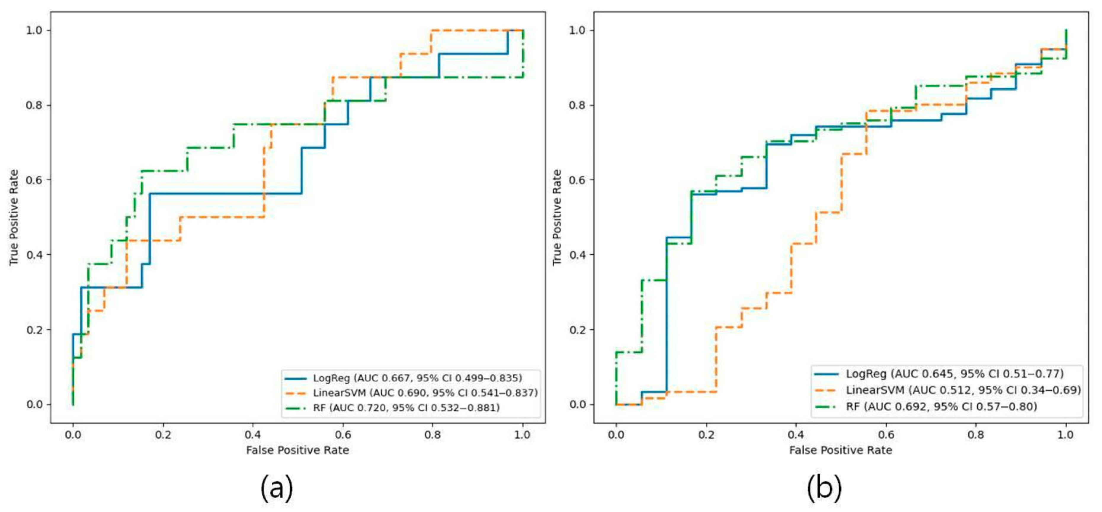
| LR | 0.667 (0.499–0.835) | 0.518 (0.285–0.732) | 0.772 (0.680–0.867) | 0.561 (0.308–0.812) | 0.829 (0.732–0.917) | 0.505 (0.276–0.698) |
| SVM | 0.690 (0.541–0.837) | 0.486 (0.259–0.700) | 0.786 (0.693–0.880) | 0.436 (0.188–0.700) | 0.882 (0.793–0.964) | 0.457 (0.222–0.667) |
| RF | 0.720 (0.532–0.881) | 0.566 (0.322–0.774) | 0.802 (0.707–0.880) | 0.622 (0.375–0.867) | 0.850 (0.754–0.934) | 0.565 (0.345–0.745) |
| Model | AUC (95% CI) | PR-AUC (95% CI) | Accuracy (95% CI) | Sensitivity (95% CI) | Specificity (95% CI) | F1-Score (95% CI) |
|---|---|---|---|---|---|---|
| LR | 0.645 (0.506–0.772) | 0.906 (0.834–0.971) | 0.597 (0.511–0.676) | 0.562 (0.472–0.650) | 0.831 (0.625–1.000) | 0.707 (0.630–0.776) |
| SVM | 0.512 (0.342–0.686) | 0.854 (0.769–0.943) | 0.740 (0.662–0.813) | 0.785 (0.708–0.855) | 0.444 (0.200–0.684) | 0.839 (0.783–0.889) |
| RF | 0.692 (0.573–0.801) | 0.940 (0.895–0.975) | 0.604 (0.518–0.683) | 0.570 (0.481–0.655) | 0.836 (0.631–1.000) | 0.714 (0.638–0.784) |
Disclaimer/Publisher’s Note: The statements, opinions and data contained in all publications are solely those of the individual author(s) and contributor(s) and not of MDPI and/or the editor(s). MDPI and/or the editor(s) disclaim responsibility for any injury to people or property resulting from any ideas, methods, instructions or products referred to in the content. |
© 2026 by the authors. Licensee MDPI, Basel, Switzerland. This article is an open access article distributed under the terms and conditions of the Creative Commons Attribution (CC BY) license.
Share and Cite
Kim, S.-B.; Kim, Y.J.; Kim, K.G. Radiomics-Based Machine Learning for Sarcopenia Detection in Abdominal and Low-Dose CT. Diagnostics 2026, 16, 1617. https://doi.org/10.3390/diagnostics16111617
Kim S-B, Kim YJ, Kim KG. Radiomics-Based Machine Learning for Sarcopenia Detection in Abdominal and Low-Dose CT. Diagnostics. 2026; 16(11):1617. https://doi.org/10.3390/diagnostics16111617
Chicago/Turabian StyleKim, Soo-Been, Young Jae Kim, and Kwang Gi Kim. 2026. "Radiomics-Based Machine Learning for Sarcopenia Detection in Abdominal and Low-Dose CT" Diagnostics 16, no. 11: 1617. https://doi.org/10.3390/diagnostics16111617
APA StyleKim, S.-B., Kim, Y. J., & Kim, K. G. (2026). Radiomics-Based Machine Learning for Sarcopenia Detection in Abdominal and Low-Dose CT. Diagnostics, 16(11), 1617. https://doi.org/10.3390/diagnostics16111617